-
From Cellular Radiosensitivity to Precision Radiotherapy: Integrating Functional Assays, Genomics, and Clinical Modeling -
Head-to-Head Comparison of [68Ga]Ga-PSMA-11 PET Interpreted with Non-Contrast CT Versus Excretory-Phase CT Urography in Biochemical Recurrence of Prostate Cancer -
Advances in Immune Checkpoint Inhibitors for Cancer Treatment
Journal Description
Cancers
Cancers
is a peer-reviewed, open access journal of oncology published semimonthly online. The North-East German Society for Gynecological Oncology (NOGGO), Irish Association for Cancer Research (IACR), Spanish Association for Cancer Research (ASEICA), Biomedical Research Centre (CIBM), British Neuro-Oncology Society (BNOS) and more are affiliated with Cancers and their members receive a discount on the article processing charges.
- Open Access— free for readers, with article processing charges (APC) paid by authors or their institutions.
- High Visibility: indexed within Scopus, SCIE (Web of Science), PubMed, PMC, Embase, CAPlus / SciFinder, and other databases.
- Journal Rank: JCR - Q2 (Oncology) / CiteScore - Q1 (Oncology)
- Rapid Publication: manuscripts are peer-reviewed and a first decision is provided to authors approximately 17.5 days after submission; acceptance to publication is undertaken in 2.7 days (median values for papers published in this journal in the first half of 2026).
- Recognition of Reviewers: Reviewers whose reports are timely and of high quality receive an APC discount voucher for a future publication in an MDPI journal. Become a reviewer.
- Sections: published in 17 topical sections.
- Companion journals for Cancers include: Cancer Survivorship, BCRC, Radiation and Onco.
- Journal Clusters of Oncology: Cancers, Current Oncology, Onco and Targets.
Impact Factor:
4.8 (2025);
5-Year Impact Factor:
5.1 (2025)
Latest Articles
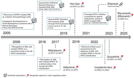
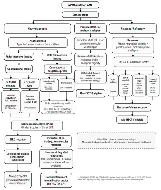
Two Decades of NPM1-Mutated Acute Myeloid Leukemia: From Molecular Insights to Clinical Decision-Making
Cancers 2026, 18(18), 3046; https://doi.org/10.3390/cancers18183046 (registering DOI) - 19 Sep 2026
Abstract
NPM1-mutated acute myeloid leukemia (AML) represents one of the most frequent and biologically distinct AML entities, characterized by the aberrant cytoplasmic localization of the NPM1 mutant protein. The recent advances in molecular biology and translational research have progressively redefined the clinical management
[...] Read more.
NPM1-mutated acute myeloid leukemia (AML) represents one of the most frequent and biologically distinct AML entities, characterized by the aberrant cytoplasmic localization of the NPM1 mutant protein. The recent advances in molecular biology and translational research have progressively redefined the clinical management of this disease. This narrative review summarizes the evolution of NPM1-mutated AML over the last two decades, focusing on the transition from immunohistochemical observations to modern genetics-based and measurable residual disease (MRD)-driven approaches. We discuss the biological role of mutant NPM1, the prognostic impact of co-occurring genetic alterations, and the integration of molecular monitoring into routine clinical practice. Particular attention is given to the prognostic and therapeutic implications of the co-mutational landscape. We also discuss emerging targeted therapies directed against key pathogenic pathways, including the menin–KMT2A axis and nuclear export machinery. Despite significant advances in risk stratification and treatment, considerable heterogeneity in clinical outcomes persists among patients with NPM1-mutated AML, indicating that genomic information obtained at diagnosis alone is insufficient to fully capture prognostic differences. Future advances will likely depend on the integration of genetic, transcriptional, and broader cellular-state information to better define biologically relevant disease states and guide more personalized therapeutic strategies.
Full article
(This article belongs to the Special Issue Precision Medicine in Acute Myeloid Leukemia)
►
Show Figures
Open AccessArticle
Survival Impact of Bevacizumab According to Chemotherapy Response Score in Advanced High-Grade Serous Ovarian Carcinoma Treated with Neoadjuvant Chemotherapy
by
Hasan Volkan Ege, Haticegul Tuncer, Utku Akgör, Murat Gultekin, Zafer Selcuk Tuncer and Derman Basaran
Cancers 2026, 18(18), 3045; https://doi.org/10.3390/cancers18183045 (registering DOI) - 19 Sep 2026
Abstract
Introduction: Chemotherapy response score (CRS) is a prognostic marker in high-grade serous ovarian carcinoma treated with neoadjuvant chemotherapy (NACT). This study evaluated whether the survival benefit associated with sustained bevacizumab exposure differs according to CRS group. Methods: We retrospectively evaluated patients who underwent
[...] Read more.
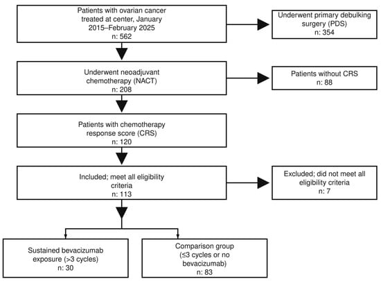
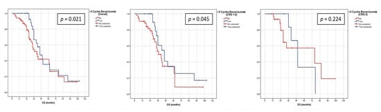
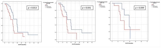
Introduction: Chemotherapy response score (CRS) is a prognostic marker in high-grade serous ovarian carcinoma treated with neoadjuvant chemotherapy (NACT). This study evaluated whether the survival benefit associated with sustained bevacizumab exposure differs according to CRS group. Methods: We retrospectively evaluated patients who underwent interval debulking surgery after platinum-taxane-based NACT between January 2015 and February 2025. Sustained bevacizumab exposure was defined as receipt of more than three cycles. Overall survival (OS) and OS after first recurrence (OS-FR) were assessed using Kaplan–Meier and Cox regression analyses. Results: The cohort included 113 patients: 30 with sustained bevacizumab exposure and 83 in the comparison group; 74 had CRS 1–2 and 39 had CRS 3. Sustained bevacizumab was associated with longer median OS in the overall cohort (57.4 vs. 50.3 months; p = 0.021) and in patients with CRS 1–2 (57.1 vs. 43.6 months; p = 0.045), but not in those with CRS 3 (p = 0.224). OS-FR was also longer with sustained bevacizumab overall (35.0 vs. 23.0 months; p = 0.013) and in CRS 1–2 (35.0 vs. 23.0 months; p = 0.041). In multivariable analysis, sustained bevacizumab was independently associated with improved OS-FR (landmark-corrected HR 0.23, 95% CI 0.07–0.75; p = 0.015). Conclusions: Sustained bevacizumab exposure was associated with longer OS and OS-FR, particularly in patients with CRS 1–2. CRS may help identify patients most likely to benefit from bevacizumab, although these findings require prospective validation.
Full article
(This article belongs to the Special Issue Locally Advanced and Recurrent Pelvic Malignancy)
►▼
Show Figures

Figure 1
Open AccessReview
Current Management and the New Paradigm for ALK+ NSCLC After CROWN
by
Shuo Shi and Petros Christopoulos
Cancers 2026, 18(18), 3044; https://doi.org/10.3390/cancers18183044 (registering DOI) - 19 Sep 2026
Abstract
The 7-year update of the phase 3 CROWN study is the most impressive milestone in anaplastic lymphoma kinase (ALK)+ non-small-cell lung cancer (NSCLC) since the discovery of the oncogene 20 years ago and inaugurates an entire new paradigm. The progression-free survival (PFS) plateau
[...] Read more.
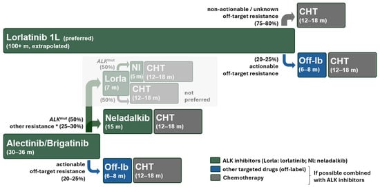
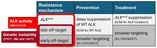
The 7-year update of the phase 3 CROWN study is the most impressive milestone in anaplastic lymphoma kinase (ALK)+ non-small-cell lung cancer (NSCLC) since the discovery of the oncogene 20 years ago and inaugurates an entire new paradigm. The progression-free survival (PFS) plateau at a high level > 50%, with an estimated very low annual progression rate < 2% after the third year, suggests that lorlatinib can inactivate the disease in most patients. The complete abrogation of on-target resistance abolishes ALK tyrosine kinase inhibitor (TKI) sequencing and will seriously impact future drug development. In the new era of extraordinary drug efficacy, the design of clinical trials, pathways of marketing authorization, and dosing optimization policies will also need to be reconsidered. Lorlatinib will dominate the therapeutic landscape of ALK+ NSCLC until the advent of combination therapies, which are the most reasonable next step. Further strengthening of the ALK blockade with additional ALK-directed drugs could likely eliminate the small residual “leak” of late off-target resistance after the third year, while combination with emerging neoantigen-directed immunotherapies may also help control the highly aggressive and more immunogenic early-progressing tumors and facilitate cure of the disease. Universal state-of-the-art DNA/RNA NGS for newly diagnosed NSCLC is now imperative to identify and treat all ALK+ patients according to the latest therapeutic advances, which will hopefully soon also spread to earlier stages and other oncogenic drivers.
Full article
(This article belongs to the Special Issue ALK in Cancer: Lessons From the Future (2nd Edition))
►▼
Show Figures

Figure 1
Open AccessReview
Circulating Tumor DNA-Based Molecular Residual Disease in Early-Stage Non-Small-Cell Lung Cancer: Biology, Assays, and the Path Toward MRD-Guided Care
by
Rajan Yadav, Masaoki Ito, Hideki Ujiie, Shuta Ohara, Yasuhiro Tsutani and Goutham Sunny
Cancers 2026, 18(18), 3043; https://doi.org/10.3390/cancers18183043 (registering DOI) - 19 Sep 2026
Abstract
Complete surgical resection remains the cornerstone of curative therapy for early-stage non-small-cell lung cancer (NSCLC), yet a substantial proportion of patients relapse despite a macroscopically complete operation and negative conventional staging. This residual risk reflects micrometastatic disease that is below the resolution of
[...] Read more.
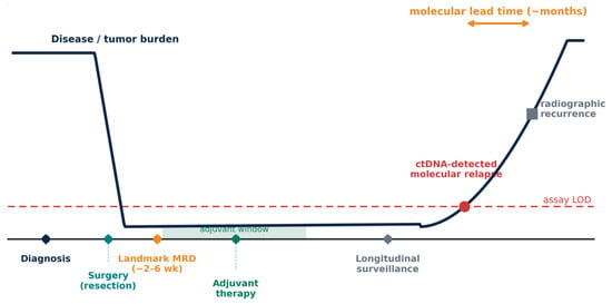
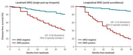
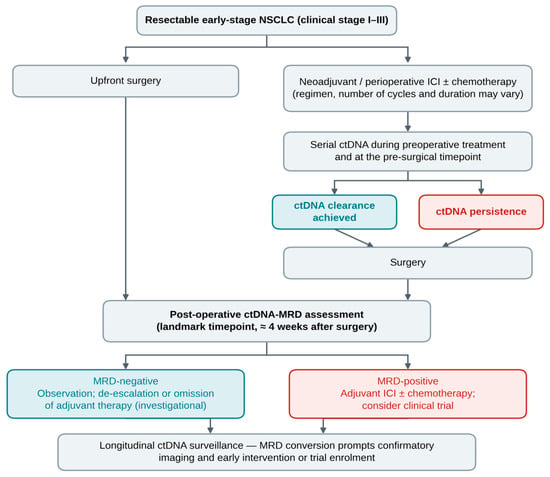
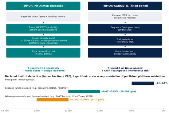
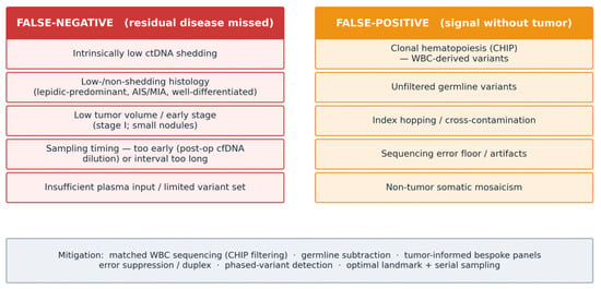
Complete surgical resection remains the cornerstone of curative therapy for early-stage non-small-cell lung cancer (NSCLC), yet a substantial proportion of patients relapse despite a macroscopically complete operation and negative conventional staging. This residual risk reflects micrometastatic disease that is below the resolution of computed tomography and that current clinicopathologic risk factors only coarsely predict. Circulating tumor DNA (ctDNA) provides a direct, blood-based readout of this molecular residual disease (MRD), its detection after definitive treatment is among the strongest known predictors of recurrence, and ctDNA frequently rises months before radiographic progression. In this narrative review, based on a literature search closed on 12 September 2026, we synthesize the foundational and translational literature underpinning ctDNA-MRD in resected NSCLC. We outline the biology of ctDNA shedding and metastatic dissemination established by serial-sampling and phylogenetic studies; the prognostic performance of landmark and longitudinal MRD, tabulated quantitatively across the major cohorts, meta-analyses and trial-nested analyses; the perioperative kinetics that dictate when sampling is informative; the analytical design of tumor-informed versus tumor-agnostic assays and the determinants of their sensitivity; and the biological and technical sources of false-negative and false-positive results, of which clonal hematopoiesis and low-shedding histologies are the most consequential. We then position MRD within the contemporary perioperative treatment landscape—adjuvant targeted therapy and immunotherapy, and neoadjuvant and perioperative chemoimmunotherapy—and review the emerging, still-investigational paradigm of MRD-guided escalation and de-escalation, including the early termination of the MRD-selected MERMAID trials. We conclude with the analytical, evidentiary and implementation barriers—and the current positions of guideline bodies and regulators—that separate a powerful prognostic biomarker from a validated predictive tool, and we set out the trial-design requirements needed to close that gap.
Full article
(This article belongs to the Special Issue A New Era in the Treatment of Early-Stage Non-Small Cell Lung Cancer (2nd Edition))
►▼
Show Figures

Figure 1

Open AccessArticle
Clinical Significance of Tumor Infiltrating Lymphocytes and Modulation by Neoadjuvant Chemotherapy in Colorectal Cancer Liver Metastases
by
Yasaman Rezaie, Kerryan Ashley, Janie Zhang, Thejal Srikumar, Jassim DiPalermo, Kurt A. Schalper and Michael Cecchini
Cancers 2026, 18(18), 3042; https://doi.org/10.3390/cancers18183042 (registering DOI) - 19 Sep 2026
Abstract
Background: Colorectal cancer liver metastases (CRCLMs) are associated with treatment resistance, and poor survival. We aimed to measure the differences in the immune composition of primary colorectal tumors and CRCLMs to identify prognostic and potentially actionable differences in the tumor microenvironment (TME).
[...] Read more.
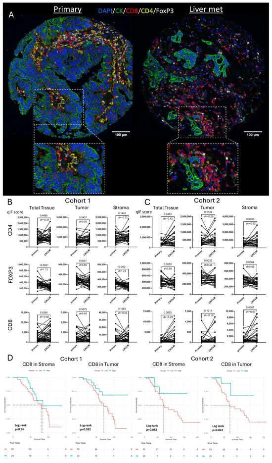
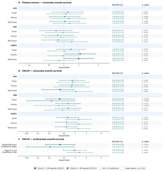
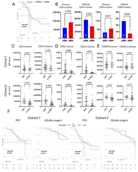
Background: Colorectal cancer liver metastases (CRCLMs) are associated with treatment resistance, and poor survival. We aimed to measure the differences in the immune composition of primary colorectal tumors and CRCLMs to identify prognostic and potentially actionable differences in the tumor microenvironment (TME). Methods: We created two independent cohorts of patients that had surgical resection of primary colorectal tumor and CRCLM resection at the Yale Cancer Center, comprising of 84 and 95 cases arranged in tissue microarray (TMA) format. Using multiplex quantitative immunofluorescence (QIF) we measured CD4+, CD8+, and FOXP3+ immune cells across spatially-resolved compartments within the tumor samples. We then assessed the association of clinicopathologic variables and treatment-specific outcomes with immune cell composition. Results: CRCLMs showed significantly higher CD8+ effector T-cells and significantly lower FOXP3+ regulatory T-cell infiltration than primary tumors. In addition, high CD8+ T-cell infiltration within the cancer cell compartment of CRCLMs was prominently associated with longer overall survival in both cohorts (Cohort1: HR = 0.38, p = 0.032; Cohort2: HR = 0.23, p = 0.046). Neoadjuvant chemotherapy (NAC) was associated with a significant increase in CD8+ T-cells across all tumor tissue regions in CRCLMs (p < 0.01 in both cohorts). However, NAC was not associated with improved three-year progression-free survival in either cohort (Cohort1: HR = 0.75, p = 0.43; Cohort2: HR = 0.97, p = 0.93). Conclusions: The TME of CRCLMs is characterized by reduced Tregs and increased TILs in NAC-treated patients. Furthermore, increased CD8+ infiltration in the tumoral compartment within CRCLMs is associated with improved survival. Our results describe local adaptive anti-tumor immune responses seen in CRCLM and underscore the need for novel therapeutic strategies that leverage TME alterations.
Full article
(This article belongs to the Section Cancer Metastasis)
►▼
Show Figures

Figure 1
Open AccessReview
Immune Checkpoint Inhibitor-Associated Cardiovascular Toxicity in Melanoma: Current Evidence and Practical Clinical Management
by
Andrea Bottardi, Gabriele Busin, Lam Nguyen, Paolo Del Fiore, Giorgia Alberti, Andrea Danese, Michele Nori, Raimondo Pittorru and Fortunato Cassalia
Cancers 2026, 18(18), 3041; https://doi.org/10.3390/cancers18183041 (registering DOI) - 19 Sep 2026
Abstract
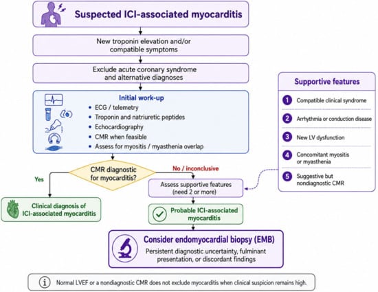
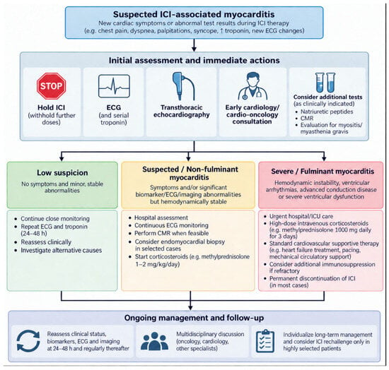
Immune checkpoint inhibitors (ICIs) have transformed the therapeutic landscape of advanced melanoma, significantly improving long-term survival. Nevertheless, these therapies may induce immune-related cardiovascular adverse events, which are uncommon but potentially life-threatening. Myocarditis is the best-characterized and most severe manifestation, although a broad spectrum
[...] Read more.
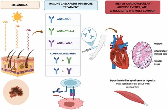
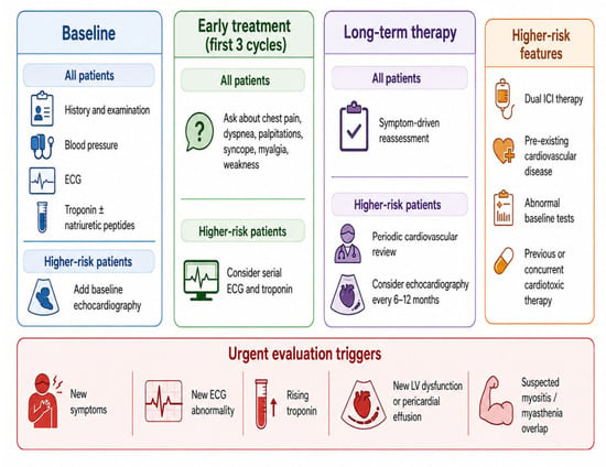
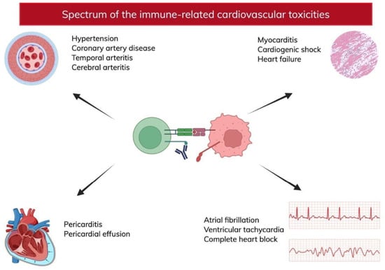
Immune checkpoint inhibitors (ICIs) have transformed the therapeutic landscape of advanced melanoma, significantly improving long-term survival. Nevertheless, these therapies may induce immune-related cardiovascular adverse events, which are uncommon but potentially life-threatening. Myocarditis is the best-characterized and most severe manifestation, although a broad spectrum of cardiovascular complications—including pericardial disease, arrhythmias, conduction abnormalities, heart failure, Takotsubo syndrome, and vascular events—has increasingly been recognized. Emerging evidence suggests that melanoma itself and dual immune checkpoint blockade may confer a higher risk of cardiotoxicity than other malignancies or single-agent immunotherapy. This narrative review summarizes current evidence regarding the epidemiology, pathophysiological mechanisms, clinical presentation, diagnostic evaluation, cardiovascular surveillance, and management of ICI-associated cardiotoxicity in patients with melanoma. Diagnosis relies on integrating clinical findings with electrocardiography, cardiac biomarkers, echocardiography, cardiac magnetic resonance imaging, and, in selected cases, endomyocardial biopsy. Prompt interruption of ICI therapy, early administration of high-dose corticosteroids, and escalation to additional immunosuppressive therapies in refractory cases remain the cornerstone of treatment. As the use of ICIs continues to expand, improving cardiovascular risk stratification and implementing evidence-based surveillance strategies will become increasingly important. A multidisciplinary cardio-oncology approach is essential to ensure early diagnosis, optimize cardiovascular outcomes, and preserve the anticancer efficacy of immunotherapy.
Full article
(This article belongs to the Special Issue New Strategies for the Treatment of Melanoma in Neoadjuvant and Adjuvant Settings)
►▼
Show Figures

Figure 1
Open AccessArticle
Clinical and Biological Factors Associated with Pseudoprogression Under Immune Checkpoint Inhibitors: A Retrospective Pan-Tumor Cohort Study
by
Amélie Toulet, Valérie Seegers, Frédéric Bigot, Sylvère Guillemois, Damien Vansteene, Rémy Delva and Manon de Vries-Brilland
Cancers 2026, 18(18), 3040; https://doi.org/10.3390/cancers18183040 (registering DOI) - 19 Sep 2026
Abstract
Background: Immune checkpoint inhibitors (ICIs) have transformed cancer treatment but may induce pseudoprogression (PP), an atypical response pattern that may mimic progressive disease (PD). Although iRECIST criteria were developed to characterize such atypical responses, distinguishing PP from PD when progression is first
[...] Read more.
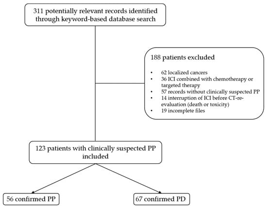
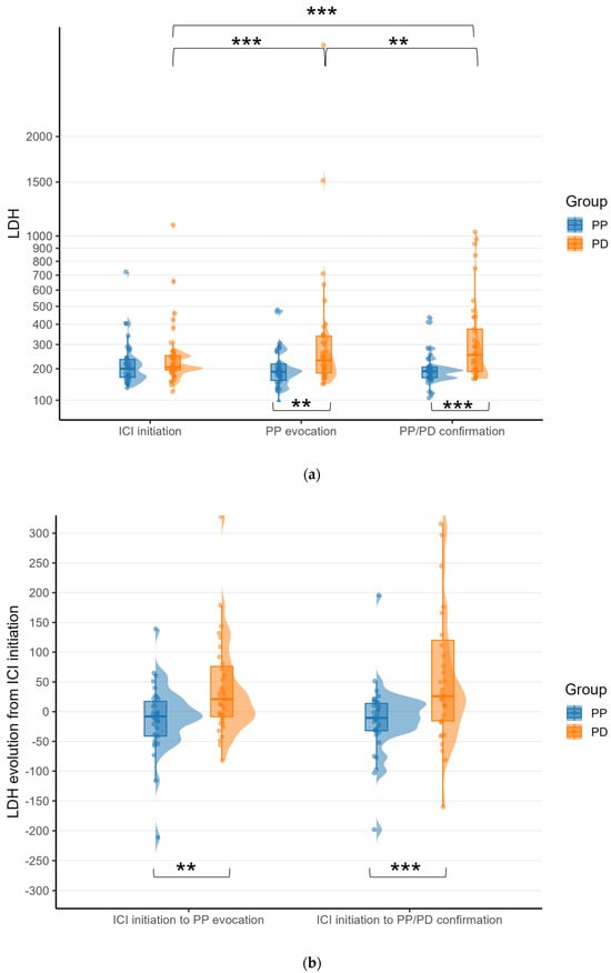
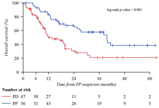
Background: Immune checkpoint inhibitors (ICIs) have transformed cancer treatment but may induce pseudoprogression (PP), an atypical response pattern that may mimic progressive disease (PD). Although iRECIST criteria were developed to characterize such atypical responses, distinguishing PP from PD when progression is first observed remains challenging. We aimed to characterize patients with clinically suspected PP and explore clinical and biological factors associated with subsequent classification as PP versus PD. Methods: We conducted a retrospective single-center study including patients with metastatic cancer treated with ICIs at the Institut de Cancérologie de l’Ouest (ICO) in whom PP was clinically suspected. Clinical, biological, treatment, and outcome data were collected at ICI initiation, PP suspicion, and subsequent PP/PD classification. Associations with PP versus PD were explored using univariable and multivariable logistic regression. Results: Among 123 patients with clinically suspected PP, 56 were subsequently classified as PP and 67 as PD. The most frequent primary tumors were lung (38%), kidney (24%), and bladder (11%) cancers. Median time from ICI initiation to PP suspicion was 79 days. ECOG performance status of 0 at ICI initiation was more frequent in the PP than in the PD group (55% vs. 32%; p = 0.046). At PP suspicion, median LDH levels were lower in the PP than in the PD group (189 vs. 232.5 U/L; p = 0.003). After multivariable adjustment, higher LDH levels at PP suspicion remained associated with PD (adjusted OR 2.78 per 100 U/L increase; 95% CI, 1.19–6.52; p = 0.019). Conclusions: In patients with clinically suspected PP, LDH may provide complementary information but cannot reliably distinguish PP from PD when considered alone. Confirmatory imaging therefore remains essential for treatment decision-making.
Full article
(This article belongs to the Section Cancer Immunology and Immunotherapy)
►▼
Show Figures

Figure 1
Open AccessReview
Therapeutic Time Migration in Hepatocellular Carcinoma: Rethinking the Timing of Systemic Therapy Across the Disease Continuum
by
Fausto Meriggi, Ester Oneda, Sara Cherri and Alberto Zaniboni
Cancers 2026, 18(18), 3039; https://doi.org/10.3390/cancers18183039 (registering DOI) - 19 Sep 2026
Abstract
The management of hepatocellular carcinoma has traditionally followed a stage-dependent sequence in which surgery or thermal ablation is prioritized for early disease, transarterial approaches are used for intermediate-stage tumors, and systemic treatment is introduced after progression to advanced disease or loss of suitability
[...] Read more.
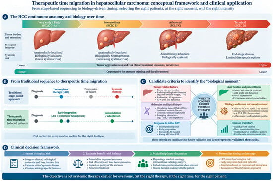
The management of hepatocellular carcinoma has traditionally followed a stage-dependent sequence in which surgery or thermal ablation is prioritized for early disease, transarterial approaches are used for intermediate-stage tumors, and systemic treatment is introduced after progression to advanced disease or loss of suitability for locoregional therapy. This paradigm was developed when systemic treatments had limited antitumor activity. Modern immune checkpoint inhibitors and immune–antiangiogenic combinations have substantially changed these premises. Randomized trials now demonstrate clinical benefit when systemic treatment is integrated with transarterial chemoembolization before conventional locoregional failure, while perioperative studies suggest that systemic treatment can also be moved into selected high-risk resectable disease. Conversely, updated adjuvant data indicate that simply administering systemic therapy at an earlier stage does not necessarily improve outcome after complete tumor eradication. We propose the concept of therapeutic time migration, whereby systemic treatment is introduced at an earlier biological point in selected patients, while hepatic reserve is preserved, occult systemic risk is becoming clinically relevant, and effective local treatment remains feasible. We explicitly distinguish this framework from the established concept of treatment stage migration and therapeutic hierarchy. This framework emphasizes treatment sequence rather than treatment escalation and suggests that timing itself may function as a dynamic therapeutic biomarker. Prospective studies should determine whether changing the order of systemic and locoregional therapy can modify immune activation, preserve liver function, increase conversion to definitive local treatment, and ultimately improve survival.
Full article
(This article belongs to the Section Cancer Therapy)
►▼
Show Figures

Figure 1
Open AccessReview
Primary Cutaneous Lymphomas and Lymphoproliferative Disorders: Current Insights for Classification and Management
by
Uğur Hatipoğlu, Mert Seyhan, Hakan Eminoglu, Turgay Ulaş and Mehmet Sinan Dal
Cancers 2026, 18(18), 3038; https://doi.org/10.3390/cancers18183038 (registering DOI) - 19 Sep 2026
Abstract
Background: Primary cutaneous lymphomas (PCLs) represent a heterogeneous group of extranodal non-Hodgkin lymphomas with distinct clinical, histological, and molecular profiles. Recent advancements in genomic profiling have led to significant revisions in diagnostic frameworks and therapeutic paradigms. Objective: This review aims to summarize the
[...] Read more.
Background: Primary cutaneous lymphomas (PCLs) represent a heterogeneous group of extranodal non-Hodgkin lymphomas with distinct clinical, histological, and molecular profiles. Recent advancements in genomic profiling have led to significant revisions in diagnostic frameworks and therapeutic paradigms. Objective: This review aims to summarize the current landscape of primary cutaneous B-cell and T-cell lymphomas and lymphoproliferative disorders (LPDs), focusing on the diagnostic updates in the WHO 5th edition and the International Consensus Classification (ICC 2022), alongside evolving risk-adapted treatment strategies. Methods: A comprehensive review of the recent literature and major international consensus guidelines was conducted, evaluating diagnostic shifts, disease reclassifications, and novel therapeutic interventions across various PCL subtypes. Results: The latest WHO 5th and ICC 2022 classifications emphasize the clinical behavior of indolent entities, shifting terminology from “lymphoma” to “lymphoproliferative disorder” for conditions such as primary cutaneous acral CD8+ T-cell LPD and primary cutaneous CD4+ small/medium T-cell LPD to prevent overtreatment. Several previously provisional categories (e.g., primary cutaneous gamma/delta T-cell lymphoma and EBV-positive mucocutaneous ulcer) have achieved definitive diagnostic status. Parallel to these revisions, therapeutic management has shifted toward toxicity-reducing, risk-adapted strategies. For indolent subtypes, ultra-low-dose radiotherapy (4 Gy) and skin-directed therapies remain highly effective. In aggressive or refractory subtypes, such as Mycosis Fungoides and primary cutaneous diffuse large B-cell lymphoma, leg type (PCDLBCL, LT), precision oncology—utilizing targeted agents (e.g., brentuximab vedotin, mogamulizumab, lenalidomide, ibrutinib) and cellular/immunotherapies—is rapidly changing outcomes. Conclusions: Integrating the updated diagnostic classifications with modern risk-stratified and targeted treatment paradigms is crucial for optimizing clinical management and avoiding overtreatment in indolent PCLs while improving survival in aggressive subtypes.
Full article
(This article belongs to the Section Cancer Therapy)
Open AccessArticle
CD84 Expression Across Disease Stages and Leukemic Subpopulations in Acute Myeloid Leukemia
by
Beatriz Martín-Herreros, Lourdes Cordón, Rebeca Rodríguez-Veiga, Isabel Cano-Ferri, Evelyn Acuña-Cruz, Laura Torres-Miñana, Irene Navarro, Pilar Lloret-Madrid, Amparo Sempere, María Dolores Linares, Sara Torres-Sánchez, Elisa González-Romero, Nela Klein-González, Lorena Pérez-Amill, Leonor Senent, José Luis Poveda-Andrés, Javier De La Rubia, Pau Montesinos and Manuel Guerreiro
Cancers 2026, 18(18), 3037; https://doi.org/10.3390/cancers18183037 (registering DOI) - 18 Sep 2026
Abstract
Background/Objectives: In recent years, increasing evidence has supported CD84 as a promising therapeutic target in acute myeloid leukemia (AML), including its potential application in CAR T-cell therapy. CD84 is consistently reported to be highly expressed on AML cells in both adult and pediatric
[...] Read more.
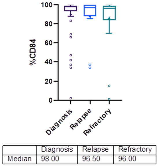
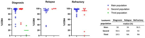
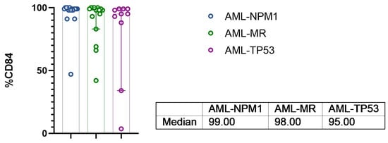
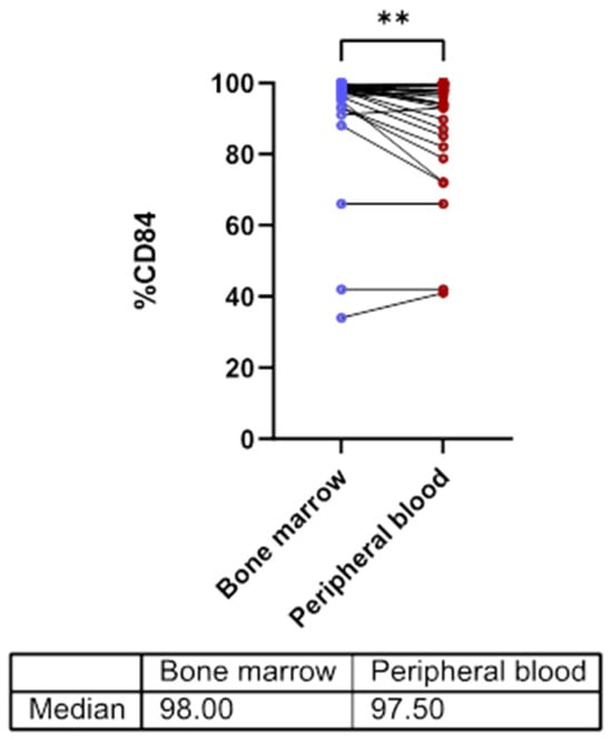
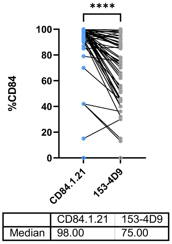
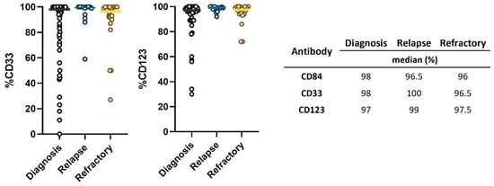
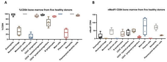
Background/Objectives: In recent years, increasing evidence has supported CD84 as a promising therapeutic target in acute myeloid leukemia (AML), including its potential application in CAR T-cell therapy. CD84 is consistently reported to be highly expressed on AML cells in both adult and pediatric patients, providing a strong biological rationale for the development of CD84-directed immunotherapies. Methods: We characterized CD84 expression in bone marrow and peripheral blood samples from 61 adult patients with AML at diagnosis, relapse, and refractory disease using multiparametric flow cytometry. Results: Our results demonstrate that CD84 is highly expressed on AML cells throughout the course of the disease. More than 80% of leukemic populations showed CD84 expression above 80%, with median expression levels remaining consistently high at diagnosis (98%), relapse (96.5%), and refractory disease (96%). A novel aspect of our study is the evaluation of CD84 expression across distinct leukemic subpopulations within individual samples; only three of the 94 leukemic populations analyzed showed CD84 expression below 20%. Similarly, CD84 expression remained consistently high across the major AML subtypes analyzed, including AML with NPM1 mutation, AML with myelodysplasia-related gene mutations, and TP53-mutated AML. Comparable median CD84 expression was observed in bone marrow and peripheral blood; however, paired analysis identified significant differences between both compartments. CD84 expression assessed with antibody clone 153-4D9 was significantly lower than that detected with clone CD84.1.21 across AML leukemic populations (median, 75% vs. 98%; p < 0.0001). When compared with CD33 and CD123, CD84 showed a similarly broad prevalence of expression across AML leukemic populations. Healthy bone marrow samples were additionally evaluated to characterize the physiological distribution of CD84 expression across normal hematopoietic populations and provide a framework for the assessment of CD84-targeted therapeutic strategies. Conclusions: Together with the available preclinical and clinical evidence, these findings support the continued evaluation of CD84-directed immunotherapies, including CAR T-cell therapy, in AML.
Full article
(This article belongs to the Special Issue Advances in Immunotherapy for Acute Myeloid Leukemia)
►▼
Show Figures

Figure 1
Open AccessReview
Ovarian Precancerous Lesions and Their Molecular Landscape: A Structured Scoping Review Focused on STIC and Related Precursors of Epithelial Ovarian Cancer
by
Katarzyna Kwas-Sarnacka, Wiktoria Lisińska, Edward Koziróg, Kinga Stanek, Daniel Wolder, Maria Szubert and Jacek Wilczyński
Cancers 2026, 18(18), 3036; https://doi.org/10.3390/cancers18183036 (registering DOI) - 18 Sep 2026
Abstract
Ovarian cancer remains one of the deadliest gynecologic malignancies because of late-stage diagnosis, biological heterogeneity, and the lack of effective early detection strategies. Epithelial ovarian cancer (EOC) comprises several histotypes, including high-grade serous carcinoma (HGSC), low-grade serous carcinoma (LGSC), endometrioid ovarian carcinoma (ENOC),
[...] Read more.
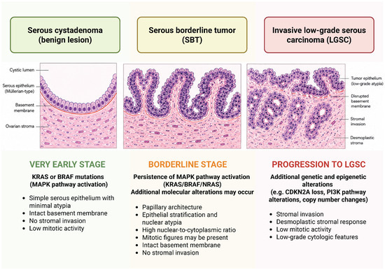
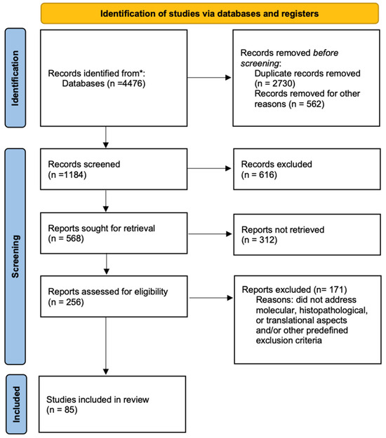
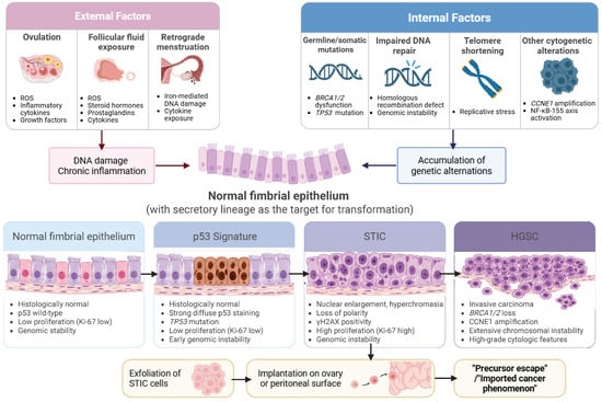
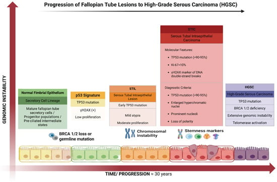
Ovarian cancer remains one of the deadliest gynecologic malignancies because of late-stage diagnosis, biological heterogeneity, and the lack of effective early detection strategies. Epithelial ovarian cancer (EOC) comprises several histotypes, including high-grade serous carcinoma (HGSC), low-grade serous carcinoma (LGSC), endometrioid ovarian carcinoma (ENOC), clear cell ovarian carcinoma (CCOC), and mucinous ovarian carcinoma (MOC), each with distinct molecular pathways and precursor lesions. In recent years, serous tubal intraepithelial carcinoma (STIC) has emerged as the principal precursor lesion of HGSC, supporting the fallopian tube origin paradigm of ovarian carcinogenesis. This structured scoping review summarizes current evidence on the molecular and cellular origins of epithelial ovarian cancer, with particular emphasis on precursor lesions, including p53 signatures, SCOUTs, STILs, cortical inclusion cysts, endometriosis-associated lesions, and borderline ovarian tumors. Particular attention is given to the molecular events that drive progression toward invasive carcinoma, including TP53 mutations, BRCA1/2 dysfunction, homologous recombination deficiency, chromosomal instability, telomere shortening, cell-cycle dysregulation, stemness-associated pathways, and inflammatory signaling. A structured literature search was conducted in PubMed/MEDLINE, Scopus, and Web of Science to identify studies published from 2000 to 2026 that addressed ovarian cancer precursor lesions, molecular pathogenesis, and early carcinogenic mechanisms. Articles were selected for relevance to epithelial ovarian carcinogenesis, precursor biology, and translational implications. Current evidence supports substantial biological heterogeneity among precursor lesions, particularly STICs, with only a subset likely to progress to invasive HGSC. Emerging molecular approaches, including genome-wide aneuploidy profiling and integrated genomic classifiers, may improve risk stratification and facilitate earlier detection of clinically significant precursor lesions. A better understanding of precursor biology may ultimately contribute to personalized prevention strategies and improved clinical outcomes in ovarian cancer.
Full article
(This article belongs to the Special Issue Advances in Pathogenesis and Pathology Diagnosis of Gynecologic Cancers)
►▼
Show Figures

Figure 1
Open AccessReview
Circulating microRNAs at the Interface of Obesity and Breast Cancer: From Molecular Machinery, Diagnostic Potential, and Therapeutic Perspectives
by
Georgina Victoria-Acosta, Stephanie I. Núñez-Olvera, María Elizbeth Alvarez-Sánchez, Yarely M. Salinas-Vera and Jonathan Puente-Rivera
Cancers 2026, 18(18), 3035; https://doi.org/10.3390/cancers18183035 (registering DOI) - 18 Sep 2026
Abstract
Obesity influences breast cancer risk, progression, and therapeutic response through endocrine, inflammatory, metabolic, stromal, and vesicular mechanisms. Circulating and extracellular vesicle (EV)-associated microRNAs are attractive liquid biopsy candidates; however, in obesity-associated breast cancer, they should not be interpreted as tumor-only biomarkers. This narrative
[...] Read more.
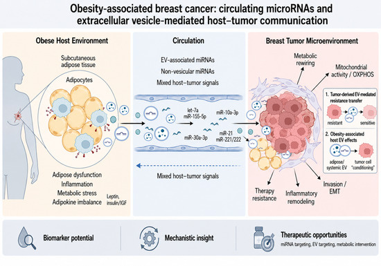
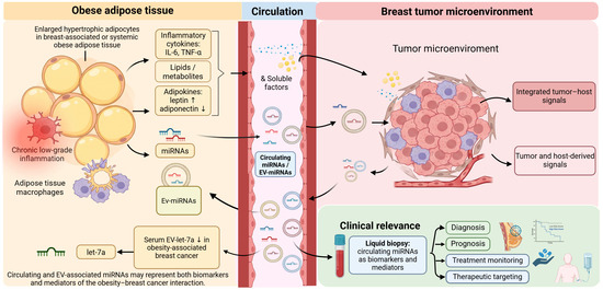
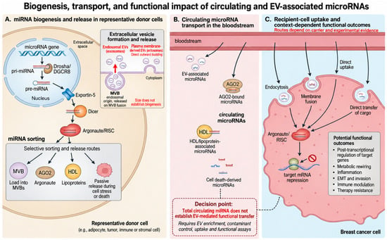
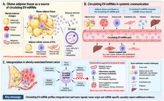
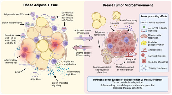
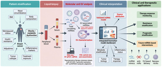
Obesity influences breast cancer risk, progression, and therapeutic response through endocrine, inflammatory, metabolic, stromal, and vesicular mechanisms. Circulating and extracellular vesicle (EV)-associated microRNAs are attractive liquid biopsy candidates; however, in obesity-associated breast cancer, they should not be interpreted as tumor-only biomarkers. This narrative review analyzes circulating/EV-miRNAs as mixed host–tumor signals and separates association-based biomarker evidence from functional EV-mediated transfer. We performed a narrative and integrative review of studies on miRNA biogenesis, circulating miRNA transport, EV biology, adipose tissue dysfunction, adipose-tumor communication, biomarker development, therapy response, and methodological reporting standards. The current literature is read along two independent axes: human relevance and source attribution, from non-breast models to obesity-stratified human cohorts, and mechanistic causality, from association only to EV-level function, miRNA-level function, and causality with rescue; clinical validation is treated as a separate qualifier rather than as the top of a single scale. For example, EV-let-7a in overweight/obese breast cancer patients mainly supports an obesity-stratified biomarker candidate. Breast adipose tissue-derived EV cargoes enriched in miR-155-5p, miR-10a-3p, and miR-30a-3p provide stronger functional support for metabolic reprogramming, whereas EV-associated miR-221/222 has mediator-level evidence in endocrine resistance but is not obesity specific. Conclusions: The main contribution of this review is an operational interpretation framework in which circulating miRNAs are evaluated according to probable source, evidence level, and clinical purpose. Future studies should report EV workflow quality, sample type, hemolysis and platelet control, normalization strategy, external validation, tumor subtype, and obesity phenotyping beyond BMI, including waist circumference or waist-to-hip ratio, insulin resistance, diabetes, menopausal status, and inflammatory markers.
Full article
(This article belongs to the Special Issue Breast Cancer Microenvironment: Molecular Mechanisms and Therapeutic Targeting)
►▼
Show Figures

Graphical abstract
Open AccessArticle
Somatic Mutation Profile in Normal Tissue Adjacent to Tumor in Colorectal Carcinoma Patients: Functional Effect on Gene Expression and Potential Implications for Precision Medicine
by
Farzana Jasmine, Daniil Vasiljevs, Armando Almazan, Habibul Ahsan and Muhammad G. Kibriya
Cancers 2026, 18(18), 3034; https://doi.org/10.3390/cancers18183034 (registering DOI) - 18 Sep 2026
Abstract
Background: The somatic mutation (SM) profile of normal colon tissue in colorectal carcinoma (CRC) patients is not well-known. We examined (a) SMs in resected normal colonic tissue adjacent to tumors from non-metastatic CRC patients, (b) identified potentially malignant mutations, and (c) their functional
[...] Read more.
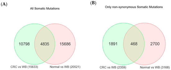
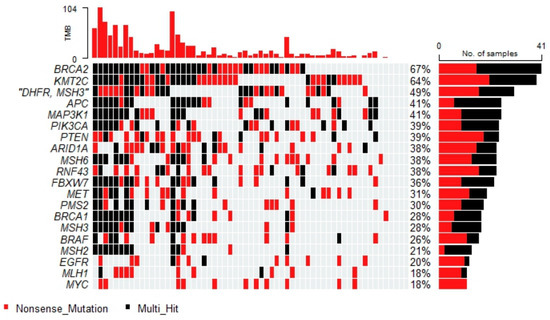
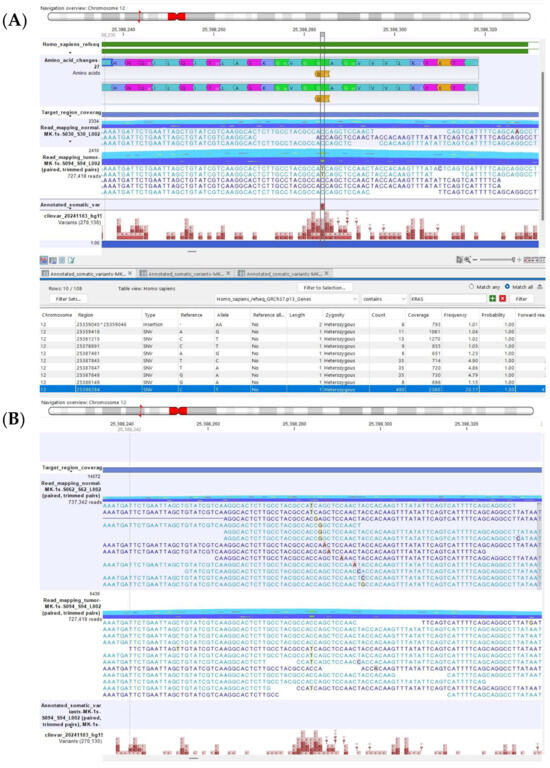
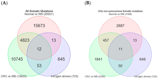
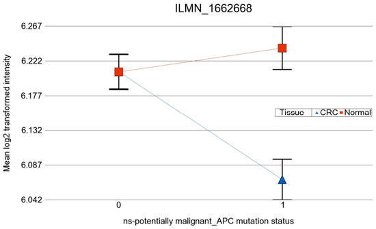
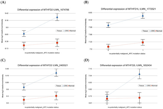
Background: The somatic mutation (SM) profile of normal colon tissue in colorectal carcinoma (CRC) patients is not well-known. We examined (a) SMs in resected normal colonic tissue adjacent to tumors from non-metastatic CRC patients, (b) identified potentially malignant mutations, and (c) their functional effects and utility for precision medicine. Materials and Methods: Surgically resected fresh paired tumor and adjacent normal colon tissues from 61 patients were used as sources of tissue DNA. Whole blood (WB) DNA was used as a germline reference. Next-generation sequencing using a custom hybridization capture panel was performed on each. Differential gene expression was assessed and stratified by mutation status in normal tissue. Results: Compared to WB DNA, similar numbers of SMs were detected in tumor and adjacent normal colon tissue. Of all SMs found in adjacent tissue, more than 23% of those same mutations were also detected in the matched tumor. Of the 61 patients, 57 (93.4%) had at least one potentially malignant non-synonymous SM in their normal tissue. The most frequently involved genes were BRCA2 (67% of the patients), KMT2C (64%), DHFR (49%), APC (41%), MAP3K1 (41%), PIK3CA (39%), PTEN (39%), ARID1A (38%), and MSH6 (38%). The use of WB as a germline reference was important, as using only adjacent normal tissue as a reference caused false negatives for mutations shared between tumor and normal tissue. Mutation status in normal tissue was associated with differential gene expressions in matched tumors. APC mutations were associated with more marked down-regulation of APC, SLC26A3, SELENBP1, KRT20, and CXCL14, and more marked up-regulation of MMP1, MMP12, MTHFD1L and MTHFD2, the latter two suggesting the folate pathway as a potential therapeutic target. Conclusions: For the first time, we document a comprehensive SM profile of normal colon tissue adjacent to tumors in CRC patients and observe its functional effect on differential gene expression in matched tumor tissue to identify potential therapeutic applications in precision medicine.
Full article
(This article belongs to the Special Issue Oncogenetics of Colorectal Cancer (2nd Edition))
►▼
Show Figures

Figure 1
Open AccessArticle
Early Longitudinal Changes in Radiomic Tumor Heterogeneity Predict Progression-Free Survival in Advanced Non-Small Cell Lung Cancer
by
Zachary Thompson, Junmin Whiting, Olyo Stringfield, Mahmoud Abdalah, Sebastian Viracacha, Jhanelle Gray, Andreas Saltos and Dung-Tsa Chen
Cancers 2026, 18(18), 3033; https://doi.org/10.3390/cancers18183033 (registering DOI) - 18 Sep 2026
Abstract
Radiomic features derived from longitudinal imaging offer a non-invasive approach to quantify tumor heterogeneity, but their integration with clinical variables in small cohorts remains methodologically challenging. We analyzed 23 patients with advanced non-small cell lung cancer (NSCLC) enrolled in a Phase I study
[...] Read more.
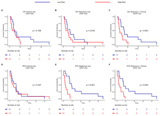
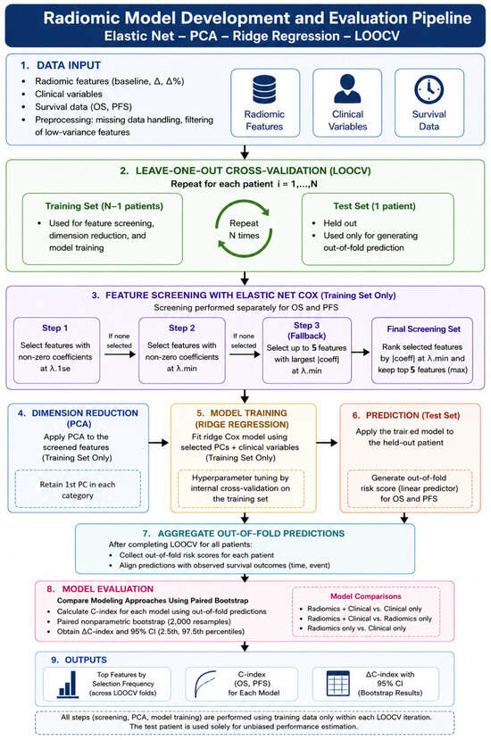
Radiomic features derived from longitudinal imaging offer a non-invasive approach to quantify tumor heterogeneity, but their integration with clinical variables in small cohorts remains methodologically challenging. We analyzed 23 patients with advanced non-small cell lung cancer (NSCLC) enrolled in a Phase I study of pembrolizumab and vorinostat. Radiomic features were extracted from baseline and two-month follow-up computed tomography scans, aggregated to the patient level, and transformed into delta features representing early changes over time. A unified modeling framework was implemented using strict leave-one-out cross-validation (LOOCV), incorporating both radiomic and clinical variables. Within each training fold, outcome-guided feature screening was performed using elastic net penalized Cox regression, followed by category-aware principal component analysis and ridge-penalized Cox modeling. Model performance was evaluated using out-of-fold concordance indices for overall survival (OS) and progression-free survival (PFS). Radiomics-only models demonstrated moderate discrimination for OS (C-index 0.609) and favorable discrimination for PFS (C-index 0.770), whereas clinical-only models showed weaker performance (OS 0.605; PFS 0.571). The combined radiomics and clinical model demonstrated numerically higher discrimination without statistically distinguishable differences for OS (C-index 0.648) while maintaining favorable discrimination for PFS (C-index 0.718). Stable features were predominantly texture-based and included Laws filter and co-occurrence-derived metrics associated with spatial heterogeneity. In this exploratory analysis, early changes in radiomic features reflecting tumor heterogeneity suggest potential associations with survival outcomes in advanced NSCLC. Integration of radiomic and clinical variables showed trends toward improved performance for overall survival while maintaining favorable discrimination for progression-free survival. These findings support further investigation of longitudinal radiomic features as candidate imaging biomarkers in larger, independently validated cohorts.
Full article
(This article belongs to the Section Cancer Biomarkers)
►▼
Show Figures

Figure 1
Open AccessArticle
Circulating Tumor DNA (ctDNA) Clearance After Immune Checkpoint Inhibition (ICI) with Radiotherapy (RT) as a Prognostic Biomarker for Advanced Melanoma
by
Alyssa K. Steimle, Amir M. Forati, Alice Y. Zhou, Trevor McCracken, Meena M. Hosny, Golbarg Rahimi, Janmesh D. Patel, Caroline Burkey, Jessica Caraway, Taylor Huppe, Andrew Wood, Madison S. Harris, Alexander Birbrair, Jose M. Ayuso, Deepak M. Sahasrabudhe, Fauzia Hollnagel, Gary C. Doolittle, Adam R. Burr, Nina S. Mathew, Gino K. In, Adrienne Victor and Vincent T. Maadd
Show full author list
remove
Hide full author list
Cancers 2026, 18(18), 3032; https://doi.org/10.3390/cancers18183032 (registering DOI) - 18 Sep 2026
Abstract
Background/Objectives: ctDNA is evolving as an important biomarker for prognostic assessment in advanced melanoma. However, the utility of ctDNA monitoring during concurrent ICI with RT remains incompletely explored. Methods: In this multicenter retrospective study, patients with unresectable stage III/IV melanoma treated with ICI
[...] Read more.
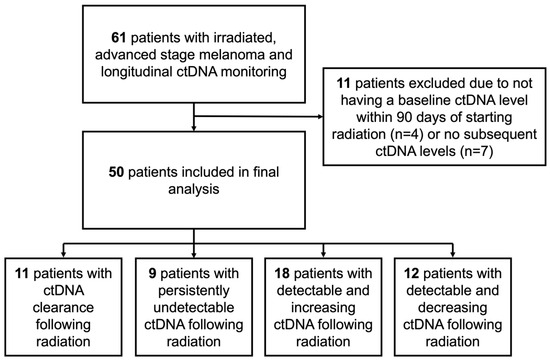
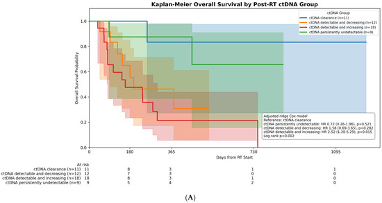
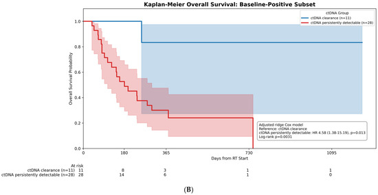
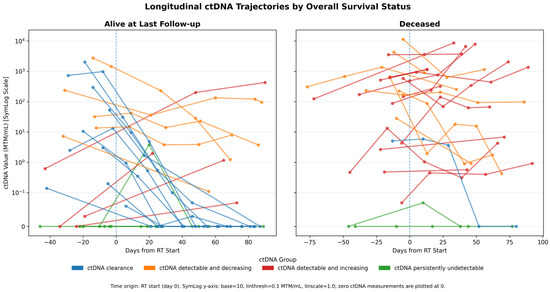
Background/Objectives: ctDNA is evolving as an important biomarker for prognostic assessment in advanced melanoma. However, the utility of ctDNA monitoring during concurrent ICI with RT remains incompletely explored. Methods: In this multicenter retrospective study, patients with unresectable stage III/IV melanoma treated with ICI and palliative RT were identified. Patients had retrospectively collected, tumor-informed, exome-based, ctDNA monitoring within 3 months following RT. Results: A total of 50 patients treated with ICI and palliative RT were analyzed. Compared with ctDNA clearance, ctDNA detectable and increasing was associated with worse OS (adjusted HR 2.52, 95% CI 1.20–5.29; p = 0.015), whereas ctDNA detectable and decreasing was not significantly different from ctDNA clearance (adjusted HR 1.58, 95% CI 0.69–3.65; p = 0.282). One-year OS was 87.5% with persistently undetectable ctDNA, 83.3% for ctDNA clearance, 46.3% for detectable and decreasing ctDNA, and 21.4% for detectable and increasing ctDNA. In a multivariable analysis, detectable and increasing ctDNA was associated with worse OS compared with ctDNA clearance (HR 2.52, 95% CI, 1.20–5.29, p = 0.015). Conclusions: Among patients with advanced melanoma treated with ICI and palliative RT, ctDNA clearance is associated with significantly improved OS compared to patients with detectable and increasing ctDNA within 3 months following RT. Ongoing prospective studies are needed to understand the role of ctDNA in this population.
Full article
(This article belongs to the Special Issue Melanoma Combination Therapy: New Horizons, Challenges and Clinical Breakthroughs)
►▼
Show Figures

Figure 1

Open AccessArticle
External Validation of the ARCAD Nomogram in a Real-World Cohort with Stage 4 Colorectal Cancer
by
James Yu, Jiannong Li, Michael J. Schell, Pablo Gonzalez Ginestet, John Raymond Zalcberg, R John Simes, Ian Marschner, Richard D. Kim and Katrin Marie Sjoquist
Cancers 2026, 18(18), 3031; https://doi.org/10.3390/cancers18183031 (registering DOI) - 18 Sep 2026
Abstract
Introduction: The ARCAD-CRC nomogram predicts 1-year survival in clinical trial populations with stage 4 colorectal cancer (CRC). This study evaluated its performance in a real-world cohort. Methods: Patients with stage 4 CRC were identified from the Flatiron database. A multivariable Cox proportional hazards
[...] Read more.
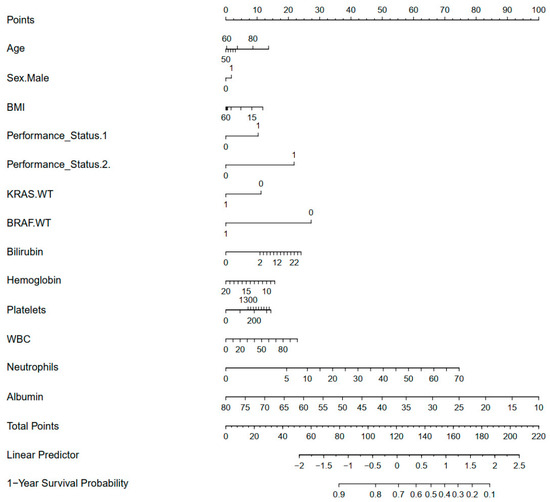
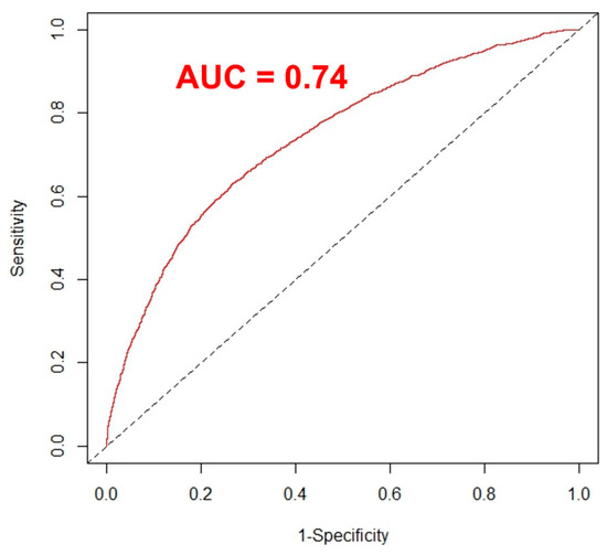
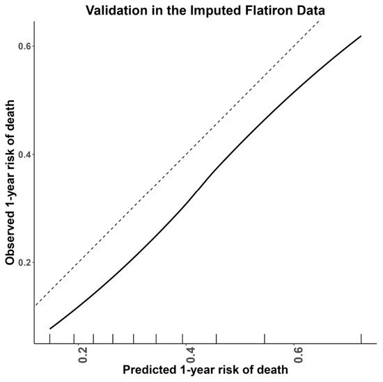
Introduction: The ARCAD-CRC nomogram predicts 1-year survival in clinical trial populations with stage 4 colorectal cancer (CRC). This study evaluated its performance in a real-world cohort. Methods: Patients with stage 4 CRC were identified from the Flatiron database. A multivariable Cox proportional hazards model for overall survival was applied to fit the reduced model derived from the published full ARCAD nomogram, as some variables were unavailable in the database. Validation of the reduced model used C-index, while predictive performance was assessed using time-dependent ROC analysis and calibration in the Flatiron real-world cohort (FRWC). Results: A total 9710 patients, including 5740 deceased, were analyzed. The FRWC was older (64 vs. 62 years) and had poorer ECOG-PS (15% vs. 4% with ECOG 2+) compared to the ARCAD cohort. The reduced nomogram’s calibration was comparable to the full model (C-index 0.67 vs. 0.68). In the FRWC, the reduced nomogram showed good discrimination, with time-dependent AUROC at the 12-month time horizon of 0.741 (95% CI 0.729–0.753). Calibration plots indicated the reduced ARCAD-CRC nomogram performed quantitively well with a calibration slope of 1.010 (95% CI 0.966–1.054). The calibration intercept was −0.160 (95% CI −0.189, −0.131), indicating systematic underestimation of 1-year survival in the FRWC. The predicted and observed 1-year OS using the reduced ARCAD nomogram in the FRWC were 0.640 (95% CI 0.637, 0.644) and 0.694 (95% CI 0.684–0.703), respectively. Conclusions: Our findings show that the ARCAD-CRC nomogram is a useful tool for predicting 1-year survival in a real-world cohort. The observed underestimation of survival may be due to advances in CRC treatments, as the FRWC cohort (2013–2020) was collected later than the original ARCAD cohort (1997–2012).
Full article
(This article belongs to the Section Clinical Research in Cancer)
►▼
Show Figures

Figure 1
Open AccessArticle
Tumor Inflammation Confounds Radiographic Determination of Extranodal Extension in HPV-Associated Head and Neck Squamous Cell Carcinoma
by
Sri Vemulamanda, Yasine Mirmozaffari, Alan H. Zhao, Noman Khan, Wendell G. Yarbrough, Natalia Issaeva, Benjamin Y. Huang and Travis P. Schrank
Cancers 2026, 18(18), 3030; https://doi.org/10.3390/cancers18183030 (registering DOI) - 18 Sep 2026
Abstract
Background/Objectives: Pathologically identified extranodal extension (pENE) predicts outcome and guides the intensity of adjuvant therapy in HPV-associated HNSCC. ENE determined by imaging (iENE) is a criterion for upstaging HPV+ HNSCC in the AJCC Cancer Staging System, 9th edition; however, its prognostic value
[...] Read more.
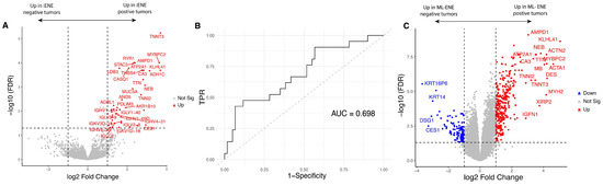
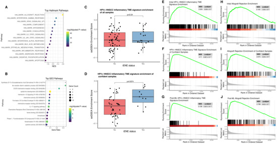
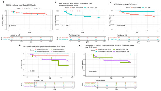
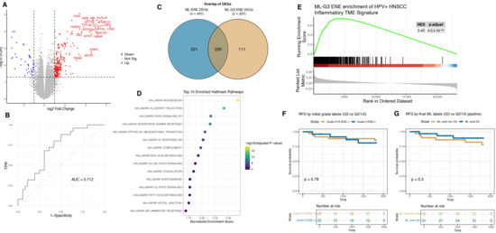
Background/Objectives: Pathologically identified extranodal extension (pENE) predicts outcome and guides the intensity of adjuvant therapy in HPV-associated HNSCC. ENE determined by imaging (iENE) is a criterion for upstaging HPV+ HNSCC in the AJCC Cancer Staging System, 9th edition; however, its prognostic value is less than that of pENE, varies between studies, and the correlation between pENE and iENE is less than optimal. This study explores biological characteristics of tumors that can decrease the predictive value of iENE. Methods: Pre-treatment CT scans from patients with HPV-associated HNSCC were evaluated for iENE status. RNA-Seq data from primary tumor samples were analyzed using edgeR v4.6.2 and transcriptional differences were categorized by ssGSEA. Machine learning was applied to refine gene expression changes associated with iENE and survival was analyzed with log-rank tests. Results: Recurrence-free survival did not differ between patients with and without iENE. Both ML-ENE status and enrichment for inflammatory signaling were associated with improved RFS (p < 0.01 and p < 0.0001). When stratified by the ML-ENE signature or the inflammatory signature, iENE status did not further separate outcomes, indicating that its prognostic information was largely captured by these gene sets. These stratified comparisons were exploratory and limited by subgroup size. Conclusions: These analyses suggest that tumor biology associated with an inflamed tumor microenvironment may confound determination of ENE by imaging and that accounting for tumors with inflammatory signaling could improve the correlation of iENE with outcomes.
Full article
(This article belongs to the Special Issue Surgery and Clinical Outcomes in Head and Neck Squamous Cell Carcinoma)
►▼
Show Figures

Figure 1
Open AccessReview
Contemporary Multimodal Management of Cutaneous Malignancies: Surgery, Radiotherapy, Systemic Therapy, Molecular Targeting and Immunotherapy
by
Dariusz Kowalczyk, Jolanta Jaworek, Andrzej Ciborek, Pawel Antoni Kolodziejski, Ewa Pruszyńska-Oszmałek and Hanna Krauss
Cancers 2026, 18(18), 3029; https://doi.org/10.3390/cancers18183029 (registering DOI) - 18 Sep 2026
Abstract
Background: The therapeutic management of cutaneous malignancies—cutaneous melanoma, cutaneous squamous-cell carcinoma (cSCC), basal-cell carcinoma (BCC) and Merkel-cell carcinoma (MCC)—has shifted toward multimodal, stage-adapted treatment integrating surgery, radiotherapy, molecularly targeted therapy and immune checkpoint inhibition. Methods: We conducted a structured narrative review with a
[...] Read more.
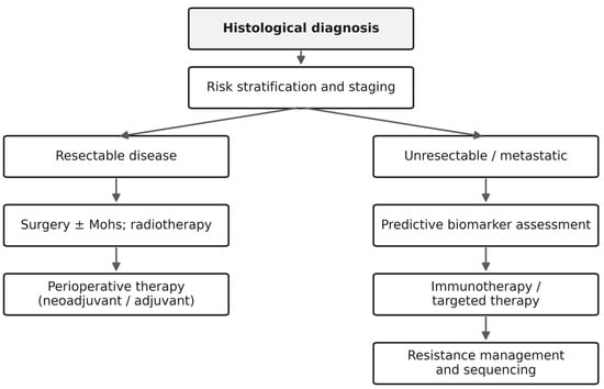
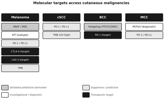
Background: The therapeutic management of cutaneous malignancies—cutaneous melanoma, cutaneous squamous-cell carcinoma (cSCC), basal-cell carcinoma (BCC) and Merkel-cell carcinoma (MCC)—has shifted toward multimodal, stage-adapted treatment integrating surgery, radiotherapy, molecularly targeted therapy and immune checkpoint inhibition. Methods: We conducted a structured narrative review with a structured literature search of PubMed/MEDLINE, Scopus and Web of Science (January 2015–6 September 2026), prioritising current clinical practice guidelines, randomised controlled trials and phase II–III studies; landmark older trials were included separately. Synthesis: Surgery with adequate histological margins, including Mohs micrographic surgery where appropriate, remains definitive for localised disease. In resectable macroscopic stage III melanoma, neoadjuvant ipilimumab plus nivolumab with response-driven postoperative management improved event-free survival versus adjuvant nivolumab in NADINA. SWOG S1801 independently demonstrated an event-free-survival advantage for perioperative pembrolizumab over adjuvant-only pembrolizumab in resectable stage III–IV melanoma. In advanced melanoma, dual immune-checkpoint strategies and BRAF/MEK inhibition require patient-specific sequencing; in the European Union, nivolumab plus relatlimab is authorised for first-line advanced melanoma with tumour-cell PD-L1 expression <1%. In high-risk cSCC after surgery and radiotherapy, adjuvant cemiplimab improved disease-free survival. In advanced BCC, Hedgehog-pathway inhibitors remain the principal first systemic option, with anti-PD-1 therapy after HHI failure or intolerance. Clinical implications: Molecular testing should be indication-driven: BRAF V600 is a validated treatment-selecting biomarker, while broader NGS profiling (including NRAS, KIT, NF1, TERT and co-occurring alterations), PD-L1, tumour mutational burden and circulating tumour DNA have context-dependent, supportive or investigational roles. Conclusions: The major unresolved issues are optimal sequencing, response-adapted perioperative treatment, resistance, biomarker validation and treatment of transplant, frail and other under-represented populations.
Full article
(This article belongs to the Section Cancer Therapy)
►▼
Show Figures

Figure 1
Open AccessArticle
Standardized Upstream Processing of Ovarian Cancer Surgical Specimens for TIL-Oriented Cellular Workflows
by
Anna Biernacka, Joanna Kacperczyk-Bartnik, Ewa Witkowska, Julia Foremniak, Mariusz Bidziński, Paweł Derlatka and Justyna Marynowska
Cancers 2026, 18(18), 3027; https://doi.org/10.3390/cancers18183027 (registering DOI) - 18 Sep 2026
Abstract
Background: Fresh ovarian cancer tissue is a demanding starting material for cellular workflows, particularly when the aim is to obtain tumor-derived suspensions suitable for further TIL-oriented procedures. In this study, we evaluated post-isolation cell concentration and viability in ovarian cancer specimens processed according
[...] Read more.
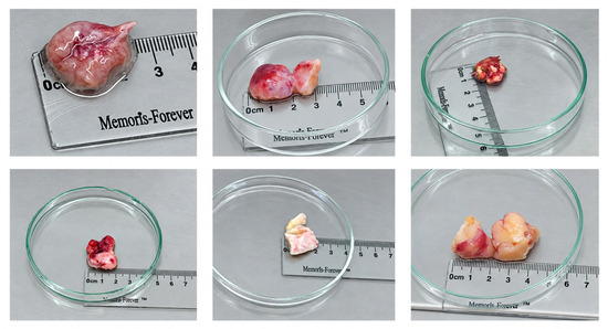
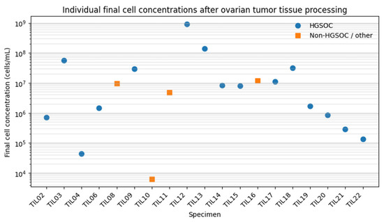
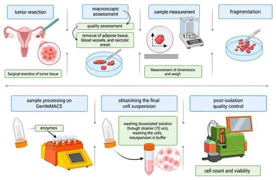
Background: Fresh ovarian cancer tissue is a demanding starting material for cellular workflows, particularly when the aim is to obtain tumor-derived suspensions suitable for further TIL-oriented procedures. In this study, we evaluated post-isolation cell concentration and viability in ovarian cancer specimens processed according to a standardized mechanical–enzymatic protocol. Methods: Biological material was collected from 24 patients undergoing routine surgery for ovarian cancer or suspected advanced ovarian malignancy. After macroscopic assessment and exclusion of extensively necrotic tissue, 19 specimens were included in the final analysis. The cohort was dominated by high-grade serous ovarian carcinoma, which accounted for 15 of 19 analyzed cases. Tissue specimens varied markedly in mass, with a median processed tissue weight of 2.05 g and a range from 0.11 to 14.93 g. Results: The median final cell concentration was 8.10 × 10⁶ cells/mL, with values ranging from 6.40 × 10³ to 9.26 × 10⁸ cells/mL. Median viability was 80.8%, although individual results ranged widely from 2.3% to 98.7%. The calculated median concentration of viable cells was 6.17 × 10⁶ viable cells/mL. Tissue mass showed a moderate positive association with final cell concentration (Spearman’s rho = 0.50, p = 0.028), but not with viability. Conclusions: These findings show that standardized processing of ovarian cancer surgical specimens can generate cell suspensions suitable for post-isolation quality assessment, even when the input material is highly variable. The present study focused on early post-isolation quality-control readouts obtained after tumor dissociation and did not evaluate TIL expansion capacity, detailed TIL phenotype, or antitumor function. The wide range of final cell concentrations underlines the biological and practical heterogeneity of ovarian cancer tissue and supports the need for careful reporting of early tissue-processing parameters in TIL-oriented workflows.
Full article
(This article belongs to the Special Issue Gynecological Cancer: Prevention, Diagnosis, Prognosis and Treatment (2nd Edition))
►▼
Show Figures

Figure 1
Open AccessArticle
Payload-Free Epitope Imprinted Polymer Nanoparticles Selectively Targeting IL-13Rα2 and Inhibition of Diffuse Midline Glioma Cells
by
Damla Ulker, Adem Ozleyen, Dmitry Pshezhetskiy, Elena Piletska, Salvador Macip, Nikolai A. Barlev and Sergey Piletsky
Cancers 2026, 18(18), 3028; https://doi.org/10.3390/cancers18183028 (registering DOI) - 17 Sep 2026
Abstract
Background/ Objectives: Diffuse midline gliomas (DMGs), including diffuse intrinsic pontine glioma (DIPG), present a significant medical challenge due to their aggressive nature and poor prognosis. Widespread overexpression of the interleukin-13 receptor subunit alpha-2 (IL-13Rα2) across gliomas makes it a promising therapeutic target. Method:
[...] Read more.
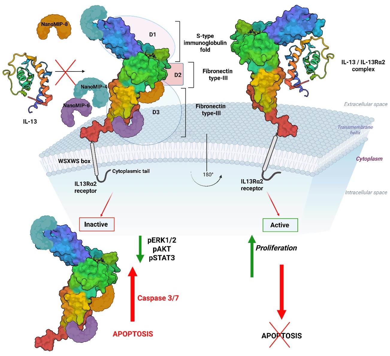
Background/ Objectives: Diffuse midline gliomas (DMGs), including diffuse intrinsic pontine glioma (DIPG), present a significant medical challenge due to their aggressive nature and poor prognosis. Widespread overexpression of the interleukin-13 receptor subunit alpha-2 (IL-13Rα2) across gliomas makes it a promising therapeutic target. Method: Using epitope mapping through a molecular imprinting (EMMI) technique, we developed nanosized molecularly imprinted polymers (nanoMIPs) that specifically recognize different IL-13Rα2 domains. Results: Functional assays showed that nanoMIPs binding to the surface of IL-13Rα2 reduced glioma cell viability, while sparing non-tumor cells. Consistent with receptor-mediated interactions, these nanoMIPs were selectively accumulated onto glioma cells, a specificity validated using a reverse-epitope nanoMIP negative control (nanoMIP-1*). IL-13Rα2-specific nanoMIPs suppressed phosphorylation of ERK1/2, AKT, and STAT3, disrupting major survival pathways. Moreover, these nanoMIPs activated the pro-apoptotic caspase-3/7 pathway, confirming apoptosis induction. Conclusions: Our findings demonstrate that IL-13Rα2-specific nanoMIPs, without a pharmacological payload, exert strong anti-proliferative and pro-apoptotic effects on DMG cells. Collectively, nanoMIPs represent a robust, reproducible synthetic antibody nanoplatform for targeted therapies in cancer and other diseases characterized by cell-surface protein dysregulation.
Full article
(This article belongs to the Section Methods and Technologies Development)
►▼
Show Figures

Graphical abstract
Journal Menu
► ▼ Journal Menu-
- Cancers Home
- Aims & Scope
- Editorial Board
- Reviewer Board
- Topical Advisory Panel
- Early Career Editorial Board
- Instructions for Authors
- Special Issues
- Topics
- Sections & Collections
- Article Processing Charge
- Indexing & Archiving
- Editor’s Choice Articles
- Most Cited & Viewed
- Journal Statistics
- Journal History
- Journal Awards
- Society Collaborations
- Conferences
- Editorial Office
Journal Browser
► ▼ Journal BrowserHighly Accessed Articles
Latest Books
E-Mail Alert
News
14 July 2025
Meet Us Virtually at the 1st International Online Conference on Hematology, 28–30 October 2026
18 September 2026
MDPI World Cancer Day 2026 Webinar Series | September—Childhood Cancer Awareness Month, 25 September 2026
Topics
Topic in
Cancers, Current Oncology, Diseases, Onco
Genomic Signature of Ocular Tumors
Topic Editors: Bita Esmaeli, Natalie WolkowDeadline: 20 October 2026
Topic in
BioMed, Cancers, Diagnostics, JCM, J. Imaging
Machine Learning and Deep Learning in Medical Imaging
Topic Editors: Rafał Obuchowicz, Michał Strzelecki, Adam Piórkowski, Karolina NurzynskaDeadline: 31 October 2026
Topic in
Cancers, Diagnostics, Diseases, IJMS, JPM
Advances in Genetics and Precision Medicine in Human Diseases: 2nd Edition
Topic Editors: Shun-Fa Yang, Shih-Chi SuDeadline: 14 November 2026
Topic in
Cancers, Diagnostics, JCM, Life, Medical Sciences, Current Oncology
Triple-Negative Breast Cancer: Genetic Vulnerabilities and Emerging Targets for Precision Oncology
Topic Editors: Tatiana de Almeida Simão, Sheila Coelho Soares-LimaDeadline: 30 November 2026
Conferences
Special Issues
Special Issue in
Cancers
Clinical Outcomes in Upper GI Cancers
Guest Editor: Yoshiaki ShojiDeadline: 20 September 2026
Special Issue in
Cancers
Advances in Targeting Drug-Resistant Tumors: Approaches Mitigating Inflammation and Oxidative Stress Pathways
Guest Editors: SubbaRao Madhunapantula, Arati SharmaDeadline: 20 September 2026
Special Issue in
Cancers
Development of Hepatitis C Virus-Related Cancers
Guest Editor: Anna linda ZignegoDeadline: 20 September 2026
Special Issue in
Cancers
Advances in Salivary Gland Carcinoma: 2nd Edition
Guest Editor: Giulio CantùDeadline: 20 September 2026
Topical Collections
Topical Collection in
Cancers
Regulatory and Non-Coding RNAs in Cancer Epigenetic Mechanisms
Topical Collection in
Cancers
Recent Advances in Pancreatic Ductal Adenocarcinoma
Collection Editors: Sumit Sahni, Anubhav Mittal, Jaswinder Samra
Topical Collection in
Cancers
Factors Regulating Cancer Cell Growth, Migration, Invasion, and Apoptosis
Collection Editors: Masahide Takahashi, Atsushi Enomoto