- Article
Pharmacological Profiling of Cannabinoid CB2 Receptor Ligands in Non-Stimulated BV-2 Microglia Cells
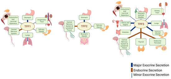
Microglia are key regulators of central nervous system homeostasis and neuroinflammation, and cannabinoid type 2 receptors (CB2Rs) have emerged as important modulators of microglial function. Although the pharmacology of CB2Rs has been extensively characterised in heterologous expression systems and activated microglia, comparatively little is known about the behaviour of CB2R ligands in non-stimulated microglia. The present study therefore aimed to characterise the pharmacological properties of a panel of CB2R ligands in non-stimulated BV-2 microglial cells and to determine whether constitutive receptor activity and ligand-dependent signalling bias could be detected under basal conditions. Classical CB2R agonists (CP 55,940, WIN 55,212-2, JWH 133 and JWH 015), putative protean agonists ((R)-AM 1241 and GW 405833), and inverse agonists (SR 144528, AM 630 and JTE 907) were evaluated using [35S]GTPγS binding and forskolin-stimulated cAMP assays. In the [35S]GTPγS assay, WIN 55,212-2 displayed the highest intrinsic activity, whereas (R)-AM 1241 and GW 405833 behaved as partial agonists. Inverse agonists reduced basal signalling, indicating constitutive CB2R activity in resting BV-2 cells. In contrast, cAMP measurements revealed greater signal amplification, with (R)-AM 1241 and GW 405833 exhibiting full agonist behaviour and SR 144528 producing pronounced inverse agonism. Marked differences in ligand efficacy and rank order between assays highlighted the influence of downstream signalling mechanisms and ligand-dependent signalling bias. Notably, GW 405833 displayed a strongly biased signalling profile, whereas SR 144528 consistently exhibited the greatest inverse agonist activity. These findings demonstrate that CB2Rs are functionally active in non-stimulated microglia and that constitutive receptor activity and signalling bias contribute significantly to their pharmacological profile under basal conditions. By focusing on non-stimulated microglia, this study provides new insights into CB2R signalling in a homeostatic cellular environment and establishes a framework for understanding how CB2R pharmacology may be altered during neuroinflammatory and neurodegenerative disease states.

Targets
2 September 2026


![Concentration–response curves of CB2 receptor ligands WIN 55,212-2, CP 55,940, JWH 133 and JWH 015, (R)-AM 1241 and GW 405833, on [35S]GTPγS binding. Data are expressed as the percentage stimulation of specific [35S]GTPγS binding above the unstimulated basal level. Each point represents the mean ± S.E.M. of independent experiments performed in duplicate (n = 3).](https://mdpi-res.com/cdn-cgi/image/width=470%2Cheight=317/https://mdpi-res.com/targets/targets-04-00031/article_deploy/html/images/targets-04-00031-ag-550.jpg)

![Metal-graphitic nanocapsules for SERS detection. (A) Schematic diagram and properties of GIAN [25]. (Copyright © 2014, the authors.) (B) Schematic diagram of SERS quantification of CV in fish muscles by GIAN substrate [26]. (Copyright © 2016, American Chemical Society.) (C) FDTD simulation of AGNs, RSD (%) with or without IS, and SERS mapping images with various concentrations of RhB [23]. (Copyright © 2016, Tsinghua University Press and Springer-Verlag Berlin Heidelberg.) (D) Schematic diagram of enrichment process and hot map of different concentration of CN− before and after enrichment [37]. (Copyright © 2016, American Chemical Society.) (E) Au@NGs SERS tags for rapid Raman imaging in the cellular silent region [56]. (Copyright © 2024 American Chemical Society.).](https://mdpi-res.com/cdn-cgi/image/width=281%2Cheight=192/https://mdpi-res.com/targets/targets-04-00029/article_deploy/html/images/targets-04-00029-ag-550.jpg)


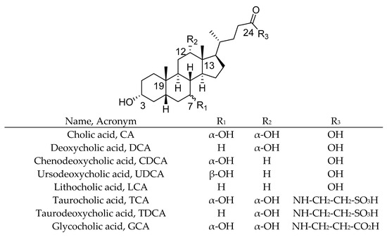
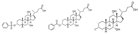
![Structure and names of common BAs [16].](https://mdpi-res.com/cdn-cgi/image/width=281%2Cheight=192/https://mdpi-res.com/targets/targets-04-00018/article_deploy/html/images/targets-04-00018-ag-550.jpg)