Article

Late Morbidity and Mortality in Survivors of Childhood Ependymoma: A Report from the Childhood Cancer Survivor Study (CCSS)

1
Hackensack Meridian School of Medicine, Nutley, NJ 07110, USA
2
Department of Pediatrics, Hackensack University Medical Center, Hackensack, NJ 07601, USA
3
College of Medicine, The University of Cincinnati, Cincinnati, OH 45229, USA
4
Cincinnati Children’s Hospital Medical Center, Cincinnati, OH 45229, USA
5
St. Jude Children’s Research Hospital, Memphis, TN 38105, USA
6
Department of Medicine, Duke University, Durham, NC 27705, USA
7
Department of Pediatrics, University of Minnesota, Minneapolis, MN 55454, USA
8
The Hospital for Sick Children, Toronto, ON M5G 1E8, Canada
9
Department of Radiation Physics, The University of Texas at MD Anderson Cancer Center, Houston, TX 77030, USA
10
Fred Hutchinson Cancer Research Center, Seattle, WA 98109, USA
11
Department of Pediatrics, University of Texas Southwestern Medical Center, Dallas, TX 75235, USA
12
Texas Children’s Hospital, Baylor College of Medicine, Houston, TX 77030, USA
*
Author to whom correspondence should be addressed.
These authors contributed equally to this work.
These authors also contributed equally to this work.
Cancers 2025, 17(22), 3669; https://doi.org/10.3390/cancers17223669 (registering DOI)
Submission received: 26 September 2025 / Revised: 10 November 2025 / Accepted: 11 November 2025 / Published: 15 November 2025
(This article belongs to the Section Cancer Survivorship and Quality of Life)

Simple Summary

This study provides the most comprehensive evaluation to date of long term outcomes among five year survivors of pediatric ependymoma. Between 1970 and 1999, treatment for pediatric ependymoma evolved to reduce cranial radiation volumes and incorporate chemotherapy for some patients. But, late morbidity and mortality have not improved among pediatric ependymoma survivors despite these treatment changes. Reduced cranial radiation volume was associated with a reduced risk for late mortality and decreased grade 3–4 chronic health conditions. Our findings reveal that rates of late mortality and serious chronic health conditions have not substantially improved over the decades studied though reduced whole-brain radiation exposure is associated with lower late mortality and morbidity, supporting efforts to minimize radiation fields when safe and feasible.

Abstract

Background/Objectives: Treatment of childhood ependymoma evolved from 1970 to 1999 by reducing radiation volumes and incorporating chemotherapy. The impact of these changes on long-term health outcomes remains unknown. In this report, we evaluated temporal changes in all-cause and cause-specific late mortality, chronic health conditions (CHCs), and subsequent neoplasms (SNs) in the Childhood Cancer Survivor Study (CCSS) cohort of adult survivors of pediatric ependymoma, diagnosed between 1970 and 1999. Methods: A total of 404 five-year survivors of ependymoma (47.5% female, 80.7% non-Hispanic White, median 6 (range 0–20) years at diagnosis, 22 (5–49) years from diagnosis) diagnosed between 1970 and 1999 and enrolled in the Childhood Cancer Survivor Study were evaluated for late (>5 years from diagnosis) mortality, SNs, and CHCs. Outcomes were analyzed by diagnosis decade, radiotherapy, and chemotherapy exposure. Gray’s test compared cumulative incidences. Multivariable piecewise exponential models estimated relative risks (RRs). Results: Whole-brain radiation exposure decreased over time (42.9% (1970s) to 2.7% (1990s)), while focal radiation (21.4% to 68.9%), and chemotherapy (29.5% to 50.2%) use increased. Fifteen-year all-cause late mortality (incidence, 95% CI) remained similar across decades: 1970s (9.3%, 3.4–18.8%), 1980s (14.7%, 9.4–21.2%), 1990s (10.3%, 6.7–14.9%). All-cause late mortality was higher after treatment with whole-brain radiation (22.5%, 11.2–36.5%) compared to focal radiation (11.4%, 7.5–16.1%) or no brain radiation (3.5%, 0.9–9.1%) (p < 0.001), and with chemotherapy (14.4%, 9.6–20.0%) versus without (6.8%, 3.8–11.0%) (p = 0.004). Compared to no brain radiation, the RR (95% CI) of grade 3–4 CHCs increased among survivors treated with focal (2.6, 1.3–5.4) and whole-brain radiation (3.5, 1.5–8.1), while chemotherapy was not associated with CHCs or SNs. Conclusions: Despite reduced radiation volumes and increased use of chemotherapy, late mortality and morbidity among pediatric ependymoma survivors remained largely unchanged across treatment decades.

1. Introduction

Pediatric ependymoma accounts for approximately 190 new cases annually in the United States, representing 5.5% of childhood brain tumors [1], and the underlying causes for this rare disease are largely unknown. Overall survival for most pediatric cancers has improved over the decades, and as many as 70% of newly diagnosed pediatric ependymoma patients in the current era will become five-year survivors [1]. However, late onset mortality and morbidity, occurring after five years from diagnosis, and the effect of recent advances in treatment on ependymoma survivors are poorly understood.
Contemporary therapy for pediatric ependymoma consists of maximal surgical resection followed by radiation therapy. Although some ependymomas can be cured without radiation exposure, most require radiation to achieve a lasting cure [2]. Radiation therapy is often accepted as a standard of care because of its importance in achieving a cure, despite its effects on cognitive development and long-term health outcomes [3,4,5]. Over the past decades, radiation therapy for non-metastatic ependymoma has moved from whole-brain irradiation, often including spinal irradiation, to more focal therapy [6]. Current radiation therapies do not work in all cases and various chemotherapy regimens have attempted to augment or replace radiation therapy in children with ependymoma, but few have demonstrated prolonged survival [7,8,9].
Due to their tumor location and treatment, survivors of childhood brain tumors are at high risk for developing late morbidity or mortality. Common morbidities following ependymoma treatment include radiation-related subsequent neoplasms (SNs) (such as meningioma and high-grade glioma) and chronic health conditions (CHCs), especially endocrine and neurologic conditions, which may increase in cumulative incidence across the lifespan [7,10,11]. Younger age at diagnosis and treatment, as well as radiation dose, have been associated with increased risk of neurologic late effects [12,13].
In this report, we evaluated temporal changes in all-cause and cause-specific late mortality, CHCs, and SNs in the Childhood Cancer Survivor Study (CCSS) cohort of adult survivors of pediatric ependymoma, diagnosed between 1970 and 1999. This time period was chosen to allow for mature follow up data and to understand the effect of treatment changes on survivorship care for adult survivors of childhood ependymoma. Outcomes were compared by treatment era and by historical changes in treatment modality across three decades. Prior studies of late mortality and morbidity among pediatric cancer survivors enrolled in the CCSS have demonstrated continued improvements in outcomes over time that have been attributed to changes in therapy: specifically, reductions in therapeutic exposure for low-risk populations, technical advancements in surgery and radiation, and increased access to survivorship care [14,15,16].

2. Materials and Methods

The CCSS is a National Cancer Institute (NCI)-funded retrospective cohort with a longitudinal follow-up of five-year survivors of childhood cancer diagnosed between 1970 and 1999. Participants were treated at 31 participating institutions in the United States and Canada and were younger than 21 years at diagnosis. The study methods and design have been described in previous publications [17,18]. The institutional review boards at each of the centers participating in the CCSS approved the study, and participants or guardians provided informed consent.
The ICD-O diagnosis codes for ependymoma as well as distribution per era included in the cohort are listed in Table 1. Demographic and treatment characteristics were reported for three treatment eras: the 1970s, 1980s, and 1990s. Treatment variables included surgery, chemotherapy, and brain radiation. Treatment variables were only considered if they occurred within 5 years after diagnosis. Chemotherapy exposure was defined as yes or no. Table S1 shows chemotherapy drug exposure by era. We chose radiation treatment to the brain as an exposure variable due to the known late effects on childhood brain tumor survivors, though in this analysis, many patients who were categorized as receiving whole-brain radiation had cranio-spinal irradiation. Brain radiation was characterized as focal or whole-brain, based on radiotherapy record abstraction. For each individual, the maximum target dose to four previously defined brain regions [19], the frontal cortex, temporal lobe, posterior fossa, and parietal and occipital cortex were determined by summing the delivered doses from all overlapping treatment fields with irradiation to more than 50% of each region [20]. Treatments were defined as whole-brain radiation when all four regions were irradiated and the dose to each region was greater than or equal to 40Gy [19]. Focal brain radiation was defined as greater than or equal to 40Gy for at least one but not all four brain regions. Radiation to the spine was not considered in this analysis. Surgery was defined as any surgery, yes or no.

2.1. Mortality

Data regarding late mortality were obtained from the National Death Index, including cause of death according to the International Classification of Diseases, Ninth and Tenth Revisions (ICD-9 and ICD-10). Late mortality was measured from the cohort entry to either death or 31 December 2017, the last date of the National Death Index search. For deaths predating the National Death Index (1975–1979), death certificates were obtained. Deaths were categorized into three mutually exclusive groups: (1) ependymoma recurrence/progression, (2) external causes (accidents, injuries, trauma, suicide), or (3) other health-related causes (including subsequent neoplasms [SNs], cardiac, pulmonary, and all other causes). No cause of death was available for 11 survivors, including Canadian survivors, so they were excluded from analyses involving cause of death.

2.2. Chronic Health Conditions (CHCs)

A comprehensive set of questionnaires administered at baseline and up to four subsequent follow-up assessments were used by participants to report on various health conditions and their age at first diagnosis. Chronic health conditions (CHCs) were classified according to a modified framework based on the Common Terminology Criteria for Adverse Events (CTCAE), version 4.03, based on self or proxy reports. CHCs were categorized as mild (grade 1), moderate (grade 2), severe or disabling (grade 3), life threatening (grade 4), or fatal (grade 5) [21]. A panel of physician experts reviewed all reported conditions and determined severity grades [22]. The questionnaires captured only the first reported occurrence of each condition, except for subsequent neoplasms, which were documented separately. The follow-up for CHCs continued until the earliest occurrence of a CHC (or second CHC in multiple CHC analyses), late recurrence, subsequent neoplasm [SN], death, or the last questionnaire.

2.3. Subsequent Neoplasms (SNs)

SNs were defined as new neoplasms occurring five or more years from diagnosis. They were classified into two mutually exclusive categories: (1) subsequent malignant neoplasms (SMNs), consisting of invasive neoplasms with an International Classification of Diseases for Oncology (ICD-O, 3rd version) behavior code of 3, excluding nonmelanoma skin cancers, or (2) non-invasive neoplasms, including nonmelanoma skin cancers and benign meningiomas. When a pathology report was unavailable, the diagnosis was confirmed by death certificate, other medical records, or both [23]. SN/SMN were tracked until the date of diagnosis of SN/SMN, death, or the last questionnaire.

2.4. Statistical Methods

Demographic and treatment characteristics of survivors were described and compared across treatment eras. Continuous variables were analyzed using the Kruskal–Wallis test and categorical variables were assessed using Chi-square or Fisher exact tests.
Cumulative incidence of late mortality, CHCs, SNs, and SMNs was estimated 15 years from diagnosis, omitting events that occurred before cohort entry (up to 5 years after diagnosis). Gray’s test was used to compare cumulative incidence across treatment groups.
Piecewise exponential models were used to assess associations between chemotherapy and radiation exposures with late mortality, CHCs, SNs, and SMNs among survivors. Models were adjusted for attained age, sex, race, and ethnicity; for late mortality analyses, early recurrence and the ependymoma subtype were additionally included as covariates. Statistical analysis was completed using SAS v.9.4. A two-sided p-value of <0.05 was considered statistically significant.

3. Results

Among 25,665 CCSS participants, there were 404 five-year survivors of pediatric ependymoma. Ependymoma survivors were 47.5% female and 80.7% non-Hispanic White. The median age at diagnosis was 6 years (range 0–20 years), and survivors were followed for a median of 22 years (5–49 years) from diagnosis to last follow-up. Descriptive characteristics of the study population are shown in Table 1. Among survivors with known treatment information, 47.7% of patients were exposed to chemotherapy, 100% had surgery, and 75.7% were exposed to cranial radiation. Across the three decades, whole-brain radiation exposure decreased from 42.9% in the 1970s to 15.2% in the 1980s and 2.7% in the 1990s, while focal brain radiation exposure increased from 21.4% in the 1970s to 68.8% in the 1980s and 68.9% in the 1990s. Chemotherapy exposure increased from 29.5% in the 1970s to 50% in the 1980s and 50.2% in the 1990s. Across the three decades, the median age at diagnosis decreased from 10 years in the 1970s to 6 years in the 1980s and 5 years in the 1990s (p < 0.001). While the proportion of anaplastic ependymoma increased over the decades from 3.6% in the 1970s to 7.4% in the 1980s and 11.3% in the 1990s (p = 0.153), the distribution of ependymoma histopathological subtypes was not significantly different across the decades. Early recurrence within 5 years (n = 39) did not differ by treatment era (p = 0.25). Age at radiation did not differ across the decades.
Characteristics that might predict more aggressive disease or treatment choice were also investigated among survivors by treatment exposure, Table S2. Survivors exposed to chemotherapy were significantly younger at diagnosis compared to those not exposed (3 years (range 0–20 years) vs. 9 years (0–20 years), p < 0.0001). Compared to those not treated with chemotherapy, chemotherapy-exposed survivors were also more likely to be diagnosed with anaplastic ependymoma (p < 0.0001), be exposed to radiation (p < 0.0001), and have a recurrence within 5 years (p = 0.05). On the other hand, survivors treated with focal radiation were younger than survivors treated with whole-brain radiation (4 years (range 0–20 years) vs. 7.5 years (1–19 years), p = 0.017). Among all three radiation categories, survivors exposed to focal radiation were also more likely to be diagnosed with anaplastic ependymoma (14.6% compared to 5% (whole-brain radiation) and 0% (no brain radiation), p < 0.0001) and more likely to be exposed to chemotherapy (55.5% compared to 50% (whole-brain) and 27.4% (no brain radiation), p < 0.0001), (Table 1).

3.1. Late Mortality

Fifteen-year all-cause late mortality (incidence, 95% CI) estimates were similar across treatment eras: 1970s (9.3%, 3.4–18.8%), 1980s (14.7%, 9.4–21.2%), and 1990s (10.3%, 6.7–14.9%), (p = 0.9, comparison of cumulative incidence curves), Figure 1A. Fifteen-year all-cause late mortality by brain radiation treatment type was higher with whole-brain radiation (22.5%, 11.0–36.5%) compared to focal radiation (11.4%, 7.5–16.1%) or no brain radiation (3.5%, 0.9–9.1%), (p < 0.001), Figure 1B. Mortality was also higher with chemotherapy (14.4%, 9.6–20.0%) compared to no chemotherapy (6.8%, 3.8–11.0%) (p = 0.004), Figure 1C. Cause-specific mortality by treatment era is provided in Table S3. There were no differences in relative risk of all-cause or cause-specific late mortality by treatment era, Table 2. Relative risk of all-cause late mortality was increased with chemotherapy (RR 1.87, 1.11–3.14) (p = 0.02) compared to no chemotherapy, and with whole-brain radiation (3.93, 1.81–8.54) and focal brain radiation (2.25, 1.11–4.58) compared to no brain radiation (p = 0.002), Table 3.
Because perceived risk factors and early tumor recurrence may have influenced treatment exposure in the first five years from diagnosis, we attempted to adjust for characteristics that might predict more aggressive disease. When adjusted for anaplastic subtype and recurrence within 5 years, the relative risk of all-cause late mortality remained increased following chemotherapy exposure (RR 1.76, 1.02–3.01) compared to no chemotherapy, and with whole-brain radiation (3.69, 1.64–8.29) compared to no brain radiation, Table S4.

3.2. Chronic Health Conditions

Prevalence of CHCs by grade and by system are reported in Table S5. The relative risk of having any grade 3–4 CHCs, two or more grade 3–4 CHCs, or any hearing CHCs was not different across the three decades, Table S6. Compared to no brain radiation, there were increased risks (RR, 95% CI) of any grade 3–4 CHCs for focal (2.6, 1.3–5.4) and whole-brain radiation (3.5, 1.5–8.1), and two or more Grade 3–4 CHCs for whole-brain radiation (6.5, 1.3–31.6), Table 4. Relatively few chronic health conditions were reported for specific organ systems, making the calculation of relative risk impossible or not significant other than auditory. There was an increased risk of any grade 3–4 hearing loss with focal and whole-brain radiation. There were no differences in CHCs by chemotherapy exposure.

3.3. Subsequent Neoplasm

There were 42 SNs identified in the cohort, including 28 SMNs. The most common SNs were Meningioma (n = 13) and Basal Cell Carcinoma (n = 10). The median time to development of SN was 19.0 years (range 8.2–36.2). A list of the SN identified in the cohort is detailed in Table S7. Risk of SN or SMN was not different across treatment decades, Table S8. There was no difference in relative risk of any SMN between radiation treatment groups. The relative risk of any SN was 2.8 (1.0–7.7) for focal and 3.8 (1.2–12.4) for whole-brain RT (p = 0.07) compared to no brain radiation, Table 5. Chemotherapy did not affect the risk of SN or SMN.

4. Discussion

With 404 survivors followed for a median of 22 years, this manuscript describes the largest reported cohort of five-year survivors of childhood ependymoma. The major finding of this work is the lack of improvement in late morbidity and mortality in children and adolescents diagnosed and treated with ependymoma between 1970 and 1999. This is disappointing, considering the improvements in long term survival that have been documented in other pediatric malignancies over the same decades, with reductions in late mortality for survivors of acute lymphocytic leukemia, Wilms tumors and Hodgkin lymphoma treated in more recent decades [14]. Among childhood brain tumor survivors, recent reports from the CCSS of pediatric glioma and medulloblastoma survivors have demonstrated reductions in all-cause mortality and CHCs by nearly half over this period [15,16], but similar improvements were not observed among pediatric ependymoma survivors. Improvements for other pediatric malignancies have been attributed to changes in therapy, including a reduction in radiation exposure. Therapy for pediatric ependymoma during this time also evolved to reduce radiation exposure from whole-brain to focal therapy and increase exposure to chemotherapy. The latter possibly contributed to a lack of reduction in overall mortality. Our study shows a reduction in risk of late mortality, severe or life-threatening (grade 3–4) CHCs, and SNs among survivors of pediatric ependymoma who were exposed to reduced cranial radiation, similar to other studies. However, despite a reduction in cranial radiation exposure over time, survivors of pediatric ependymoma did not demonstrate improvements in late morbidity or mortality across the three eras.
Unlike radiation, the role of chemotherapy in ependymoma therapy is less certain. Decades of chemotherapy trials have investigated multiple chemotherapy regimens in an effort to improve outcomes. While some patients had objective tumor responses, there was no survival benefit [7]. The most recent phase III Children’s Oncology Group trial of newly diagnosed pediatric ependymoma added four cycles of maintenance chemotherapy to radiation in patients who had undergone a gross total or near total resection but survival statistics are not yet available [9]. The current International Society of Pediatric Oncology (SIOP) ependymoma study also investigates whether the addition of chemotherapy improves outcomes [24]. Neither study was stratified upfront by ependymoma subgroups based on molecular differences, and it is unknown whether a specific subgroup may benefit from chemotherapy [25].
The effect of chemotherapy exposure, as well as cranial radiation, in our study may be subject to confounding by indication, for which we cannot fully adjust. Survivors with high-risk features, such as young age or anaplastic subtype, may have been exposed to expanded therapy because of their perceived risk factors. Survivors with early recurrence (prior to cohort entry) may have been treated with additional therapy following relapse. Although adjustment of mortality analyses for these factors demonstrated no significant change in the unadjusted estimates, we were unable to adjust for all risk factors, such as tumor location and the extent of resection, which are not captured in the CCSS. We identified few differences in risk factors over the treatment decades. While survivors were younger at diagnosis in the 1980s and 1990s, other risk factors such as anaplastic histology and early tumor recurrence were not different. Survivors exposed to whole-brain radiation also did not appear to have increased risk factors compared to those exposed to focal radiation: they were older at diagnosis, tended toward less frequent anaplastic histology, and showed no difference in early tumor recurrence. On the other hand, survivors exposed to chemotherapy were associated with increased risk factors: they were younger at diagnosis, more likely to have anaplastic histology, and more frequently had early tumor recurrence. It seems reasonable that some of the worse outcomes associated with chemotherapy may be due to more aggressive disease in this treatment group. A prospective study that controls for these risk factors would be required to confirm these findings associated with chemotherapy exposure.
Regardless of the cause, the lack of improvement in late mortality over three decades is concerning. In addition to treatment changes, there are other improvements in care during this period. The incorporation of MRI into clinical care in the 1980s, improved understanding of the importance of complete resection, and improved survivorship care would be expected to improve mortality from the 1970s to 2000. The Central Brain Tumor Registry of the United States reported improvement in the overall 5-year survival of patients diagnosed with ependymoma from 1977 (29.5%) to 2011 (71.1%). The trend towards improved overall 5-year survival would have been expected to correlate with improved late mortality. The persistent late mortality, despite these advancements, highlights a critical gap in translating improved treatments and survivorship care into long-term survival benefits for pediatric ependymoma survivors. Knowledge about potential late complications should inform therapeutic strategies for patients with ependymoma going forward. Focal compared to whole-brain should be prioritized when appropriate and feasible. Survivorship care should focus on early screening for CHCs known to impact survivors. And, given the auditory impact of ependymoma therapy, investigation of otoprotectants should be prioritized in future protocols.
This study confirms the poor late outcomes found in smaller cohorts with a shorter follow-up time. Late mortality among survivors of childhood ependymoma diagnosed in the 1970s is greater than 50% 45 years from diagnosis, Figure 1A. Cranial radiation, especially whole-brain radiation, is associated with an increased risk of severe or life-threatening CHCs, as well as an increased risk of SN. These findings are similar to a recent population-based study from Canada evaluating 96 survivors of pediatric ependymoma diagnosed between 1987 and 2015 that showed an increased risk of death and disability compared to matched population controls [11]. Our study also agrees with prior studies demonstrating the significant risk of late mortality among survivors of pediatric ependymoma [26,27]. However, our study is the first to identify the lack of improvement for survivors of pediatric ependymoma over three decades, as well as identify potential late risks of chemotherapy exposure.
This study is subject to potential limitations. Chronic medical conditions in the CCSS are self-reported. This concern is partially mitigated by restricting the analysis to severe outcomes such as grade 3–4 CHCs. In addition, the years 1970–1999 saw considerable advances in diagnostic imaging and surgical techniques that may impact tumor resection, and therefore late mortality and CHCs. The extent of surgical resection is not captured in the CCSS. The concern for variable rates of gross total resection is partially addressed by reports that rates of gross total resection did not change significantly until the late 1990s. The relative stability of these rates over the decades studied may mitigate the effect this information could have added to this analysis [4,28,29,30,31]. It is likely that the majority of long-term survivors did undergo gross total resection, as survival rates for subtotal resection during this time period were poor. Almost a quarter of ependymoma patients in this cohort did not receive brain radiation. A proportion of these patients (6%) received isolated spinal radiation, which was not included in this analysis. Like other cohorts of pediatric ependymoma [11], these tumors may have been treated with surgery alone which has been shown to successfully treat some cases of pediatric ependymoma [2]. Information on molecular classification of tumors is also unavailable, although the full impact of this classification on risk stratification is still unknown. While there were changes in therapy over the decades studied, the initiation of a large cooperative trial in North America for ependymoma patients, ACNS0121, and the publication by Merchant et al. detailing the benefits of focal radiation both occurred after [3,32]. Studying these decades may have been too early to demonstrate a difference as another, albeit smaller, cohort studied through 2014 showed survival improvement [33].

5. Conclusions

Our study focuses on outcomes occurring five or more years after diagnosis. While prior studies have shown improved survival in pediatric ependymoma, these gains are largely limited to the first five years post-diagnosis [1,26]. In contrast, late morbidity and mortality are increasingly driven by CHCs and SNs, in addition to the late relapse that is particularly seen in ependymoma. Other CCSS studies have demonstrated long-term improvements in outcomes across cancer types, attributed to evolving therapies, technological advances and enhanced survivorship care. However, despite treatment changes in pediatric ependymoma therapy between the 1970s and 1990s, late outcomes remained unchanged. Reductions in cranial radiation treatment volumes have reduced risks of late mortality, CHC, and SN. In contrast, the introduction of chemotherapy was associated with increased risks of late mortality, although these results should be interpreted with caution. Further research is needed to understand this lack of progress in late outcomes and to evaluate the long-term risks of emerging therapies in future trials.
For oncologists, these results underscore the need for long-term surveillance and multidisciplinary survivorship care that extends well beyond the traditional five-year window, as adult survivors of childhood ependymoma remain at risk for late mortality, chronic health conditions, and subsequent neoplasms related to prior treatments. Importantly, this study demonstrates that focusing only on early survival endpoints fails to capture the true burden of disease. Given that approximately 70% of patients with pediatric ependymoma survive at least five years but remain at risk for recurrence and late morbidity, future clinical trials should incorporate longer observation periods to more accurately assess the lasting risks and benefits of novel therapies. Despite therapeutic advances, expected improvements in late outcomes have not yet been realized, underscoring the need for continued research and comprehensive survivorship strategies to improve long-term quality of life.

Supplementary Materials

The following supporting information can be downloaded at: https://www.mdpi.com/article/10.3390/cancers17223669/s1. Table S1: Chemotherapy agents; Table S2: Characteristics of ependymoma survivors by treatment type; Table S3: Cause-specific late mortality by era; Table S4: Relative risk of late mortality among survivors of ependymoma, adjusted for treatment, recurrence within 5 years, and ependymoma subtype (9391.3 and 9392.3) a; Table S5: Prevalence of chronic health conditions in five-year survivors of ependymoma by decade; Table S6: Relative risk of chronic health conditions in five-year survivors of ependymoma adjusted for treatment decade a; Table S7: Subsequent neoplasms among patients with known treatment information; Table S8: Multivariable relative risk of subsequent neoplasm by treatment era a.

Author Contributions

Conceptualized and designed the study, K.R.L., P.d.B., D.C.B. and M.F.O.; collection and assembly of data, K.R.L., P.d.B., M.X., S.M., D.K.S., K.O., J.N., K.K., P.C.N., R.H., K.K.N., L.M.T., W.L., G.T.A., T.B., D.C.B. and M.F.O.; data analysis was provided by M.X., S.M. and D.K.S.; K.R.L., P.d.B., D.C.B., M.F.O., M.X. and S.M., and D.K.S. provided data interpretation. K.R.L. wrote the first draft of the manuscript and all authors contributed to manuscript editing and revision. All authors have read and agreed to the published version of the manuscript.

Funding

This research was funded by the National Cancer Institute (CA55727, G.T. Armstrong, Principal Investigator). Support to St. Jude Children’s Research Hospital was also provided by the Cancer Center Support (CORE) grant (CA21765, C. Roberts, Principal Investigator) and the American Lebanese-Syrian Associated Charities (ALSAC).

Institutional Review Board Statement

The study was conducted in accordance with the Declaration of Helsinki and approved by the Institutional Review Board of St. Jude Children’s Research Hospital (protocol code CCSS and approval date 30 January 2025).

Informed Consent Statement

Informed consent was obtained from all participants or guardians involved in the study.

Data Availability Statement

The Childhood Cancer Survivor Study is a US National Cancer Institute funded resource (U24 CA55727) to promote and facilitate research among long-term survivors of cancer diagnosed during childhood and adolescence. CCSS data are publicly available on dbGaP at https://www.ncbi.nlm.nih.gov/gap/ (accessed on 9 November 2025) through its accession number phs001327.v2.p1., and on the St Jude Survivorship Portal within the St. Jude Cloud, at https://survivorship.stjude.cloud/ (accessed on 9 November 2025). In addition, utilization of the CCSS data that leverages the expertise of CCSS Statistical and Survivorship research and resources will be considered on a case-by case basis. For this utilization, a research application of intent, followed by an analysis concept proposal, must be submitted for evaluation by the CCSS Publications Committee. Users interested in utilizing this resource are encouraged to visit http://ccss.stjude.org (accessed on 9 November 2025). Full analytical data sets associated with CCSS publications since January of 2023 are also available on the St. Jude Survivorship Portal at https://viz.stjude.cloud/community/cancer-survivorship-community~4/publications (accessed on 9 November 2025).

Acknowledgments

Presented at the International Symposium on Late Complications after Childhood Cancer (ISLCCC); 27 June–29 June 2024; Lucerne, Switzerland. Also presented at the 21st International Symposium on Pediatric Neuro-Oncology (ISPNO); 28 June–2 July 2024; Philadelphia, PA.

Conflicts of Interest

The authors declare no conflicts of interest.

Abbreviations

The following abbreviations are used in this manuscript:
CCSSChildhood Cancer Survivor Study
NCINational Cancer Institute
ICD-9 and ICD-10International Classification of Diseases, Ninth and Tenth Revisions
CHCsChronic Health Conditions
CTCAECommon Terminology Criteria for Adverse Events
SNsSubsequent Neoplasms
SMNsSubsequent Malignant Neoplasms
ICD-O, 3rd versionInternational Classification of Diseases for Oncology
RRRelative Risk
SIOPInternational Society of Pediatric Oncology

References

  1. Ostrom, Q.T.; de Blank, P.M.; Kruchko, C.; Petersen, C.M.; Liao, P.; Finlay, J.L.; Stearns, D.S.; Wolff, J.E.; Wolinsky, Y.; Letterio, J.J.; et al. Alex’s Lemonade Stand Foundation Infant and Childhood Primary Brain and Central Nervous System Tumors Diagnosed in the United States in 2007–2011. Neuro-Oncology 2014, 16, x1–x36. [Google Scholar] [CrossRef]
  2. Hukin, J.; Epstein, F.; Lefton, D.; Allen, J. Treatment of Intracranial Ependymoma by Surgery Alone. Pediatr. Neurosurg. 1998, 29, 40–45. [Google Scholar] [CrossRef]
  3. Merchant, T.E.; Bendel, A.E.; Sabin, N.D.; Burger, P.C.; Shaw, D.W.; Chang, E.; Wu, S.; Zhou, T.; Eisenstat, D.D.; Foreman, N.K.; et al. Conformal Radiation Therapy for Pediatric Ependymoma, Chemotherapy for Incompletely Resected Ependymoma, and Observation for Completely Resected, Supratentorial Ependymoma. J. Clin. Oncol. 2019, 37, 974–983. [Google Scholar] [CrossRef] [PubMed]
  4. E Merchant, T.; Zhu, Y.; Thompson, S.J.; Sontag, M.R.; Heideman, R.L.; E Kun, L. Preliminary results from a Phase II trail of conforml radiation therapy for pediatric patients with localised low-grade astrocytoma and ependymoma. Int. J. Radiat. Oncol. 2002, 52, 325–332. [Google Scholar] [CrossRef]
  5. Merchant, T.E.; Kiehna, E.N.; Li, C.; Xiong, X.; Mulhern, R.K. Radiation dosimetry predicts IQ after conformal radiation therapy in pediatric patients with localized ependymoma. Int. J. Radiat. Oncol. Biol. Phys. 2005, 63, 1546–1554. [Google Scholar] [CrossRef] [PubMed]
  6. Merchant, T.E.; Fouladi, M. Ependymoma: New Therapeutic Approaches Including Radiation and Chemotherapy. J. Neuro-Oncology 2005, 75, 287–299. [Google Scholar] [CrossRef] [PubMed]
  7. Bouffet, E.; Foreman, N. Chemotherapy for intracranial ependymomas. Child’s Nerv. Syst. 1999, 15, 563–570. [Google Scholar] [CrossRef]
  8. Grill, J.; Kalifa, C.; Doz, F.; Schoepfer, C.; Sainte-Rose, C.; Couanet, D.; Terrier-Lacombe, M.; Valteau-Couanet, D.; Hartmann, O. A High-Dose Busulfan-Thiotepa Combination Followed by Autologous Bone Marrow Transplantation in Childhood Recurrent Ependymoma. Pediatr. Neurosurg. 1996, 25, 7–12. [Google Scholar] [CrossRef]
  9. Smith, A.; Onar-Thomas, A.; Ellison, D.; Owens-Pickle, E.; Wu, S.; Leary, S.E.S.; Fouladi, M.; Merchant, T.; Gajjar, A.; Foreman, N. EPEN-54. ACNS0831, PHASE III RANDOMIZED TRIAL OF POST-RADIATION CHEMOTHERAPY IN PATIENTS WITH NEWLY DIAGNOSED EPENDYMOMA AGES 1 TO 21 YEARS. Neuro-Oncology 2020, 22, iii318–iii319. [Google Scholar] [CrossRef]
  10. Armstrong, G.T.; Liu, Q.; Yasui, Y.; Huang, S.; Ness, K.K.; Leisenring, W.; Hudson, M.M.; Donaldson, S.S.; King, A.A.; Stovall, M.; et al. Long-Term Outcomes Among Adult Survivors of Childhood Central Nervous System Malignancies in the Childhood Cancer Survivor Study. JNCI J. Natl. Cancer Inst. 2009, 101, 946–958. [Google Scholar] [CrossRef]
  11. Coltin, H.; Pequeno, P.; Liu, N.; Tsang, D.S.; Gupta, S.; Taylor, M.D.; Bouffet, E.; Ramaswamy, V.; Nathan, P.C. The medical and functional burden of surviving childhood ependymoma: A population-based study in Ontario, Canada. Pediatr. Blood Cancer 2024, 71, e31275. [Google Scholar] [CrossRef] [PubMed]
  12. Kenborg, L.; Winther, J.F.; Linnet, K.M.; Krøyer, A.; Albieri, V.; Holmqvist, A.S.; Tryggvadottir, L.; Madanat-Harjuoja, L.M.; Stovall, M.; Hasle, H.; et al. Neurologic disorders in 4858 survivors of central nervous system tumors in childhood—An Adult Life after Childhood Cancer in Scandinavia (ALiCCS) study. Neuro-Oncology 2019, 21, 125–136. [Google Scholar] [CrossRef]
  13. Roddy, E.; Mueller, S. Late Effects of Treatment of Pediatric Central Nervous System Tumors. J. Child Neurol. 2015, 31, 237–254. [Google Scholar] [CrossRef]
  14. Armstrong, G.T.; Chen, Y.; Yasui, Y.; Leisenring, W.; Gibson, T.M.; Mertens, A.C.; Stovall, M.; Oeffinger, K.C.; Bhatia, S.; Krull, K.R.; et al. Reduction in Late Mortality among 5-Year Survivors of Childhood Cancer. New Engl. J. Med. 2016, 374, 833–842. [Google Scholar] [CrossRef] [PubMed]
  15. de Blank, P.M.K.; Lange, K.R.; Xing, M.; Salehabadi, S.M.; Srivastava, D.; Brinkman, T.M.; Ness, K.K.; Oeffinger, K.C.; Neglia, J.; Krull, K.R.; et al. Temporal changes in treatment and late mortality and morbidity in adult survivors of childhood glioma: A report from the Childhood Cancer Survivor Study. Nat. Cancer 2024, 5, 590–600. [Google Scholar] [CrossRef] [PubMed]
  16. Salloum, R.; Chen, Y.; Yasui, Y.; Packer, R.; Leisenring, W.; Wells, E.; King, A.; Howell, R.; Gibson, T.M.; Krull, K.R.; et al. Late Morbidity and Mortality Among Medulloblastoma Survivors Diagnosed Across Three Decades: A Report From the Childhood Cancer Survivor Study. J. Clin. Oncol. 2019, 37, 731–740. [Google Scholar] [CrossRef]
  17. Leisenring, W.M.; Mertens, A.C.; Armstrong, G.T.; Stovall, M.A.; Neglia, J.P.; Lanctot, J.Q.; Boice, J.D.; Whitton, J.A.; Yasui, Y. Pediatric Cancer Survivorship Research: Experience of the Childhood Cancer Survivor Study. J. Clin. Oncol. 2009, 27, 2319–2327. [Google Scholar] [CrossRef]
  18. Robison, L.L.; Armstrong, G.T.; Boice, J.D.; Chow, E.J.; Davies, S.M.; Donaldson, S.S.; Green, D.M.; Hammond, S.; Meadows, A.T.; Mertens, A.C.; et al. The Childhood Cancer Survivor Study: A National Cancer Institute–Supported Resource for Outcome and Intervention Research. J. Clin. Oncol. 2009, 27, 2308–2318. [Google Scholar] [CrossRef]
  19. Packer, R.J.; Gurney, J.G.; Punyko, J.A.; Donaldson, S.S.; Inskip, P.D.; Stovall, M.; Yasui, Y.; Mertens, A.C.; Sklar, C.A.; Nicholson, H.S.; et al. Long-Term Neurologic and Neurosensory Sequelae in Adult Survivors of a Childhood Brain Tumor: Childhood Cancer Survivor Study. J. Clin. Oncol. 2003, 21, 3255–3261. [Google Scholar] [CrossRef]
  20. Howell, R.M.; Smith, S.A.; Weathers, R.E.; Kry, S.F.; Stovall, M. Adaptations to a Generalized Radiation Dose Reconstruction Methodology for Use in Epidemiologic Studies: An Update from the MD Anderson Late Effect Group. Radiat. Res. 2019, 192, 169–188. [Google Scholar] [CrossRef] [PubMed]
  21. Oeffinger, K.C.; Mertens, A.C.; Sklar, C.A.; Kawashima, T.; Hudson, M.M.; Meadows, A.T.; Friedman, D.L.; Marina, N.; Hobbie, W.; Kadan-Lottick, N.S.; et al. Chronic health conditions in adult survivors of childhood cancer. New Engl. J. Med. 2006, 355, 1572–1582. [Google Scholar] [CrossRef]
  22. Gibson, T.M.; Mostoufi-Moab, S.; Stratton, K.L.; Leisenring, W.M.; Barnea, D.; Chow, E.J.; Donaldson, S.S.; Howell, R.M.; Hudson, M.M.; Mahajan, A.; et al. Temporal patterns in the risk of chronic health conditions in survivors of childhood cancer diagnosed 1970–99: A report from the Childhood Cancer Survivor Study cohort. Lancet Oncol. 2018, 19, 1590–1601. [Google Scholar] [CrossRef]
  23. Neglia, J.P.; Friedman, D.L.; Yasui, Y.; Mertens, A.C.; Hammond, S.; Stovall, M.; Donaldson, S.S.; Meadows, A.T.; Robison, L.L. Second Malignant Neoplasms in Five-Year Survivors of Childhood Cancer: Childhood Cancer Survivor Study. JNCI J. Natl. Cancer Inst. 2001, 93, 618–629. [Google Scholar] [CrossRef]
  24. Leblond, P.; Massimino, M.; English, M.; Ritzmann, T.A.; Gandola, L.; Calaminus, G.; Thomas, S.; Pérol, D.; Gautier, J.; Grundy, R.G.; et al. Toward Improved Diagnosis Accuracy and Treatment of Children, Adolescents, and Young Adults with Ependymoma: The International SIOP Ependymoma II Protocol. Front. Neurol. 2022, 13, 887544. [Google Scholar] [CrossRef]
  25. Pajtler, K.W.; Witt, H.; Sill, M.; Jones, D.T.; Hovestadt, V.; Kratochwil, F.; Wani, K.; Tatevossian, R.; Punchihewa, C.; Johann, P.; et al. Molecular Classification of Ependymal Tumors across All CNS Compartments, Histopathological Grades, and Age Groups. Cancer Cell 2015, 27, 728–743. [Google Scholar] [CrossRef]
  26. Marinoff, A.E.; Ma, C.; Guo, D.; Snuderl, M.; Wright, K.D.; Manley, P.E.; Al-Sayegh, H.; Sinai, C.E.; Ullrich, N.J.; Marcus, K.; et al. Rethinking childhood ependymoma: A retrospective, multi-center analysis reveals poor long-term overall survival. J. Neuro-Oncology 2017, 135, 201–211. [Google Scholar] [CrossRef]
  27. Shu, H.G.; Sall, W.F.; Maity, A.; Tochner, Z.A.; Janss, A.J.; Belasco, J.B.; Rorke-Adams, L.B.; Phillips, P.C.; Sutton, L.N.; Fisher, M.J. Childhood intracranial ependymoma. Cancer 2007, 110, 432–441. [Google Scholar] [CrossRef] [PubMed]
  28. Robertson, P.L.; Zeltzer, P.M.; Boyett, J.M.; Rorke, L.B.; Allen, J.C.; Geyer, J.R.; Stanley, P.; Li, H.; Albright, A.L.; McGuire-Cullen, P.; et al. Survival and prognostic factors following radiation therapy and chemotherapy for ependymomas in children: A report of the Children’s Cancer Group. J. Neurosurg. 1998, 88, 695–703. [Google Scholar] [CrossRef]
  29. Garvin, J.H.; Selch, M.T.; Holmes, E.; Berger, M.S.; Finlay, J.L.; Flannery, A.; Goldwein, J.W.; Packer, R.J.; Rorke-Adams, L.B.; Shiminski-Maher, T.; et al. Phase II study of pre-irradiation chemotherapy for childhood intracranial ependymoma. Children’s Cancer Group protocol 9942: A report from the Children’s Oncology Group. Pediatr. Blood Cancer 2012, 59, 1183–1189. [Google Scholar] [CrossRef] [PubMed]
  30. A Healey, E.; Barnes, P.D.; Kupsky, W.J.; Scott, R.M.; E Sallan, S.; Black, P.M.; Tarbell, N.J. The prognostic significance of postoperative residual tumor in ependymoma. Neurosurgery 1991, 28, 666. [Google Scholar] [CrossRef] [PubMed]
  31. Nazar, G.B.; Hoffman, H.J.; Becker, L.E.; Jenkin, D.; Humphreys, R.P.; Hendrick, E.B. Infratentorial ependymomas in childhood: Prognostic factors and treatment. J. Neurosurg. 1990, 72, 408–417. [Google Scholar] [CrossRef] [PubMed]
  32. Merchant, T.E.; Mulhern, R.K.; Krasin, M.J.; Kun, L.E.; Williams, T.; Li, C.; Xiong, X.; Khan, R.B.; Lustig, R.H.; Boop, F.A.; et al. Preliminary Results From a Phase II Trial of Conformal Radiation Therapy and Evaluation of Radiation-Related CNS Effects for Pediatric Patients with Localized Ependymoma. J. Clin. Oncol. 2004, 22, 3156–3162. [Google Scholar] [CrossRef] [PubMed]
  33. Zapotocky, M.; Beera, K.; Adamski, J.; Laperierre, N.; Guger, S.; Janzen, L.; Lassaletta, A.; Nobre, L.F.; Bartels, U.; Tabori, U.; et al. Survival and functional outcomes of molecularly defined childhood posterior fossa ependymoma: Cure at a cost. Cancer 2019, 125, 1867–1876. [Google Scholar] [CrossRef] [PubMed]
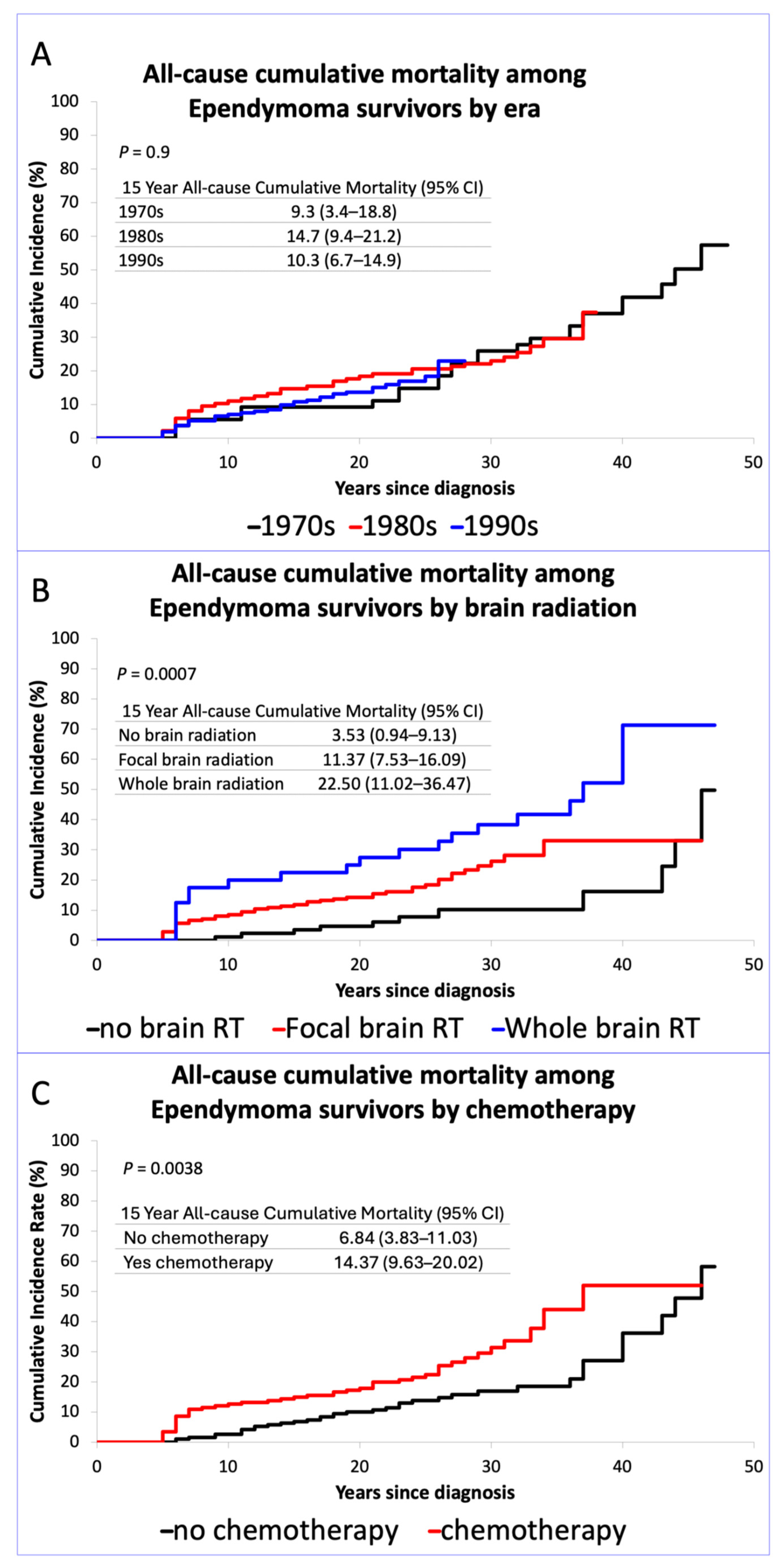
Figure 1. (A) Cumulative incidence curve for all-cause mortality by era, Inset table shows cumulative incidence of mortality at 15 years by treatment era; (B) Cumulative incidence curve for all-cause mortality by brain radiation treatment, Inset table shows cumulative incidence of mortality at 15 years by brain radiation treatment; (C) Cumulative incidence curve for all-cause mortality by chemotherapy, Inset table shows cumulative incidence of mortality at 15 years by chemotherapy.
Cancers 17 03669 g001
Table 1. Demographic and treatment characteristics for five-year survivors of childhood ependymoma: overall and by treatment era.
Total1970–19791980–19891990–1999
Characteristicn = 404n = 55n = 136n = 213p-Value a
Diagnosis
9383.1—subependymoma 4 (0.99)1 (1.8)1 (0.7)2 (0.9)0.6
9391.3—ependymoma NOS342 (84.65)48 (87.3)118 (86.8)176 (82.6)
9392.3—anaplastic ependymoma, 36 (8.91)2 (3.6)10 (7.4)24 (11.3)
9393.1—papillary ependymoma6 (1.49)2 (3.6)1 (0.7)3 (1.4)
9394.1—myxopapillary ependymoma16 (3.96)2 (3.6)6 (4.4)8 (3.8)
Sex (n,%): Male212 (52.5)32 (58.2)65 (47.8)115 (54.0)0.35
Female192 (47.5)23 (41.8)71 (52.2)98 (46.0)
Race/Ethnicity (n,%):
Non-Hispanic White
326 (80.7)50 (90.9)117 (86.0)159 (74.6)0.006
Non-Hispanic Black30 (7.4)0 (0.0)6 (4.4)24 (11.3)
Hispanic33 (8.2)2 (3.6)8 (5.9)23 (10.8)
Other15 (3.7)3 (5.5)5 (3.7)7 (3.3)
Age at diagnosis (median, min–max) years6 (0–20)10 (0–19)6 (0–20)5 (0–20)0.0001
Time since diagnosis (median, min–max) years22 (5–49)34 (6–49)27.5 (5–37)20 (5–28)<0.001
Chemotherapy Exposure (n,%)174 (47.7)13 (29.5)59 (50)102 (50.2)0.037
Any surgery (n,%)367 (100)45 (100)118 (100)204 (100)NA b
Any brain RT (n,%)265 (75.7)28 (65.1)97 (84.3)140 (72.9)0.0174
Radiation treatment group, dose ≥40 Gy
Dose in Gy, among those ≥40 Gy
(median, min–max)
54 (40–300)50 (40–69)54 (43.2–90)56 (40–300)<0.0001
No brain radiation85 (25.2)15 (35.7)18 (16.1)52 (28.4)<0.0001
Focal brain radiation212 (62.9)9 (21.4)77 (68.8)126 (68.9)
Whole-brain radiation40 (11.9)18 (42.9)17 (15.2)5 (2.7)
Recurrence within 5 years (n,%)48 (11.9)3 (5.5)16 (11.8)29 (13.6)0.25
Age at radiation in years (median, min–max)5.45 (0.8–21.35)9.33 (1.19–16.96)5.39 (0.8–20.96)5.41 (0.92–21.35)0.1815
a Overall p value. b NA—All patients with known treatment information had surgery, therefore p-value cannot be calculated. The percentages were based on the total number of participants for whom information was available.
Table 2. Relative risk of late mortality among survivors of ependymoma, adjusted for treatment decade a.
All-Cause MortalityDeath due to Recurrence or Progression of Primary MalignancyHealth-Related Cause Mortality
RR [95%CI]RR [95%CI]RR [95%CI]
Treatment Era
1970–19791.01.01.0
1980–19890.95 (0.54–1.69)1.74 (0.54–5.64)0.68 (0.31–1.5)
1990–19990.88 (0.47–1.65)1.51 (0.46–4.99)0.85 (0.35–2.09)
Sex
Male1.01.01.0
Female0.95 (0.63–1.41)1.21 (0.64–2.28)0.84 (0.46–1.51)
Race
Non-Hispanic White1.01.01.0
Other0.92 (0.52–1.61)0.93 (0.4–2.13)0.85 (0.35–2.04)
a Multivariable models adjusted for attained age.
Table 3. Relative risk of late mortality among survivors of ependymoma, adjusted for chemotherapy and brain radiation a.
All-CauseRecurrence or Progression of Primary MalignancyHealth-Related Causes
RR [95%CI]RR [95%CI]RR [95%CI]
Sex
Male1.01.01.0
female0.92
(0.58–1.46)
1.24
(0.58–2.67)
0.78 (0.39–1.55)
Race
Non-Hispanic White1.01.01.0
Other0.71 (0.36–1.4)1.13 (0.45–2.83)0.29 (0.07–1.21)
Chemotherapy
No1.01.01.0
Yes1.87 (1.11–3.14)1.56 (0.68–3.58)2.93 (1.31–6.54)
Brain Radiation
No brain radiation1.01.01.0
Focal brain radiation2.25 (1.11–4.58)3.98 (0.92–17.33)1.44 (0.54–3.85)
Whole-brain radiation3.93 (1.81–8.54)5.2 (0.94–28.6)3.86 (1.43–10.43)
a Multivariable models adjusted for attained age.
Table 4. Relative risk of chronic health conditions among survivors of ependymoma, adjusted for chemotherapy and radiation a.
Any Grade 3–4>1 Grade 3–4Any Hearing CHC Grade 3–4
RR [95%CI]RR [95%CI]RR [95%CI]
Sex
Male1.01.01.0
Female1.13 (0.69–1.84)1.09 (0.52–2.27)0.43 (0.18–1.06)
Race
Non-Hispanic White1.01.01.0
Other1.13 (0.61–2.1)1.42 (0.60–3.37)0.56 (0.17–1.90)
Chemotherapy
No1.01.01.0
Yes1.15 (0.69–1.91)2.17 (0.99–4.73)0.55 (0.22–1.33)
Brain Radiation
No Brain Radiation1.01.01.0
Focal Brain Radiation2.6 (1.25–5.39)4.39 (1.0–19.31)12.54 (1.60–98.14)
Whole-Brain Radiation3.5 (1.48–8.11)6.47 (1.32–31.64)15.65 (1.79–137.07)
a Multivariable models adjusted for attained age.
Table 5. Relative risk of subsequent neoplasm in five-year survivors of childhood ependymoma, adjusted for treatment exposure a.
Any Subsequent NeoplasmAny Subsequent Malignant Neoplasm
RR [95%CI]RR [95%CI]
Sex
Male1.01.0
Female1.73 (0.86–3.49)1.16 (0.49–2.75)
Race
Non-Hispanic White1.01.0
Other0.45 (0.14–1.47)0.48 (0.11–2.08)
Chemotherapy
No1.01.0
Yes1.26 (0.62–2.58)1.67 (0.68–4.11)
RT
No Brain Radiation1.01.0
Focal Brain Radiation2.78 (1.0–7.73)2.70 (0.74–9.93)
Whole-Brain Radiation3.82 (1.17–12.41)3.05 (0.66–13.98)
a Multivariable models adjusted for attained age.
Disclaimer/Publisher’s Note: The statements, opinions and data contained in all publications are solely those of the individual author(s) and contributor(s) and not of MDPI and/or the editor(s). MDPI and/or the editor(s) disclaim responsibility for any injury to people or property resulting from any ideas, methods, instructions or products referred to in the content.