


Cancers 2023, 15(20), 4915; https://doi.org/10.3390/cancers15204915 - 10 Oct 2023
Cited by 8 | Viewed by 2300
Abstract
►
Show Figures
Biomarkers are biomolecules used to identify or predict the presence of a specific disease or condition. They play an important role in early diagnosis and may be crucial for treatment. MicroRNAs (miRNAs), a group of small non-coding RNAs, are more and more regarded
[...] Read more.
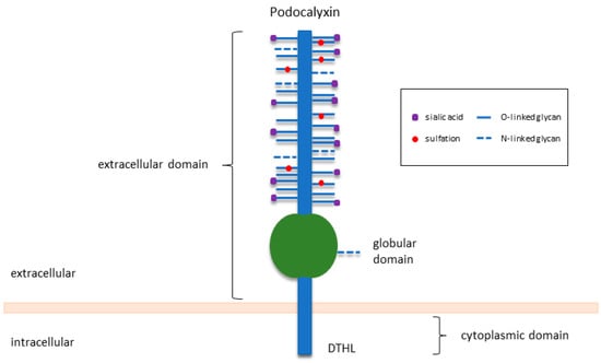
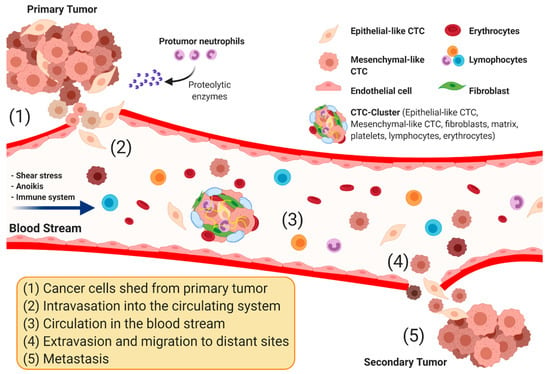
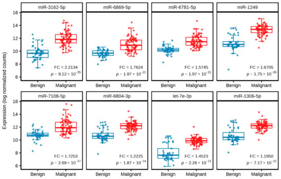
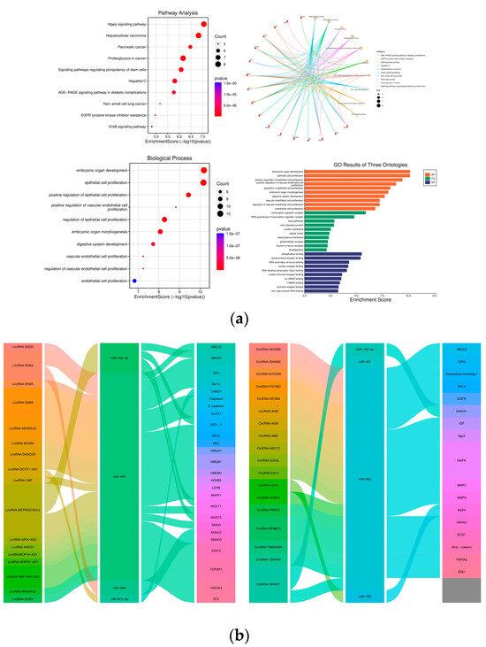
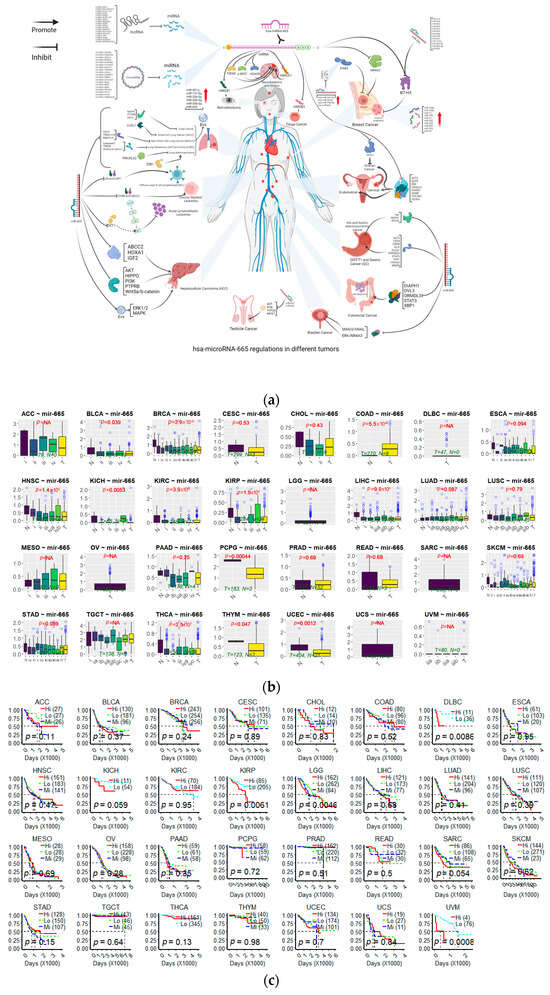
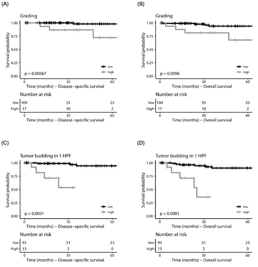
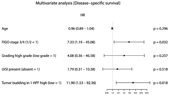
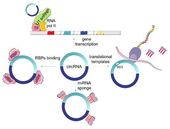
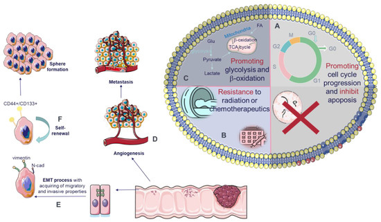
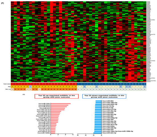
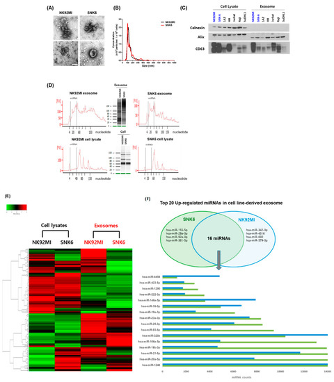
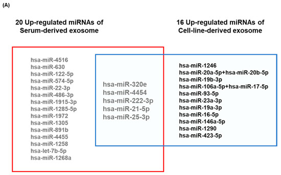
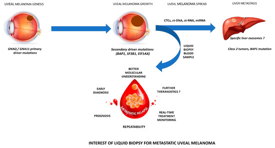
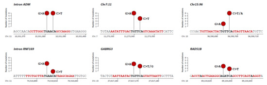
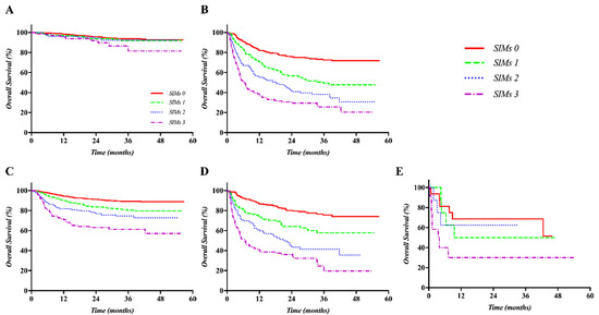
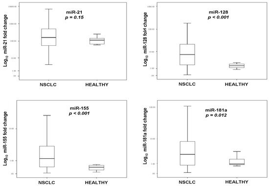
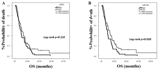
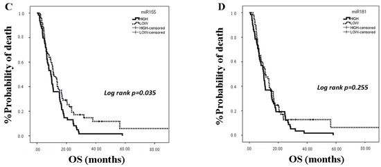
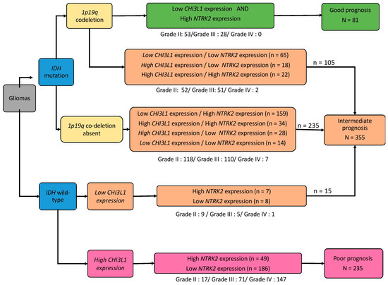
Biomarkers are biomolecules used to identify or predict the presence of a specific disease or condition. They play an important role in early diagnosis and may be crucial for treatment. MicroRNAs (miRNAs), a group of small non-coding RNAs, are more and more regarded as promising biomarkers for several reasons. Dysregulation of miRNAs has been linked with development of several diseases, including many different types of cancer, and abnormal levels can be present in early stages of tumor development. Because miRNAs are stable molecules secreted and freely circulating in blood and urine, they can be sampled with little or no invasion. Here, we present an overview of the current literature, focusing on the types of cancers for which dysregulation of miR-665 has been associated with disease progression, recurrence, and/or prognosis. It needs to be emphasized that the role of miR-665 sometimes seems ambiguous, in the sense that it can be upregulated in one cancer type and downregulated in another and can even change during the progression of the same cancer. Caution is thus needed before using miR-665 as a biomarker, and extrapolation between different cancer types is not advisable. Moreover, more detailed understanding of the different roles of miR-665 will help in determining its potential as a diagnostic and prognostic biomarker.
Full article

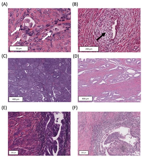
Figure 1


