Article

Association of miR-34a Expression with Quality of Life of Glioblastoma Patients: A Prospective Study

by
Paulina Vaitkiene
1,2,*,
Aiste Pranckeviciene
3,
Rytis Stakaitis
1,
Giedrius Steponaitis
1,
Arimantas Tamasauskas
1 and
Adomas Bunevicius
3
1
Laboratory of Molecular Neurooncology, Neuroscience Institute, Lithuanian University of Health Sciences, Eiveniu str. 4, LT-50161 Kaunas, Lithuania
2
Institute of Biology Systems and Genetic Research, Lithuanian University of Health Sciences, Tilzes str. 18, LT-47181 Kaunas, Lithuania
3
Laboratory of Behavioral Medicine, Neuroscience Institute, Lithuanian University of Health Sciences, Eiveniu str. 4, LT-50161 Kaunas, Lithuania
*
Author to whom correspondence should be addressed.
Cancers 2019, 11(3), 300; https://doi.org/10.3390/cancers11030300
Submission received: 4 February 2019 / Revised: 20 February 2019 / Accepted: 26 February 2019 / Published: 4 March 2019
(This article belongs to the Collection Cancer Biomarkers)

Abstract

:
MiR-34a acts as tumor-suppressor by targeting many oncogenes related to proliferation, apoptosis, and invasion of gliomas. We studied the relationships between health-related quality of life (HRQOL), depression, and miR-34a expression status in patients with newly diagnosed glioblastoma (GBM). A comprehensive HRQOL assessment was completed by 38 patients with glioblastoma prior to surgical resection and included the European Organization for Research and Treatment of Cancer (EORTC) questionnaire for cancer patients (QLQ-C30) and the Brain Cancer-Specific Quality of Life Questionnaire (QLQ-BN20), the Patient Health Questionnaire-9 (PHQ-9), the Karnofsky performance index (KPS), and The Glasgow Outcome Scale (GOS). The miR-34a expression in glioblastoma tissue was measured using quantitative reverse transcription PCR. Our findings show that lower miR-34a expression is significantly associated with higher tumor volume, worse physical functioning, lower KPS, and greater depressive symptom severity of GBM patients. Moreover, analysis reveals that miR-34a effects might be gender specific, as stronger relationships between miR-34a and patient functioning measures were observed in males when compared to females. Despite the fact that, due to small sample size, our results should be considered as preliminary, our study suggests that miR-34a is associated with tumor burden and can be important for health-related quality of life, functional status, and mood symptoms of glioblastoma patients.

1. Introduction

Glioma is a rare and often devastating disease associated with significant functional impairment and short survival time [1,2]. Prediction of outcomes after brain tumor surgery is critical for treatment guidance and optimized use of healthcare resources. Currently, besides traditional outcome measures such as overall survival, progression-free survival, and radiological response to treatment, the value of patient-centered outcome measures is widely acknowledged [3]. Health-related quality of life (HRQOL), as an outcome measure, reflects the patient’s perspective on his or her disease, covering physical, psychological, and social aspects of patient’s functioning, as well as symptoms induced by the disease and/or its treatment [4]. Decreased HRQOL and depression in patients are sensitive predictors of shorter survival in glioma patients [3]. However, patient-centered outcome measures are rarely investigated in relation to biological biomarkers.
The need for glioma biomarkers with improved sensitivity and specificity has inspired research of small non-coding micro-RNAs (miRNAs). Previous studies report that MIR34A (miR-34a) can directly down-regulate several target mRNAs which encode proteins required for cell cycle transition (E2F3, MYCN, CCND1, c-MET, Notch1/Notch2), invasion and metastasis (Fra-1), mitogen-activated protein kinase pathways (MAP3K9), and anti-apoptotic function (Bcl-2) [5]. Expression profiling identifies miR-34a as one of the several microRNAs that are down-regulated in various types of cancer including neuroblastoma, leukemia, pancreatic and hepatocellular carcinomas, glioblastoma, breast, lung, and colon cancers [6]. On the contrary, other studies find that it functions as an oncogene promoting tumorigenesis in renal cell carcinoma, papillary thyroid carcinoma (PTC), and uterine cancers [7,8,9]. These studies across different types of cancers have contradictory results regarding miR-34a role in tumor progression.
Gender is an important factor that affects the risk of cancer occurrence and development, incidence, prognosis, and treatment response and sex-specific therapeutic strategies should be quite urgent in cancer treatment [10]. There is increasing evidence that miR-34a expression may be related to gender [10,11]. Sex and treatment-dependent regulation of miRNAs may explain the different treatment response of males and females. Therefore, it is important to examine the impact of miRNA expression in both sexes under different disease conditions. Although extensive studies explore the role of miR-34a in the glioblastoma cell lines [12], little is known about the relationship between the expression level of miR-34a in glioblastoma tissues and the quality of patient functioning. Therefore, in this study, we examine the associations between expression level of miR-34a in glioblastoma tissue and the spectrum of a patients’ presenting symptoms.

2. Results

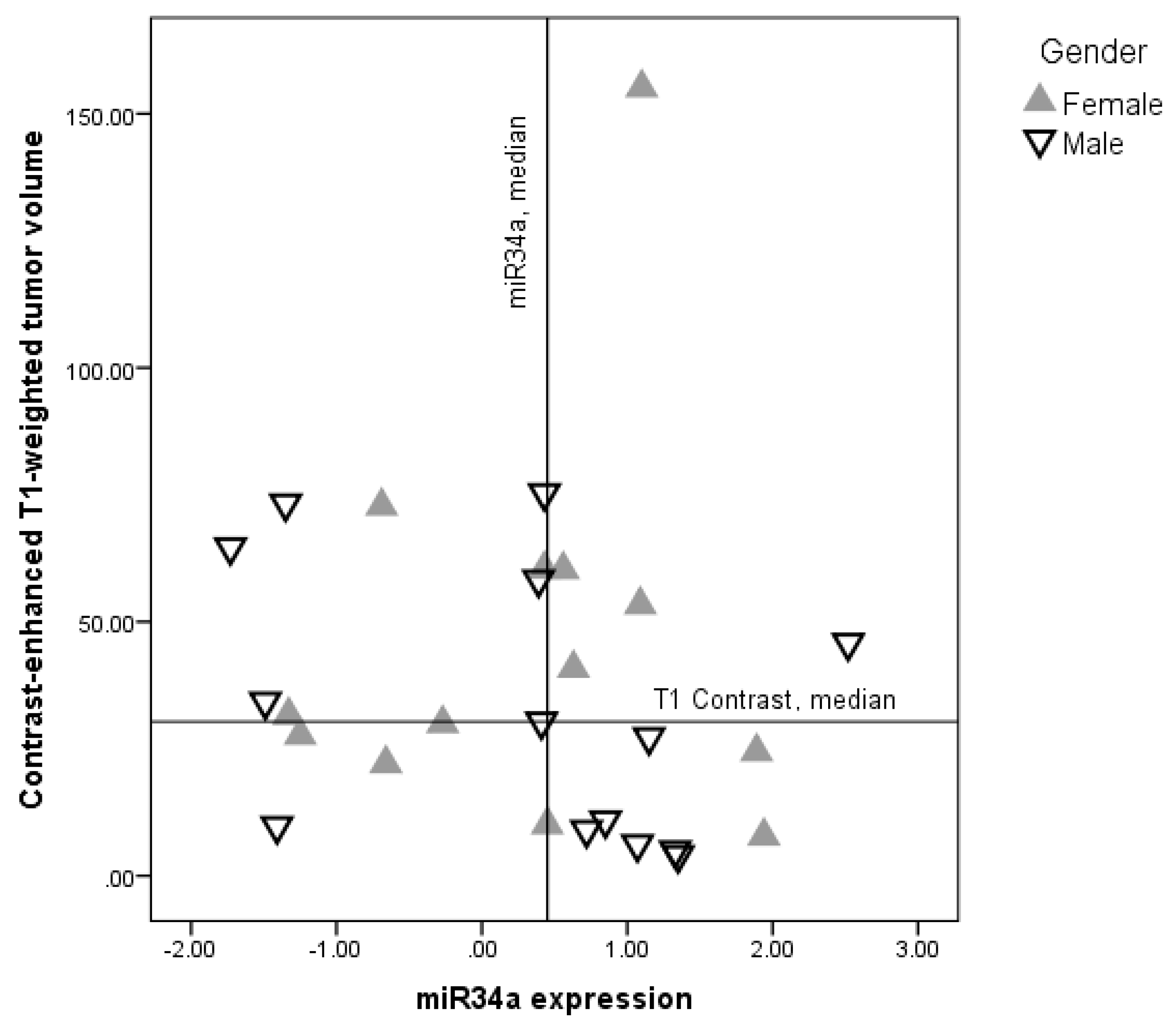
2.1. Demographic and Clinical Characteristics Relationship with miR-34a Expression Levels

Social, demographic, and clinical characteristics of the sample are presented by miR-34a expression status in Table 1. Expression of miR-34a in tumor tissue was not related to any of demographic variables. The tendency that patients with lower miR-34a expression more frequently had frontal tumors can be observed and less of them were diagnosed with tumors located in more than one lobe of the brain. Patients with higher miR-34a expression were more frequently diagnosed with multifocal tumors, however none of these differences were statistically significant, most likely due to a small number of patients in the subgroups.
Patients with lower miR-34 expression had significantly greater tumor volume in contrast-enhanced T1-weighted sequences, when compared with patients with higher miR-34a expression (U = 34.0, p = 0.03), however no volume differences were found in T2 fluid-attenuated inversion recovery (FLAIR) image sequences (Table 1).
Additional analysis of tumor volume and miR-34a expression relationships in gender subgroups revealed stronger relationship between miR-34a expression and tumor volume in males when compared with females.
In males, miR-34a expression correlated negatively with T1-weighted contrast-enhanced tumor volume (Spearman rho = −0.53, p = 0.05) (Figure 1). The correlation between miR-34a expression and tumor volume on FLAIR sequences was insignificant (Spearman rho = −0.25, p = 0.31). In females, there was no correlation between miR-34a expression and T1 contrast volume (Spearman rho = −0.09, p = 0.78), as well as no correlation between miR-34a expression and FLAIR tumor volume (Spearman rho = 0.18, p = 0.54).

2.2. Health Related Quality of Life and miR-34a Expression

Relationships between miR-34a expression levels and HRQOL indicators are shown in Table 2. Correlation analysis revealed a statistical tendency for higher miR-34a expression in tumor tissue to be positively related with physical functioning and total HRQOL reported by glioblastoma patients (Table 2). Correlation between miR-34a expression and physical functioning was stronger in males. Tendency for positive correlation between miR-34a expression and cognitive and social functioning was also observed in males, but not in females. Higher miR-34a expression was significantly negatively related to subjectively reported complaints in drowsiness. Similarly, the relationship between drowsiness and miR-34a expression was stronger in the male subsample.

2.3. Depression Correlation with miR-34a Expression

In addition to making criteria-based diagnoses of depressive disorders, the PHQ-9 is a reliable and valid measure of depression severity. The examination was conducted before the operation. Higher miR-34a expression was statistically significantly negatively correlated with depressive symptom severity, preoperatively (Table 2), both in the total sample of GBM patients and in males and females separately.

2.4. Functional Status

The Karnofsky Performance Scale (KPS) allows patients to be classified as to their functional impairment. This can be used to compare effectiveness of different therapies and to assess the prognosis in individual patients. Higher miR-34a expression in tumor tissue is significantly positively correlated with the KPS score on admission, indicating that patients with higher miR-34a expression have a better functional status before surgery. This association was slightly stronger in males when compared to females (Table 2). The miR-34a expression was not related to functional outcomes at discharge, assessed with the GOS.

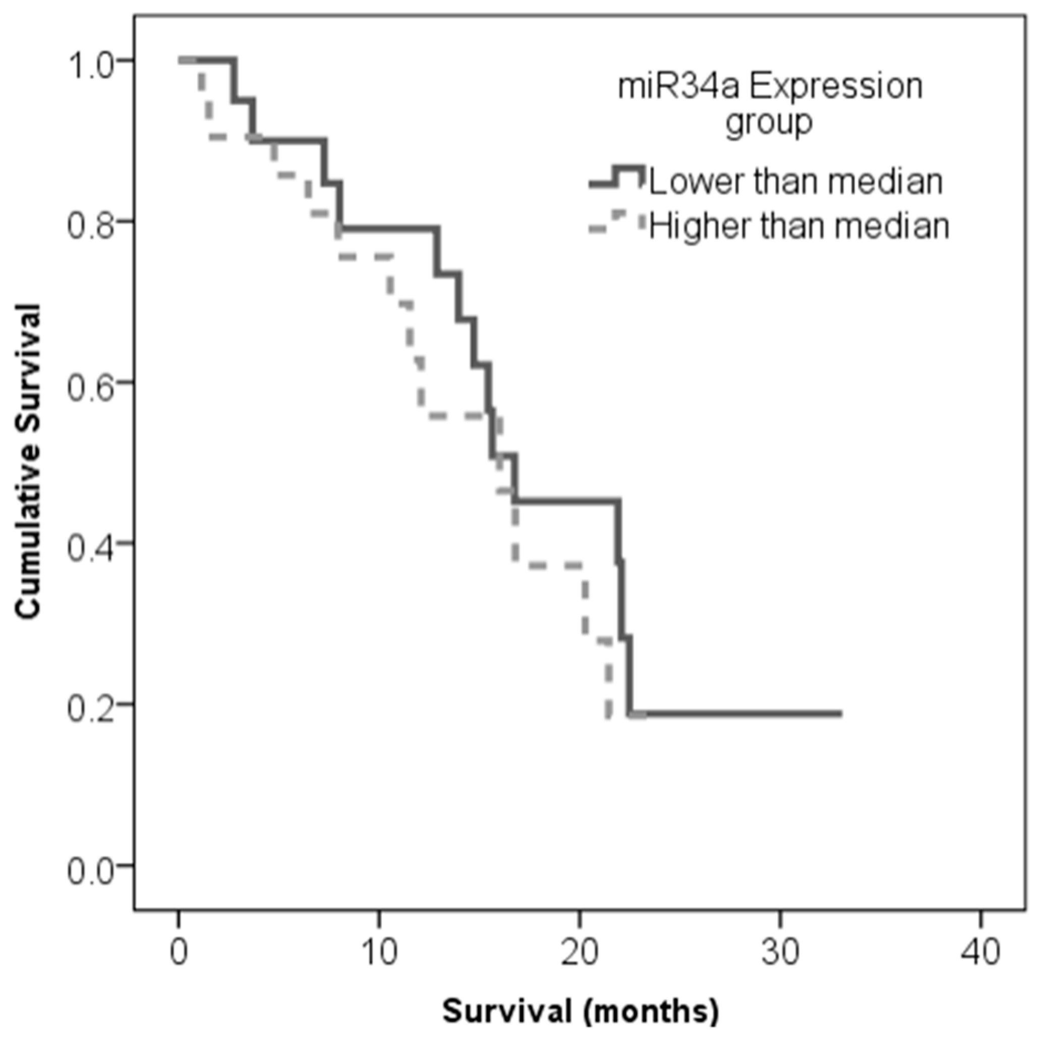
2.5. Correlation of miR-34a Expression and Patient Survival

The observed association of miR-34a expression with health-related quality of life and functional status indicated for us to check the association between patient survival and miR-34a expression. For this purpose, the miR-34a expression level values obtained from the complete set of 41 glioblastoma samples were divided into two categories as follows: Values that were lower than or equal to the median expression were ranked as “low” miR-34a expression levels and values that were higher than the median were ranked as “high” miR-34a expression levels. The Kaplan–Meier analysis using the log-rank test showed no association between overall patient survival and miR-34a expression (Log-rank test, χ2 = 0,471, df = 1, p = 0.493) (see Figure 2).

3. Discussion

This study, for the first time, reveals the relationships between HRQOL and miR-34a expression in patients with newly diagnosed glioblastomas. Previous studies indicate that miR-34a expression might be decreased in glioblastomas, as compared to lower grade gliomas and non-tumor brain tissue. Moreover, low levels of miR-34a were associated with a poor survival prognosis. However, more comprehensive studies are needed to confirm the significance of miR-34a expression levels for the glioblastoma patients [6,13,14,15].
Firstly, we analyzed the relationship between social, demographic, and clinical characteristics of glioblastoma patients and miR-34a expression levels in their tumor tissue and found that patients with lower miR-34a expression had significantly higher tumor volumes. These results are expected as previous studies show that miR-34a may act as tumor suppressor gene by targeting many oncogenes related to proliferation, differentiation, growth, apoptosis, and invasion [16]. The association of miR-34a expression with tumor size is also reported in other types of tumors, e.g. low miR-34a levels are associated with larger sizes of hepatocellular carcinoma and prostate cancer [17,18]. Meanwhile, Gao and colleagues did not notice any association between miR-34a expression and glioma tumor sizes [13]. Different results in the Gao et al. study may be attributed to a different study design, as gliomas of different grades are analyzed together. We analyze only glioblastoma patients, thus making our sample more homogeneous. Behaviour of low-grade gliomas is different than that of high-grade gliomas, thus different relationship between tumor volume and miR-34a expression might be expected as a function of tumor grade. Gao and colleagues also evaluate tumor size by using tumor diameter while we employ volumetric analysis [13]. More detailed research in larger patient samples is needed to confirm these results.
Some gender differences in GBM risk and the course of the illness were recently reported. GBM incidence rates in males are slightly higher when compared to females, however males have some survival advantages over females during the first year after diagnosis but with no difference thereafter [19,20]. Gender might be important for treatment-dependent regulation of miRNA expression and may explain the differential treatment response of males and females [10]. Thus, it is important to examine the impact of miRNA-expression in both sexes individually and under different disease conditions. There is increasing evidence that the expression of miR-34a may be related to gender and in response to therapy. For example, miR-34a was up-regulated in prostate adenocarcinoma, male group, and was not abnormally expressed in the other related cancer groups [10]. There are sex differences in response to miRNA-34a therapy in mouse models of cardiac disease [11]. Thus, it is important to examine the impact of miRNA expression in both sexes individually. In our study, additional analysis of tumor volume and miR-34a expression relationships in gender subgroups reveals stronger relationship between miR-34a expression and tumor volume in males, when compared to females. Although the exact mechanism of miR-34a regulation across genders still needs to be discovered, preliminary findings indicate that various hormone factors might participate in miR-34a expression regulation. For example, the thyroid hormone 3,3,5-triiodo-L-thyronine (T3) is shown to induce the expression of miR-34a [21] and reduced T3 levels are linked to worse HRQOL and shorter survival of brain tumor patients, including those with gliomas [22,23]. Additional hormones to be considered in miR-34a regulation are estradiol (E2), as shown in human breast cancer [24]. However, in our sample of glioblastomas, the difference in expression between miR-34a and gender has not been established. Nevertheless, with increasing evidence that miR-34a expression can be controlled by the hormone, we have decided to explore the differences of HRQOL, functional status, or depression linking to miR-34a expression in more detail, not only across all the glioblastoma samples but also between genders.
Worse perceived HRQOL is shown to predict shorter survival of glioma patients [25]. Identification of molecular markers, which could act as predictors of patients’ health status, is important in order to develop novel therapeutic strategies aiming to improve prognosis and to optimize the health status of glioblastoma patients. There is a tendency for an association between higher miR-34a expression and better physical functioning and overall HRQOL. The correlation between higher miR-34a expression and better physical functioning was stronger in males. The tendency for a positive correlation between miR-34a expression and cognitive and social functioning is also observed in males, but not in females. Patients with a higher miR-34a expression also scored higher on the KPS before surgery. However, miR34a expression was not significantly related to short term patients’ functional outcomes at the time of discharge.
Given the poor prognosis of glioblastoma, depression stands to worsen outcomes when it develops concomitantly [26]. Despite this common interaction, relatively little research has been performed on the development of depression associated with glioblastoma. One reason for this is that the pathophysiological development of depression and glioblastoma share several pathways, including altered regulation of the 5-HT receptor, norepinephrine, and 3′:5′-cyclic monophosphate [26]. We find that patients with a lower expression of miR-34a reported more severe depressive symptoms. These results are in line with Azavedo et al. [27], who report an association between miR-34a expression in postmortem brain tissue of patients with Major Depressive Disorder and Bipolar Disorder. In animal models, miR-34 family is related to stress and anxiety response [28]. Current evidence suggests that the miR-34 family might have a critical function in regulating the behavioral and neurochemical response to acute stress and in inducing stress-related amygdala neuroplasticity [29]. However, studies with many more cases will be needed to carefully elucidate the better awareness of depression when it occurs in conjunction with miR-34a expression and to encourage optimal patient care and future research to identify potential molecular pathways between them.
Previous studies provide contradictory results on the importance of miR-34a expression for the survival of glioma patients. Gao and colleagues find that grade III glioma and glioblastomas with lower miR-34a expressions correlates with worse progression-free patient survival and overall survival [10]. Meanwhile, Toraih and colleagues do not find any significant associations between miR-34a expression levels and overall survival of glioblastoma patients [6]. In contrast to these previous studies, Genovese and colleagues, in two independent cohorts of glioblastoma, show that glioblastomas with low-expressing miR-34a have better outcomes, with longer survival overall [15]. In our sample of glioblastoma patients, no statistically significant associations between miR-34a expression and overall survival are found. Further studies are needed to confirm miR-34a expression significance in glioblastomas.
Several limitations of the current study should be acknowledged. A relatively small sample size limited the statistical power of our analysis and prevented us from employing more sophisticated and multivariate statistics. Preoperative MRI images were collected retrospectively and they were available for only 61% of total sample. It might be expected that the size of the tumor is a significant covariate linking miR-34a expression with various aspects of patients functioning, thus, further studies investigating miR-34a in the context of clinical factors are needed. However, this study presents one of the first attempts to link molecular tumor data with patients functioning, assessed by patients themselves as well as their doctors. Patient assessments were performed prospectively and provide us with interesting relationships between micro and macro levels of patient functioning.

4. Materials and Methods

4.1. Procedures

The study protocol and consent procedures were approved by the Ethics Committee for Biomedical Research of the Lithuanian University of Health Sciences (LUHS) (P2-9/2003 and BE-2-3). Written informed consent was obtained from each study patient before inclusion in the study.
Consecutive adult patients admitted for surgery for suspected glioblastoma based on brain MRI, at the Department of Neurosurgery, Hospital of LUHS, Kaunas, Lithuania in a period from October 2015 until May 2017, were invited to participate in this prospective observational cohort study. The study exclusion criteria included severe cognitive deficits and/or neurological impairment leading to inability to complete all study tasks. Neuropsychological assessment was performed, from two to three days before brain tumor surgery, by a certified medical psychologist. The medical history, clinical characteristics, and functional status of the study patients were recorded by the study neurosurgeon. Histological brain tumor diagnoses were verified from postoperative pathology reports. Pre-operative MRI images were obtained from medical documentation.

4.2. Samples

Forty-six patients with histologically confirmed glioblastoma participated in the study. Data of five patients was excluded due to failed miRNA analysis. Thirty-eight (92.7%) patients completed health related quality of life (HRQOL) and depression questionnaires. Functional status was assessed in 37 (90.2%) patients. Preoperative MRI data was available for 25 (61.0%) patients.

4.3. Questionnaires

The European Organization for Research and Treatment of Cancer Quality of Life Questionnaire QLQ-30 [30] and QLQ-BN20 questionnaires [31,32] were used to evaluate preoperative health related quality of life (HRQOL) and brain tumor related symptoms. Both questionnaires were previously validated for HRQOL assessment in Lithuanian brain tumor patients [33].
The QLQ-C30 contains 30 items that were designed to assess global health status, functional status, role functioning, emotional functioning, cognitive functioning, social functioning, and various cancer related symptoms. Raw scores were linearly transformed to 0–100 scales with higher scores indicating better global health, functional status, and greater general HRQOL.
The QLQ-BN20 is a 20-item self-rating instrument that was designed as the QLQ-C30 supplement for evaluation of HRQOL specifically in brain tumor patients. It addresses future uncertainty, visual disorder, motor dysfunction, communication deficits, and other common BT-related symptoms. The QLQ-BN20 scores were linearly transformed to a 0–100 scale with higher score indicating greater BT-related symptom severity.
The Patient Health Questionnaire-9 (PHQ-9) [34] is a brief self-report tool for screening, diagnosing, monitoring, and measuring the severity of depression. The PHQ-9 is based on the Diagnostic Statistical Manual-IV depression diagnostic criteria and it is recommended for depression screening in glioma patients [35,36].
The Karnofsky performance scale (KPS) [37], was used for assessment of functional status. The KPS is an 11-point rating scale that is designed to measure a patient’s ability to carry his/her normal activities and dependence on help and nursing care. The total KPS score ranges from 100 (normal functioning) to 0 (death).
Functional outcomes at hospital discharge were evaluated by a neurosurgeon using The Glasgow Outcome Scale (GOS) [38]. The GOS ranges from 1 (death) to 5 (good recovery) and it is widely used for research purposes in neurosurgical patients.

4.4. Tumour Volume Measurements

Tumor segmentation and volume measurements were performed using the 3D Slicer medical image computing platform, version 4.3.1 (www.slicer.org) [39]. We used the most recent pre-operative structural MRI imaging data, which was performed on 1.5T or 3T MRI scanners. For the purpose of this study, we used T1-weighted contrast enhanced and T2-FLAIR weighted sequences. T1-contrast enhancing tumor volume (in cm3), representing tumor necrotic core, and T2-FLAIR hyper-intense tumor volume, representing tumor infiltrations/edema, were calculated. All volumetric analyses were performed by a trained neurosurgeon. The rater was blinded to microRNA and psychological assessment data.

4.5. Small RNA Extraction, Micro RNA cDNA Synthesis and qPCR Performance

Small RNA (<200 nt) was extracted from snap-frozen (−196 °C) post-surgical tumor samples applying cryogenic mechanical grinding, ultrasonic homogenization, and using a “mirVana™ miRNA Isolation Kit” (Catalog nr: AM1560). Quality and quantity of extracted small RNAs were evaluated with Agilent “2100 Bioanalyzer” (Part nr: G2939BA) and “Small RNA analysis kit” (Part nr: 5067-1548). A measure of 10 ng of purified micro RNA was synthesized to cDNA using a “TaqMan™ Advanced miRNA cDNA Synthesis Kit” (Catalog nr: A28007) and the expression profile of mature micro RNA 34a (hsa-miR-34a-5p) was detected by performing quantitative RT-PCR (qPCR) on “Applied Biosystems 7500 Fast Real-Time PCR System” in 3 replicates using “TaqMan™ Fast Advanced Master Mix” (Catalog nr: 4444557) in addition to hsa-miR-191-5p, hsa-miR-361-5p (as referenced), and hsa-miR-34a-5p probes from “TaqMan™ miRNA Advanced Assay” product line (Assays ID: 477952_mir, 478056_mir, and 478048_mir, respectively). Fluorescent data were converted to cycle threshold (Ct) measurements and relative quantitation of hsa-miR-34a-5p was calculated according to the following formulas:
1)
Δ C t m i R N A = M e a n C t m i R N A M e a n C t m i R 191 × M e a n C t m i R 361
2)
2 Δ C t m i R N A
In order to quantify samples in 95% of the cases, samples with a standard deviation of more than 0.25 were eliminated from the analysis.

4.6. Statistical Analysis

The SPSS Statistics 19 (SPSS Inc., Chicago, IL, USA) software package was used for statistical analysis. Chi-square and Mann–Whitney tests were used to evaluate associations among miR-34a expression levels and clinical parameters. The relationship between patients’ functioning and miR-34a expression was evaluated using Spearman correlation analysis. A Kruskal–Wallis test was used to reveal the difference across medians of miR-34a expression. The significance level was defined as a p value less than 0.05. The Kaplan–Meier method was used to estimate survival functions. For comparing survival time distribution between groups, the log-rank test was used.

5. Conclusions

Taken together, the findings of our study suggest that some molecular markers might be important for health-related quality of life, functional status, and depressive symptoms of glioblastoma patients. That is, the slower proliferation rate of tumors with higher miR-34a expression may allow for greater neuroplasticity by offering the brain more time for reorganization in response to invading tumors. Due to the small sample size, our results should be considered as preliminary. Thus, further studies in miR-34a expression in glioblastoma patients, addressing possible gender differences, are strongly encouraged.

Author Contributions

P.V., A.B., A.P., and G.S. generated the idea; P.V. and A.P. drafted the manuscript; R.S. and G.S. performed molecular analysis; A.P. and A.B. collected patient clinicopathological data, performed neuropsychological tests, and survey according to questionnaires; A.P. and P.V. performed all computational and statistical analysis of the data; A.T. and A.B. coordinated postoperative patient sample acquisition. All authors have read and approved the final version submitted.

Funding

This work was funded by the Research Council of Lithuania (grant no. S-MIP-17-108).

Acknowledgments

We kindly thank Jūratė Žeglienė for assistance in tissue sampling and patient clinical data gathering.

Conflicts of Interest

The authors declare no conflict of interest.

References

  1. Crocetti, E.; Trama, A.; Stiller, C.; Caldarella, A.; Soffietti, R.; Jaal, J.; Weber, D.C.; Ricardi, U.; Slowinski, J.; Brandes, A.; et al. Epidemiology of glial and non-glial brain tumours in Europe. Eur. J. Cancer 2012, 48, 1532–1542. [Google Scholar] [CrossRef] [PubMed]
  2. Porter, K.R.; McCarthy, B.J.; Freels, S.; Kim, Y.; Davis, F.G. Prevalence estimates for primary brain tumors in the United States by age, gender, behavior, and histology. Neuro. Oncol. 2010, 12, 520–527. [Google Scholar] [CrossRef] [PubMed]
  3. Gately, L.; McLachlan, S.; Dowling, A.; Philip, J. Life beyond a diagnosis of glioblastoma: A systematic review of the literature. J. Cancer Surviv. 2017, 11, 447–452. [Google Scholar] [CrossRef] [PubMed]
  4. Dirven, L.; Aaronson, N.K.; Heimans, J.J.; Taphoorn, M.J.B. Health-related quality of life in high-grade glioma patients. Chin. J. Cancer 2014, 33, 40–45. [Google Scholar] [CrossRef] [PubMed] [Green Version]
  5. Ma, Z.-L.; Hou, P.-P.; Li, Y.-L.; Wang, D.-T.; Yuan, T.-W.; Wei, J.-L.; Zhao, B.-T.; Lou, J.-T.; Zhao, X.-T.; Jin, Y.; et al. MicroRNA-34a inhibits the proliferation and promotes the apoptosis of non-small cell lung cancer H1299 cell line by targeting TGFβR2. Tumor Biol. 2015, 36, 2481–2490. [Google Scholar] [CrossRef] [PubMed]
  6. Toraih, E.A.; Alghamdi, S.A.; El-Wazir, A.; Hosny, M.M.; Hussein, M.H.; Khashana, M.S.; Fawzy, M.S. Dual biomarkers long non-coding RNA GAS5 and microRNA-34a co-expression signature in common solid tumors. PLoS ONE 2018, 13, e0198231. [Google Scholar] [CrossRef] [PubMed]
  7. Liu, H.; Brannon, A.R.; Reddy, A.R.; Alexe, G.; Seiler, M.W.; Arreola, A.; Oza, J.H.; Yao, M.; Juan, D.; Liou, L.S.; et al. Identifying mRNA targets of microRNA dysregulated in cancer: With application to clear cell Renal Cell Carcinoma. BMC Syst. Biol. 2010, 4, 51. [Google Scholar] [CrossRef] [PubMed]
  8. Ma, Y.; Qin, H.; Cui, Y. MiR-34a targets GAS1 to promote cell proliferation and inhibit apoptosis in papillary thyroid carcinoma via PI3K/Akt/Bad pathway. Biochem. Biophys. Res. Commun. 2013, 441, 958–963. [Google Scholar] [CrossRef] [PubMed]
  9. Marsh, E.E.; Lin, Z.; Yin, P.; Milad, M.; Chakravarti, D.; Bulun, S.E. Differential expression of microRNA species in human uterine leiomyoma versus normal myometrium. Fertil. Steril. 2008, 89, 1771–1776. [Google Scholar] [CrossRef] [PubMed] [Green Version]
  10. Guo, L.; Zhang, Q.; Ma, X.; Wang, J.; Liang, T. miRNA and mRNA expression analysis reveals potential sex-biased miRNA expression. Sci. Rep. 2017, 7, 39812. [Google Scholar] [CrossRef] [PubMed] [Green Version]
  11. Bernardo, B.C.; Ooi, J.Y.Y.; Matsumoto, A.; Tham, Y.K.; Singla, S.; Kiriazis, H.; Patterson, N.L.; Sadoshima, J.; Obad, S.; Lin, R.C.Y.; et al. Sex differences in response to miRNA-34a therapy in mouse models of cardiac disease: Identification of sex-, disease- and treatment-regulated miRNAs. J. Physiol. 2016, 594, 5959–5974. [Google Scholar] [CrossRef] [PubMed]
  12. Dong, X.; Jin, Z.; Chen, Y.; Xu, H.; Ma, C.; Hong, X.; Li, Y.; Zhao, G. Knockdown of long non-coding RNA ANRIL inhibits proliferation, migration, and invasion but promotes apoptosis of human glioma cells by upregulation of miR-34a. J. Cell. Biochem. 2018, 119, 2708–2718. [Google Scholar] [CrossRef] [PubMed]
  13. Gao, H.; Zhao, H.; Xiang, W. Expression level of human miR-34a correlates with glioma grade and prognosis. J. Neurooncol. 2013, 113, 221–228. [Google Scholar] [CrossRef] [PubMed]
  14. Li, Y.; Guessous, F.; Zhang, Y.; DiPierro, C.; Kefas, B.; Johnson, E.; Marcinkiewicz, L.; Jiang, J.; Yang, Y.; Schmittgen, T.D.; et al. MicroRNA-34a Inhibits Glioblastoma Growth by Targeting Multiple Oncogenes. Cancer Res. 2009, 69, 7569–7576. [Google Scholar] [CrossRef] [PubMed] [Green Version]
  15. Genovese, G.; Ergun, A.; Shukla, S.A.; Campos, B.; Hanna, J.; Ghosh, P.; Quayle, S.N.; Rai, K.; Colla, S.; Ying, H.; et al. microRNA regulatory network inference identifies miR-34a as a novel regulator of TGF-β signaling in glioblastoma. Cancer Discov. 2012, 2, 736–749. [Google Scholar] [CrossRef] [PubMed]
  16. Li, X.J.; Ren, Z.J.; Tang, J.H. MicroRNA-34a: A potential therapeutic target in human cancer. Cell Death Dis. 2014, 5, e1327. [Google Scholar] [CrossRef] [PubMed]
  17. Li, J.; Lam, M.; Iorns, E.; Gunn, W.; Tan, F.; Lomax, J.; Errington, T. Registered report: The microRNA miR-34a inhibits prostate cancer stem cells and metastasis by directly repressing CD44. Elife 2015, 4, e06434. [Google Scholar] [CrossRef] [PubMed]
  18. Cui, X.; Wu, Y.; Wang, Z.; Liu, X.; Wang, S.; Qin, C. MicroRNA-34a expression is predictive of recurrence after radiofrequency ablation in early hepatocellular carcinoma. Tumor Biol. 2015, 36, 3887–3893. [Google Scholar] [CrossRef] [PubMed]
  19. Thakkar, J.P.; Dolecek, T.A.; Horbinski, C.; Ostrom, Q.T.; Lightner, D.D.; Barnholtz-Sloan, J.S.; Villano, J.L. Epidemiologic and molecular prognostic review of glioblastoma. Cancer Epidemiol. Biomarkers Prev. 2014, 23, 1985–1996. [Google Scholar] [CrossRef] [PubMed]
  20. Brodbelt, A.; Greenberg, D.; Winters, T.; Williams, M.; Vernon, S.; Collins, V.P.; (UK) National Cancer Information Network Brain Tumour Group. Glioblastoma in England: 2007–2011. Eur. J. Cancer 2015, 51, 533–542. [Google Scholar] [CrossRef] [PubMed] [Green Version]
  21. Lu, X.; Chen, Z.; Liang, H.; Li, Z.; Zou, X.; Luo, H.; Guo, W.; Xu, L. Thyroid hormone inhibits TGFβ1 induced renal tubular epithelial to mesenchymal transition by increasing miR34a expression. Cell. Signal. 2013, 25, 1949–1954. [Google Scholar] [CrossRef] [PubMed]
  22. Bunevicius, A.; Laws, E.R.; Deltuva, V.; Tamasauskas, A. Association of thyroid hormone concentrations with quality of life of primary brain tumor patients: A pilot study. J. Neurooncol. 2017, 131, 385–391. [Google Scholar] [CrossRef] [PubMed]
  23. Bunevicius, A.; Deltuva, V.P.; Tamasauskas, S.; Smith, T.; Laws, E.R.; Bunevicius, R.; Iervasi, G.; Tamasauskas, A. Preoperative low tri-iodothyronine concentration is associated with worse health status and shorter five year survival of primary brain tumor patients. Oncotarget 2017, 8, 8648–8656. [Google Scholar] [CrossRef] [PubMed]
  24. Zhao, G.; Guo, J.; Li, D.; Jia, C.; Yin, W.; Sun, R.; Lv, Z.; Cong, X. MicroRNA-34a suppresses cell proliferation by targeting LMTK3 in human breast cancer mcf-7 cell line. DNA Cell Biol. 2013, 32, 699–707. [Google Scholar] [CrossRef] [PubMed]
  25. Mainio, A.; Tuunanen, S.; Hakko, H.; Niemelä, A.; Koivukangas, J.; Räsänen, P. Decreased quality of life and depression as predictors for shorter survival among patients with low-grade gliomas: A follow-up from 1990 to 2003. Eur. Arch. Psychiatry Clin. Neurosci. 2006, 256, 516–521. [Google Scholar] [CrossRef] [PubMed]
  26. Mugge, L.; Mansour, T.R.; Crippen, M.; Alam, Y.; Schroeder, J. Depression and glioblastoma, complicated concomitant diseases: A systemic review of published literature. Neurosurg. Rev. 2018, 1–15. [Google Scholar] [CrossRef] [PubMed]
  27. Azevedo, J.A.; Carter, B.S.; Meng, F.; Turner, D.L.; Dai, M.; Schatzberg, A.F.; Barchas, J.D.; Jones, E.G.; Bunney, W.E.; Myers, R.M.; et al. The microRNA network is altered in anterior cingulate cortex of patients with unipolar and bipolar depression. J. Psychiatr. Res. 2016, 82, 58–67. [Google Scholar] [CrossRef] [PubMed] [Green Version]
  28. Andolina, D.; Di Segni, M.; Ventura, R. MiRNA-34 and stress response. Oncotarget 2017, 8, 5658–5659. [Google Scholar] [CrossRef] [PubMed]
  29. Andolina, D.; Di Segni, M.; Bisicchia, E.; D’Alessandro, F.; Cestari, V.; Ventura, A.; Concepcion, C.; Puglisi-Allegra, S.; Ventura, R. Effects of lack of microRNA-34 on the neural circuitry underlying the stress response and anxiety. Neuropharmacology 2016, 107, 305–316. [Google Scholar] [CrossRef] [PubMed] [Green Version]
  30. Fayers, P.; Aaronson, N.; Bjordal, K.; Groenvold, M.; Curran, D.; Bottomley, A. EORTC QLQ-C30 Scoring Manual, 3rd ed.; EORTC Data Center: Brussels, Belgium, 2001; Volume 30. [Google Scholar]
  31. Taphoorn, M.J.B.; Claassens, L.; Aaronson, N.K.; Coens, C.; Mauer, M.; Osoba, D.; Stupp, R.; Mirimanoff, R.O.; van den Bent, M.J.; Bottomley, A.; et al. An international validation study of the EORTC brain cancer module (EORTC QLQ-BN20) for assessing health-related quality of life and symptoms in brain cancer patients. Eur. J. Cancer 2010, 46, 1033–1040. [Google Scholar] [CrossRef] [PubMed]
  32. Osoba, D.; Aaronson, N.K.; Muller, M.; Sneeuw, K.; Hsu, M.; Yung, W.K.A.; Brada, M.; Newlands, E. The development and psychometric validation of a brain cancer quality-of-life questionnaire for use in combination with general cancer-specific questionnaires. Qual. Life Res. 1996, 5, 139–150. [Google Scholar] [CrossRef] [PubMed]
  33. Bunevičius, A.; Tamašauskas, Š.; Tamašauskas, A.; Deltuva, V. Evaluation of health-related quality of life in Lithuanian brain tumor patients using the EORTC brain cancer module. Medicina (Kaunas) 2012, 48, 588–594. [Google Scholar] [CrossRef] [PubMed]
  34. Kroenke, K.; Spitzer, R.L.; Williams, J.B. The PHQ-9: Validity of a brief depression severity measure. J. Gen. Intern. Med. 2001, 16, 606–613. [Google Scholar] [CrossRef] [PubMed]
  35. Pranckeviciene, A.; Bunevicius, A. Depression screening in patients with brain tumors: A review. CNS Oncol. 2015, 4, 71–78. [Google Scholar] [CrossRef] [PubMed]
  36. Rooney, A.G.; McNamara, S.; Mackinnon, M.; Fraser, M.; Rampling, R.; Carson, A.; Grant, R. Screening for major depressive disorder in adults with cerebral glioma: An initial validation of 3 self-report instruments. Neuro. Oncol. 2013, 15, 122–129. [Google Scholar] [CrossRef] [PubMed]
  37. Karnofsky, D.A.; Abelmann, W.H.; Craver, L.F.; Burchenal, J.H. The use of the nitrogen mustards in the palliative treatment of carcinoma.With particular reference to bronchogenic carcinoma. Cancer 1948, 1, 634–656. [Google Scholar] [CrossRef] [Green Version]
  38. Jennett, B.; Bond, M. Assessment of outcome after severe brain damage. Lancet (London, England) 1975, 1, 480–484. [Google Scholar] [CrossRef]
  39. Egger, J.; Kapur, T.; Fedorov, A.; Pieper, S.; Miller, J.V.; Veeraraghavan, H.; Freisleben, B.; Golby, A.J.; Nimsky, C.; Kikinis, R. GBM Volumetry using the 3D Slicer Medical Image Computing Platform. Sci. Rep. 2013, 3, 1364. [Google Scholar] [CrossRef] [PubMed]
Figure 1. Relationship between miR-34a expression and contrast-enhanced T1-weighted glioblastoma tumor volume. Solid lines represent median values of either miR-34a expression (vertical) or T1 contrast median (horizontal). Gender dependent correlation between T1-weighted contrast-enhanced tumor volume and miR-34a expression was found in the male (Spearman rho = −0.53, p = 0.05) but not in the female ▲ (Spearman rho = −0.09, p = 0.78) subgroup.
Cancers 11 00300 g001
Figure 2. Kaplan–Meier survival curves in high and low miR-34a expression groups. No association between overall patient survival and miR-34a expression was found (Log-rank test, χ2 = 0.471, df = 1, p = 0.493).
Cancers 11 00300 g002
Table 1. Social, demographic and clinical characteristics in total study sample and miR-34 subgroups.
CharacteristicsTotal SampleLower than Median miR34 ExpressionEqual or Higher than Median miR34 Expression
N (%)
Gender
Females
Males

23 (56.1%)
18 (43.9%)

13 (56.5%)
7 (38.9%)

10 (43.5%)
11 (61.1%)
Marital status
Living alone
With partner

6 (14.6%)
35 (85.4%)

3 (50.0%)
17 (48.6%)

3 (50.0%)
18 (51.4%)
Education
Lower than university
University degree

18 (43.9%)
23 (56.1%)

8 (44.4%)
12 (52.2%)

10 (55.6%)
11 (47.8%)
Tumor location
Frontal
Temporal
Parietal
Occipital
Two or three lobes

14 (34.1%)
8 (19.5%)
6 (14.6%)
-
13 (31.7%)

10 (71.4%)
3 (37.5%)
3 (50.0%)
-
4 (30.8%)

4 (28.6%)
5 (62.5%)
3 (50.0%)
-
9 (69.2%)
Tumor side
Right
Left
Bilateral

19 (46.3%)
19 (46.3%)
3 (7.3%)

10 (52.6%)
9 (47.4%)
1 (33.3%)

9 (47.4%)
10 (52.6%)
2 (66.7%)
Lesion
Solitary
Multifocal

34 (82.9%)
7 (17.1%)

19 (55.9%)
1 (14.3%)

15 (44.1%)
6 (85.7%)
Median
Volume
T1 Contrast enhanced
T2 FLAIR weighted

31.0
116.7

58.3
144.3

24.4*
114.2
* U = 34.0, p = 0.03.
Table 2. Relationship between health-related quality of life indicators, clinical evaluation of patient’s functioning, and miR-34 expression in glioblastoma patients. Spearman rho.
Scales and DomainsmiR-34 Expression
Total SampleFemalesMales
rhoSig.rhoSig.rhoSig.
Health-related Quality of life
EORTC QLQ-C30 A
Global evaluation of health−0.050.76−0.110.620.050.86
Physical functioning0.300.060.180.400.660.01
Role functioning0.050.78−0.130.560.270.32
Emotional functioning0.140.390.150.490.270.32
Cognitive functioning0.110.52−0.090.680.440.09
Social functioning0.260.120.090.670.440.09
QLQ C30 Total Score0.310.060.260.250.240.36
EORTC QLQ-BN20 B
Future uncertainty−0.110.50−0.140.52−0.070.81
Visual difficulties0.060.740.250.25−0.220.43
Communication0.150.380.180.42−0.140.62
Motor difficulties0.130.420.230.30−0.170.52
Headaches−0.080.640.070.70−0.190.49
Seizures0.150.36−0.160.460.420.12
Drowsiness−0.340.03−0.200.37−0.490.05
Hair loss−0.010.970.330.13−0.420.11
Itchy skin−0.060.710.130.54−0.270.32
Leg weakness−0.260.11−0.170.45−0.42−0.11
Bladder control0.190.240.370.08−0.010.98
Depression
PHQ-9 C−0.360.03−0.360.09−0.370.16
Level of functioning
KPS at time of admission D0.360.030.190.410.340.22
GOS at time of discharge E0.170.300.110.620.090.74
A The European Organization for Research and Treatment of Cancer Quality of Life Questionnaire QLQ-30. Higher scores represent better functioning; B The European Organization for Research and Treatment of Cancer Quality of Life Questionnaire, Brain tumor module QLQ-BN20. Higher scores represent higher symptom burden. C Patient Health Questionnaire-9. Higher scores indicate higher levels of depression. D Karnofsky Performance Scale. Higher scores represent better functioning.E Glasgow Outcome Scale. Bolded values indicate significant associations. Higher scores represent better functional outcomes after surgical treatment.

