Next Issue
Volume 17, January
Previous Issue
Volume 16, November


J. Funct. Biomater., Volume 16, Issue 12 (December 2025) – 45 articles

Cover Story (view full-size image): 
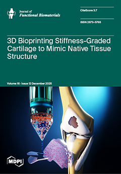
3D bioprinting offers a unique opportunity to fabricate tissues with complex, spatially controlled functionality that closely resembles native tissues. Articular cartilage is a clear example, as its zonal organization has marked variations in composition, cell density, and mechanical properties through its thickness—features absent in conventional homogeneous scaffolds. By enabling precise control over material deposition and crosslinking, 3D bioprinting allows the generation of stiffness gradients to reproduce, to a certain extent, the native environment, guiding cell distribution and matrix deposition in a spatially regulated manner. Our findings highlight how advanced 3D bioprinting strategies can move tissue engineering beyond uniform designs toward biomimetic scaffolds with enhanced mechano-functional relevance. View this paper
  • Issues are regarded as officially published after their release is announced to the table of contents alert mailing list.
  • You may sign up for e-mail alerts to receive table of contents of newly released issues.
  • PDF is the official format for papers published in both, html and pdf forms. To view the papers in pdf format, click on the "PDF Full-text" link, and use the free Adobe Reader to open them.
Order results
Result details
Section
Select all
Export citation of selected articles as:
15 pages, 2822 KB  
Article
Does Printing Orientation Matter in PolyJet 3D Printed Teeth for Endodontics? A Micro-CT Analysis
by Cláudia Barbosa, Tiago Reis, José B. Reis, Margarida Franco, Catarina Batista, Rui B. Ruben, Benjamín Martín-Biedma and Jose Martín-Cruces
J. Funct. Biomater. 2025, 16(12), 471; https://doi.org/10.3390/jfb16120471 - 18 Dec 2025
Viewed by 339
Abstract
This study aimed to identify the optimal printing orientation (X, Y, or Z axis) and positioning of a mandibular molar presenting an isthmus using PolyJet™ technology. The influence of these parameters on dimensional accuracy and on the behavior of 3D-printed teeth (3DPT) during [...] Read more.
This study aimed to identify the optimal printing orientation (X, Y, or Z axis) and positioning of a mandibular molar presenting an isthmus using PolyJet™ technology. The influence of these parameters on dimensional accuracy and on the behavior of 3D-printed teeth (3DPT) during endodontic preparation with ProTaper Gold® system was evaluated. Six groups (XA, XB, YA, YB, ZA, ZB; n = 10) were printed with different axis orientations and distinct isthmus positions relative to the build platform. All samples underwent micro-computed tomography scanning before and after endodontic preparation. Regarding preoperative analyses—canal volume, centroids, and total tooth volume and area—no significant differences were found between groups XA–YA or XB–YB (p > 0.05), supporting their comparability. In contrast, groups ZA and ZB differed significantly from all others (p < 0.05), failing to meet equivalence required for further comparison, and were therefore excluded. Postoperative evaluation—volume change, centroid displacement, transportation, and unprepared areas—revealed no significant differences between XA–YA and XB–YB. Within the limitations of this study, both printing orientation and position affected the accuracy and repeatability of 3DPT, with positioning exerting the greatest influence, while their behavior towards endodontic preparation remained consistent across orientations. Full article
(This article belongs to the Special Issue Digital Design and Biomechanical Analysis of Dental Materials)
Show Figures

Figure 1

15 pages, 1294 KB  
Article
Sodium Hypochlorite/Amino Acid Gel in the Non-Surgical Treatment of Periodontitis—Clinical and Molecular Results of Randomized Clinical Trial
by Ewa Dolińska, Katarzyna Golińska, Violetta Dymicka-Piekarska, Robert Milewski, Magdalena Sulewska and Małgorzata Pietruska
J. Funct. Biomater. 2025, 16(12), 470; https://doi.org/10.3390/jfb16120470 - 18 Dec 2025
Viewed by 364
Abstract
Due to the limitations of SRP, new methods are being sought to support non-surgical periodontal therapy. One of them is the use of antiseptics such as low-concentration sodium hypochlorite gel buffered with amino acids (NaOCl/AA). The aim of the study was to evaluate [...] Read more.
Due to the limitations of SRP, new methods are being sought to support non-surgical periodontal therapy. One of them is the use of antiseptics such as low-concentration sodium hypochlorite gel buffered with amino acids (NaOCl/AA). The aim of the study was to evaluate periodontal parameters and the concentration of metalloproteinase 8 (MMP-8) and interleukin 8 (IL-8) in the gingival crevicular fluid (GCF) after SRP with or without NaOCL/AA gel. The study included 40 periodontal patients randomized to study and control groups. Before SRP, the study group had a gel introduced into pockets with PD ≥ 5 mm. After treatment in both groups, the pocket depth (PD) decreased, there was a CAL gain, and unnoticeable changes in the gingival recession (GR). In the study group, deep pockets accounted for 25% of the sites examined prior to therapy, whereas after therapy, they decreased to 12%. In the control group, the proportion of deep periodontal pockets (PD ≥ 5 mm) fell from 17.46% to 9.05%. No differences were noted between groups. In the study group, there was a significant reduction in the amount of MMP-8 in GCF from 8.32 ng/mL to 5.14 ng/mL after 3 months. No statistically significant difference was observed in the control group. The concentration of IL-8 decreased significantly over time in both groups without differences between them. A single application of the NaOCl/AA gel in deep periodontal pockets does not affect clinical results and IL-8 levels. However, it had a significant effect on the amount of MMP-8. Full article
(This article belongs to the Special Issue New Biomaterials in Periodontology and Implantology)
Show Figures

Figure 1

20 pages, 6412 KB  
Article
Neo-Dermis Formation and Graft Timing After ADM Reconstruction: A Cohort Study with Histological Validation
by Daniel Pit, Teodora Hoinoiu, Bogdan Hoinoiu, Cristian Suciu, Panche Taskov, Zorin Petrisor Crainiceanu, Daciana Grujic, Isabela Caizer-Gaitan, Miruna Samfireag, Oana Suciu and Razvan Bardan
J. Funct. Biomater. 2025, 16(12), 469; https://doi.org/10.3390/jfb16120469 - 18 Dec 2025
Viewed by 348
Abstract
Acellular dermal matrices (ADMs) are widely used in soft-tissue reconstruction, yet the optimal timing for split-thickness skin grafting (STSG) remains unsettled. We conducted a single-center retrospective cohort study (January 2023–August 2025) of adults undergoing ADM-based reconstruction with Integra® Double Layer (IDL), Integra [...] Read more.
Acellular dermal matrices (ADMs) are widely used in soft-tissue reconstruction, yet the optimal timing for split-thickness skin grafting (STSG) remains unsettled. We conducted a single-center retrospective cohort study (January 2023–August 2025) of adults undergoing ADM-based reconstruction with Integra® Double Layer (IDL), Integra® Single Layer (ISL), or Nevelia®. Primary endpoints included length of stay (LOS), STSG requirement and timing, and in-hospital complications; secondary endpoints included spontaneous epithelialization. Prespecified adjusted analyses (linear/logistic models) controlled for age, sex, etiology, anatomical site, diabetes/PAOD, smoking, wound size (when available), wound contamination, and matrix type. Histology and immunohistochemistry (H&E, Masson trichrome, CD105, D2-40) assessed matrix integration and vascular/lymphatic maturation. Seventy-five patients were included (IDL n = 40; ISL n = 20; Nevelia n = 15). On multivariable analysis, matrix type was not an independent predictor of LOS (ISL vs. IDL β = +2.84 days, 95% CI −17.34 to +23.02; Nevelia vs. IDL β = −4.49 days, 95% CI −16.24 to +7.26). Complications were infrequent (6/75, 8.0%) and comparable across matrices; spontaneous epithelialization occurred in 3/75 patients (4.0%). A day-14 grafting strategy, applied only after documented clinical integration, was feasible in 30/75 (40.0%) patients without excess complications. Histology/IHC at 3–4 weeks demonstrated CD105-positive, perfused capillary networks with abundant collagen; at 4–6 weeks, D2-40-positive lymphatic structures confirmed progressive neo-dermis maturation, supporting the biological plausibility of earlier grafting once integration criteria are met. In this cohort, outcomes were broadly similar across matrices after adjustment. A criteria-based early STSG approach (~day 14) appears safe and operationally advantageous when integration is confirmed, while a minority of defects may heal without grafting. Prospective multicenter studies with standardized scar/functional measures and cost analyses are needed to refine patient selection and graft timing strategies. Full article
Show Figures

Figure 1

16 pages, 1804 KB  
Article
Flexural Strength and Hardness Analysis of 3D-Printed vs. Milled Resin Composites Indicated for Definitive Crowns
by Hunaida Khaled Tayeb, Nick Silikas, Abdulrahman Jafar Alhaddad and Julian Satterthwaite
J. Funct. Biomater. 2025, 16(12), 468; https://doi.org/10.3390/jfb16120468 - 18 Dec 2025
Viewed by 429
Abstract
The growing use of 3D-printed dental restorations has created a need to understand how the mechanical behaviour of definitive 3D-printed resin composites compares with milled counterparts. This study compared the mechanical properties of 3D-printed and milled definitive crowns and examined the factors affecting [...] Read more.
The growing use of 3D-printed dental restorations has created a need to understand how the mechanical behaviour of definitive 3D-printed resin composites compares with milled counterparts. This study compared the mechanical properties of 3D-printed and milled definitive crowns and examined the factors affecting these properties. The 3D-printed materials (Permanent Crown Resin: PCR, VarseoSmile Crown Plus: VCP, and Crowntec: CT) and milled blocks (Brilliant Crios: BC, Shofu Block HC: HC, and Grandio Blocs: Gr) were tested. Filler content was evaluated using the ash method (n = 3), and flexural strength (FS) and flexural modulus (Ef) were assessed using a three-point bending test (n = 10). Martens hardness (HM), indentation modulus, and Vickers hardness were determined using the Martens indentation test (n = 24). Vickers hardness was also measured with the Vickers indenter tester (n = 24). Statistical analysis investigated differences between materials and methods, and correlations between filler weight and mechanical properties (α = 0.05). FS of milled blocks, Gr (244.5 MPa), BC (225.5 MPa), and HC (155 MPa), were higher than that of the 3D-printed resin composites: PCR (143.6 MPa), CT (140.9 MPa), and VCP (128 MPa). Measured mechanical properties of the milled blocks were significantly higher than those of the 3D-printed materials (p < 0.001). HM of the 3D-printed materials was similar (≈217 N/mm2), while HM of the milled blocks ranged from 434.7 to 858.4 N/mm2. The 3D-printed materials showed comparable properties; however, milled blocks differed significantly (p < 0.05). Filler content was strongly associated with FS and HM. Within the limitations of this study, the milled resin composites showed markedly higher strength and hardness, while 3D-printed materials may be suitable for low-to-moderate load clinical scenarios. Further studies to evaluate the long-term performance of the 3D-printed resin composites are recommended. Full article
Show Figures

Figure 1

14 pages, 5186 KB  
Article
Split Crest—Is It Necessary to Fill the Gap?—A Controlled Trial
by Vladimir Biocanin, Zoran Tambur, Djordje Pejanovic, Marija Biocanin, Mihailo Ostojic, Marija Lalovic and Svetislav Zaric
J. Funct. Biomater. 2025, 16(12), 467; https://doi.org/10.3390/jfb16120467 - 18 Dec 2025
Viewed by 324
Abstract
Introduction: The split crest (SC) is a technique for horizontal ridge augmentation that enables simultaneous implant placement. While the use of bone grafts within the osteotomy gap is well-documented, the efficacy of dentin as a graft material in SC procedures has not been [...] Read more.
Introduction: The split crest (SC) is a technique for horizontal ridge augmentation that enables simultaneous implant placement. While the use of bone grafts within the osteotomy gap is well-documented, the efficacy of dentin as a graft material in SC procedures has not been thoroughly evaluated. Objective: This study aimed to assess whether the addition of bone graft or dentin to the osteotomy gap during the SC procedure improves bone width, density, and implant stability compared to SC without grafting. Materials and Methods: A partially randomized prospective study was conducted on 24 implants divided into three groups: SC only, SC and bone graft, and SC and dentin graft. Clinical and radiographic evaluations, including CBCT-based bone measurements and implant stability (ISQ) values, were performed preoperatively, immediately postoperatively, and three months after surgery. All patients were followed for one year. Results: Significant increases in bone width were recorded in all groups, with gains exceeding 2 mm in the SC and SC and bone graft groups. The SC and dentin group showed the least bone gain and the greatest horizontal bone resorption (p < 0.05). Conclusions: While bone grafting may offer modest advantages in bone preservation, the use of dentin grafting may not demonstrate significant benefits. Full article
(This article belongs to the Special Issue Functional Dental Materials for Orthodontics and Implants)
Show Figures

Figure 1

17 pages, 7531 KB  
Article
L-Serine–Incorporated Collagen Scaffolds for Modulating In Vivo Degradation Behavior
by Su-Young Kim, Ji-Hyeon Oh, Min-Ho Hong, Joon Ha Lee, You-Young Jo and Seong-Gon Kim
J. Funct. Biomater. 2025, 16(12), 466; https://doi.org/10.3390/jfb16120466 - 18 Dec 2025
Viewed by 318
Abstract
Collagen-based biomaterials are widely used, but their relatively rapid biodegradation can limit functional duration. Such collagen constructs are widely used as barrier membranes in guided tissue and bone regeneration, where controlled degradation is essential for maintaining function. Although conventional crosslinking methods extend stability, [...] Read more.
Collagen-based biomaterials are widely used, but their relatively rapid biodegradation can limit functional duration. Such collagen constructs are widely used as barrier membranes in guided tissue and bone regeneration, where controlled degradation is essential for maintaining function. Although conventional crosslinking methods extend stability, they may introduce cytotoxicity, alter mechanical behavior, or hinder tissue integration. This study evaluated whether incorporating L-serine, a polar amino acid capable of hydrogen bonding, could modulate collagen structure and slow degradation without chemical crosslinking. L-Serine was selected because its hydroxyl-containing side chain can engage in biocompatible, hydrogen-bond–mediated interactions that offer a mild, non-crosslinking means of stabilizing collagen. Collagen scaffolds, prepared by incorporating L-serine into a collagen hydrogel followed by drying, were produced with 0–40 wt% L-serine and characterized using X-ray diffraction, Fourier-transform infrared spectroscopy, circular dichroism, and scanning electron microscopy. In vivo degradation was assessed in a subcutaneous mouse model comparing unmodified collagen, collagen containing 40 wt% L-serine, and a commercially available bilayer porcine collagen membrane (Bio-Gide®, composed of type I and III collagen), with residual area quantified by serial sonography and histological evaluation. Low-to-moderate L-serine incorporation preserved triple-helical features, while 40 wt% led to crystalline domain formation and β-sheet enrichment. L-serine–treated collagen exhibited significantly greater residual area (2.70 ± 1.45 mm2) than unmodified collagen (0.37 ± 0.22 mm2, p < 0.05), although Bio-Gide® remained the most persistent (5.64 ± 2.76 mm2). These findings demonstrate that L-serine incorporation can modulate collagen structure and degradation kinetics through a simple, aqueous, and non-crosslinking approach. The results provide preliminary feasibility data supporting amino acid–assisted tuning of collagen resorption properties and justify further evaluation using membrane-specific fabrication and application-relevant testing. Full article
Show Figures

Graphical abstract

15 pages, 1909 KB  
Article
The Carbon Dots from Seabuckthorn (Hippophae rhamnoides L.) Leaves: Recycle the Herbal Waste Products for a Nano-Formulation in Delivering Bioactive Compounds
by Chen-Xi Xia, Xiong Gao, Queenie Wing-Sze Lai, Zheng-Qi Wang, Lish Sheng-Yin Lin, Janet Yuen-Man Ho, Jia-Yu Zhu, Roy Wai-Lun Tang, Tina Ting-Xia Dong and Karl Wah-Keung Tsim
J. Funct. Biomater. 2025, 16(12), 465; https://doi.org/10.3390/jfb16120465 - 17 Dec 2025
Viewed by 277
Abstract
Carbon dots have emerged as promising nanocarriers for drug delivery due to their unique physicochemical properties and biocompatibilities. Here, the potential of leaf-derived carbon dots (named as SBLCD), derived from Seabuckthorn (Hippophae rhamnoides L.), was illustrated as a novel nano-formulation [...] Read more.
Carbon dots have emerged as promising nanocarriers for drug delivery due to their unique physicochemical properties and biocompatibilities. Here, the potential of leaf-derived carbon dots (named as SBLCD), derived from Seabuckthorn (Hippophae rhamnoides L.), was illustrated as a novel nano-formulation for bioactive compound delivery. Seabuckthorn leaves, rich in flavonoids, are the waste product during the production of Seabuckthorn fruits. The wasted leaves were utilized to synthesize carbon dots via a hydrothermal method. The resulting SBLCD, characterized by TEM, FT-IR and Raman spectroscopy, exhibited a diameter of ~5 nm in both amorphous and quasi-crystalline forms. Applications of SBLCD in cultures demonstrated robust properties of anti-inflammation and inducing neuronal cell differentiation. Furthermore, SBLCD was able to encapsulate luteolin, a bioactive flavonoid. The enhanced delivery efficiency translated to superior biological activity, with SBLCD-luteolin requiring only 1.50 μg/mL in achieving the EC50 efficacy, as compared to 6.82 μg/mL for free luteolin in pNF200-Luc expression assays. This approach not only valorizes Seabuckthorn leaf by-products but also potentially improves the efficacy of encapsulated flavonoids. The development of SBLCD as a multifunctional platform for flavonoid delivery represents a promising strategy in enhancing the efficacy of neuroactive compounds, combining anti-inflammatory effects (>70% cytokine suppression) with enhanced cellular uptake (4.5-fold increase). Full article
(This article belongs to the Section Biomaterials for Drug Delivery)
Show Figures

Graphical abstract

2 pages, 319 KB  
Correction
Correction: Rodrigues et al. Physicochemical, Morphological, and Cytotoxic Properties of Brazilian Jackfruit (Artocarpus heterophyllus) Starch Scaffold Loaded with Silver Nanoparticles. J. Funct. Biomater. 2023, 14, 143
by José Filipe Bacalhau Rodrigues, Valeriano Soares Azevedo, Rebeca Peixoto Medeiros, Gislaine Bezerra de Carvalho Barreto, Maria Roberta de Oliveira Pinto, Marcus Vinicius Lia Fook and Maziar Montazerian
J. Funct. Biomater. 2025, 16(12), 464; https://doi.org/10.3390/jfb16120464 - 17 Dec 2025
Viewed by 170
Abstract
In the original publication [...] Full article
Show Figures

Figure 4

17 pages, 6202 KB  
Article
Three-Dimensional Printing of Calcium Phosphate-Mesoporous Bioactive Glass Scaffolds for Bone Tissue Engineering
by Ana Beatriz Gomes de Carvalho, Lais Medeiros Cardoso, Igor Paulino Mendes Soares, Joyce Rodrigues de Souza, Arpita Roy, Prabaha Sikder, Aldo R. Boccaccini, Eliandra de Sousa Trichês and Marco C. Bottino
J. Funct. Biomater. 2025, 16(12), 463; https://doi.org/10.3390/jfb16120463 - 16 Dec 2025
Viewed by 1384
Abstract
Calcium phosphate cements (CPCs) and biomaterials, such as mesoporous bioactive glass (MBG), are critical for bone tissue engineering. This study aimed to 3D-print CPC scaffolds modified with MBG to enhance their osteogenic potential and regenerative ability. MBG powder was synthesized and characterized using [...] Read more.
Calcium phosphate cements (CPCs) and biomaterials, such as mesoporous bioactive glass (MBG), are critical for bone tissue engineering. This study aimed to 3D-print CPC scaffolds modified with MBG to enhance their osteogenic potential and regenerative ability. MBG powder was synthesized and characterized using transmission electron microscopy (TEM), X-ray diffraction (XRD), and nitrogen adsorption–desorption techniques. A commercial CPC ink (hydroxyapatite/α-tricalcium phosphate) was mixed with 5% MBG (w/w; CPC/MBG), and, after rheological assessment, the mixture was used to obtain scaffolds via 3D printing. These scaffolds were then tested for chemical, morphological, and mechanical properties, as well as ion release analysis. Unmodified CPC 3D-printed scaffolds served as controls. Biological experiments, including cell viability, DNA content, cell adhesion/spreading, and osteogenic gene expression, were performed by seeding alveolar bone-derived mesenchymal stem cells onto the scaffolds. Statistics were performed using Student’s t-test and ANOVA with post hoc tests (α = 5%). MBG characterization showed a typical mesoporous structure with aligned microchannels and an amorphous structure. Both formulations released calcium and phosphate ions; however, CPC/MBG also released silicon. Cell viability, adhesion/spreading, and DNA content were significantly greater in CPC/MBG scaffolds compared to CPC (p < 0.05) after 3 and 7 days of culture. Furthermore, CPC/MBG supported increased expression of key osteogenic genes, including collagen (COL1A1), osteocalcin (OCN), and Runt-related transcription factor 2 (RUNX2), after 14 days (p < 0.05). The combination of CPC ink with MBG particles effectively enhances the biocompatibility and osteogenic potential of the scaffold, making it an innovative bioceramic ink formulation for 3D printing personalized scaffolds for bone regeneration. Full article
Show Figures

Figure 1

16 pages, 10065 KB  
Article
Effect of Immersion in Quaternary Ammonium Methacryloxy Silane Mixed Monomer on the Mechanical Properties and Antibacterial Activity of a 3D-Printed Urethane Dimethacrylate Denture Base Resin
by Kun-Min Kim, Yeseul Park, Jimin Kim, Mu-Yeol Cho and Jee-Hwan Kim
J. Funct. Biomater. 2025, 16(12), 462; https://doi.org/10.3390/jfb16120462 - 14 Dec 2025
Viewed by 360
Abstract
Denture base resins are susceptible to microbial colonization, and current antibacterial additives often lose effectiveness and may weaken material properties. This study evaluated whether immersion in a quaternary ammonium methacryloxy silane (QAMS)-containing monomer can enhance antibacterial activity without compromising the mechanical properties of [...] Read more.
Denture base resins are susceptible to microbial colonization, and current antibacterial additives often lose effectiveness and may weaken material properties. This study evaluated whether immersion in a quaternary ammonium methacryloxy silane (QAMS)-containing monomer can enhance antibacterial activity without compromising the mechanical properties of digital light processing–printed urethane dimethacrylate denture base resin. Specimens of printed denture base resin were immersed in mixtures of denture base resin and a QAMS-containing monomer at ratios of 10:0 (Control), 7:3 (K3), 5:5 (K5), 3:7 (K7), and 0:10 (K10), followed by post-curing. Flexural strength and modulus were measured by three-point bending, and surface hardness was assessed by Vickers microhardness testing. Antibacterial activity against Streptococcus mutans was assessed by inhibition-zone and colony-counting assays. All QAMS-treated groups preserved flexural strength, with a slight reduction in modulus in K5 (p < 0.05), while hardness remained unchanged. Antibacterial activity improved in all QAMS-treated groups; K5 and K7 showed the strongest results. Surface analyses using scanning electron microscopy and energy-dispersive X-ray spectroscopy verified formation of a Si-rich modified layer. QAMS immersion followed by post-curing produced a stable, contact-active antibacterial surface without reducing mechanical properties. Among the formulations, K7 (~21 wt% QAMS) provided the most favorable balance of antibacterial activity and mechanical performance. Full article
Show Figures

Figure 1

17 pages, 2516 KB  
Article
Cationic Surface Modification Combined with Collagen Enhances the Stability and Delivery of Magnetosomes for Tumor Hyperthermia
by Yu Wang, Conghao Lin, Yubing Zhang, Wenjun Li, Hongli Cui, Bohan Li, Zhengyi Liu, Kang Wang, Qi Wang, Yinchu Wang, Kangning Lv, Yandi Huang, Hongqin Zhuang and Song Qin
J. Funct. Biomater. 2025, 16(12), 461; https://doi.org/10.3390/jfb16120461 - 12 Dec 2025
Viewed by 1216
Abstract
Magnetosomes (MTS), membrane-enclosed magnetic nanoparticles naturally biomineralized by magnetotactic bacteria, are promising materials for tumor hyperthermia owing to their good biocompatibility and heating efficiency. However, their application is limited by poor suspension stability and low injectability at high concentrations. This study aimed to [...] Read more.
Magnetosomes (MTS), membrane-enclosed magnetic nanoparticles naturally biomineralized by magnetotactic bacteria, are promising materials for tumor hyperthermia owing to their good biocompatibility and heating efficiency. However, their application is limited by poor suspension stability and low injectability at high concentrations. This study aimed to enhance magnetosome stability and delivery performance through surface cationization combined with collagen matrix stabilization. The resulting cationic magnetosomes (CMTS) exhibited an increased positive charge on the outer membrane. Collagen, functioning as a negatively charged matrix under mildly alkaline conditions, effectively stabilized the cationic magnetosomes, forming CMTS–collagen aqueous suspensions (CMTS-Colas) that remained well-suspended for over 24 h and could be easily resuspended after 10 days of storage. Compared with native magnetosome suspensions, CMTS in collagen displayed smaller hydrodynamic diameters and significantly improved injectability through 26G and 31G fine needles. Under an alternating magnetic field, 2 mg/mL CMTS-Colas efficiently induced over 98% apoptosis in hepatoma cells after two treatment sessions and led to complete loss of cell viability after three sessions. These findings demonstrate that CMTS-Colas substantially improve the suspension stability and injectability of magnetosomes while maintaining strong hyperthermic efficacy, suggesting a promising strategy for stabilizing magnetosomes and potentially benefiting other charged, aggregation-prone magnetic biomaterials. Full article
(This article belongs to the Special Issue Biomaterials for Drug Delivery and Cancer Therapy)
Show Figures

Graphical abstract

19 pages, 1616 KB  
Review
Porcine Corneal Models as Translational Platforms for Innovative Therapies: Current Insights and Future Directions
by Patrícia Nápoles, Mónica Faria and Elisa Julião Campos
J. Funct. Biomater. 2025, 16(12), 460; https://doi.org/10.3390/jfb16120460 - 9 Dec 2025
Viewed by 392
Abstract
The development of advanced biomaterials for corneal applications requires robust translational platforms that faithfully replicate human characteristics. Porcine corneas are increasingly recognized for ophthalmic research. Their unique combination of anatomical similarity, biomechanical comparability, and accessibility make them highly suitable for preclinical evaluation of [...] Read more.
The development of advanced biomaterials for corneal applications requires robust translational platforms that faithfully replicate human characteristics. Porcine corneas are increasingly recognized for ophthalmic research. Their unique combination of anatomical similarity, biomechanical comparability, and accessibility make them highly suitable for preclinical evaluation of innovative therapies, bridging the gap between preclinical discovery and clinical application. This review outlines the utility of porcine corneal models in validating advanced biomaterials, particularly in ex vivo settings, focusing on current methodologies, while addressing challenges and future directions. We aim to underscore the potential of porcine corneal models to accelerate the translation of next-generation biomaterials into clinically relevant corneal therapies. Full article
(This article belongs to the Special Issue Biocompatible Research of Materials in Biomedical Applications)
Show Figures

Graphical abstract

19 pages, 1229 KB  
Article
Cytotoxicity and Genotoxicity of Resin-Based Dental Composites Modified with Quaternary Ammonium Salts
by Izabela Szymczak-Pajor, Maja Zalega, Joanna Nowak, Agnieszka Śliwińska, Katarzyna Woźniak and Kinga Bociong
J. Funct. Biomater. 2025, 16(12), 459; https://doi.org/10.3390/jfb16120459 - 9 Dec 2025
Viewed by 427
Abstract
The primary reason of dental restoration failure is the recurrence of caries, driving research to incorporate quaternary ammonium salts (QASs) into resin-based composites (RBCs). Given the prolonged contact of these materials with oral tissue, this in vitro study assessed the biocompatibility (cytotoxicity and [...] Read more.
The primary reason of dental restoration failure is the recurrence of caries, driving research to incorporate quaternary ammonium salts (QASs) into resin-based composites (RBCs). Given the prolonged contact of these materials with oral tissue, this in vitro study assessed the biocompatibility (cytotoxicity and genotoxicity) profiles of experimental RBCs modified with cetyltrimethylammonium bromide (CTAB) and dimethyldioctadecylammonium bromide (DODAB), using two restorative materials: an unmodified–experimental composite, KE, and Flow-Art (FA) as comparative standards. The primarily novelty of this study is the direct comparison of the cellular safety profiles of CTAB vs. DODAB when incorporated into RBCs. Human fibroblast BJ cells were exposed to composite eluates for 24 h, and cell viability (MTT assay), the percentage of apoptotic and necrotic cells (the Annexin V/Propidium Iodide (PI) flow cytometry method), and DNA damage (the alkaline comet assay) were quantified. Among the compounds evaluated, only CTAB caused a significant, dose-dependent decrease in BJ cell viability, primarily by inducing late apoptosis or necrosis. Cell viability was severely reduced, dropping by 84% at 2 wt% CTAB (p < 0.001) compared to control. Consistent with this effect, CTAB also induced a dose-dependent increase in DNA damage. In contrast, the DODAB-modified composites, along with the KE and FA controls, exhibited non-cytotoxic and non-genotoxic profiles across all tested concentrations. This innovative comparative assessment highlights that DODAB exhibits superior cellular safety, offering vital guidance to prioritize its use for developing safe and effective next-generation antibacterial dental composites. Conversely, CTAB is precluded for clinical use at these concentrations due to its observed toxicity. Full article
Show Figures

Figure 1

27 pages, 6969 KB  
Article
Evaluation of Composites Comprising Spherical, Porous, Sintered β-Tricalcium Phosphate Particles and Cyanoacrylate as Bone Cement
by Kazuaki Hashimoto, Shuhei Aida, Iori Takigawa, Hirobumi Shibata, Satoshi Kobune, Toshiisa Konishi, Takashi Meguro, Shigeo Fukuyama and Shinya Tanaka
J. Funct. Biomater. 2025, 16(12), 458; https://doi.org/10.3390/jfb16120458 - 9 Dec 2025
Viewed by 326
Abstract
Bone cements based on polymethyl methacrylate (PMMA) remain the clinical standard for joint replacement and vertebral augmentation but suffer from several major challenges. These include excessive stiffness compared with cancellous bone, lack of resorption and osteoconductivity, and thermal necrosis during curing. Calcium phosphate [...] Read more.
Bone cements based on polymethyl methacrylate (PMMA) remain the clinical standard for joint replacement and vertebral augmentation but suffer from several major challenges. These include excessive stiffness compared with cancellous bone, lack of resorption and osteoconductivity, and thermal necrosis during curing. Calcium phosphate cements (CPCs) are bioactive and resorbable but tend to exhibit low mechanical strength, poor injectability and brittle fracture. The work reported herein developed an injectable composite bone cement by combining spherical, porous, sintered β-tricalcium phosphate (β-TCP) particles with a cyanoacrylate adhesive. The β-TCP granules provided bioactivity and a favorable microarchitecture while the cyanoacrylate ensured strong adhesion and rapid setting. Ion substitution with Mg, Na and Si was found to modify the surface acidity of the material while also inhibiting cyanoacrylate polymerization, thereby extending the setting time and lowering the exotherm temperature. This composite exhibited high chemical stability, smooth injectability and early surface reactivity indicative of osteoconductivity. The compressive strength of the material stabilized at approximately 40 MPa and so exceeded that of cancellous bone. This new material also showed ductility, energy absorption and superior impact resistance, although its tensile and fatigue resistance remained limited. Importantly, the composite provided strength comparable to that of PMMA in cemented models during fixation tests and significantly outperformed CPCs in cementless tibial tray fixation experiments. These findings demonstrate that the present β-TCP/cyanoacrylate cement bridges the gap between PMMA and CPCs by combining injectability and mechanical reliability with bioactivity. This cement is therefore a promising next-generation option for minimally invasive osteoporotic fracture treatment and revision arthroplasty. Full article
(This article belongs to the Section Bone Biomaterials)
Show Figures

Figure 1

20 pages, 2850 KB  
Article
Influence of Sandblasting Particle Size on the Shear Bond Strength of Orthodontic Brackets to Milled and 3D-Printed Provisional Crowns or Materials After Artificial Aging
by Khurshid Mattoo, Mohammed E. Sayed, Marwan Someli, Ahmed Alhazmi, Mohammed Khawajy, Shroog A. Almasoudi, Ebrahim Fihaid Alsubaiy, Saeed M. Alqahtani, Mohammed A. Alfaifi and Raghdah M. Alshaibani
J. Funct. Biomater. 2025, 16(12), 457; https://doi.org/10.3390/jfb16120457 - 8 Dec 2025
Viewed by 473
Abstract
Purpose: This in vitro study ascertained the impact of three distinct alumina particle sizes on the shear bond strength (SBS) between two distinct provisional crowns (milled and 3D-printed) and stainless-steel orthodontic brackets following artificial aging. Materials and methods: Eighty specimens [disc 10 mm [...] Read more.
Purpose: This in vitro study ascertained the impact of three distinct alumina particle sizes on the shear bond strength (SBS) between two distinct provisional crowns (milled and 3D-printed) and stainless-steel orthodontic brackets following artificial aging. Materials and methods: Eighty specimens [disc 10 mm diameter/15 mm height] were fabricated with two provisional crown materials, milled (CopraTemp) [group (GP) M] and three-dimensionally printed (Asiga DentaTooth) (GP P), and divided into eight subgroups based on alumina oxide (sandblasting) particle size surface treatments of 25 μm [P25, M25], 50 μm [P50, M50], and 100 μm [P100, M100], with no surface treatment specimens serving as control [PC, MC]. After thermocycling (2200 cycles), the SBS and Adhesive Remnant Index (ARI) were calculated. Statistical tests included one-way analysis of variance (ANOVA) (Kruskal–Wallis), followed by post hoc tests [Tukey HSD, Dunn’s], with the probability ‘p’ value being significant at 0.05 (p ≤ 0.05). Results: Without surface treatment, the 3D-printed provisional crown had the lowest SBS [median (IQR); 12.8 (2.74)]. The highest SBS was found in both milled and 3D-printed PMs with 50-micron particle sizes [Milled = 23.10 (2.3); Printed = 20.72 (2.31)], followed by 100-micron [Milled = 20 (2.36); Printed = 17.99 (3.45)] and 25-micron [Milled = 16.13 (2.71); Printed = 15.08 (1.55)]. The majority of cohesive failures were seen in the milled subgroups, while all subgroups of 3D-printed provisional material had adhesive bond failures. Conclusions: Sandblasting, irrespective of particle size, enhances SBS in both milled and 3D-printed provisional restorations; however, 50-micron alumina particles are recommended since they enhance SBS substantially. Full article
(This article belongs to the Section Dental Biomaterials)
Show Figures

Figure 1

15 pages, 2354 KB  
Article
3D-Printed Multifunctional Multicompartment Polymer-Based Capsules for Tunable and Spatially Controlled Drug Release
by Antonio Minopoli, Giordano Perini, Davide Evangelista, Matteo Marras, Alberto Augello, Valentina Palmieri, Marco De Spirito and Massimiliano Papi
J. Funct. Biomater. 2025, 16(12), 456; https://doi.org/10.3390/jfb16120456 - 8 Dec 2025
Viewed by 488
Abstract
The development of polymer-based systems is central to the design of next-generation drug delivery carriers, as polymers enable versatile tuning of physicochemical properties and responsiveness. In this work, we introduce a 3D printing-based strategy for the fabrication of multicompartment capsules that integrate multiple [...] Read more.
The development of polymer-based systems is central to the design of next-generation drug delivery carriers, as polymers enable versatile tuning of physicochemical properties and responsiveness. In this work, we introduce a 3D printing-based strategy for the fabrication of multicompartment capsules that integrate multiple polymers within a unique one-step process. This approach allows precise spatial organization and structural complexity, yielding capsules with customizable features such as compartmentalization, polymer-specific responsiveness, and localized release control. In particular, pH-triggered release can be programmed across distinct polymeric regions of the capsules, enabling site-specific delivery along different intestinal segments, including the small intestine and colon. The use of 3D printing thus provides a scalable and adaptable platform to generate multifunctional polymer-based carriers with finely tunable drug release profiles, paving the way for new directions in polymer-enabled controlled delivery technologies. Full article
(This article belongs to the Special Issue Polymer-Based Materials in Biomedical Applications)
Show Figures

Graphical abstract

22 pages, 8541 KB  
Article
The Impact of Post-Printing Hydration in NaCl Solution on the Properties of Binder Jet 3D-Printed Calcium Sulfate and Its Converted Hydroxyapatite
by Faungchat Thammarakcharoen, Autcharaporn Srion, Waraporn Suvannapruk, Wiroj Limtrakarn and Jintamai Suwanprateeb
J. Funct. Biomater. 2025, 16(12), 455; https://doi.org/10.3390/jfb16120455 - 8 Dec 2025
Viewed by 441
Abstract
Binder jet 3D printing of calcium sulfate-based materials combined with phase transformation offers a versatile route for fabricating customized bone grafts; however, controlling the transformation process remains a key challenge. This study investigates the effect of post-printing hydration in sodium chloride (NaCl) solutions [...] Read more.
Binder jet 3D printing of calcium sulfate-based materials combined with phase transformation offers a versatile route for fabricating customized bone grafts; however, controlling the transformation process remains a key challenge. This study investigates the effect of post-printing hydration in sodium chloride (NaCl) solutions on the phase transformation, dimension, and compressive properties of binder jet-printed calcium sulfate (3DPCaS) toward hydroxyapatite (3DPHA) formation. The as-printed 3DPCaS primarily consisted of bassanite with minor gypsum, which progressively transformed into gypsum upon immersion in NaCl solutions of varying concentrations (1–5 M) and durations (2–30 min). Increased immersion time and moderate NaCl concentrations (2–4 M) promoted gypsum formation without inducing dimensional instability. Subsequent transformation in phosphate solution produced 3DPHA with high hydroxyapatite (HA) purity, reaching 100% conversion. Microstructural analysis revealed recrystallized, plate-like gypsum crystals that served as favorable templates for HA nucleation. The resulting 3DPHA exhibited enhanced specific modulus (up to 274.9 MPa.m3/kg) and specific strength (up to 7.5 MPa.m3/kg). The optimal condition, immersion in 4 M NaCl solution for 30 min, achieved a balance between complete HA transformation, mechanical enhancement, and dimensional stability. Controlled ionic hydration thus represents a simple, low-cost, and effective strategy for improving properties of 3DPHA bone grafts. Full article
(This article belongs to the Special Issue Three-Dimensional-Printable Biomaterials for Bone Regeneration)
Show Figures

Figure 1

17 pages, 2753 KB  
Article
Three-Dimensional Human Neurovascular Unit Modeling Reveals Cell-Specific Mechanisms of Traumatic Brain Injury
by Liam H. Power, Evan C. Marcet, Zihong Chen, Jinpeng Chen, Artem Arkhangelskiy, Michael J. Whalen, Ying Chen and David L. Kaplan
J. Funct. Biomater. 2025, 16(12), 454; https://doi.org/10.3390/jfb16120454 - 7 Dec 2025
Viewed by 859
Abstract
Severe traumatic brain injury includes neurovascular unit (NVU) damage that is linked to the later development of neurodegenerative diseases. Cell-type-specific contributions and crosstalk between cells of the neurovascular unit following brain injury remain poorly defined in human cells. Here, we developed a three-dimensional [...] Read more.
Severe traumatic brain injury includes neurovascular unit (NVU) damage that is linked to the later development of neurodegenerative diseases. Cell-type-specific contributions and crosstalk between cells of the neurovascular unit following brain injury remain poorly defined in human cells. Here, we developed a three-dimensional (3D) human NVU model using silk–collagen scaffolds to examine cellular responses to controlled cortical impact (CCI). Using this platform, we show that CCI induced acute cell death in astrocytes, microglia, and endothelial cells but spared pericytes, which occurred independently of classical apoptotic or necroptotic pathways. Astrocytes and microglia were the primary sources of early bioactive IL-1β release, while endothelial junctional integrity was differentially regulated by support cells: astrocytes destabilized VE-cadherin, pericytes preserved barrier proteins, and microglia contributed to Claudin-5 loss in multicellular settings. Conditioned media experiments demonstrated that soluble factors from injured support cells alone were sufficient to disrupt endothelial junctional proteins (ZO-1 and Occludin) and induce inflammatory adhesion molecules (ICAM-1 and VCAM-1). Together, these findings define cell-type-specific injury responses and reveal how NVU interactions regulate vascular dysfunction after trauma, providing a human-based framework for understanding blood–brain barrier (BBB) disruption following traumatic brain injury (TBI). Full article
Show Figures

Graphical abstract

12 pages, 513 KB  
Article
Fluorescence as a Quantitative Indicator of Cariogenic Bacteria During Chemo-Mechanical Caries Excavation with BRIX 3000 in Primary Teeth
by Zornitsa Lazarova, Raina Gergova and Nadezhda Mitova
J. Funct. Biomater. 2025, 16(12), 453; https://doi.org/10.3390/jfb16120453 - 6 Dec 2025
Viewed by 402
Abstract
Minimally invasive treatment is increasingly successful in managing carious lesions in primary teeth, owing to the regenerative capacity of the dental pulp and the possibility to influence the pulp–dentin complex. Chemo-mechanical caries excavation (CME) with Brix 3000, a papain-based enzymatic agent, allows selective [...] Read more.
Minimally invasive treatment is increasingly successful in managing carious lesions in primary teeth, owing to the regenerative capacity of the dental pulp and the possibility to influence the pulp–dentin complex. Chemo-mechanical caries excavation (CME) with Brix 3000, a papain-based enzymatic agent, allows selective removal of infected dentin while preserving affected dentin for potential remineralization. Fluorescence-aided caries excavation (FACE) enables visualization of porphyrins produced by cariogenic microorganisms, guiding selective dentin removal. In this study, 42 children aged 4–7 years with ICDAS II code 05–06 lesions in primary molars were treated, and the correlation between fluorescence intensity and cariogenic microbial load was evaluated. CME was performed using Brix 3000, and residual dentin was categorized by fluorescence as red, red with pale-pink areas, pale-pink, or non-fluorescent. Microbiological samples were collected pre- and post-excavation, cultured under standardized laboratory conditions, and quantitatively analyzed. Results showed that higher fluorescence intensity corresponded to increased presence of S. mutans (ρ = 0.945, p < 0.001), while other species were present in lower quantities. CME with Brix 3000 significantly reduced microbial load, and fluorescence reliably indicated areas requiring removal. These findings demonstrate that combining FACE with Brix 3000 allows precise, minimally invasive caries removal in primary teeth, providing an objective method to guide tissue-preserving excavation while effectively controlling cariogenic microorganisms. Full article
(This article belongs to the Special Issue Biomaterials for Management of Dental Caries and Periodontal Disease)
Show Figures

Figure 1

16 pages, 1608 KB  
Article
Injectable Piezoelectric Hydrogel for Vital Pulp Therapy
by Varun Solanki, Carolina Montoya, Prasanna Neelakantan, Maobin Yang and Santiago Orrego
J. Funct. Biomater. 2025, 16(12), 452; https://doi.org/10.3390/jfb16120452 - 5 Dec 2025
Viewed by 706
Abstract
Vital pulp therapy (VPT) seeks to preserve pulp vitality by using biocompatible with regenerative potential. This study tested the hypothesis that an injectable gelatin methacryloyl (GelMA) hydrogel containing piezoelectric barium titanate promotes odontogenic differentiation of dental pulp stem cells (DPSC) significantly better than [...] Read more.
Vital pulp therapy (VPT) seeks to preserve pulp vitality by using biocompatible with regenerative potential. This study tested the hypothesis that an injectable gelatin methacryloyl (GelMA) hydrogel containing piezoelectric barium titanate promotes odontogenic differentiation of dental pulp stem cells (DPSC) significantly better than a commercially available tricalcium silicate material used for vital pulp therapy. First, the light-curable, injectable piezoelectric hydrogel was engineered and characterized for its physicomechanical, piezoelectric properties and biocompatibility to DPSCs. The effect of this gel on the odontogenic differentiation of DPSCs was determined by measuring the expression level of key genes, compared to Biodentine XP. The hydrogel exhibited excellent injectability (<1 kgf of force), mechanical stability, and generated physiologically relevant voltages under cyclic loading mimicking mastication. MTT and ROS assays show no cytotoxic or damaging oxidative stress effects. When DPSCs were cultured over the materials under cyclic loading, the piezoelectric hydrogel significantly enhanced cell viability and upregulated COL1A1, DSPP, and DMP1 expression compared to Biodentine XP and non-piezoelectric hydrogel controls. These findings establish piezoelectric hydrogel as a self-powered, bioactive platform that converts physiological forces into regenerative bioelectric cues, offering a promising next-generation material for vital pulp therapy. Full article
(This article belongs to the Special Issue Advanced Biomaterials and Engineered Systems in Endodontics)
Show Figures

Figure 1

12 pages, 1447 KB  
Article
Three-Dimensional Bioprinting of Regenerative Cartilage Constructs with Directional Ionically Derived Stiffness Gradients
by Maryam Hosseini, Angeliki Dimaraki, Gerjo. J. V. M. van Osch, Lidy E. Fratila-Apachitei, Pedro J. Díaz-Payno and Amir A. Zadpoor
J. Funct. Biomater. 2025, 16(12), 451; https://doi.org/10.3390/jfb16120451 - 3 Dec 2025
Viewed by 2099
Abstract
Tissue engineering approaches for cartilage tissue regeneration are expanding to include the complex features of the tissue, such as the biological and mechanical gradients. Many of these approaches are, however, based on the use of multiple biomaterials or concentrations, and crosslinking methods that [...] Read more.
Tissue engineering approaches for cartilage tissue regeneration are expanding to include the complex features of the tissue, such as the biological and mechanical gradients. Many of these approaches are, however, based on the use of multiple biomaterials or concentrations, and crosslinking methods that make it difficult to integrate and control the properties of the resulting scaffolds. In this study, a 3D bioprinted scaffold with a stiffness gradient was fabricated by using a single biomaterial type and concentration combined with a directional ionic crosslinking method. The scaffolds revealed a gradient in stiffness from 39.8 ± 6.6 kPa at the top to 60.6 ± 10.9 kPa at the bottom of the scaffolds. Live/dead analysis of human chondrocytes embedded in the scaffolds showed no negative effects of the stiffness gradient on cell viability over 28 days. The induced stiffness gradient led to a gradient in cell density and sulfated glycosaminoglycan deposition in the bioprinted tissue constructs with enhanced values in the softer top region of the scaffolds as compared to the stiffer bottom part. This study showed a novel method to generate scaffolds with stiffness gradients from a single biomaterial and indicates that such scaffolds could be used to spatially regulate the behavior of chondrocytes and the associated deposition of the cartilage matrix. Full article
Show Figures

Graphical abstract

18 pages, 3761 KB  
Article
Hexapeptide-Liposome Nanosystem for the Delivery of Endosomal pH Modulator to Treat Acute Lung Injury
by Yuting Ji, Qian Wang, Rujing Lin, Mimi Pang, Liya Sun, Jiameng Gong, Huiqiang Ma, Shan-Yu Fung and Hong Yang
J. Funct. Biomater. 2025, 16(12), 450; https://doi.org/10.3390/jfb16120450 - 1 Dec 2025
Viewed by 625
Abstract
The overactivation of endosomal Toll-like receptor (TLR) in macrophages plays an important role in the pathogenesis of acute lung injury (ALI). There is currently still a lack of nano-formulated and macrophage-targeted endosomal TLR inhibitors that have been approved for clinical uses. We previously [...] Read more.
The overactivation of endosomal Toll-like receptor (TLR) in macrophages plays an important role in the pathogenesis of acute lung injury (ALI). There is currently still a lack of nano-formulated and macrophage-targeted endosomal TLR inhibitors that have been approved for clinical uses. We previously discovered that the elevation of endosomal pH using nanodevices provides a promising strategy to specifically inhibit endosomal TLRs in macrophages. The weakly basic drug hydroxychloroquine (HCQ) has been reported for its capability to accumulate in endolysosomes and modulate the acidity in these compartments. To enhance its macrophage-targeting ability and the therapeutic efficacy in vivo, herein we formulated HCQ into a nanoform using liposomes, named HCQ-L. We found that HCQ-L was less cytotoxic and more effective in inhibiting endosomal TLRs (including TLR3, TLR4, TLR 7/8) than the molecular HCQ. Subsequently, a hexapeptide, Pep12, was inserted onto the surface of HCQ-L to form HCQ-L-P12. Interestingly, Pep12 modification significantly improved the stability of liposomes in aqueous solution for at least 2 years; while having enhanced inhibitory effects on TLR7/8 signaling, HCQ-L-P12 displayed similar effects on inhibiting the TLR4 pathway and down-stream pro-inflammatory cytokine production when compared with HCQ-L. Furthermore, both HCQ nanoformulations potently elevated the endosomal pH. In vivo evaluation showed that HCQ-L-P12 and HCQ-L (but not molecular HCQ) were able to alleviate lung inflammation and injuries by decreasing inflammatory cell infiltration upon intratracheal instillation in a lipopolysaccharide (LPS)-induced acute lung injury (ALI) mouse model. This research provides a new strategy to fabricate lipid-based nanocarriers for targeted delivery of endosomal pH modulators to treat ALI and other acute and chronic inflammatory disorders. Full article
(This article belongs to the Special Issue Nanomaterials for Drug Delivery Systems)
Show Figures

Graphical abstract

17 pages, 4639 KB  
Article
Comparative Animal Study of Zirconia-Coated Titanium Implants: Effect on Bone Formation and Collagen Fiber Orientation
by Kohei Osawa, Masatsugu Hirota, Toshitsugu Sakurai, Yohei Iinuma, Chikahiro Ohkubo, Hiroki Nagai, Takatsugu Yamamoto and Kenji Mitsudo
J. Funct. Biomater. 2025, 16(12), 449; https://doi.org/10.3390/jfb16120449 - 29 Nov 2025
Viewed by 727
Abstract
Tissue responses to zirconia-coated implants treated with molecular precursor method were evaluated. The zirconia film was characterized using scanning electron microscopy (SEM), atomic force microscopy (AFM), energy dispersive X-ray spectroscopy (EDX), X-ray photoelectron spectroscopy (XPS), and X-ray diffraction (XRD). Cylindrical titanium (ZrO2 [...] Read more.
Tissue responses to zirconia-coated implants treated with molecular precursor method were evaluated. The zirconia film was characterized using scanning electron microscopy (SEM), atomic force microscopy (AFM), energy dispersive X-ray spectroscopy (EDX), X-ray photoelectron spectroscopy (XPS), and X-ray diffraction (XRD). Cylindrical titanium (ZrO2/Ti) specimens were sandblasted, acid-etched, and coated with zirconia using the molecular precursor method. Control specimens were sandblasted and acid-etched only (SLA/Ti). After maxillary first molar extraction, four ZrO2/Ti and four SLA/Ti implants were placed in the alveolar bone of the rats, and tissue responses were observed after 3 weeks. Surface analysis using SEM and AFM showed zirconia was present on ZrO2/Ti surface, with coating not affecting surface morphology compared to SLA/Ti. EDX, XPS, and XRD measurements confirmed the ZrO2 coating on the roughened Ti. The amount of new bone was greater in ZrO2/Ti (77.0 ± 7.2%) than in SLA/Ti (59.7 ± 5.8%) (p = 0.807). Collagen fibers oriented perpendicular to implant surface were observed more frequently in ZrO2/Ti (67.3 ± 9.5%) than in SLA/Ti (18.8 ± 10.01%) (p < 0.001). The area of perpendicular collagen fibers was significantly larger in ZrO2/Ti (53.1 ± 13.4%) than in SLA/Ti (16.8 ± 2.6%) (p = 0.002). Zirconia-coated implants maintained surface morphology and improved bone formation and fiber orientation in the gingiva compared to conventional titanium implants in short-term animal experiments. Full article
(This article belongs to the Section Dental Biomaterials)
Show Figures

Figure 1

14 pages, 3165 KB  
Article
Fracture Load of Polyaryletherketone for 4-Unit Posterior Fixed Dental Prostheses: An In Vitro Study
by Dalea M. Bukhary, Hasan Y. Asiri, Ruwaida Z. Alshali, Walaa A. Babaeer, Thamer Y. Marghalani, Ghadeer I. Basunbul and Osama A. Qutub
J. Funct. Biomater. 2025, 16(12), 448; https://doi.org/10.3390/jfb16120448 - 29 Nov 2025
Viewed by 634
Abstract
Background/Objectives: This study aimed to evaluate the mechanical properties, particularly the fracture load, modulus of elasticity, and fracture patterns, of four-unit posterior tooth-supported fixed dental prostheses (FDPs) fabricated from various computer-aided design/computer-aided manufacturing (CAD-CAM) materials. Understanding the mechanical behavior of these materials [...] Read more.
Background/Objectives: This study aimed to evaluate the mechanical properties, particularly the fracture load, modulus of elasticity, and fracture patterns, of four-unit posterior tooth-supported fixed dental prostheses (FDPs) fabricated from various computer-aided design/computer-aided manufacturing (CAD-CAM) materials. Understanding the mechanical behavior of these materials is crucial for optimizing prosthesis performance in high-load-bearing posterior regions. Methods: A total of 68 standardized FDP frameworks were fabricated, each consisting of two abutments (first premolar and second molar) and two pontics (second premolar and first molar). The specimens were divided into four groups (n = 17): polyetheretherketone (PEEK), polyetherketoneketone (PEKK), 3Y zirconia (control 1), and 4Y zirconia (control 2). All samples underwent three-point bending tests using a universal testing machine with a crosshead speed of 0.5 mm/min. Fracture patterns were assessed visually and documented. Fractured specimens were examined using scanning electron microscopy (SEM). Data were analyzed using the SPSS v20. Normality was assessed with the Shapiro–Wilk test. The fracture loads were compared using the Kruskal–Wallis test with Bonferroni correction, and the modulus of elasticity was analyzed via a one-way ANOVA with Dunnett’s T3 post hoc test. A significance level of α = 0.05 was applied. Results: Significant differences were observed among the groups. The 3Y zirconia demonstrated the highest fracture load (2275 ± 511.03 N), followed by the 4Y zirconia (1034.28 ± 221.55 N), PEEK (883.21 ± 172.24 N), and PEKK (402.01 ± 127.98 N). PEEK showed ductile fracture behavior, while PEKK exhibited brittle failure. Both zirconia groups demonstrated brittle fracture patterns. Conclusions: PEEK and 4Y zirconia presented comparable fracture loads, but with differing fracture behaviors—ductile in PEEK and brittle in 4Y zirconia. The 3Y zirconia offered the highest fracture load, but with limited flexibility. PEKK showed the lowest mechanical performance. These findings highlight the importance of material selection for FDPs in posterior load-bearing areas, considering both fracture load and failure mode. Full article
(This article belongs to the Section Dental Biomaterials)
Show Figures

Graphical abstract

28 pages, 18645 KB  
Article
Biomimetic Glycosaminoglycan-Enriched Electrospun Polymeric Scaffolds for Enhanced Early Tissue Regeneration
by Morgane Meyer, Rana Smaida, Henri Favreau, Cristina Yus, Hervé Gegout, Manuel Arruebo, Nadia Bahlouli, Guy Ladam, Guillaume Conzatti, Stephan Lemmens, Guoqiang Hua, Florence Fioretti and Nadia Benkirane-Jessel
J. Funct. Biomater. 2025, 16(12), 447; https://doi.org/10.3390/jfb16120447 - 29 Nov 2025
Viewed by 596
Abstract
Implantable scaffolds are increasingly recognized as transformative tools in regenerative medicine, offering the potential to prevent or mitigate tissue degeneration. Osteoarthritis is a widespread degenerative joint disease that often progresses from early focal lesions to severe joint damage, creating substantial clinical and socioeconomic [...] Read more.
Implantable scaffolds are increasingly recognized as transformative tools in regenerative medicine, offering the potential to prevent or mitigate tissue degeneration. Osteoarthritis is a widespread degenerative joint disease that often progresses from early focal lesions to severe joint damage, creating substantial clinical and socioeconomic burdens. Preventive strategies for early-stage lesions remain limited. This study reports the design and development of a functional polymeric scaffold intended to support early tissue regeneration and potentially prevent lesion progression. The scaffold consists of an electrospun poly (ε-caprolactone) nanofibrous membrane enriched with glycosaminoglycans, including hyaluronic acid and chondroitin sulfate, to mimic essential features of the cartilage extracellular matrix and provide a supportive microenvironment. Complete structural, physicochemical, and mechanical characterization was performed to assess the scaffold architecture, stability, hydration properties, and suitability for tissue environments. In vitro investigations were conducted to evaluate cytocompatibility and the interaction of the scaffold with relevant cell types. The scaffold is designed as a potential future preventive strategy to support cartilage integrity and limit disease progression. This approach represents a promising strategy to preserve joint integrity and function, addressing a critical unmet clinical need and enabling translation toward clinical application. Full article
Show Figures

Graphical abstract

13 pages, 3986 KB  
Article
Effects of Disinfectant Solutions Against COVID-19 on Surface Roughness, Gloss, and Color of Removable Denture Materials
by Aikaterini Mikeli, Nick Polychronakis, Xanthippi Barmpagadaki, Gregory Polyzois, Panagiotis Lagouvardos and Spiros Zinelis
J. Funct. Biomater. 2025, 16(12), 446; https://doi.org/10.3390/jfb16120446 - 29 Nov 2025
Viewed by 563
Abstract
The objective of this study was to investigate the effect of surface disinfectant solutions against COVID-19 on the surface roughness, gloss, and color of removable denture materials. Fifty rectangular metallic specimens made of Co-Cr alloy and fifty disk-shaped specimens made of PMMA were [...] Read more.
The objective of this study was to investigate the effect of surface disinfectant solutions against COVID-19 on the surface roughness, gloss, and color of removable denture materials. Fifty rectangular metallic specimens made of Co-Cr alloy and fifty disk-shaped specimens made of PMMA were prepared according to the manufacturers’ instructions. Fifty maxillary right central incisors were also included in the study. The above-mentioned specimens were equally divided into five groups (n = 10). Four disinfectant solutions were tested (0.1 wt% NaOCl, 0.5 wt% H2O2, 78 wt% ethanol, and 1 wt% Povidone Iodine), and freshly distilled water was used as the control. To simulate clinical practice, each specimen was immersed in the disinfectant solution 15 times. All specimens were marked, and surface roughness, gloss, and color were measured before and after immersion. All roughness parameters (Sa, Sq, Sz, Sc, and Sv) and gloss values before and after immersion were statistically compared. ΔE*ab values were statistically compared with perception and acceptability thresholds according to ISO/TR 28642. No significant differences were identified for surface roughness parameters for all groups tested. All materials demonstrated a significant increase in gloss after aging regime, while only the metallic specimens illustrated ΔE*ab values higher than the acceptability threshold after disinfection with H2O2. None of the surface properties deteriorated after exposure to tested disinfectants, and thus, all of them can be effectively implemented in everyday practice. Full article
Show Figures

Figure 1

14 pages, 1908 KB  
Article
Enhancing Radiopacity and Antibacterial Activity of Osteogenic Calcium Silicate Cement by Incorporating Strontium
by Ting-Yi Chiang, Yu-Ci Lu, Chun-Cheng Chen and Shinn-Jyh Ding
J. Funct. Biomater. 2025, 16(12), 445; https://doi.org/10.3390/jfb16120445 - 28 Nov 2025
Viewed by 618
Abstract
Calcium silicate-based cement is commonly used for bone repair and regeneration. Current research focuses on developing innovative antibacterial materials with radiopacity, which is essential for ensuring successful clinical outcomes in procedures like vertebroplasty and endodontic treatments. Strontium (Sr) has emerged as a powerful [...] Read more.
Calcium silicate-based cement is commonly used for bone repair and regeneration. Current research focuses on developing innovative antibacterial materials with radiopacity, which is essential for ensuring successful clinical outcomes in procedures like vertebroplasty and endodontic treatments. Strontium (Sr) has emerged as a powerful additive, stimulating bone formation and inhibiting bone resorption. In this study, we evaluated the impact of varying levels of Sr—5, 10, and 20 mol% (designated as CSSr5, CSSr10, and CSSr20) on critical attributes of bone cement, including radiopacity, setting time, in vitro bioactivity, antibacterial efficacy, and osteogenic activity. The findings indicated that as the Sr content increased, the setting time and radiopacity of the cement increased. Remarkably, the cement formulations containing over 10 mol% Sr achieved radiopacity values surpassing the 3 mm aluminum threshold mandated by ISO 6876:2001 standards. Furthermore, incorporating Sr significantly improved MG63 cell attachment, proliferation, differentiation, and mineralization, while also boosting antibacterial properties in a dose-dependent manner. After 48 h of inoculation with E. coli or S. aureus, the CSSr10 and CSSr20 cements showed a bacteriostatic ratio exceeding 1.7 or 2 times that of the control without Sr. In conclusion, the CSSr10 cement could be a promising bone filler, exhibiting favorable setting time, radiopacity, antibacterial ability, and osteogenic activity. Full article
Show Figures

Figure 1

17 pages, 940 KB  
Article
The Impact of Coffee, Matcha, Protein Drinks, and Water Storage on the Microhardness and Color Stability of a Nano-Ceramic Hybrid Composite CAD/CAM Blank
by Hanin E. Yeslam, Atheer Alkhaldi, Ghadah Alshehri and Maher S. Hajjaj
J. Funct. Biomater. 2025, 16(12), 444; https://doi.org/10.3390/jfb16120444 - 28 Nov 2025
Viewed by 588
Abstract
This study aimed to evaluate the effect of popular beverages, coffee, matcha, and protein isolate, on the microhardness and color stability of feldspar glass ceramic (VB) and nano-ceramic hybrid composite (GD) CAD/CAM materials. Three hundred specimens were prepared and divided into control and [...] Read more.
This study aimed to evaluate the effect of popular beverages, coffee, matcha, and protein isolate, on the microhardness and color stability of feldspar glass ceramic (VB) and nano-ceramic hybrid composite (GD) CAD/CAM materials. Three hundred specimens were prepared and divided into control and immersion groups (water, coffee, matcha, protein). Vicker’s microhardness (HN) was recorded for the control group and post-immersion groups, while color changes were measured before and after immersion. Microhardness values (HN) and color change (ΔE00) were statistically analyzed using the Kruskal–Wallis test followed by Dunn’s post hoc test (p < 0.05). Results: The HN values of all VB and GD immersion subgroups were significantly lower than those of the control groups (p < 0.001). The VB water immersion group had a significantly lower HN than the protein and matcha immersion groups. The GD immersion groups showed no significant difference in HN between them (p > 0.05). VB had a significantly lower ΔE00 (>3.5) and higher HN (790.8 ± 123.62 kgf/mm2) than GD (175.22 ± 28.95 kgf/mm2) (p < 0.001). Coffee caused the greatest ΔE00 in both VB and GD, whereas protein caused the lowest ΔE00 in GD. Conclusion: The study revealed that the feldspar glass ceramic CAD/CAM material had higher microhardness and color stability than the nano-ceramic hybrid composite. Immersion reduces the microhardness and color stability of CAD/CAM ceramics. Matcha and protein have less impact on glass ceramic microhardness, with protein causing less discoloration in nano-ceramic hybrid composites than other immersion media. Full article
(This article belongs to the Special Issue Dental Composite Resin: Characteristics and Future Perspectives)
Show Figures

Figure 1

14 pages, 2766 KB  
Article
Encapsulation of Snail Slime in Metal–Organic Framework ZIF-8
by Maria Cristina Cassani, Francesca Bonvicini, Maria Francesca Di Filippo, Barbara Ballarin, Silvia Panzavolta and Valentina Di Matteo
J. Funct. Biomater. 2025, 16(12), 443; https://doi.org/10.3390/jfb16120443 - 28 Nov 2025
Viewed by 911
Abstract
The literature consistently identifies Zeolitic Imidazolate Framework-8 (ZIF-8) as an excellent material for on-demand drug delivery. Its appeal results from its superior loading capacity, inherent stability within physiological environments, and the ability to fine-tune its drug release kinetics. In this work, we investigated [...] Read more.
The literature consistently identifies Zeolitic Imidazolate Framework-8 (ZIF-8) as an excellent material for on-demand drug delivery. Its appeal results from its superior loading capacity, inherent stability within physiological environments, and the ability to fine-tune its drug release kinetics. In this work, we investigated the encapsulation of snail slime extracted from Cornu aspersum mucus into ZIF-8. PXRD, SEM microscopy, ATR-FTIR spectroscopy, and fluorescence microscopy were used for a detailed characterization of the nanoparticles. The antibacterial potential of the ZIF-8-based biocomposite was assayed in vitro against Staphylococcus epidermidis. Overall, the results indicate that encapsulating the snail slime within ZIF-8 enhances its antibacterial activity, yielding a potent antimicrobial material. Full article
(This article belongs to the Section Bone Biomaterials)
Show Figures

Graphical abstract

16 pages, 2949 KB  
Article
In Vitro Study Regarding Cytotoxic and Inflammatory Response of Gingival Fibroblasts to a 3D-Printed Resin for Denture Bases
by Miruna Dinescu, Lucian Toma Ciocan, Ana Maria Cristina Țâncu, Vlad Gabriel Vasilescu, Bianca Voicu-Balasea, Florentina Rus, Alexandra Ripszky, Silviu-Mirel Pițuru and Marina Imre
J. Funct. Biomater. 2025, 16(12), 442; https://doi.org/10.3390/jfb16120442 - 27 Nov 2025
Viewed by 687
Abstract
In clinical practice, the selection of dental material is a crucial factor for the final success of the treatment, regarding mechanical properties and biocompatibility. Our study aimed to evaluate the cytotoxicity of a PMMA dental resin used for denture base fabrication and to [...] Read more.
In clinical practice, the selection of dental material is a crucial factor for the final success of the treatment, regarding mechanical properties and biocompatibility. Our study aimed to evaluate the cytotoxicity of a PMMA dental resin used for denture base fabrication and to investigate whether autophagy might be involved in the response of the exposed cells. In vitro tests, such as assessments of cell viability and metabolism, nitric oxide (NO), lactate dehydrogenase (LDH), and autophagy, were conducted. The results showed that exposure to PMMA-based material decreased cell viability by 35% after 24 h and 36% after 48 h. NO levels increased by 10% after 24 h and 2% after 48 h. LDH levels increased by 8% after 24 h and 31% after 48 h. Within the limits of this present study, our results suggest a significant activation of autophagy in the exposed fibroblasts, possibly as a survival mechanism, based on the viability and cell metabolic activity results. Full article
(This article belongs to the Section Dental Biomaterials)
Show Figures

Figure 1

Previous Issue
Next Issue
Back to TopTop