Next Issue
Volume 15, June-1
Previous Issue
Volume 15, May-1


Diagnostics, Volume 15, Issue 10 (May-2 2025) – 117 articles

Cover Story (view full-size image): 
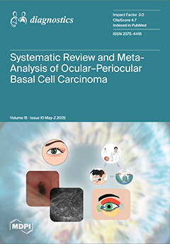
Basal cell carcinoma (BCC) is the most common keratinocytic malignant tumor that affects sun exposure areas, especially in the head-and-neck region. Early diagnosis can limit the extent of facial tissue involvement and subsequent resection, resulting in improved cosmetic and functional results. In vivo reflectance confocal microscopy (RCM) is a non-invasive imaging technique of high diagnostic value used to support the early diagnosis of BCC. The aim of this study was to provide the largest and most up-to-date overview of ocular and periocular BCCs. We also reported the first case of caruncle BCC investigated by dermoscopy and RCM. A systematic review and meta-analysis were carried out, including 236 articles encompassing a total of 71,730 patients with ocular and periocular BCCs. View this paper
  • Issues are regarded as officially published after their release is announced to the table of contents alert mailing list.
  • You may sign up for e-mail alerts to receive table of contents of newly released issues.
  • PDF is the official format for papers published in both, html and pdf forms. To view the papers in pdf format, click on the "PDF Full-text" link, and use the free Adobe Reader to open them.
Order results
Result details
Section
Select all
Export citation of selected articles as:
12 pages, 712 KiB  
Article
Postoperative Radiologic Changes in Early Recurrent Lumbar Foraminal Stenosis After Transforaminal Endoscopic Lumbar Foraminotomy for Lower Lumbar Segments
by Chi-Ho Kim, Pius Kim, Chang-Il Ju and Jong-Hun Seo
Diagnostics 2025, 15(10), 1299; https://doi.org/10.3390/diagnostics15101299 - 21 May 2025
Viewed by 130
Abstract
Background/Objectives: One of the surgical treatments for lumbar foraminal stenosis, full endoscopic foraminotomy, is known for its numerous advantages and favourable clinical outcomes. While previous studies have analyzed preoperative radiological risk factors associated with recurrence within one year after endoscopic foraminal decompression, no [...] Read more.
Background/Objectives: One of the surgical treatments for lumbar foraminal stenosis, full endoscopic foraminotomy, is known for its numerous advantages and favourable clinical outcomes. While previous studies have analyzed preoperative radiological risk factors associated with recurrence within one year after endoscopic foraminal decompression, no research has investigated postoperative radiological changes. The aim of this study is to analyze the radiological changes occurring in cases of early recurrence within six months after endoscopic foraminal decompression. Methods: A retrospective review was conducted on patients with unilateral lumbar foraminal stenosis who underwent full endoscopic foraminotomy at a single institution. The study included 11 recurrent patients who initially experienced symptomatic improvement and sufficient neural decompression on radiological evaluation, but exhibited recurrent radicular pain and radiological restenosis within six months postoperatively. Additionally, 33 control patients with favourable clinical outcomes and no evidence of restenosis were analyzed. Preoperative and postoperative plain X-ray imaging was used to evaluate sagittal and coronal parameters reflecting spinal anatomical characteristics, including disc height, foraminal height, disc wedging, coronal Cobb’s angle, total lumbar lordosis angle, segmental lumbar lordosis angle, and dynamic segmental lumbar lordosis angle. The study aimed to analyze postoperative changes in these parameters between the recurrent and control groups. Clinical outcomes were assessed using the Visual Analog Scale (VAS). Results: There were no significant differences between the groups in terms of age, sex distribution, presence of adjacent segment disease, or existence of Grade 1 spondylolisthesis. Analysis of preoperative and postoperative radiological changes revealed that, in the recurrent group, disc height and foraminal height showed a significant decrease postoperatively, while disc wedging and the coronal Cobb’s angle demonstrated a significant increase. In contrast, the control group exhibited a significant postoperative increase in the total lumbar lordosis angle and segmental lumbar lordosis angle. Conclusions: Progressive worsening of disc wedging and the coronal Cobb’s angle, and reductions in disc and foraminal height, along with minimal improvement in lumbar lordosis following TELF, suggest the presence of irreversible preoperative degenerative changes. Careful radiologic assessment and close postoperative monitoring are essential to identify patients at risk of early recurrence. Full article
(This article belongs to the Special Issue Recent Advances in Bone and Joint Imaging—2nd Edition)
Show Figures

Figure 1

16 pages, 2562 KiB  
Article
Diagnostic Utility of Podoplanin Immunohistochemistry Combined with the NanoSuit-Correlative Light and Electron Microscopy Method for Thoracic Malignant Tumors
by Shin-ya Katsuragi, Yuri Sakano, Isao Ohta, Hisami Kato, Rei Ishikawa, Hirofumi Watanabe, Ryosuke Miyazaki, Katsuhiro Yoshimura, Hidetaka Yamada, Yasuhiro Sakai, Yusuke Inoue, Yusuke Takanashi, Keigo Sekihara, Kazuhito Funai, Yoshiro Otsuki, Hideya Kawasaki and Kazuya Shinmura
Diagnostics 2025, 15(10), 1298; https://doi.org/10.3390/diagnostics15101298 - 21 May 2025
Viewed by 131
Abstract
Background/Objectives: Differentiating thoracic malignant tumors, such as epithelioid malignant pleural mesothelioma (EMPM) and non-small-cell lung carcinoma (NSCLC), primarily comprising lung adenocarcinoma (LAC) and lung squamous cell carcinoma (LSCC), remains a challenge in routine pathological diagnosis. This study aimed to evaluate whether podoplanin (PDPN) [...] Read more.
Background/Objectives: Differentiating thoracic malignant tumors, such as epithelioid malignant pleural mesothelioma (EMPM) and non-small-cell lung carcinoma (NSCLC), primarily comprising lung adenocarcinoma (LAC) and lung squamous cell carcinoma (LSCC), remains a challenge in routine pathological diagnosis. This study aimed to evaluate whether podoplanin (PDPN) immunohistochemistry combined with scanning electron microscopy (SEM) using the NanoSuit-correlative light and electron microscopy (CLEM) methods could serve as a reliable tool for distinguishing these thoracic malignancies. Methods/Results: Initially, PDPN expression was assessed by immunohistochemical analysis in 11 EMPM, 100 LAC, and 23 LSCC cases. PDPN positivity was predominantly observed in the cell membrane and was significantly more frequent in EMPM (100%) than in LAC (2%; p < 0.0001) or LSCC (43.5%; p = 0.0018). Subsequently, field emission–SEM (FE-SEM) observations of PDPN-positive sites on immunohistochemical slides, conducted using the NanoSuit-CLEM method, revealed distinctive ultrastructural features. EMPM exhibited densely packed, elongated microvilli, whereas such structures were absent in LAC and LSCC. Furthermore, analysis of thick-cut sections (20 μm) demonstrated extensive microvilli coverage characteristic of EMPM. Conclusions: These findings suggest that the combined approach of PDPN immunohistochemistry and FE-SEM observation of PDPN-positive sites, using the NanoSuit-CLEM method, constitutes an effective diagnostic strategy for enhancing the accuracy of distinguishing EMPM from NSCLCs. Full article
(This article belongs to the Special Issue Diagnosis, Classification, and Monitoring of Pulmonary Diseases)
Show Figures

Figure 1

12 pages, 1051 KiB  
Article
The Train-Line Pattern on Duplex Ultrasound Helps Differentiate Intramural Thrombus from Atheromatous Plaque in Common Carotid Artery Dissection
by Ming-Hsing Chang, Yen-Yu Huang, Fang-I Hsieh, Kuan-Yu Lin, Hsu-Ling Yeh, Kai-Jing Yeh and Li-Ming Lien
Diagnostics 2025, 15(10), 1297; https://doi.org/10.3390/diagnostics15101297 - 21 May 2025
Viewed by 134
Abstract
Background/Objectives: Common carotid artery dissection (CCAD) can result in severe neurological sequelae; however, its diagnosis may be challenging due to consciousness disturbance and aphasia. The objective of this article is to propose a new imaging feature to assist in the identification of CCAD. [...] Read more.
Background/Objectives: Common carotid artery dissection (CCAD) can result in severe neurological sequelae; however, its diagnosis may be challenging due to consciousness disturbance and aphasia. The objective of this article is to propose a new imaging feature to assist in the identification of CCAD. Methods: This retrospective case series enrolled 139 patients with discharge diagnoses of aortic dissection who underwent carotid ultrasound during admission over a period of three years. Results: Among these patients, 23 had type A aortic dissection, and 113 had type B aortic dissection or related conditions. Notably, among the 23 patients with type A aortic dissection, eight had associated common carotid artery dissection (CCAD), and a total of nine CCAD events were identified. Meanwhile, a B-mode ultrasound revealed six double lumens with intimal flaps and three intramural thrombi. The ‘train-line’ pattern in ultrasonography was discerned by detecting a hypoechoic thickened wall, which was characterized by a margin formed by two parallel linear reflections in close proximity. This distinctive “train-line” pattern was identified in three intimal flaps and two intramural thrombi. Conclusions: While double lumens and intramural thrombus are prevalent findings, the latter may be misinterpreted as atherosclerotic plaque. The “train-line” pattern may aid in distinguishing intramural thrombus from atheromatous plaque, offering an additional diagnostic tool alongside the identification of double lumens with intimal flaps. Full article
(This article belongs to the Special Issue Neurological Disorders: Diagnosis and Management)
Show Figures

Figure 1

16 pages, 1980 KiB  
Article
Paramedics Performed Sonographic Identification of the Conic Ligament—A Prospective Controlled Trial
by Johannes Weimer, Christopher David Chrissostomou, Christopher Jonck, Andreas Michael Weimer, Carlotta Ille, Lukas Müller, Liv Annebritt Lorenz, Marie Stäuber, Thomas Vieth, Holger Buggenhagen, Julia Weinmann-Menke, Maximilian Rink and Julian Künzel
Diagnostics 2025, 15(10), 1296; https://doi.org/10.3390/diagnostics15101296 - 21 May 2025
Viewed by 106
Abstract
Background/Objectives: Acute obstructions of the upper respiratory tract are emergencies that may require a cricothyrotomy as ultima ratio. For this, precise identification of the conic ligament is essential. Point-of-care ultrasound (POCUS) offers a reliable tool for anatomical localization in challenging cases and [...] Read more.
Background/Objectives: Acute obstructions of the upper respiratory tract are emergencies that may require a cricothyrotomy as ultima ratio. For this, precise identification of the conic ligament is essential. Point-of-care ultrasound (POCUS) offers a reliable tool for anatomical localization in challenging cases and could be used by a range of emergency medicine workers. This prospective, controlled observational study assesses the development of competencies of paramedics (PMs) in point-of-care ultrasound (POCUS) assisted identification of the conic ligament after structured training, and compares their competence level to emergency physicians (EPs). Methods: PMs and a control group of EPs received an identical structured training program as part of an ultrasound course. It included a 10-min theoretical introduction, a 10-min video, and a 45-min practical session with ultrasound devices. Questionnaires and a practical test assessed both group’s previous experiences, satisfaction with training, and the development of subjective and objective competencies before (T1) and after (T2) the training. Results: A total of 120 participants (N = 92 PMs and N = 28 EPs) participated. A minority had previously performed a cricothyrotomy even in training settings (PMs 17%; EPs 11%), and none had identified the conic ligament using POCUS. The study group’s subjective and objective competencies increased significantly (p < 0.001). At T2, the study group demonstrated comparable subjective (p = 0.22) and objective (p = 0.81) competencies to those of the control group. The study group needed significantly (p < 0.01) less time to perform the DOPS. While both groups were satisfied with the study material (PMs 2.2 ± 1.2 vs. Eps 1.6 ± 1.0; p = 0.02) and the training (PMs 1.8 ± 1.0 vs. EPs 1.4 ± 0.7, p = 0.03), the study group rated both significantly better. Conclusions: After structured training, paramedics successfully identified the conic ligament using POCUS comparably to emergency physicians. Integrating POCUS into paramedic training may improve prehospital airway management and enhance patient safety. Further studies should investigate long-term skill retention and real-life application. Full article
(This article belongs to the Special Issue The Utility of Ultrasound in Emergency Medicine)
Show Figures

Figure 1

24 pages, 836 KiB  
Article
Utility of Biometric Measurements from Fetal Magnetic Resonance Imaging for Improved Antenatal Diagnosis of Dandy–Walker Spectrum Posterior Fossa Lesions
by Rakhee M. Bowker, Kranthi K. Marathu, Marissa Pharel, Jubril O. Adepoju, Farzan Vahedifard, Seth Adler, Mehmet Kocak, Xuchu Liu and Sharon E. Byrd
Diagnostics 2025, 15(10), 1295; https://doi.org/10.3390/diagnostics15101295 - 21 May 2025
Viewed by 150
Abstract
Background/Objective: The accurate diagnosis of congenital central nervous system abnormalities is critical to pre- and postnatal prognostication and management. When an abnormality is found in the posterior fossa of the fetal brain, parental counseling is challenging because of the wide spectrum of clinical [...] Read more.
Background/Objective: The accurate diagnosis of congenital central nervous system abnormalities is critical to pre- and postnatal prognostication and management. When an abnormality is found in the posterior fossa of the fetal brain, parental counseling is challenging because of the wide spectrum of clinical and neurodevelopmental outcomes in patients with Dandy–Walker (DW) spectrum posterior malformations. The objective of this study was to evaluate the utility of biometric measurements obtained from fetal magnetic resonance imaging (MRI) to facilitate the prenatal differentiation of Dandy–Walker (DW) spectrum malformations, including vermian hypoplasia (VH), Blake’s pouch cyst (BPC), and classic Dandy–Walker malformation (DWM). Methods: This retrospective single-center study evaluated 34 maternal–infant dyads referred for fetal MRI evaluation of suspected DW spectrum malformations identified on antenatal ultrasound. Radiologists took posterior fossa measurements, including the vermis anteroposterior (AP) diameter, vermis height (VH), and tegmento–vermian angle (TVA). The posterior fossa, fourth ventricle, and cisterna magna were classified as normal, large, or dilated. The postnatal imaging findings were evaluated for concordance. The acquired values were compared between the groups and with normative data. The genetic testing results are reported when available. Results: A total of 27 DW spectrum fetal MRI cases were identified, including 7 classic DWMs, 14 VHs, and 6 BPCs. The TVA was significantly higher in the DWM group compared with the VH and BPC groups (p < 0.001). All three groups had reduced AP vermis measurements for gestational age compared with normal fetal brains, as well as differences in the means across the groups (p = 0.002). Conclusions: Biometric measurements derived from fetal MRI can effectively facilitate the prenatal differentiation of VH, BPC, and classic DWM when assessing DW spectrum posterior fossa lesions. Standardizing biometric measurements may increase the diagnostic utility of fetal MRI and facilitate improved antenatal counseling and clinical decision-making. Full article
(This article belongs to the Special Issue Advances in Fetal Imaging)
Show Figures

Figure 1

27 pages, 908 KiB  
Review
Forensic Microbiology: Challenges in Detecting Sexually Transmitted Infections
by Ana Eira, Jennifer Fadoni, António Amorim and Laura Cainé
Diagnostics 2025, 15(10), 1294; https://doi.org/10.3390/diagnostics15101294 - 21 May 2025
Viewed by 185
Abstract
Sexual assault crimes consist of acts committed without consent and represent a major global issue with serious implications for victims. These acts have both short- and long-term consequences on the physical, mental, and sexual health of victims, with sexually transmitted infections (STIs) being [...] Read more.
Sexual assault crimes consist of acts committed without consent and represent a major global issue with serious implications for victims. These acts have both short- and long-term consequences on the physical, mental, and sexual health of victims, with sexually transmitted infections (STIs) being one of the direct outcomes of such crimes. Sexually transmitted infections constitute a serious global public health problem and can lead to severe consequences. These infections may be caused by bacteria, viruses, or parasites and are transmitted through sexual contact. Some of the most common STIs among the general population and victims of sexual crimes include gonorrhoea, chlamydia, trichomoniasis, and syphilis. In most carriers, these infections are asymptomatic, making their detection particularly challenging. Considering the importance of further research in this field, the primary objectives of this study are to review the existing literature on the incidence of major STIs in victims of sexual crimes, to identify the various risk factors associated with these infections, and to explore their public health implications. Additionally, this study aims to assess different STI detection techniques, analyzing their advantages and disadvantages. Studies on this topic are crucial for better understanding the role of sexually transmitted infections in the context of sexual crimes. However, throughout this work, it was verified that point-of-care methods are a good option to allow the diagnosis to be faster and more accurate, when compared to other methods of detecting sexually transmitted infections. Full article
(This article belongs to the Special Issue Autopsy for Medical Diagnostics: 3rd Edition)
Show Figures

Figure 1

11 pages, 731 KiB  
Article
Comparative Evaluation of Four Different Anti-CCP Assays for the Diagnosis of Rheumatoid Arthritis: A Diagnostic Performance Analysis
by Lydia Lamara Mahammed, Tamazouzt Hadjout, Asma Bensaci, Ryma Hamma, Ghalya Bousbia, Nawel Dahmani, Halima Ismail, Nadia Tamechmacht and Reda Djidjik
Diagnostics 2025, 15(10), 1293; https://doi.org/10.3390/diagnostics15101293 - 21 May 2025
Viewed by 124
Abstract
Background/Objectives: Anti-cyclic citrullinated peptide (anti-CCP) antibodies are highly specific markers for rheumatoid arthritis (RA). Over the past decade, novel automating detection systems have been developed for anti-CCP detection. The present study aimed to evaluate the diagnostic performances of three fully automated anti-CCP [...] Read more.
Background/Objectives: Anti-cyclic citrullinated peptide (anti-CCP) antibodies are highly specific markers for rheumatoid arthritis (RA). Over the past decade, novel automating detection systems have been developed for anti-CCP detection. The present study aimed to evaluate the diagnostic performances of three fully automated anti-CCP assays in comparison to a conventional manual enzyme-linked immunosorbent assay (ELISA). Methods: One hundred ninety-nine patients with rheumatic symptoms (100 with RA and 99 without RA) were tested for anti-CCP autoantibodies using four assays: a manual-ELISA (EUROIMMUN®), two chemiluminescence immunoassays (CLIAs) performed on the MAGLUMI X3® and iFlash 1800® platforms, and an enzyme immunoassay (EIA) run on the UNI® analyzer. Results: The Kappa statistic indicated a moderate qualitative agreement among the EUROIMMUN, iFlash, and UNI assays (0.734 ≤ ĸ ≤ 0.778), while the MAGLUMI anti-CCP assay showed only weak-to-moderate agreement with the others (0.510 ≤ ĸ ≤ 0.628). A strong positive correlation was observed between anti-CCP levels measured by the four assays (0.747 ≤ rho ≤ 0.839). At the manufacturers’ cut-off values, sensitivities ranged from 76% to 99% and specificities from 69.7% to 99%, depending on the assay. However, at a fixed specificity of 95%, all the four assays showed good diagnostic performances for RA, with sensitivities ranging from 80% to 89% and positive likelihood ratios (LRs+) from 16 to 17.8. Conclusions: Our results revealed that at the manufacturers’ cut-offs, the UNI anti-CCP assay was the most valuable alternative to the conventional ELISA for diagnosing RA in our cohort. Nevertheless, after an appropriate adjustment of the thresholds, all the evaluated assays showed good diagnostic performances for RA. Full article
(This article belongs to the Section Clinical Laboratory Medicine)
Show Figures

Figure 1

13 pages, 1584 KiB  
Article
Radiomics and AI-Based Prediction of MGMT Methylation Status in Glioblastoma Using Multiparametric MRI: A Hybrid Feature Weighting Approach
by Erdal Tasci, Ying Zhuge, Longze Zhang, Holly Ning, Jason Y. Cheng, Robert W. Miller, Kevin Camphausen and Andra V. Krauze
Diagnostics 2025, 15(10), 1292; https://doi.org/10.3390/diagnostics15101292 - 21 May 2025
Viewed by 133
Abstract
Background/Objectives: Glioblastoma (GBM) is a highly aggressive primary central nervous system tumor with a median survival of 14 months. MGMT (O6-methylguanine-DNA methyltransferase) promoter methylation status is a key biomarker as a prognostic indicator and a predictor of chemotherapy response in GBM. Patients [...] Read more.
Background/Objectives: Glioblastoma (GBM) is a highly aggressive primary central nervous system tumor with a median survival of 14 months. MGMT (O6-methylguanine-DNA methyltransferase) promoter methylation status is a key biomarker as a prognostic indicator and a predictor of chemotherapy response in GBM. Patients with MGMT methylated disease progress later and survive longer (median survival rate 22 vs. 15 months, respectively) as compared to patients with MGMT unmethylated disease. Patients with GBM undergo an MRI of the brain prior to diagnosis and following surgical resection for radiation therapy planning and ongoing follow-up. There is currently no imaging biomarker for GBM. Studies have attempted to connect MGMT methylation status to MRI imaging appearance to determine if brain MRI can be leveraged to provide MGMT status information non-invasively and more expeditiously. Methods: Artificial intelligence (AI) can identify MRI features that are not distinguishable to the human eye and can be linked to MGMT status. We employed the UPenn-GBM dataset patients for whom methylation status was available (n = 146), employing a novel radiomic method grounded in hybrid feature selection and weighting to predict MGMT methylation status. Results: The best MGMT classification and feature selection result obtained resulted in a mean accuracy rate value of 81.6% utilizing 101 selected features and five-fold cross-validation. Conclusions: This compared favorably with similar studies in the literature. Validation with external datasets remains critical to enhance generalizability and propagate robust results while reducing bias. Future directions include multi-channel data integration with radiomic features and deep and ensemble learning methods to improve predictive performance. Full article
(This article belongs to the Special Issue The Applications of Radiomics in Precision Diagnosis)
Show Figures

Figure 1

12 pages, 2843 KiB  
Article
Effect of Strabismus Surgery on Meibomian Glands, Ocular Surface Parameters, and Conjunctival Impression Cytology
by Şenol Sabancı, Canan Sadullahoğlu, Sibel Yavuz, Mehmet Fatih Küçük, Esra Kızıldağ Özbay, Muhammet Kazım Erol and Berna Doğan
Diagnostics 2025, 15(10), 1291; https://doi.org/10.3390/diagnostics15101291 - 21 May 2025
Viewed by 93
Abstract
Background/Objectives: To investigate the effect of strabismus surgery on ocular surface parameters, meibomian glands, and conjunctival impression cytology. Methods: Preoperative and postoperative (10th day, first month, and third month) tear break-up time (TBUT) tests, Schirmer 1 tests, corneal staining scores (CSS), [...] Read more.
Background/Objectives: To investigate the effect of strabismus surgery on ocular surface parameters, meibomian glands, and conjunctival impression cytology. Methods: Preoperative and postoperative (10th day, first month, and third month) tear break-up time (TBUT) tests, Schirmer 1 tests, corneal staining scores (CSS), meibomian gland (MG) loss rates, ocular surface disease index (OSDI) scores, and conjunctival impression cytology (IC) results of 30 patients who underwent strabismus surgery were compared. Results: Significant differences were found between preoperative TBUT test results and those evaluated on the postoperative 10th day and at the postoperative first month (p < 0.0001 for both). There were also significant differences between the preoperative and postoperative first- and third-month Schirmer 1 test results (p = 0.02 and p < 0.0001, respectively). Furthermore, mean OSDI scores significantly differed between preoperative and postoperative 10th-day measurements (p < 0.0001). The mean postoperative 10th-day CSS was found to be significantly higher than the preoperative mean CSS (p < 0.0001). The stages in preoperative conjunctival IC samples were found to be significantly lower than those evaluated at all postoperative times (p < 0.0001 for all). Significant differences were observed between the preoperative lower eyelid MG loss rate and all postoperative MG loss rates (p < 0.0001 for the 10th day and first month and p < 0.001 for the third month). Lastly, the preoperative upper eyelid MG loss rate significantly differed from all postoperative MG loss rates (p < 0.0001 for the 10th day, p < 0.003 for the first month, and p < 0.0001 for the third month). Conclusions: We observed changes indicative of dry eye in the mean OSDI score, TBUT, Schirmer 1 test, MG loss rates, and conjunctival IC findings up to the postoperative third month in patients who underwent strabismus surgery. Therefore, we believe that patients undergoing strabismus surgery should be followed up for ocular surface diseases, particularly dry eye. Full article
(This article belongs to the Section Clinical Diagnosis and Prognosis)
Show Figures

Figure 1

9 pages, 202 KiB  
Article
Post Mortem Paliperidone Blood Concentrations Following Long-Acting Injectable Treatments
by Pietro Zuccarello, Giulia Carnazza, Antonino Petralia and Nunziata Barbera
Diagnostics 2025, 15(10), 1290; https://doi.org/10.3390/diagnostics15101290 - 21 May 2025
Viewed by 87
Abstract
Background/Objectives: I Paliperidone is an antipsychotic recently added into the market in various formulations. There are few data about safety and on therapeutic, toxic, or lethal blood concentrations. Currently, the published analytical methods are often applied to serum or plasma that are not [...] Read more.
Background/Objectives: I Paliperidone is an antipsychotic recently added into the market in various formulations. There are few data about safety and on therapeutic, toxic, or lethal blood concentrations. Currently, the published analytical methods are often applied to serum or plasma that are not obtained from cadaveric blood. Alternatively, aliquots of high volume of whole blood are used, but often in forensic investigations using samples at very small quantities. The aims of the present study were (a) to develop an analytical method to detect and quantify paliperidone in whole blood using only a small sample volume (10 µL) and (b) to summarize data on post-mortem blood analysis obtained from authentic autopsy cases. Methods: Method validation was carried out on 10 µL of whole blood, extracted by LLE and analyzed by LC-MS. Paliperidone concentrations obtained from blood analysis of 16 authentic autopsy cases were reported. Results: The method showed a good linearity and sensitivity, a normal distribution, the absence of anomalous values, an interday RSD% always less than 10%, and an 80–120% recovery, as required by AAFS guidelines. Femoral blood concentrations obtained from authentic autopsy cases ranged between 23.4 and 146.9 ng/mL. Conclusions: This method is to be used properly in all cases where it is necessary (a) to monitor the therapeutic adherence of patients, (b) to establish the psycho-physical conditions of the treated subject at the time of the death, and (c) to ascertain if the drug may have played a causal role in the obitus. This study reported the first data obtained from post-mortem investigation of subjects treated with paliperidone LAI. Cadaveric blood concentrations could be higher than ante-mortem reference values due to post-mortem redistribution. Full article
17 pages, 559 KiB  
Review
The Application of Circulating Tumour DNA (ctDNA) in the Diagnosis, Prognosis, and Treatment Monitoring of Gynaecological and Breast Cancers (Review)
by Aleksandra Englisz, Marta Smycz-Kubańska, Patrycja Królewska-Daszczyńska, Magdalena Błaut, Agnieszka Duszyc and Aleksandra Mielczarek-Palacz
Diagnostics 2025, 15(10), 1289; https://doi.org/10.3390/diagnostics15101289 - 21 May 2025
Viewed by 79
Abstract
Gynaecological cancers, including endometrial, ovarian, and cervical cancers as well as breast cancer, despite numerous studies, still constitute a challenge for modern oncology. For this reason, research aimed at the application of modern diagnostic methods that are useful in early detection, prognosis, and [...] Read more.
Gynaecological cancers, including endometrial, ovarian, and cervical cancers as well as breast cancer, despite numerous studies, still constitute a challenge for modern oncology. For this reason, research aimed at the application of modern diagnostic methods that are useful in early detection, prognosis, and treatment monitoring deserves special attention, Great hopes are currently being placed on the use of liquid biopsy (LB), which examines various tumour components, including cell-free RNA (cfRNA), circulating tumour cells (CTCs), circulating tumour DNA (ctDNA), exosomes, and tumour-educated platelets (TEPs). LB has shown promise as a minimally invasive means of early diagnosis of cancers, detection of recurrence, prediction of therapy response, treatment monitoring, and drug selection. The integration of this test into clinical practice in modern oncology is challenging, but offers many benefits, including reducing the risks associated with invasive procedures, improving diagnostic and therapeutic efficacy, and improving the quality of life of oncology patients. The aim of this review is to present recent reports on the use of ctDNA in diagnosing, predicting the outcome of, and monitoring the treatment of gynaecological and breast cancers. Full article
(This article belongs to the Special Issue Diagnosis and Management of Gynecological Cancers: Third Edition)
Show Figures

Figure 1

11 pages, 1310 KiB  
Article
Diagnostic Value of Multimodal Lymphatic Imaging Techniques in Thoracic Duct Outlet Obstruction
by Ying Fei, Yanli Lu, Zhichao Yao, Kongxiang Yin, Dayong Zhou and Zhanao Liu
Diagnostics 2025, 15(10), 1288; https://doi.org/10.3390/diagnostics15101288 - 20 May 2025
Viewed by 144
Abstract
Objectives: To investigate the diagnostic value of various lymphatic imaging techniques for thoracic duct (TD) outlet obstruction in patients with chylous leakage. Methods: A retrospective analysis was conducted on 23 patients with chylous leakage who were radiologically diagnosed with a TD outlet obstruction [...] Read more.
Objectives: To investigate the diagnostic value of various lymphatic imaging techniques for thoracic duct (TD) outlet obstruction in patients with chylous leakage. Methods: A retrospective analysis was conducted on 23 patients with chylous leakage who were radiologically diagnosed with a TD outlet obstruction and underwent a TD exploration and reconstruction between January 2022 and February 2025. Non-enhanced magnetic resonance lymphangiography (MRL), 99Tcm-DX lymphoscintigraphy, and intranodal lymphangiography were employed to detect abnormalities in the central lymphatic vessels. The Receiver Operating Characteristic (ROC) curve was utilized to analyze the diagnostic performance of these imaging methods for TD outlet obstruction in lymphatic disorders. Results: Twenty-three patients (fifteen males and eight females) with chylous leakage were included in this study, with an average age of 59.78 ± 13.08 years. Non-enhanced MRL, 99Tcm-DX lymphoscintigraphy, and intranodal lymphangiography revealed TD outlet obstructions in 13, 17, and 18 patients, respectively. Twenty patients exhibited findings consistent with preoperative imaging during TD explorations; the intraoperative microscopic visualization demonstrated the difficulty of white chyle entering the bloodstream for these patients. The ROC curve analysis indicated that “at least two imaging modalities were positive” and had the highest Area Under the Curve (AUC) value (0.90); “intranodal lymphangiography” and “non-enhanced magnetic resonance lymphangiography” followed closely with respective AUC values of 0.76 and 0.73, and 99Tcm-DX lymphoscintigraphy exhibited a lower AUC value 0.63. Conclusions: The combined utilization of multimodal lymphatic imaging techniques demonstrated a high diagnostic accuracy in identifying TD outlet obstruction in patients with chylous leakage. Full article
Show Figures

Figure 1

18 pages, 1567 KiB  
Article
Immune Cells as Mediators of Lipidome Influence on Osteoporosis: Evidence from a Mediation Analysis
by Jiheng Xiao, Wei Zhou, Jiatai He, Yanbin Zhu, Yingze Zhang and Liming Xiong
Diagnostics 2025, 15(10), 1287; https://doi.org/10.3390/diagnostics15101287 - 20 May 2025
Viewed by 116
Abstract
Background: Although clinical studies have indicated a possible association between dyslipidemia and osteoporosis, the underlying genetic basis and mechanistic pathways remain insufficiently defined. Most prior research has concentrated on conventional lipid markers, which are prone to confounding and limit causal inference. Exploring [...] Read more.
Background: Although clinical studies have indicated a possible association between dyslipidemia and osteoporosis, the underlying genetic basis and mechanistic pathways remain insufficiently defined. Most prior research has concentrated on conventional lipid markers, which are prone to confounding and limit causal inference. Exploring lipidomic profiles offers a more comprehensive view of lipid metabolism and may reveal novel genetic links beyond traditional lipid traits. Additionally, alterations in immune cell function, often triggered by metabolic disturbances, may contribute to osteoporosis development; however, the potential mediating role of immune cells in the lipid–bone axis has not been systematically investigated. Methods: A total of 179 lipid species across 13 lipid classes were analyzed in 7174 Finnish individuals from the GeneRISK cohort. Genome-Wide Association Study (GWAS) summary statistics for osteoporosis and 731 immune cell immunophenotypes were sourced from the GWAS Catalog. A two-step, two-sample Mendelian randomization analysis, using inverse variance weighting (IVW), was conducted to explore the potential causal effects of lipids on osteoporosis and the mediating role of immune cells in the relationship between lipids and osteoporosis. Results: Mendelian randomization analysis indicated that triacylglycerol levels of 48:0 were possibly associated with an increased risk of osteoporosis (IVW: odds ratio [OR] 1.1320, 95% CI 1.0401–1.2321; p = 0.004), while triacylglycerol levels of 48:3 appeared to be associated with a reduced risk of osteoporosis (IVW: OR 0.9053, 95% CI 0.8364–0.9800; p = 0.014). Two statistically significant mediating effects were identified: First, IgD− CD38dim %B cells appeared to partially negatively mediate the association between triacylglycerol levels of 48:3 and osteoporosis, with a negative mediating effect of −0.00669 (95% CI: −0.0214, 0.00805), which accounted for 6.73% of the total effect. That is, the protective effect of triacylglycerol levels of 48:3 against osteoporosis was attenuated by IgD− CD38dim %B cells. Second, HLA DR++ monocytes% leukocytes also partially negatively mediated this relationship, with a mediating effect of −0.023 (95% CI: −0.0434, −0.00266), accounting for 23.2% of the total effect. This indicates that other immune cells, HLA DR++ monocytes %leukocytes, resisted the protective effect of triacylglycerol levels of 48:3 against osteoporosis, with a weakening effect stronger than that of IgD− CD38dim %B cells. Conclusions: Our findings contribute to the growing understanding of the potential causal relationships and shared pathogenic mechanisms between dyslipidemia and osteoporosis. The results suggest that the potential genetic effects of plasma lipid metabolites on osteoporosis may be partially down-regulated by specific kinds of immune cells. Full article
(This article belongs to the Section Pathology and Molecular Diagnostics)
Show Figures

Figure 1

16 pages, 254 KiB  
Article
Sexual Functioning and Depressive Symptoms in Women with Postpartum Thyroiditis
by Karolina Kowalcze, Gaspare Cucinella, Giuseppe Gullo and Robert Krysiak
Diagnostics 2025, 15(10), 1286; https://doi.org/10.3390/diagnostics15101286 - 20 May 2025
Viewed by 127
Abstract
Background/Objectives: The presence of autoimmune thyroiditis was found to be associated with an increased prevalence of sexual dysfunction. Women’s sexual health was not investigated in postpartum disorders of the thyroid gland. The aim of this study was to assess female sexual functioning [...] Read more.
Background/Objectives: The presence of autoimmune thyroiditis was found to be associated with an increased prevalence of sexual dysfunction. Women’s sexual health was not investigated in postpartum disorders of the thyroid gland. The aim of this study was to assess female sexual functioning and depressive symptoms in postpartum thyroiditis. Methods: This study compared four groups of non-lactating women who gave birth within 12 months before the beginning of the study: women with postpartum thyroiditis and overt hypothyroidism (group A), women with postpartum thyroiditis and subclinical hypothyroidism (group B), euthyroid females with postpartum thyroiditis (group C) and healthy euthyroid females without thyroid disease (group D). All patients completed questionnaires assessing female sexual function (FSFI), and the presence and severity of depressive symptoms (BDI-II). Moreover, we assessed thyroid peroxidase and thyroglobulin antibodies, as well as serum levels of thyroid-stimulating hormone (TSH), free thyroid hormones, testosterone, dehydroepiandrosterone sulfate (DHEAS), estradiol and prolactin. Results: The mean total FSFI score was lower in women with overt hypothyroidism (22.74 ± 4.12) than in the remaining groups of women, lower in groups B (25.71 ± 3.84) and C (29.67 ± 4.00) than in group D (32.15 ± 2.98), as well as lower in group B than in group C. Compared to healthy controls, both groups of women with postpartum thyroiditis and thyroid hypofunction had lower scores for all domains, while euthyroid patients with postpartum thyroiditis had lower scores for sexual desire, sexual arousal and lubrication. The total BDI-II score was highest in group A (15.6 ± 3.2) and lowest in group D (7.8 ± 3.2). Serum testosterone and DHEAS levels were lower while serum prolactin levels were higher in women with postpartum thyroiditis than in healthy subjects. The lowest testosterone levels (1.02 ± 0.35 nmol/L) and estradiol levels (190 ± 80 pmol/L) and the highest prolactin concentration (39.9 ± 13.9 ng/mL) were found in group A. Conclusions: The obtained results show that postpartum thyroiditis is complicated by multidimensional impairment of female sexual functioning, which is accompanied by mood deterioration. Severity of sexual dysfunction and depressive symptoms in this clinical entity depends on the degree of thyroid autoimmunity and hypothyroidism. It seems that assessment of sexual functioning and mood should be recommended to all women with postpartum thyroiditis. Full article
(This article belongs to the Special Issue Diagnosis and Prognosis of Gynecological and Obstetric Diseases)
28 pages, 8822 KiB  
Article
Multiclassification of Colorectal Polyps from Colonoscopy Images Using AI for Early Diagnosis
by Jothiraj Selvaraj, Kishwar Sadaf, Shabnam Mohamed Aslam and Snekhalatha Umapathy
Diagnostics 2025, 15(10), 1285; https://doi.org/10.3390/diagnostics15101285 - 20 May 2025
Viewed by 164
Abstract
Background/Objectives: Colorectal cancer (CRC) remains one of the leading causes of cancer-related mortality worldwide, emphasizing the critical need for the accurate classification of precancerous polyps. This research presents an extensive analysis of the multiclassification framework leveraging various deep learning (DL) architectures for the [...] Read more.
Background/Objectives: Colorectal cancer (CRC) remains one of the leading causes of cancer-related mortality worldwide, emphasizing the critical need for the accurate classification of precancerous polyps. This research presents an extensive analysis of the multiclassification framework leveraging various deep learning (DL) architectures for the automated classification of colorectal polyps from colonoscopy images. Methods: The proposed methodology integrates real-time data for training and utilizes a publicly available dataset for testing, ensuring generalizability. The real-time images were cautiously annotated and verified by a panel of experts, including post-graduate medical doctors and gastroenterology specialists. The DL models were designed to categorize the preprocessed colonoscopy images into four clinically significant classes: hyperplastic, serrated, adenoma, and normal. A suite of state-of-the-art models, including VGG16, VGG19, ResNet50, DenseNet121, EfficientNetV2, InceptionNetV3, Vision Transformer (ViT), and the custom-developed CRP-ViT, were trained and rigorously evaluated for this task. Results: Notably, the CRP-ViT model exhibited superior capability in capturing intricate features, achieving an impressive accuracy of 97.28% during training and 96.02% during validation with real-time images. Furthermore, the model demonstrated remarkable performance during testing on the public dataset, attaining an accuracy of 95.69%. To facilitate real-time interaction and clinical applicability, a user-friendly interface was developed using Gradio, allowing healthcare professionals to upload colonoscopy images and receive instant classification results. Conclusions: The CRP-ViT model effectively predicts and categorizes colonoscopy images into clinically relevant classes, aiding gastroenterologists in decision-making. This study highlights the potential of integrating AI-driven models into routine clinical practice to improve colorectal cancer screening outcomes and reduce diagnostic variability. Full article
Show Figures

Figure 1

10 pages, 1247 KiB  
Article
The Clinical and Laboratory Profiles of a Deletional α2-Globin Gene Polyadenylation Signal Sequence (AATAAA > AATA--) [HBA2:c.*93_*94delAA]: The Malaysian Experience
by Norafiza Mohd Yasin, Syahzuwan Hassan, Nur Aisyah Aziz, Faidatul Syazlin Abdul Hamid, Ezalia Esa, Ezzanie Suffya Zulkefli, Rohana Ghazali, Syirah Nazirah Tajuddin, Mohd Nazif Darawi, Yuslina Mat Yusoff and Cornelis L. Harteveld
Diagnostics 2025, 15(10), 1284; https://doi.org/10.3390/diagnostics15101284 - 20 May 2025
Viewed by 166
Abstract
Poly A (AATAAA > AATA--) [HBA2:c.*93_*94delAA] is a rare α-variant reported in our population. It is caused by 2 bp deletion (--AA) in the α2 poly A sequence, leading to a significant α–thalassaemia phenotype. Background/Objectives: This study describes the haematological parameters, phenotype, [...] Read more.
Poly A (AATAAA > AATA--) [HBA2:c.*93_*94delAA] is a rare α-variant reported in our population. It is caused by 2 bp deletion (--AA) in the α2 poly A sequence, leading to a significant α–thalassaemia phenotype. Background/Objectives: This study describes the haematological parameters, phenotype, and genotype characteristics of AATA(--AA) in the Malaysian population. Methods: The study was carried out on 17 177 cases referred to the Institute for Medical Research, Malaysia, for further diagnosis of α-thalassaemia in a five-year period. Alpha-Gap and ARMS-PCR were performed to detect common α-thalassaemia, followed by HBA1 and HBA2 genes sequencing and multiplex ligation-dependent probe amplification (MLPA). Haematological parameters among various groups with the AATA(--AA) allele were presented in this study. Results: Thirty-two patients with AATA(--AA) displaying an α–thalassaemia-like phenotype were analysed. They comprised 22 (68.75%) AATA(--AA) carriers, 2 (6.25%) compounds with 3.7 deletion, 2 (6.25%) compounds with --SEA deletion, 1 (3.12%) AATA(--AA) homozygote, and 3 (9.37%) compounds of Hb Adana, Hb CS, and Hb Pakse with co-inheritance Hb E, respectively. Most of the patients with AATA(--AA) compounds with the α-variant exhibited a significant phenotype between moderate to severe thalassaemia, especially cases with compound α−AAα/αAdanaα. Conclusions: AATA(--AA) is a significant pathogenic variant that should be diagnosed to prevent significant thalassaemia phenotype or transfusion-dependent thalassaemia. Full article
(This article belongs to the Section Clinical Laboratory Medicine)
Show Figures

Figure 1

16 pages, 1186 KiB  
Article
Exploring the Role of Peritumoral Edema in Predicting Lung Cancer Subtypes Through T2-FLAIR Digital Subtraction Imaging of Metastatic Brain Tumors
by Okan Dilek, Emin Demırel, Zeynel Abidin Tas and Emre Bılgın
Diagnostics 2025, 15(10), 1283; https://doi.org/10.3390/diagnostics15101283 - 20 May 2025
Viewed by 207
Abstract
Background/Objectives: This study aimed to investigate whether small-cell lung cancer (SCLC) and non-small-cell lung cancer (NSCLC) can be distinguished based on radiomics data derived from T2-FLAIR digital subtraction images of the peritumoral edema region in patients with brain metastases. Methods: A total [...] Read more.
Background/Objectives: This study aimed to investigate whether small-cell lung cancer (SCLC) and non-small-cell lung cancer (NSCLC) can be distinguished based on radiomics data derived from T2-FLAIR digital subtraction images of the peritumoral edema region in patients with brain metastases. Methods: A total of 136 patients who underwent surgery for brain tumors, including 100 patients in the Pretreat-Metstobrain-MASKS dataset and 36 patients from our institution, were included in our study. Radiomic features were extracted from digitally subtracted T2-FLAIR images in the peritumoral edema area. Patients were divided into NSCLC and SCLC groups. The maximum relevance–minimum redundancy (mRMR) method was then used for dimensionality reduction. The Naive Bayes algorithm was used for model development, and the interpretability of the model was explored using SHapley Additive exPlanations (SHAP). The performance metrics included the area under the curve (AUC), sensitivity (SENS), and specificity (SPEC). Results: The mean age of NSCLC patients was 64.6 ± 10.3 years, and that of SCLC patients was 63.4 ± 11.7 years. In the external validation cohort, the model achieved an AUC of 0.82 (0.68–0.97), a SENS of 0.87 (0.74–0.91), and a SPEC of 0.72 (0.72–0.89). In the train cohort, the model achieved an AUC of 1.000, a SENS of 1.000, and a SPEC of 1.000. The feature providing the best effect was wavelet-HHHglcmJointEnergy, with a SHAP value of approximately 2.5. Conclusions: An artificial intelligence model developed using radiomics data from T2-FLAIR digital subtraction images of the peritumoral edema area can identify the histologic type of lung cancer in patients with associated brain metastases. Full article
(This article belongs to the Section Machine Learning and Artificial Intelligence in Diagnostics)
Show Figures

Figure 1

14 pages, 989 KiB  
Systematic Review
Efficacy of Hyperosmolar Dextrose Injection for Osgood–Schlatter Disease: A Systematic Review with Meta-Analysis
by Hye Chang Rhim, Lori B. Bjork, Jaehyung Shin, Jewel Park, Stephanie E. DeLuca, Katelyn C. McCarron, Ki-Mo Jang and Chris Ha
Diagnostics 2025, 15(10), 1282; https://doi.org/10.3390/diagnostics15101282 - 19 May 2025
Viewed by 296
Abstract
Background/Objectives: Although Osgood–Schlatter disease (OSD) is often self-limiting following apophyseal closure, it may cause persistent symptoms into adulthood, affecting physical and functional activities. The purpose of this systematic review is to summarize the current evidence on the efficacy of hyperosmolar dextrose injection for [...] Read more.
Background/Objectives: Although Osgood–Schlatter disease (OSD) is often self-limiting following apophyseal closure, it may cause persistent symptoms into adulthood, affecting physical and functional activities. The purpose of this systematic review is to summarize the current evidence on the efficacy of hyperosmolar dextrose injection for patients with OSD unresponsive to conservative treatment. Methods: Multiple databases were searched for studies investigating the efficacy of hyperosmolar dextrose injection in patients with OSD. Two reviewers independently extracted data and evaluated the risk of bias. Meta-analyses were performed to compare hyperosmolar dextrose injection with placebo injections. Results: Four studies including three randomized controlled trials (RCTs) and one case series involving a total of 166 (162 males and 4 females) patients with 184 knees were included in this review. At three months, there was no significant difference in patient-reported improvement from baseline between hyperosmolar dextrose injection and placebo injections (standardized mean difference [SMD] = 1.92, 95% confidence interval [CI], −0.12 to 3.96; I2 = 96.2%). However, a meta-analysis of two RCTs including athletic pediatric patients found a pooled risk ratio of 2.11 (95% CI: 1.12 to 3.98, I2 = 30.73%) for pain-free return to sports at three months. In addition, at one year, a meta-analysis of two RCTs showed greater patient-reported improvement from baseline with hyperosmolar dextrose injection compared to placebo (SMD = 1.09, 95% CI, 0.62 to 1.56; I2= 0%). Conclusions: Based on the limited number of RCTs, although no improvement in patient-reported outcomes is seen at three months, hyperosmolar dextrose injection may safely facilitate a pain-free return to sports at three months and lead to patient-reported improvement at one year. However, further high-quality RCTs are needed to substantiate these findings. Full article
(This article belongs to the Section Medical Imaging and Theranostics)
Show Figures

Figure 1

14 pages, 2105 KiB  
Article
Association of [18F]-FDG PET/CT-Derived Radiomic Features with Clinical Outcomes and Genomic Profiles in Patients with Chronic Lymphocytic Leukemia
by Fabiana Esposito, Luigi Manco, Guglielmo Manenti, Livio Pupo, Andrea Nunzi, Roberta Laureana, Luca Guarnera, Massimiliano Marinoni, Elisa Buzzatti, Paola Elda Gigliotti, Andrea Micillo, Giovanni Scribano, Adriano Venditti, Massimiliano Postorino and Maria Ilaria Del Principe
Diagnostics 2025, 15(10), 1281; https://doi.org/10.3390/diagnostics15101281 - 19 May 2025
Viewed by 299
Abstract
Background: The role of PET/CT imaging in chronic lymphoproliferative syndromes (CLL) is debated. This study examines the potential of PET/CT radiomics in predicting outcomes and genetic profiles in CLL patients. Methods: A retrospective analysis was conducted on 50 CLL patients treated at Policlinico [...] Read more.
Background: The role of PET/CT imaging in chronic lymphoproliferative syndromes (CLL) is debated. This study examines the potential of PET/CT radiomics in predicting outcomes and genetic profiles in CLL patients. Methods: A retrospective analysis was conducted on 50 CLL patients treated at Policlinico Tor Vergata, Rome, and screened, at diagnosis, with [18F]-FDG PET/CT. Potentially pathological lymph nodes were semi-automatically segmented. Genetic mutations in TP53, NOTCH1, and IGVH were assessed. Eight hundred and sixty-five radiomic features were extracted, with the cohort split into training (70%) and validation (30%) sets. Four machine learning models, each with Random Forest, Stochastic Gradient Descent, and Support Vector Machine learners, were trained. Results: Progression occurred in 10 patients. The selected radiomic features from CT and PET datasets were correlated with four models of progression and mutations (TP53, NOTCH1, IGVH). The Random Forest models outperformed others in predicting progression (AUC = 0.94/0.88, CA = 0.87/0.75, TP = 80.00%/87.50%, TN = 72.70%/87.50%) and the occurrence of TP53 (AUC = 0.94/0.96, CA = 0.87/0.80, TP = 87.50%/90.21%, TN = 85.70%/90.90%), and NOTCH1 (AUC = 0.94/0.85, CA = 0.87/0.67, TP = 80.00%/88.90%, TN = 80.00%/83.30%)mutations. The IGVH models showed poorer performance. Conclusions: ML models based on PET/CT radiomic features effectively predict outcomes and genetic profiles in CLL patients. Full article
(This article belongs to the Section Medical Imaging and Theranostics)
Show Figures

Figure 1

3 pages, 481 KiB  
Interesting Images
Tumor Cavitation with Anlotinib Treatment in Lung Adenocarcinoma
by Jie Huang and Xueqin Chen
Diagnostics 2025, 15(10), 1280; https://doi.org/10.3390/diagnostics15101280 - 18 May 2025
Viewed by 245
Abstract
Tumor cavitation is distinguished by the emergence of central necrosis and cavity formation within the tumor mass, which indicates a notable outcome of anti-angiogenic therapies. This case describes a 52-year-old Chinese female with advanced EGFR-mutated lung adenocarcinoma (Exon 19 deletion), which was metastatic [...] Read more.
Tumor cavitation is distinguished by the emergence of central necrosis and cavity formation within the tumor mass, which indicates a notable outcome of anti-angiogenic therapies. This case describes a 52-year-old Chinese female with advanced EGFR-mutated lung adenocarcinoma (Exon 19 deletion), which was metastatic to bilateral lungs, brain, and right adrenal gland, who exhibited a radiographic response to combination therapy with the third-generation EGFR tyrosine kinase inhibitor (TKI) aumolertinib and the anti-angiogenic agent anlotinib. The patient achieved near-complete cavitation of almost all bilateral lung nodules, manifesting as distinctive “bullet hole” lesions on the chest CT. Despite this initial response, disease progression occurred two months later with new liver metastases, culminating in the patient’s death. This case underscores the potential efficacy of EGFR TKIs and anti-angiogenic agents in inducing unique tumor microenvironment modifications, while highlighting the transient nature of such responses and the critical need to address resistance mechanisms. Tumor cavitation may serve as a radiographic marker of anti-angiogenic activity but does not preclude metastatic spread, necessitating vigilant monitoring even in the setting of favorable imaging changes. Full article
(This article belongs to the Section Medical Imaging and Theranostics)
Show Figures

Figure 1

18 pages, 3268 KiB  
Article
Experience in Diagnostic of HIV Drug Resistance in the Mekong Delta Region, Vietnam: A Comparative Analysis Before and After the COVID-19 Pandemic
by Huynh Hoang Khanh Thu, Alexandr N. Schemelev, Yulia V. Ostankova, Vladimir S. Davydenko, Diana E. Reingardt, Ton Tran, Le Chi Thanh, Thi Xuan Lien Truong and Areg A. Totolian
Diagnostics 2025, 15(10), 1279; https://doi.org/10.3390/diagnostics15101279 - 18 May 2025
Viewed by 263
Abstract
Background: Vietnam has made significant strides in reducing the prevalence of HIV infection and achievements in its antiretroviral treatment program. However, the COVID-19 pandemic and financial challenges in the healthcare system have posed significant obstacles to maintaining effective HIV treatment and monitoring, particularly [...] Read more.
Background: Vietnam has made significant strides in reducing the prevalence of HIV infection and achievements in its antiretroviral treatment program. However, the COVID-19 pandemic and financial challenges in the healthcare system have posed significant obstacles to maintaining effective HIV treatment and monitoring, particularly among vulnerable populations. This study aims to evaluate the situation of HIV drug resistance among patients who have experienced treatment failure in the Mekong Delta region and to compare data from 2019 to 2022. Methods: The study material was blood plasma samples from HIV-infected individuals with ART failure: 316 collected in 2019 and 326 collected in 2022. HIV-1 genotyping and mutation detection were performed based on an analysis of the nucleotide sequences of the Pol gene region. A total of 116 HIV-infected individuals with virological failure in 2019 and 2022 were assessed for HIV drug resistance. Results: The study revealed a high proportion of participants with viral loads exceeding 1000 copies/mL, significantly increasing from 12.0% in 2019 to 23.9% in 2022 (OR = 2.3; p = 0.0001). HIV drug resistance mutations were detected in 84.21% of cases in 2019 and 92.59% in 2022. The prevalence of concurrent resistance to NRTIs and NNRTIs was 37.5% and 30.13% in 2019 and 2022, respectively. There was a statistically significant decrease in NNRTI resistance (OR = 0.32, χ2 = 5.43, p < 0.05). In contrast, multi-drug resistance to protease inhibitors rose from 18.52% to 45.21% (φ* = 0.00403, p < 0.05). Triple-class resistance was identified only in 2022 (17.81%). The most common mutations included M184I/V, D67N, K103N, Y181C, and V82A/S/T, with D67N rising significantly from 3.13% to 21.92%. The predominant subtype was CRF01_AE. Conclusion: A high prevalence of viral non-suppression and HIV drug resistance was observed among patients in the Mekong Delta region, particularly after the onset of the COVID-19 pandemic. Our study highlights the ongoing challenges that the HIV/AIDS treatment program in Vietnam must address in the post-pandemic period to sustain its success and achieve the goals of the country’s HIV prevention strategies. Full article
(This article belongs to the Section Diagnostic Microbiology and Infectious Disease)
Show Figures

Figure 1

19 pages, 977 KiB  
Article
Sex-Specific Impact of Inflammation and Nutritional Indices on AVF Blood Flow and Maturation: A Retrospective Analysis
by Özgür Akkaya and Ümit Arslan
Diagnostics 2025, 15(10), 1278; https://doi.org/10.3390/diagnostics15101278 - 18 May 2025
Viewed by 430
Abstract
Background: Arteriovenous fistula (AVF) failure rates are consistently higher in females, although the underlying mechanisms remain incompletely understood. Inflammatory processes play a key role in AVF remodeling and venous arterialization, yet their influence may differ by sex. This study aimed to evaluate [...] Read more.
Background: Arteriovenous fistula (AVF) failure rates are consistently higher in females, although the underlying mechanisms remain incompletely understood. Inflammatory processes play a key role in AVF remodeling and venous arterialization, yet their influence may differ by sex. This study aimed to evaluate the impact of inflammatory indices on AVF blood flow and maturation, with a focus on sex-specific differences. Methods: This retrospective analytical study included 110 patients (50 females, 60 males) undergoing initial surgical AVF creation. Postoperative assessments occurred at the fourth and sixth weeks. Patients demonstrating insufficient maturation (blood flow < 600 mL/min) at the fourth week were re-evaluated after two weeks without any intervening procedures or additional interventions. Results: Intraoperative Transit-Time Flow Measurement (TTFM) revealed significantly higher median AVF blood flow in males compared to females (289 mL/min vs. 200 mL/min; p < 0.001). Doppler ultrasonography (DUS) findings confirmed these sex-related differences, demonstrating consistently lower blood flow rates in female patients. An elevated neutrophil-to-lymphocyte ratio (NLR) was associated with approximately a 31% reduction in AVF blood flow among females, whereas an increased C-reactive protein-to-albumin ratio (CrA) correlated with an approximate 9% decline. In males, an elevated systemic immune-inflammation index (SII) and systemic inflammation response index (SIRI) were significantly associated with decreased AVF blood flow. Conversely, a higher prognostic nutritional index (PNI) positively correlated with AVF blood flow in both sexes. Risk factors associated with inadequate AVF maturation (<600 mL/min at sixth week) included female sex, advanced age, obesity, smoking, anemia, low vitamin D levels, and elevated inflammatory indices (NLR, SII, and SIRI). Conclusions: Inflammatory and nutritional indices derived from routine laboratory tests may assist in estimating AVF maturation likelihood. While DUS reliably assesses AVF blood flow, complementary evaluation methods may be required to assess the broader vascular status. Further research is needed to clarify sex-specific inflammatory mechanisms influencing AVF outcomes and to guide individualized management strategies. Full article
Show Figures

Figure 1

11 pages, 2923 KiB  
Article
First-Trimester Morphological Evaluation of Fetuses and Medical Law Implications
by Răzvan Grigoraș Căpitănescu, Marius Cristian Marinaș, Larisa Pătru, Dragoș George Popa, Elena Cristina Andrei, Aura Iuliana Popa, Gabriel Florin Răzvan Mogoș, Nicolae Dragoș Mărgăritescu and Ciprian Laurențiu Pătru
Diagnostics 2025, 15(10), 1277; https://doi.org/10.3390/diagnostics15101277 - 18 May 2025
Viewed by 242
Abstract
Background/Objectives: Over the years, the potential of the first-trimester (FT) ultrasound in the detection of fetal structural defects has increased. The main objectives of the first-trimester fetal screening evaluation are the detection of major structural anomalies and the diagnosis of additional sonographic markers [...] Read more.
Background/Objectives: Over the years, the potential of the first-trimester (FT) ultrasound in the detection of fetal structural defects has increased. The main objectives of the first-trimester fetal screening evaluation are the detection of major structural anomalies and the diagnosis of additional sonographic markers for chromosomal disorders. When a fetal anomaly is diagnosed, patients have the right to be informed about the risks, necessary interventions, or alternatives. Depending on the severity of the anomalies and the pregnancy period, the legality of the pregnancy termination was evaluated. The aim of this study was to assess the impact of the first-trimester morphological screening of the fetus using an ultrasound protocol according to the latest international protocols (the ISUOG protocol). Methods: Between 1 January 2024 and 31 December 2024, 854 pregnancies with gestational ages between 11 weeks and 13 weeks + 6 days were morphologically evaluated during the nuchal scan in the Obstetrics and Gynecology Department of the Emergency County Hospital from Craiova. Both transabdominal and transvaginal ultrasound in 2D and in a color Doppler mode were used in the scanning technique. The ultrasound findings were correlated with the genetic testing results and pregnancy outcome. The medical law implications were related to the cases where the ultrasound was performed at about 13 weeks of gestation, and the screening genetic results showed an increased pregnancy risk, which arose during the FT. In these cases, we performed amniocentesis at about 16–17 weeks of gestation, and especially, the Non-Invasive Prenatal Testing (NIPT)-positive cases were confirmed by karyotyping. Still, at this gestational age of diagnosis, the Romanian law would not allow abortions. Results: By using this extended FT ultrasound protocol, we detected 58 cases with fetal structural anomalies. Eighteen cases were also associated with genetic syndromes after performing chorionic villous sampling (CVS). Three cases detected with minor structural anomalies (two cases with club foot and one case with a cleft upper lip) were lost to follow-up. Conclusions: Fetal morphological ultrasound evaluation is feasible in the late first trimester. By using an extended ultrasound protocol, we can detect most of the fetal structural anomalies and contribute to better medical counseling and improve pregnancy outcomes. Full article
(This article belongs to the Special Issue Echocardiography Applications in Cardiovascular Diseases)
Show Figures

Figure 1

16 pages, 1870 KiB  
Article
Artificial Intelligence as a Potential Tool for Predicting Surgical Margin Status in Early Breast Cancer Using Mammographic Specimen Images
by David Andras, Radu Alexandru Ilies, Victor Esanu, Stefan Agoston, Tudor Florin Marginean Jumate and George Calin Dindelegan
Diagnostics 2025, 15(10), 1276; https://doi.org/10.3390/diagnostics15101276 - 17 May 2025
Viewed by 357
Abstract
Background/Objectives: Breast cancer is the most common malignancy among women globally, with an increasing incidence, particularly in younger populations. Achieving complete surgical excision is essential to reduce recurrence. Artificial intelligence (AI), including large language models like ChatGPT, has potential for supporting diagnostic [...] Read more.
Background/Objectives: Breast cancer is the most common malignancy among women globally, with an increasing incidence, particularly in younger populations. Achieving complete surgical excision is essential to reduce recurrence. Artificial intelligence (AI), including large language models like ChatGPT, has potential for supporting diagnostic tasks, though its role in surgical oncology remains limited. Methods: This retrospective study evaluated ChatGPT’s performance (ChatGPT-4, OpenAI, March 2025) in predicting surgical margin status (R0 or R1) based on intraoperative mammograms of lumpectomy specimens. AI-generated responses were compared with histopathological findings. Performance was evaluated using sensitivity, specificity, accuracy, positive predictive value (PPV), negative predictive value (NPV), F1 score, and Cohen’s kappa coefficient. Results: Out of a total of 100 patients, ChatGPT achieved an accuracy of 84.0% in predicting surgical margin status. Sensitivity for identifying R1 cases (incomplete excision) was 60.0%, while specificity for R0 (complete excision) was 86.7%. The positive predictive value (PPV) was 33.3%, and the negative predictive value (NPV) was 95.1%. The F1 score for R1 classification was 0.43, and Cohen’s kappa coefficient was 0.34, indicating moderate agreement with histopathological findings. Conclusions: ChatGPT demonstrated moderate accuracy in confirming complete excision but showed limited reliability in identifying incomplete margins. While promising, these findings emphasize the need for domain-specific training and further validation before such models can be implemented in clinical breast cancer workflows. Full article
(This article belongs to the Section Machine Learning and Artificial Intelligence in Diagnostics)
Show Figures

Figure 1

10 pages, 471 KiB  
Article
Outcomes and Prognostic Markers in Extracorporeal Cardiopulmonary Resuscitation: 10-Year Experience from a Rural Tertiary Care Center
by Kamran Namjouyan, Aastha Mittal, Evan Gajkowski, Amanda Young, Sudheer Penupolu and Brendan Carry
Diagnostics 2025, 15(10), 1275; https://doi.org/10.3390/diagnostics15101275 - 17 May 2025
Viewed by 264
Abstract
Background: Extracorporeal cardiopulmonary resuscitation (eCPR) is a method for initiation of cardiopulmonary bypass during resuscitation of a patient with refractory cardiac arrest to support end-organ perfusion. This retrospective study evaluates which prognostic markers are seen in patients with poor outcomes who underwent eCPR [...] Read more.
Background: Extracorporeal cardiopulmonary resuscitation (eCPR) is a method for initiation of cardiopulmonary bypass during resuscitation of a patient with refractory cardiac arrest to support end-organ perfusion. This retrospective study evaluates which prognostic markers are seen in patients with poor outcomes who underwent eCPR in our rural tertiary care center. Study Design/Methods: All patients who underwent eCPR at our center from May 2013 to January 2023 were analyzed in a retrospective manner. We then compared outcomes in patients who survived to discharge (survivors) versus those who did not survive to discharge (non-survivors). Demographic factors, body mass index, peak serum lactate in 24 h, initial rhythm, lowest mean arterial pressure within the first six hours, a requirement of renal replacement therapy, and the number of blood transfusions required during the hospitalization were analyzed. Results: 37 patients (24 males and 13 females) with a median age of 58 years (IQR: 48–65) were included. The overall mortality rate was 75.7%, and all survivors had good neurological outcomes, which were defined as Cerebral Performance Category (CPC) scores of 1 or 2. The most significant factors seen in non-survivors were obesity as measured by BMI more than 30 (odds ratio = 7.33; 95% CI 1.40–38.33; p = 0.02), and lowest MAP <65 within the first 6 h despite being on extracorporeal membrane oxygenation (0% vs. 74.1%; p = <0.01). Conclusions: This retrospective study demonstrates that initial presentations of patients who underwent eCPR with obesity and MAPS < 65 within the first 6 h despite ECMO support were seen in patients with higher mortality. Full article
(This article belongs to the Special Issue Advances in the Diagnosis and Management of Cardiovascular Disease)
Show Figures

Figure 1

13 pages, 1713 KiB  
Article
Risk Factors Influencing Right and Left Ventricular Variables Assessed with Gated Cadmium–Zinc–Telluride Equilibrium Radionuclide Angiocardiography in Oncology Patients
by Olav Monsson, Marc Nielsen, Thomas Kümler, Christian Haarmark and Bo Zerahn
Diagnostics 2025, 15(10), 1274; https://doi.org/10.3390/diagnostics15101274 - 17 May 2025
Viewed by 272
Abstract
Background: Left ventricular ejection fraction remains the primary focus in cardiac monitoring for oncology patients undergoing potentially cardiotoxic chemotherapy, while right ventricular function is seldom examined. This study evaluates how established risk factors for left ventricular dysfunction affect right ventricular function. Methods [...] Read more.
Background: Left ventricular ejection fraction remains the primary focus in cardiac monitoring for oncology patients undergoing potentially cardiotoxic chemotherapy, while right ventricular function is seldom examined. This study evaluates how established risk factors for left ventricular dysfunction affect right ventricular function. Methods: This retrospective cohort study included 1770 patients undergoing cadmium–zinc–telluride equilibrium radionuclide angiocardiography before chemotherapy. Patients were categorized based on risk factors for left ventricular dysfunction—diabetes (DM), atrial fibrillation (AF), coronary heart disease (CHD), and previous oncological therapy—and compared to controls using independent t-tests. Results: Patients with previous oncological therapy exhibited a significantly lower right ventricular end-diastolic volume (RVEDV) (mean difference: −4.4 mL/m2, 95% CI: −6.1 to −2.7, p < 0.001), lower right ventricular end-systolic volume (RVESV) (−2.3 mL/m2, 95% CI: −3.4 to −1.2, p < 0.001), and lower right ventricular stroke volume (RVSV) (−2.1 mL/m2, 95% CI: −3 to −1.2, p < 0.001). In patients with CHD, there was a higher right ventricular ejection fraction (RVEF) (3.0 mL/m2, 95% CI: 0.8 to 5.2, p < 0.01), whereas patients with DM had lower RVEDV (−5.1 mL/m2, 95% CI: −9.2 to −1, p < 0.05) and RVESV (−3.0 mL/m2, 95% CI: −5.5 to −0.4, p < 0.05). No ventricular variables differed from the control group among patients with AF. Conclusions: Risk factors known to affect the left ventricle also impacted the right ventricle, with the exception of AF. Full article
(This article belongs to the Section Medical Imaging and Theranostics)
Show Figures

Figure 1

13 pages, 5630 KiB  
Case Report
Carotid Stump Syndrome: A Case That Highlights the Necessity of Digital Subtraction Angiography for the Prompt Management of the Syndrome
by Christos Stenos, Aikaterini Anastasiou, Georgia Nikolopoulou, Panagiotis Papanagiotou, Georgios Papagiannis, Aikaterini Koutroumpi, Danai Drakopoulou, Periklis Anastasiou and Konstantina Yiannopoulou
Diagnostics 2025, 15(10), 1273; https://doi.org/10.3390/diagnostics15101273 - 17 May 2025
Viewed by 322
Abstract
Background and Clinical Significance: Carotid stump syndrome (CSS) is a rare and unexpected cause of recurrent ischemic ipsilateral events in the carotid vascular territory despite the demonstrated occlusion of the internal carotid artery (ICA). It is believed to be caused by microemboli due [...] Read more.
Background and Clinical Significance: Carotid stump syndrome (CSS) is a rare and unexpected cause of recurrent ischemic ipsilateral events in the carotid vascular territory despite the demonstrated occlusion of the internal carotid artery (ICA). It is believed to be caused by microemboli due to turbulent blood flow in the patent stump of the occluded ICA that passes through anastomotic channels and retrograde flow into the middle cerebral artery circulation. Case Presentation: We describe the case of a 65-year-old male patient who suffered multiple concurrent transient ischemic attacks (TIAs) with a totally occluded ipsilateral ICA revealed by computed tomography angiography (CTA). He was diagnosed with CSS, which required the safest therapeutic approach. A further investigation with digital subtraction angiography (DSA) was performed, and a trickle of blood flow was observed in the reportedly occluded ICA. The diagnosis of a true ICA occlusion was withdrawn, and a diagnosis of pseudo-occlusion was established, affecting the final treatment strategy. Therefore, the patient underwent an ipsilateral carotid endarterectomy (CEA), and he has remained asymptomatic since then. Conclusions: The differentiation between a pseudo-occlusion and a true ICA occlusion is essential in promptly managing acute recurrent ipsilateral ischemic strokes in the carotid vascular territory. A further investigation with DSA in cases with a totally occluded ICA using CTA is essential for excluding pseudo-occlusions in ipsilaterally symptomatic patients. Full article
(This article belongs to the Special Issue Digital Imaging in Acute Ischemic Stroke)
Show Figures

Figure 1

18 pages, 592 KiB  
Review
Utility and Challenges of Imaging in Peripheral Vestibular Disorder Diagnosis: A Narrative Review
by Gabriela Cornelia Musat, Codrut Sarafoleanu, Mihai Alexandru Preda, Calin Petru Tataru, George G. Mitroi, Andreea Alexandra Mihaela Musat, Mihnea Radu and Ovidiu Musat
Diagnostics 2025, 15(10), 1272; https://doi.org/10.3390/diagnostics15101272 - 16 May 2025
Viewed by 234
Abstract
This review focuses on the contribution of medical imaging in the diagnosis of peripheral vestibular disorders. This is a narrative review based on a focused literature search conducted using PubMed and the Cochrane Library. Imaging is not usually recommended in initial consultations for [...] Read more.
This review focuses on the contribution of medical imaging in the diagnosis of peripheral vestibular disorders. This is a narrative review based on a focused literature search conducted using PubMed and the Cochrane Library. Imaging is not usually recommended in initial consultations for vestibular disorders because only 5–10% of MRI scans reveal findings directly related to the disease. The study is a review of the literature that highlights the utility and limitations of imaging such as computed tomography (CT) and magnetic resonance imaging (MRI). It follows the diagnostic approach from history and physical examination to laboratory tests and imaging. Some conditions like vestibular neuritis and benign paroxysmal positional vertigo (BPPV) have limited imaging utility due to the fine details required. Conversely, high-resolution CT and MRI are important for diagnosing Meniere’s disease, acoustic neuroma, and superior canal dehiscence. The role of imaging varies a lot among specific conditions. Advances in imaging technology, particularly high-resolution MRI, promise enhanced diagnostic capabilities. Full article
(This article belongs to the Section Medical Imaging and Theranostics)
Show Figures

Figure 1

23 pages, 11757 KiB  
Article
Comparative Study of Cell Nuclei Segmentation Based on Computational and Handcrafted Features Using Machine Learning Algorithms
by Rashadul Islam Sumon, Md Ariful Islam Mozumdar, Salma Akter, Shah Muhammad Imtiyaj Uddin, Mohammad Hassan Ali Al-Onaizan, Reem Ibrahim Alkanhel and Mohammed Saleh Ali Muthanna
Diagnostics 2025, 15(10), 1271; https://doi.org/10.3390/diagnostics15101271 - 16 May 2025
Viewed by 239
Abstract
Background: Nuclei segmentation is the first stage of automated microscopic image analysis. The cell nucleus is a crucial aspect in segmenting to gain more insight into cell characteristics and functions that enable computer-aided pathology for early disease detection, such as prostate cancer, breast [...] Read more.
Background: Nuclei segmentation is the first stage of automated microscopic image analysis. The cell nucleus is a crucial aspect in segmenting to gain more insight into cell characteristics and functions that enable computer-aided pathology for early disease detection, such as prostate cancer, breast cancer, brain tumors, and other diagnoses. Nucleus segmentation remains a challenging task despite significant advancements in automated methods. Traditional techniques, such as Otsu thresholding and watershed approaches, are ineffective in challenging scenarios. However, deep learning-based methods exhibit remarkable results across various biological imaging modalities, including computational pathology. Methods: This work explores machine learning approaches for nuclei segmentation by evaluating the quality of nuclei image segmentation. We employed several methods, including K-means clustering, Random Forest (RF), Support Vector Machine (SVM) with handcrafted features, and Logistic Regression (LR) using features derived from Convolutional Neural Networks (CNNs). Handcrafted features extract attributes like the shape, texture, and intensity of nuclei and are meticulously developed based on specialized knowledge. Conversely, CNN-based features are automatically acquired representations that identify complex patterns in nuclei images. To assess how effectively these techniques segment cell nuclei, their performance is evaluated. Results: Experimental results show that Logistic Regression based on CNN-derived features outperforms the other techniques, achieving an accuracy of 96.90%, a Dice coefficient of 74.24, and a Jaccard coefficient of 55.61. In contrast, the Random Forest, Support Vector Machine, and K-means algorithms yielded lower segmentation performance metrics. Conclusions: The conclusions suggest that leveraging CNN-based features in conjunction with Logistic Regression significantly enhances the accuracy of cell nuclei segmentation in pathological images. This approach holds promise for refining computer-aided pathology workflows, potentially leading to more reliable and earlier disease diagnoses. Full article
(This article belongs to the Special Issue Diagnostic Imaging of Prostate Cancer)
Show Figures

Figure 1

17 pages, 1748 KiB  
Review
Vulvar Hemangioma: A Review
by Wing-Yu Sharon Siu, Yen-Chang Chen and Dah-Ching Ding
Diagnostics 2025, 15(10), 1270; https://doi.org/10.3390/diagnostics15101270 - 16 May 2025
Viewed by 179
Abstract
Objectives: To review the clinical presentation, diagnostic approach, treatment strategies, and outcomes of vulvar hemangiomas, and to evaluate the consistency of management practices in the absence of standardized guidelines. Eligibility criteria: We included case reports, case series, and observational studies describing [...] Read more.
Objectives: To review the clinical presentation, diagnostic approach, treatment strategies, and outcomes of vulvar hemangiomas, and to evaluate the consistency of management practices in the absence of standardized guidelines. Eligibility criteria: We included case reports, case series, and observational studies describing vulvar hemangiomas with clinical, histological, and management data in human subjects. Reviews, editorials, and studies unrelated to vulvar hemangiomas were excluded. Information sources: Systematic searches were conducted in PubMed, Embase, Scopus, and Web of Science up to 20 February 2025, with additional citation tracking. Results: 85 studies were included. Data were synthesized narratively due to heterogeneity in the study design and outcome reporting. Most studies described symptomatic lesions presenting as swelling, bleeding, or pain, often misdiagnosed as Bartholin cysts or varicosities. Treatment varied widely, including observation, surgical excision, embolization, and medical therapies. No evidence-based guidelines were identified. Conclusions: Vulvar hemangiomas are rare and frequently misdiagnosed. While asymptomatic lesions may be observed, symptomatic cases require individualized treatment. This review highlights the need for diagnostic vigilance and the development of standardized management protocols. Further research is needed to optimize clinical outcomes. Full article
(This article belongs to the Section Clinical Diagnosis and Prognosis)
Show Figures

Figure 1

Previous Issue
Next Issue
Back to TopTop