Next Issue
Volume 15, May-2
Previous Issue
Volume 15, April-2


Diagnostics, Volume 15, Issue 9 (May-1 2025) – 131 articles

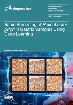
Cover Story (view full-size image): Helicobacter pylori is a major risk factor for gastric cancer. The incidence and prevalence of the pathogen are increasing worldwide. We present an algorithm combining a feature pyramid network and a ResNet architecture for automatic and rapid H. pylori detection in digitized Warthin–Starry-stained gastric biopsies. The algorithm exhibited 0.923 average precision and 0.982 average recall. The conducted efficiency study demonstrated that algorithm utilization significantly decreased (p < 0.001) diagnostic turnaround times for all participants, observing an 88.13–91.76% time reduction. Implementation of the algorithm also improved diagnostic accuracy for the resident, technician, and biotechnologist, indicating that the tool remarkably supports less experienced personnel. View this paper
  • Issues are regarded as officially published after their release is announced to the table of contents alert mailing list.
  • You may sign up for e-mail alerts to receive table of contents of newly released issues.
  • PDF is the official format for papers published in both, html and pdf forms. To view the papers in pdf format, click on the "PDF Full-text" link, and use the free Adobe Reader to open them.
Order results
Result details
Section
Select all
Export citation of selected articles as:
14 pages, 1819 KiB  
Article
Mucoepidermoid Carcinoma of the Minor Salivary Glands Diagnosed by High-Definition Ultrasound and Fine-Needle Aspiration: A Milan System-Based Retrospective Study
by Luisa Limongelli, Marta Forte, Gianfranco Favia, Fabio Dell’Olio, Giuseppe Ingravallo, Eliano Cascardi, Eugenio Maiorano, Alfonso Manfuso, Chiara Copelli, Antonio d’Amati and Saverio Capodiferro
Diagnostics 2025, 15(9), 1182; https://doi.org/10.3390/diagnostics15091182 - 7 May 2025
Viewed by 310
Abstract
Background/Objectives: Mucoepidermoid carcinoma (MEC) is the most common malignant tumor of the minor salivary glands, often affecting the hard palate. Preoperative diagnosis and surgical planning are challenging due to anatomical complexity and limitations in sampling, generally obtained by fine-needle aspiration (FNA). This [...] Read more.
Background/Objectives: Mucoepidermoid carcinoma (MEC) is the most common malignant tumor of the minor salivary glands, often affecting the hard palate. Preoperative diagnosis and surgical planning are challenging due to anatomical complexity and limitations in sampling, generally obtained by fine-needle aspiration (FNA). This study retrospectively evaluated the diagnostic and therapeutic performance of a high-definition ultrasound (HDUS)-guided fine-needle aspiration cytology/biopsy (FNAC/FNAB) protocol in diagnosing intraoral MEC, based on the Milan System for Reporting Salivary Gland Cytopathology (MSRSGC), with the relative clinical outcomes. Methods: A cohort of 64 patients with histologically confirmed MEC of the minor salivary glands, treated between 2000 and 2022, was retrospectively analyzed. All patients underwent HDUS-guided FNAC/FNAB, imaging (CT, MRI, and panoramic X-ray), and subsequent surgical treatment. The cytological specimens were classified using the MSRSGC. Surgical margins, histopathological findings, lymph node status, and follow-up outcomes were recorded. Results: Of 64 MECs, 42 cases were finally diagnosed as low-grade (LG)/intermediate grade (IG) and 22 as high-grade (HG) carcinomas, using a two-tier histological classification system. HDUS accurately delineated the lesion size, infiltration depth, and bone proximity, with excellent correlation with surgical specimens (difference ≤ 0.6 mm). MSRSGC classification distributed the cases across all categories, with 28 classified as malignant (category VI). Repeat FNAC improved the diagnostic yield in non-diagnostic and atypical cases. FNAB confirmed the cytological findings in all cases, with immunohistochemistry investigation with Ki-67 supporting tumor grading. Surgical margins were clear in all resections. Lymph node metastases were identified in all patients who underwent neck dissection (n = 18), all with HG-MEC. No recurrences occurred among the LG/IG-MEC patients during a median 2-year follow-up. Conclusions: The combined use of HDUS and FNAC/FNAB, interpreted through the MSRSGC framework, offers a highly accurate, minimally invasive approach for preoperative diagnosis and surgical planning in intraoral MEC. HDUS-guided cytology significantly improves diagnostic reliability, particularly in LG/IG and cystic variants, facilitating tailored surgical management. Also, the clinical outcomes may support the possibility of using a simplified grading classification for two histopathological types. Full article
(This article belongs to the Section Pathology and Molecular Diagnostics)
Show Figures

Figure 1

17 pages, 886 KiB  
Systematic Review
A Closer Look at Periocular Necrotizing Fasciitis: A Systematic Review of Literature
by David Oliver-Gutierrez, Elena Ros-Sanchez, Gloria Segura-Duch, Tirso Alonso, Miguel Ángel Arcediano, Alejandra Herranz-Cabarcos, Jessica Matas, Roberto Castro Seco, R. L. P. van der Veen, Anna Boixadera, José García-Arumí and Joan Oliveres
Diagnostics 2025, 15(9), 1181; https://doi.org/10.3390/diagnostics15091181 - 7 May 2025
Viewed by 259
Abstract
Background: Periocular necrotizing fasciitis (PNF) is a rare but life-threatening emergency that requires immediate recognition, as delayed diagnosis can worsen patient outcomes. To address this critical issue, we conducted the largest and most comprehensive systematic review to date, providing valuable insights into [...] Read more.
Background: Periocular necrotizing fasciitis (PNF) is a rare but life-threatening emergency that requires immediate recognition, as delayed diagnosis can worsen patient outcomes. To address this critical issue, we conducted the largest and most comprehensive systematic review to date, providing valuable insights into the diagnosis and treatment of PNF to improve clinical practice and patient prognosis. Methods: A search on Pubmed, Scopus, Embase, and WOS from January 2013 to August 2024 was performed. Only the cases of NF affecting the periocular region were included with no age limitations. Article selection and data extraction were performed independently by two investigators to avoid bias. Bias on individual studies is low as they represent case reports or case series, and publication bias is partially addressed including all the large case series even if no individual data could be retrieved. Results: The cohort included a total of 183 patients with PNF, with detailed patient-specific data for 107 individuals and only aggregated data for another 76. The average age at diagnosis was 54.2 years, and females constituted 44% of the population sample. Notably, 49.6% of the patients were immunocompromised. Streptococcus pyogenes was the predominant causative organism, identified in 79.8% of the cases. Most infections were unilateral (72.1%) without extension beyond the periocular area (54.7%). Most patients (89.6%) underwent surgical debridement alongside intravenous antibiotics. Septic shock occurred in 26.8% of the patients, and the overall mortality rate was 4.9%. Visual acuity was unaffected in 67.5% of the patients, though 18.2% progressed to blindness on the affected side. Reconstructive efforts predominantly involved skin grafting, both free and local pedunculate flaps as well as secondary healing in some instances. Conclusions: This systematic review summarizes the understanding of periocular necrotizing fasciitis’ (PNF) demographic trends, clinical manifestations, causative pathogens, and patient outcomes. Vigilance for PNF should be heightened when the clinical assessment of the patient’s eyelids reveals rapidly spreading edema and induration, subcutaneous emphysema, or necrotic bullae and/or eschar. Prompt identification and expedited intervention, including debridement and targeted antibiotic therapy, critically influence prognosis. Despite optimal management, patients may still suffer from significant aesthetic impairment, severe complications such as vision loss, or death due to septic shock. Full article
(This article belongs to the Special Issue Advances in the Diagnosis of Eye Diseases)
Show Figures

Figure 1

21 pages, 6719 KiB  
Review
Ultrasonography of the Fasciae and Common Pathologies: The Game Changer
by Carmelo Pirri, Nina Pirri, Veronica Macchi, Andrea Porzionato, Raffaele De Caro, Levent Özçakar and Carla Stecco
Diagnostics 2025, 15(9), 1180; https://doi.org/10.3390/diagnostics15091180 - 7 May 2025
Viewed by 265
Abstract
Ultrasound (US) is rapidly gaining attraction among physicians for the evaluation of fasciae. Unlike traditional imaging, which often lacks specificity of pain localization, US examination stands out as a versatile tool, capable of detecting both structural and functional information. This unique capability allows [...] Read more.
Ultrasound (US) is rapidly gaining attraction among physicians for the evaluation of fasciae. Unlike traditional imaging, which often lacks specificity of pain localization, US examination stands out as a versatile tool, capable of detecting both structural and functional information. This unique capability allows for a comprehensive assessment of fasciae—the intricate connective tissue essential for human biomechanics. US examination offers a multiparametric approach for the assessment of thickness, echogenicity, stiffness, deformation and shear strain. This comprehensive examination is invaluable for identifying fascial pathologies that may not be detected during physical examination. In this study, we render and discuss common/elementary lesions of the fascia. Full article
(This article belongs to the Section Medical Imaging and Theranostics)
Show Figures

Figure 1

17 pages, 6643 KiB  
Case Report
Calciphylaxis of the Upper Limbs—A Rare and Serious Disease with Multidisciplinary Treatments—A Case Series and Literature Review
by Mihaela Pertea, Malek Benamor, Andra-Irina Bulgaru-Iliescu, Abderrazek Abid, Said Abid and Alexandru-Hristo Amarandei
Diagnostics 2025, 15(9), 1179; https://doi.org/10.3390/diagnostics15091179 - 6 May 2025
Viewed by 336
Abstract
Background/Objectives: Calciphylaxis or calcific uremic arteriolopathy is a rare but highly lethal pathology that occurs most frequently in a uremic context, although it can also occur outside of this context. It is characterized by the appearance of necrotic skin lesions. Localization to the [...] Read more.
Background/Objectives: Calciphylaxis or calcific uremic arteriolopathy is a rare but highly lethal pathology that occurs most frequently in a uremic context, although it can also occur outside of this context. It is characterized by the appearance of necrotic skin lesions. Localization to the upper limbs is rare and has a similarly progressive evolution. Methods: We present a series of two cases—a male and a female—with calciphylaxis diagnoses (including biopsies) and with the patients undergoing dialysis for end-stage renal disease, both with infected and extensive necrotic lesions to the hands and fingers. Both cases required serial debridement treatments and amputations. A literature review was conducted using the precise search terms “calciphylaxis”, “upper limb”, “uremic calcific arteriolopathy”, and “end-stage renal disease” from January 2010 to May 2024. Results: One of the two reported cases ended with the patient’s death. The results of the literature review (comprising seven similar cases) confirmed the rarity of calciphylaxis lesion localization to the upper limbs and the high mortality rate among these patients despite administered treatments. No therapeutic protocol for these cases was confirmed. Conclusions: The treatment of calciphylaxis cases is multidisciplinary. Although surgical intervention is controversial, it is necessary in some cases, sometimes serially. Localization to the thoracic limbs has the same evolution and poor prognosis as other localizations. A standardized therapeutic protocol for these cases is still far from being established. Full article
(This article belongs to the Section Pathology and Molecular Diagnostics)
Show Figures

Figure 1

18 pages, 1414 KiB  
Review
Neurodegenerative Biomarkers in Multiple Sclerosis: At the Interface Between Research and Clinical Practice
by Alin Ciubotaru, Mădălina Irina Smihor, Cristina Grosu, Daniel Alexa, Roxana Covali, Robert-Constantin Anicăi, Ioana Păvăleanu, Andrei Ionuț Cucu, Amelian Mădălin Bobu, Cristina Mihaela Ghiciuc and Emilian Bogdan Ignat
Diagnostics 2025, 15(9), 1178; https://doi.org/10.3390/diagnostics15091178 - 6 May 2025
Viewed by 349
Abstract
Multiple sclerosis (MS) is a chronic autoimmune disorder characterized by inflammation, demyelination, and neurodegeneration within the central nervous system (CNS). While the inflammatory components of MS have been extensively studied, the progressive neurodegenerative aspect remains a critical factor contributing to long-term disability. Therefore, [...] Read more.
Multiple sclerosis (MS) is a chronic autoimmune disorder characterized by inflammation, demyelination, and neurodegeneration within the central nervous system (CNS). While the inflammatory components of MS have been extensively studied, the progressive neurodegenerative aspect remains a critical factor contributing to long-term disability. Therefore, the identification and validation of biomarkers associated with neurodegenerative processes are essential for improved diagnosis, prognosis, and treatment monitoring. This review explores cerebrospinal fluid (CSF) and blood-based biomarkers, including neurofilaments, lipid markers, kynurenines, and other molecular indicators that provide insights into neurodegeneration in MS. Full article
(This article belongs to the Special Issue Advances in the Laboratory Diagnosis)
Show Figures

Figure 1

34 pages, 15537 KiB  
Article
Explainable Artificial Intelligence for Diagnosis and Staging of Liver Cirrhosis Using Stacked Ensemble and Multi-Task Learning
by Serkan Savaş
Diagnostics 2025, 15(9), 1177; https://doi.org/10.3390/diagnostics15091177 - 6 May 2025
Viewed by 605
Abstract
Background/Objectives: Liver cirrhosis is a critical chronic condition with increasing global mortality and morbidity rates, emphasizing the necessity for early and accurate diagnosis. This study proposes a comprehensive deep-learning framework for the automatic diagnosis and staging of liver cirrhosis using T2-weighted MRI [...] Read more.
Background/Objectives: Liver cirrhosis is a critical chronic condition with increasing global mortality and morbidity rates, emphasizing the necessity for early and accurate diagnosis. This study proposes a comprehensive deep-learning framework for the automatic diagnosis and staging of liver cirrhosis using T2-weighted MRI images. Methods: The methodology integrates stacked ensemble learning, multi-task learning (MTL), and transfer learning within an explainable artificial intelligence (XAI) context to improve diagnostic accuracy, reliability, and transparency. A hybrid model combining multiple pre-trained convolutional neural networks (VGG16, MobileNet, and DenseNet121) with XGBoost as a meta-classifier demonstrated robust performance in binary classification between healthy and cirrhotic cases. Results: The model achieved a mean accuracy of 96.92%, precision of 95.12%, recall of 98.93%, and F1-score of 96.98% across 10-fold cross-validation. For staging (mild, moderate, and severe), the MTL framework reached a main task accuracy of 96.71% and an average AUC of 99.81%, with a powerful performance in identifying severe cases. Grad-CAM visualizations reveal class-specific activation regions, enhancing the transparency and trust in the model’s decision-making. The proposed system was validated using the CirrMRI600+ dataset with a 10-fold cross-validation strategy, achieving high accuracy (AUC: 99.7%) and consistent results across folds. Conclusions: This research not only advances State-of-the-Art diagnostic methods but also addresses the black-box nature of deep learning in clinical applications. The framework offers potential as a decision-support system for radiologists, contributing to early detection, effective staging, personalized treatment planning, and better-informed treatment planning for liver cirrhosis. Full article
(This article belongs to the Section Machine Learning and Artificial Intelligence in Diagnostics)
Show Figures

Figure 1

26 pages, 810 KiB  
Review
Advancing Cervical Cancer Prevention Equity: Innovations in Self-Sampling and Digital Health Technologies Across Healthcare Settings
by Michelle Gomes, Elena Provaggi, Andrea Barnabas Pembe, Adeola Olaitan and Aleksandra Gentry-Maharaj
Diagnostics 2025, 15(9), 1176; https://doi.org/10.3390/diagnostics15091176 - 6 May 2025
Viewed by 570
Abstract
Cervical cancer causes 350,000 deaths annually, with 90% occurring in low- and middle-income countries (LMICs), despite being largely preventable through vaccination and screening. This review examines innovative approaches to address screening coverage gaps worldwide, analysing both established programmes in high-income countries and implementation [...] Read more.
Cervical cancer causes 350,000 deaths annually, with 90% occurring in low- and middle-income countries (LMICs), despite being largely preventable through vaccination and screening. This review examines innovative approaches to address screening coverage gaps worldwide, analysing both established programmes in high-income countries and implementation strategies for LMICs. Self-sampling technologies demonstrate significant potential to improve the uptake of cervical screening, thereby improving cervical cancer prevention compared to traditional methods, particularly benefiting underserved populations across all healthcare settings. Among self-collection devices, vaginal brushes achieve sensitivity of 94.6% (95% CI: 92.4–96.8) for HPV detection, while novel approaches like the tampon show promising results (sensitivity 82.9–100%, specificity 91.6–96.8%) with high user acceptability. Implementation strategies vary by healthcare context, with high-income countries achieving success through integrated screening programmes and digital solutions, while LMICs demonstrate effective adaptation through community-based distribution (20–35% uptake) and innovative delivery methods. In resource-limited settings, self-sampling increases participation through enhanced patient comfort and cultural acceptability, while reducing costs by 32–48%. Progress toward WHO’s cervical cancer elimination goals require careful consideration of local healthcare infrastructure, cultural contexts and sustainable financing mechanisms. Future research priorities include optimising self-sampling technologies for sustainability and scalability, developing context-specific implementation strategies and validating artificial intelligence applications to enhance screening efficiency across diverse healthcare settings. Full article
(This article belongs to the Special Issue Exploring Gynecological Pathology and Imaging)
Show Figures

Figure 1

18 pages, 3032 KiB  
Systematic Review
Intravascular Imaging-Guided Versus Angiography-Guided Percutaneous Coronary Intervention: A Systematic Review and Meta-Analysis of Randomized Controlled Trials
by Akash Kumar, Muhammad Salman Nadeem, Sooraj Kumar, Muzamil Akhtar, Ayesha Maryam, Rubyisha Sheikh, Nomesh Kumar, Naresh Kumar Ladhwani, Nimurta Madhwani, Nisha Kumari, Muhammad Riyyan Rao, Syed Sarmad Javaid, Peter Collins and Raheel Ahmed
Diagnostics 2025, 15(9), 1175; https://doi.org/10.3390/diagnostics15091175 - 6 May 2025
Viewed by 391
Abstract
Background/Objectives: Despite the potential benefits, intravascular imaging for guiding percutaneous coronary intervention (PCI) remains underutilized. Recent trials have provided new data, prompting a need for updated insights. This study aimed to perform a comprehensive meta-analysis to compare the clinical outcomes of intravascular imaging-guided [...] Read more.
Background/Objectives: Despite the potential benefits, intravascular imaging for guiding percutaneous coronary intervention (PCI) remains underutilized. Recent trials have provided new data, prompting a need for updated insights. This study aimed to perform a comprehensive meta-analysis to compare the clinical outcomes of intravascular imaging-guided PCI versus angiography-guided PCI, thereby evaluating the relative effectiveness of these two guidance strategies in improving patient outcomes. Methods: PubMed, Cochrane Library, Embase and Clinicaltrials.gov databases were systematically searched from inception till 25 November 2024. Randomized clinical trials (RCTs) comparing intravascular imaging with coronary angiography in patients undergoing complex PCI were included. Statistical analysis was conducted using a random effects model to calculate pooled risk ratios with 95% confidence intervals (CI). Results: In this meta-analysis of 21 studies involving 18,043 patients, intravascular image-guided PCI significantly reduced the risk of all-cause mortality by 24%, cardiac mortality by 63%, MACE by 35%, target vessel myocardial infarction by 32%, stent thrombosis by 42%, target vessel revascularization by 45%, target lesion revascularization by 34% and myocardial infarction by 22% compared to angiography-guided PCI. There was no significant difference in bleeding events. Conclusions: Intravascular imaging significantly reduces cardiac events, all-cause mortality and revascularization rates in PCI patients. These findings support its broader adoption and potential updates to clinical guidelines. Full article
(This article belongs to the Special Issue Diagnosis and Management of Coronary Heart Disease)
Show Figures

Figure 1

12 pages, 762 KiB  
Review
Debunking Myths and Misinformation in Cervical Cancer: A Narrative Review on Navigating Complex Treatment Choices in Locally Advanced Cases and Exploring Beyond Standard Protocols
by Mustafa Zelal Muallem and Ahmad Sayasneh
Diagnostics 2025, 15(9), 1174; https://doi.org/10.3390/diagnostics15091174 - 6 May 2025
Viewed by 279
Abstract
Cervical cancer remains a significant health challenge globally, with locally advanced cervical cancer (LACC) representing a particularly complex subset due to its diverse definitions and varied treatment approaches. The absence of randomized controlled trials comparing the upfront radical surgery followed by concurrent chemoradiotherapy [...] Read more.
Cervical cancer remains a significant health challenge globally, with locally advanced cervical cancer (LACC) representing a particularly complex subset due to its diverse definitions and varied treatment approaches. The absence of randomized controlled trials comparing the upfront radical surgery followed by concurrent chemoradiotherapy (CCRT) or chemotherapy alone for clearly defined risk factors for LACC hinders the development of standardized treatment protocols, leading to disparities in patient outcomes across different healthcare settings. This paper seeks to underline the necessity of a consensus on the definition of LACC and aims to comprehensively and critically review the existing literature trying to harmonize treatment strategies and improve prognostic outcomes. Our analysis suggests a multimodal approach for treating FIGO IB3, IIA2, and selected IIB cases at facilities capable of delivering highly curative nerve-sparing surgical interventions, with the goal of bridging the gap in current treatment methodologies. Preliminary findings suggest that adopting a standardized definition could facilitate more consistent treatment outcomes and enhance comparative research. Full article
Show Figures

Figure 1

13 pages, 1231 KiB  
Protocol
Real-World, National Study of Palbociclib in HR+/HER2− Metastatic Breast Cancer: A 2.5-Year Follow-Up PALBO01/2021
by Cristina Marinela Oprean, Larisa Maria Badau, Ramona Petrita, Mircea Dragos Median and Alis Dema
Diagnostics 2025, 15(9), 1173; https://doi.org/10.3390/diagnostics15091173 - 5 May 2025
Viewed by 411
Abstract
Background: Palbociclib, when combined with endocrine therapy, represents a valuable treatment option for patients diagnosed with hormone receptor (HR) positive/human epidermal growth factor receptor 2 (HER2) negative advanced breast cancer (BC) or metastatic breast cancer (MBC). Approved in Europe following phase II/III trials, [...] Read more.
Background: Palbociclib, when combined with endocrine therapy, represents a valuable treatment option for patients diagnosed with hormone receptor (HR) positive/human epidermal growth factor receptor 2 (HER2) negative advanced breast cancer (BC) or metastatic breast cancer (MBC). Approved in Europe following phase II/III trials, it became the first CDK4/6 inhibitor used alongside hormone therapy. Available real-world data demonstrate the strong performance of Palbociclib in unselected, heavily pretreated patient groups. Our retrospective, observational, multicenter study, conducted in six Romanian institutions during a follow-up period of 2.5 years, aimed to assess Palbociclib’s safety and effectiveness in clinical practice. Objectives: The primary endpoints included response rate such as overall response rate (ORR), duration of response (DOR), disease control rate (DCR) and best clinical response (BCR), progression free survival (PFS) and overall survival (OS). The secondary objectives focused on treatment duration with aromatase inhibitors (AI) or fulvestrant and subsequent therapies after disease progression. Grade 3/4 adverse events were individually recorded. Exploratory analysis evaluated the potential predictive biomarkers such as Ki67, lower levels of HER2 expression (HER2-low), and histological or luminal subtype. Methods: Approximately 650 patients were planned for inclusion. PFS and OS were analyzed via the Kaplan–Meier method, with median times, 1- and 2-year estimates, and 95% confidence intervals reported. Conclusions: This study supports the integration of clinical trial evidence into real-world settings, enhancing patient selection and treatment personalization. Full article
(This article belongs to the Special Issue Advances in Diagnosis and Prognosis of Breast Cancer)
Show Figures

Figure 1

12 pages, 2092 KiB  
Article
Agreement Analysis Among Hip and Knee Periprosthetic Joint Infections Classifications
by Caterina Rocchi, Marco Di Maio, Alberto Bulgarelli, Katia Chiappetta, Francesco La Camera, Guido Grappiolo and Mattia Loppini
Diagnostics 2025, 15(9), 1172; https://doi.org/10.3390/diagnostics15091172 - 4 May 2025
Viewed by 339
Abstract
Background/Objectives: A missed periprosthetic joint infection (PJI) diagnosis can lead to implant failure. However, to date, no gold standard for PJI diagnosis exists, although several classification scores have been developed in the past years. The primary objective of the study was the [...] Read more.
Background/Objectives: A missed periprosthetic joint infection (PJI) diagnosis can lead to implant failure. However, to date, no gold standard for PJI diagnosis exists, although several classification scores have been developed in the past years. The primary objective of the study was the evaluation of inter-rater reliability between five PJI classification systems when defining a patient who is infected. Two secondary outcomes were further examined: the inter-rater reliability assessed by comparing the classifications in pairs, and the evaluation of each classification system within the subcategories defined by the World Association against Infection in Orthopaedics and Trauma (WAIOT) definition. Methods: Retrospectively collected data on patients with knee and hip PJIs were used to assess the agreement among five PJI scoring systems: the Musculoskeletal Infection Society (MSIS) 2013 definition, the Infection Consensus Group (ICG) 2018 definition, the European Bones and Joints Infection Society (EBJIS) 2018 definition, the WAIOT definition, and the EBJIS 2021 definition. Results: In total, 203 patients with PJI were included in the study, and the agreement among the examined scores was 0.90 (Krippendorff’s alpha = 0.81; p-value < 0.001), with the MSIS 2013 and ICG 2018 classification systems showing the highest agreement (Cohen’s Kappa = 0.91; p-value < 0.001). Conclusions: There is a strong agreement between the major PJI classification systems. However, a subset of patients (n = 11, 5.42%) still falls into a diagnostic grey zone, especially in cases of low-grade infections. This highlights the need for enhanced diagnostic criteria that incorporate tools that are available even with limited resources, and the potential of artificial intelligence-based techniques in improving early detection and management of PJIs. Full article
Show Figures

Figure 1

17 pages, 2130 KiB  
Article
TERT Immunohistochemistry in Thin Melanomas Compared to Melanocytic Nevi
by Iulia Zboraș, Loredana Ungureanu, Simona Corina Șenilă, Bobe Petrushev, Paula Zamfir, Doinița Crișan, Flaviu Andrei Zaharie, Ștefan Cristian Vesa and Rodica Cosgarea
Diagnostics 2025, 15(9), 1171; https://doi.org/10.3390/diagnostics15091171 - 4 May 2025
Viewed by 260
Abstract
Background/Objectives: Telomerase plays a vital role in preserving telomere length, a key process in cancer development. Human telomerase reverse transcriptase (hTERT) is commonly expressed in various cancers, including melanoma. This study evaluated hTERT protein expression in melanomas compared to melanocytic nevi. Methods [...] Read more.
Background/Objectives: Telomerase plays a vital role in preserving telomere length, a key process in cancer development. Human telomerase reverse transcriptase (hTERT) is commonly expressed in various cancers, including melanoma. This study evaluated hTERT protein expression in melanomas compared to melanocytic nevi. Methods: In total, we examined 75 melanocytic lesions using TERT immunohistochemistry on paraffin-embedded tissues; 36 of them were thin melanomas (Breslow index ≤ 1 mm) and 39 melanocytic nevi. Results: The TERT expression differed with statistical significance between the two studied groups, melanomas and melanocytic nevi, in all three aspects examined: percentage of staining (p = 0.006), intensity of staining (p = 0.035), and localisation of staining (p = 0.012). Three quarters of the melanomas stained in over 50% of the cells at cytoplasmic level, 52.78% of the melanomas exhibited an intensity of 3+, and all melanomas were stained at the cytoplasmic level, except for the two negative cases. The values were lower in the melanocytic nevi group. Still, the diagnostic values were relatively low (sensitivity = 75%, specificity = 58.97%, PPV = 62.79%, NPV = 71.88%, and ACC = 66.67%). Conclusions: TERT immunohistochemistry differed between the two studied groups; however, the diagnostic utility is low in our study. Combining with other immunohistochemical antibodies would probably increase the diagnostic power. Full article
(This article belongs to the Special Issue New Developments in the Diagnosis of Skin Tumors)
Show Figures

Figure 1

5 pages, 175 KiB  
Editorial
Editorial for the Special Issue “Medical Data Processing and Analysis—2nd Edition”
by Wan Azani Mustafa and Hiam Alquran
Diagnostics 2025, 15(9), 1170; https://doi.org/10.3390/diagnostics15091170 - 4 May 2025
Viewed by 352
Abstract
Medical data processing and analysis have become central to advancements in healthcare, driven largely by the need for accurate diagnosis, personalized treatment, and efficient healthcare system management [...] Full article
(This article belongs to the Special Issue Medical Data Processing and Analysis—2nd Edition)
3 pages, 1684 KiB  
Interesting Images
Computed Tomography, Coronary Angiography, and Intravascular Ultrasound in the Diagnosis of Left Anterior Descending Stenosis in a 38-Year-Old Woman with a Calcium Score of Zero
by Malgorzata Zalewska-Adamiec and Slawomir Dobrzycki
Diagnostics 2025, 15(9), 1169; https://doi.org/10.3390/diagnostics15091169 - 4 May 2025
Viewed by 280
Abstract
Cardiovascular diseases, including coronary artery disease, are the leading cause of mortality in the general population. The likelihood of significant coronary artery disease occurring in young women (<40 years of age) is very low. Diagnosis is challenging and often delayed, treatment tends to [...] Read more.
Cardiovascular diseases, including coronary artery disease, are the leading cause of mortality in the general population. The likelihood of significant coronary artery disease occurring in young women (<40 years of age) is very low. Diagnosis is challenging and often delayed, treatment tends to be suboptimal, and the long-term prognosis is poor. We present the case of a 38-year-old woman with typical anginal chest pain whose coronary computed tomography angiography (CCTA) revealed significant narrowing in the left anterior descending artery (LAD) despite a coronary artery calcium score (CACS) of zero. To assess the significance of the lesion, coronary angiography and intravascular ultrasound (IVUS) were performed, which revealed borderline narrowing. Conservative treatment was recommended. Full article
(This article belongs to the Special Issue Clinical Diagnosis and Management in Cardiology)
Show Figures

Figure 1

4 pages, 812 KiB  
Interesting Images
Immune Checkpoint Inhibitor-Related Sjögren’s Syndrome: An Ocular Immune-Related Adverse Event
by Hideki Fukuoka, Akifumi Matsumoto and Chie Sotozono
Diagnostics 2025, 15(9), 1168; https://doi.org/10.3390/diagnostics15091168 - 4 May 2025
Viewed by 289
Abstract
A 67-year-old male with metastatic human papillomavirus (HPV)-positive oropharyngeal cancer receiving pembrolizumab (anti-programmed cell death protein 1 [PD-1] immune checkpoint inhibitor) presented with bilateral ocular dryness. It is important to note that these symptoms appeared eight months after the initiation of the pembrolizumab [...] Read more.
A 67-year-old male with metastatic human papillomavirus (HPV)-positive oropharyngeal cancer receiving pembrolizumab (anti-programmed cell death protein 1 [PD-1] immune checkpoint inhibitor) presented with bilateral ocular dryness. It is important to note that these symptoms appeared eight months after the initiation of the pembrolizumab therapy. Ophthalmologic evaluation revealed keratoconjunctivitis sicca with characteristic bulbar fluorescein staining and the Schirmer test showed 0 mm bilaterally. Serological testing demonstrated positive antinuclear and anti-SSb/La antibodies, consistent with Sjögren’s syndrome as an immune-related adverse event (irAE). Treatment with topical fluorometholone 0.1% and diquafosol 3% led to complete symptom resolution within one year while maintaining cancer immunotherapy. Long-term follow-up over 3.5 years demonstrated sustained ocular improvement and a favorable oncologic response without development of systemic autoimmune manifestations. This case highlights that Sjögren’s syndrome as an irAE may present with isolated ocular manifestations, which could be overlooked in clinical practice. Full article
(This article belongs to the Collection Interesting Images)
Show Figures

Figure 1

26 pages, 2853 KiB  
Systematic Review
Can the Pupillary Light Reflex and Pupillary Unrest Be Used as Biomarkers of Parkinson’s Disease? A Systematic Review and Meta-Analysis
by Aleksander Dawidziuk, Emilia Butters, Daniel Josef Lindegger, Campbell Foubister, Hugo Chrost, Michal Wlodarski, John Grogan, Paulina A Rowicka, Fion Bremner and Sanjay G Manohar
Diagnostics 2025, 15(9), 1167; https://doi.org/10.3390/diagnostics15091167 - 3 May 2025
Viewed by 294
Abstract
Background/Objectives: The pathological changes preceding the onset of Parkinson’s disease (PD) commence several decades before motor symptoms manifest, offering a potential window for identifying objective biomarkers for early diagnosis and disease monitoring. Among the primary non-motor features of PD is autonomic dysfunction; however, [...] Read more.
Background/Objectives: The pathological changes preceding the onset of Parkinson’s disease (PD) commence several decades before motor symptoms manifest, offering a potential window for identifying objective biomarkers for early diagnosis and disease monitoring. Among the primary non-motor features of PD is autonomic dysfunction; however, its precise assessment remains challenging, limiting its viability as a reliable biomarker. Both the pupillary light reflex (PLR) and pupillary unrest are regulated by autonomic pathways suggesting their potential as objective non-invasive indicators of the PD prodromal phase. This review systematically evaluates studies that compare PLR and pupillary unrest in individuals with PD and healthy controls to determine their utility as potential biomarkers of the disease. Methods: A systematic search strategy was designed to identify studies reporting PLR and pupillary unrest findings in PD patients. Searches were conducted across three databases (MEDLINE, Embase PsycINFO), supplemented by cross-referencing relevant studies found on Google Scholar. The literature search was last updated on 7 December 2020. Pupillometric parameters that permitted statistical synthesis included maximum constriction velocity (VMax), constriction amplitude (CAmp), and constriction latency (CL). Pooled incidence and effect sizes were determined using a random-effects model with an inverse variance DerSimonian–Laird estimator. The I2 statistic was used to assess study heterogeneity. When meta-analysis was not feasible, a qualitative analysis was undertaken. Results: The initial search yielded 219 references. Following deduplication and exclusion of ineligible studies, 31 papers were selected for review. Pupillometric data from 11 studies were incorporated into the meta-analysis. Effect sizes for PD patients were significant for VMax −0.92, (p < 0.01), CAmp −0.58, (p < 0.05), and CL 0.46, (p < 0.05). Measures of pupillary unrest were elevated in PD patients compared to controls, but evidence was limited to two studies. Conclusions: Pupillary constriction in response to light is characterised by reduced speed and amplitude in PD, with effect sizes suggesting potential clinical applicability. However, evidence regarding baseline pupillary variability remains insufficient, underlining the necessity for further research. Pupillary metrics represent a promising avenue for early PD detection, though their clinical utility is constrained by methodological heterogeneity and variations in disease duration among studies. Full article
(This article belongs to the Section Pathology and Molecular Diagnostics)
Show Figures

Figure 1

11 pages, 1028 KiB  
Article
Potential for Misinterpretation in the Laboratory Diagnosis of Clostridioides difficile Infections
by Alexandra Kalacheva, Metodi Popov, Valeri Velev, Rositsa Stoyanova, Yordanka Mitova-Mineva, Tsvetelina Velikova and Maria Pavlova
Diagnostics 2025, 15(9), 1166; https://doi.org/10.3390/diagnostics15091166 - 3 May 2025
Viewed by 247
Abstract
Background/Objective. Toxin-producing strains of Clostridioides difficile (C. diff) are the most commonly identified cause of healthcare-associated infection in the elderly. Risk factors include advanced age, hospitalization, prior or concomitant systemic antibacterial therapy, chemotherapy, and gastrointestinal surgery. Patients with unspecified and [...] Read more.
Background/Objective. Toxin-producing strains of Clostridioides difficile (C. diff) are the most commonly identified cause of healthcare-associated infection in the elderly. Risk factors include advanced age, hospitalization, prior or concomitant systemic antibacterial therapy, chemotherapy, and gastrointestinal surgery. Patients with unspecified and new-onset diarrhea with ≥3 unformed stools in 24 h are the target population for C. diff infection (CDI) testing. To present data on the risks of laboratory misdiagnosis in managing CDI. Materials. In two general hospitals, we examined 116 clinical stool specimens from hospitalized patients with acute diarrhea suspected of nosocomial or antibiotic-associated diarrhea (AAD) due to C. diff. Enzyme immunoassay (EIA) tests for the detection of C. diff toxins A (cdtA) and B (cdtB) in stool, automated CLIA assay for the detection of C. diff GDH antigen and qualitative determination of cdtA and B in human feces and anaerobic stool culture were applied for CDI laboratory diagnosis. MALDI-TOF (Bruker) was used to identify the presumptive anaerobic bacterial colonies. The following methods were used as confirmatory diagnostics: the LAMP method for the detection of Salmonella spp. and simultaneous detection of C. jejuni and C. coli, an E. coli Typing RT-PCR detection kit (ETEC, EHEC, STEC, EPEC, and EIEC), API 20E and aerobic stool culture methods. Results. A total of 40 toxigenic strains of C. diff were isolated from all 116 tested diarrheal stool samples, of which 38/40 produced toxin B and 2/40 strains were positive for both cdtA and cdtB. Of the stool samples positive for cdtA (6/50) and/or cdtB (44/50) by EIA, 33 were negative for C. diff culture but positive for the following diarrheal agents: Salmonella enterica subsp. arizonae (1/33, LAMP, culture, API 20E); C. jejuni (2/33, LAMP, culture, MALDI TOF); ETEC O142 (1/33), STEC O145 and O138 (2/33, E. coli RT-PCR detection kit, culture); C. perfringens (2/33, anaerobic culture, MALDI TOF); hypermycotic enterotoxigenic K. pneumonia (2/33) and enterotoxigenic P. mirabilis (2/33, culture; PCR encoding LT-toxin). Two of the sixty-six cdtB-positive samples (2/66) showed a similar misdiagnosis when analyzed using the CLIA method. However, the PCR analysis showed that they were cdtB-negative. In contrast, the LAMP method identified a positive result for C. jejuni in one sample, and another was STEC positive (stx1+/stx2+) by RT-PCR. We found an additional discrepancy in the CDI test results: EPEC O86 (RT-PCR eae+) was isolated from a fecal sample positive for GHA enzyme (CLIA) and negative for cdtA and cdtB (CLIA and PCR). However, the culture of C. diff was negative. These findings support the hypothesis that certain human bacterial pathogens that produce enterotoxins other than C. diff, as well as intestinal commensal microorganisms, including Klebsiella sp. and Proteus sp., contribute to false-positive EIA card tests for C. diff toxins A and B, which are the most widely used laboratory tests for CDI. Conclusions. CDI presents a significant challenge to clinical practice in terms of laboratory diagnostic management. It is recommended that toxin-only EIA tests should not be used as the sole diagnostic tool for CDI but should be limited to detecting toxins A and B. Accurate diagnosis of CDI requires a combination of laboratory diagnostic methods on which proper infection management depends. Full article
(This article belongs to the Section Diagnostic Microbiology and Infectious Disease)
Show Figures

Figure 1

14 pages, 555 KiB  
Article
Leveraging Subjective Parameters and Biomarkers in Machine Learning Models: The Feasibility of lnc-IL7R for Managing Emphysema Progression
by Tzu-Tao Chen, Tzu-Yu Cheng, I-Jung Liu, Shu-Chuan Ho, Kang-Yun Lee, Huei-Tyng Huang, Po-Hao Feng, Kuan-Yuan Chen, Ching-Shan Luo, Chien-Hua Tseng, Yueh-His Chen, Arnab Majumdar, Cheng-Yu Tsai and Sheng-Ming Wu
Diagnostics 2025, 15(9), 1165; https://doi.org/10.3390/diagnostics15091165 - 3 May 2025
Viewed by 310
Abstract
Background/Objectives: Chronic obstructive pulmonary disease (COPD) remains a leading cause of death worldwide, with emphysema progression providing valuable insights into disease development. Clinical assessment approaches, including pulmonary function tests and high-resolution computed tomography, are limited by accessibility constraints and radiation exposure. This study, [...] Read more.
Background/Objectives: Chronic obstructive pulmonary disease (COPD) remains a leading cause of death worldwide, with emphysema progression providing valuable insights into disease development. Clinical assessment approaches, including pulmonary function tests and high-resolution computed tomography, are limited by accessibility constraints and radiation exposure. This study, therefore, proposed an alternative approach by integrating the novel biomarker long non-coding interleukin-7 receptor α-subunit gene (lnc-Il7R), along with other easily accessible clinical and biochemical metrics, into machine learning (ML) models. Methods: This cohort study collected baseline characteristics, COPD Assessment Test (CAT) scores, and biochemical details from the enrolled participants. Associations with emphysema severity, defined by a low attenuation area percentage (LAA%) threshold of 15%, were evaluated using simple and multivariate-adjusted models. The dataset was then split into training and validation (80%) and test (20%) subsets. Five ML models were employed, with the best-performing model being further analyzed for feature importance. Results: The majority of participants were elderly males. Compared to the LAA% <15% group, the LAA% ≥15% group demonstrated a significantly higher body mass index (BMI), poor pulmonary function, and lower expression levels of lnc-Il7R (all p < 0.01). Fold changes in lnc-IL7R were strongly and negatively associated with LAA% (p < 0.01). The random forest (RF) model achieved the highest accuracy and area under the receiver operating characteristic curve (AUROC) across datasets. A feature importance analysis identified lnc-IL7R fold changes as the strongest predictor for emphysema classification (LAA% ≥15%), followed by CAT scores and BMI. Conclusions: Machine learning models incorporated accessible clinical and biochemical markers, particularly the novel biomarker lnc-IL7R, achieving classification accuracy and AUROC exceeding 75% in emphysema assessments. These findings offer promising opportunities for improving emphysema classification and COPD management. Full article
(This article belongs to the Section Machine Learning and Artificial Intelligence in Diagnostics)
Show Figures

Figure 1

17 pages, 7042 KiB  
Review
Multimodal Imaging Approach to MEN-1 Syndrome-Associated Tumors
by Alice Carli, Elisa Boffa, Matteo Bonatti, Marco Chincarini, Maria Vittoria Davì and Giulia A. Zamboni
Diagnostics 2025, 15(9), 1164; https://doi.org/10.3390/diagnostics15091164 - 3 May 2025
Viewed by 286
Abstract
Multiple endocrine neoplasia type 1 (MEN-1) is an autosomal dominant inherited syndrome characterized by a genetic predisposition for the development of specific hormone-secreting tumors. Effective diagnosis and management of MEN-1 require genetic testing, regular surveillance, and imaging follow-up to detect and monitor tumor [...] Read more.
Multiple endocrine neoplasia type 1 (MEN-1) is an autosomal dominant inherited syndrome characterized by a genetic predisposition for the development of specific hormone-secreting tumors. Effective diagnosis and management of MEN-1 require genetic testing, regular surveillance, and imaging follow-up to detect and monitor tumor growth or recurrence and to plan for surgical intervention. The aim of this narrative review is to provide an overview of the current imaging modalities and their role in the diagnosis and follow-up of patients affected by MEN-1, focusing on the detection and characterization of associated neoplasms. The knowledge of the most frequent MEN-1 associated neoplasms and their imaging features is crucial for an accurate diagnosis, management, and treatment. Full article
(This article belongs to the Section Medical Imaging and Theranostics)
Show Figures

Figure 1

22 pages, 37030 KiB  
Review
The Many Faces of the Angry Peritoneum
by Maria Chiara Ambrosetti, Matilde Bariani, Giulia Angela Zamboni, Riccardo Valletta and Matteo Bonatti
Diagnostics 2025, 15(9), 1163; https://doi.org/10.3390/diagnostics15091163 - 3 May 2025
Viewed by 263
Abstract
The peritoneum is a thin membrane that lines the abdominal cavity and covers the abdominal organs. It serves as a conduit for the spread of various pathological processes, including gas and fluid collections, inflammation, infections, and neoplastic conditions. Peritoneal carcinomatosis is the most [...] Read more.
The peritoneum is a thin membrane that lines the abdominal cavity and covers the abdominal organs. It serves as a conduit for the spread of various pathological processes, including gas and fluid collections, inflammation, infections, and neoplastic conditions. Peritoneal carcinomatosis is the most common and well-known pathology involving the peritoneum, typically resulting from the dissemination of gastrointestinal and pelvic malignancies. However, numerous benign and malignant peritoneal diseases can mimic the imaging appearance of peritoneal carcinomatosis. The aim of this review is to revisit the anatomy of peritoneal compartments and elucidate the patterns of peritoneal disease spread. Emphasis is placed on identifying the distinctive imaging features of both neoplastic and non-neoplastic peritoneal diseases that differ from peritoneal carcinomatosis. Full article
(This article belongs to the Section Medical Imaging and Theranostics)
Show Figures

Figure 1

15 pages, 1224 KiB  
Article
Development of a Pulmonary Nodule Service and Clinical Pathway: A Pragmatic Approach Addressing an Unmet Need
by Georgia Hardavella, Ioannis Karampinis, Nikolaos Anastasiou, Konstantinos Stefanidis, Kyriaki Tavernaraki, Styliani Arapostathi, Nektaria Sidiropoulou, Petros Filippousis, Alexandro Patirelis, Eugenio Pompeo, Panagiotis Demertzis and Stefano Elia
Diagnostics 2025, 15(9), 1162; https://doi.org/10.3390/diagnostics15091162 - 2 May 2025
Viewed by 338
Abstract
Background/Objectives: The surveillance of patients with incidental pulmonary nodules overloads existing respiratory and lung cancer clinics, as well as multidisciplinary team meetings. In our clinical setting, until 2018, we had numerous patients with incidental pulmonary nodules inundating our outpatient clinics; therefore, the need [...] Read more.
Background/Objectives: The surveillance of patients with incidental pulmonary nodules overloads existing respiratory and lung cancer clinics, as well as multidisciplinary team meetings. In our clinical setting, until 2018, we had numerous patients with incidental pulmonary nodules inundating our outpatient clinics; therefore, the need to develop a novel service and dedicated clinical pathway arose. The aims of this study are to 1. provide (a) a model of setting up a novel pulmonary nodule service, and (b) a pragmatic clinical pathway to address the increasing need for surveillance of patients with incidental pulmonary nodules. 2. share real-world data from a dedicated pulmonary nodule service running in a tertiary setting with existing resources. Methods: A retrospective review of established processes and referral mechanisms to our tertiary pulmonary nodule service was conducted. We have also performed a retrospective collection and review of data for patients reviewed and discussed in our tertiary pulmonary nodule service between April 2018 and April 2024. Results: Our tertiary pulmonary nodule service (PNS) comprises a dedicated pulmonary nodule clinic, a nodule multidisciplinary team (MDT) meeting and a dedicated proforma referral system. Due to the current national health system legislation and relevant processes, patients are required to physically attend clinic appointments. There are various sources of referral, including other departments within the hospital, other hospitals, various specialties in primary care and self-referrals. Between 15 April 2018 and 15 April 2024, 2203 patients were reviewed in the pulmonary nodule clinic (903 females, 1300 males, mean age 64 ± 19 years). Of those patients, 65% (1432/2203) were current smokers. A total of 1365 new patients and 838 follow-up patients were reviewed in total. Emphysema was radiologically present in 72% of patients, and 75% of those (1189/1586) already had a confirmed diagnosis of chronic obstructive pulmonary disease (COPD). Coronary calcification was identified in 32% (705/2203), and 78% of those (550/705) were already known to cardiology services. Interestingly, 27% (368/1365) of the new patients were discharged following their first MDT meeting discussion, and 67% of these were discharged as the reason for their referral was an intrapulmonary lymph node which did not warrant any further action. Among all patients, 11% (246/2203) were referred to the multidisciplinary thoracic oncology service (MTOS) due to suspicious appearances/changes in their nodules that warranted further investigation, and from those, 37% were discharged (92/246) from the MTOS. The lung cancer diagnosis rate was 7% (154/2203). Conclusions: The applied pathway offers a pragmatic approach in setting up a service that addresses an increasing patient need. Its application is feasible in a tertiary care setting, and admin support is of vital importance to ensure patients are appropriately tracked and not lost to follow-up. Real-world data from pulmonary nodules services provide a clear overview and contribute to understanding patients’ characteristics and improving service provision. Full article
(This article belongs to the Section Clinical Diagnosis and Prognosis)
Show Figures

Figure 1

16 pages, 1198 KiB  
Review
Circulating Cell-Free DNA as an Epigenetic Biomarker for Early Diabetic Retinopathy: A Narrative Review
by Boaz Li, Megan M. Yim, Yu Xuan Jin, Brendan K. Tao, Jim S. Xie, Michael Balas, Haaris Khan, Wai-Ching Lam, Peng Yan and Eduardo V. Navajas
Diagnostics 2025, 15(9), 1161; https://doi.org/10.3390/diagnostics15091161 - 2 May 2025
Viewed by 412
Abstract
Diabetic retinopathy (DR), a complication of type 2 diabetes mellitus (T2DM), is typically asymptomatic in its early stages. Diagnosis typically relies on routine fundoscopy for the clinical detection of microvascular abnormalities. However, permanent retinal damage may occur well before clinical signs are appreciable. [...] Read more.
Diabetic retinopathy (DR), a complication of type 2 diabetes mellitus (T2DM), is typically asymptomatic in its early stages. Diagnosis typically relies on routine fundoscopy for the clinical detection of microvascular abnormalities. However, permanent retinal damage may occur well before clinical signs are appreciable. In the early stages of DR, the retina undergoes distinct epigenetic changes, including DNA methylation and histone modifications. Recent evidence supports unique epigenetic ‘signatures’ in patients with DR compared to non-diabetic controls. These DNA ‘signature’ sequences may be specific to the retina and may circulate in peripheral blood in the form of cell-free DNA (cfDNA). In this review, we explore the literature and clinical application of cfDNA sampling as an early, non-invasive, accessible assessment tool for early DR detection. First, we summarize the known epigenetic signatures of DR. Next, we review current sequencing technologies used for cfDNA detection, such as magnetic bead-based enrichment, next-generation sequencing, and bisulfite sequencing. Finally, we outline the current research limitations and emerging areas of study which aim to improve the clinical utility of cfDNA for DR evaluation. Full article
(This article belongs to the Special Issue New Insights into the Diagnosis and Prognosis of Eye Diseases)
Show Figures

Figure 1

13 pages, 6991 KiB  
Article
Ultrasonographic and Radiographic Evaluation of Osteoarthritic Changes in the Temporomandibular Joint
by Didem Dumanlı and Çiğdem Şeker
Diagnostics 2025, 15(9), 1160; https://doi.org/10.3390/diagnostics15091160 - 2 May 2025
Viewed by 278
Abstract
Background/Objectives: This study aims to determine the sensitivity, specificity, positive predictive value, and negative predictive value by comparing ultrasonography and panoramic radiography with the gold standard cone beam computed tomography in the diagnosis of osteoarthritic changes in the temporomandibular joint (TMJ) and [...] Read more.
Background/Objectives: This study aims to determine the sensitivity, specificity, positive predictive value, and negative predictive value by comparing ultrasonography and panoramic radiography with the gold standard cone beam computed tomography in the diagnosis of osteoarthritic changes in the temporomandibular joint (TMJ) and to determine the distribution of these degenerations in terms of age and gender. Methods: In the study, cone beam computed tomography (CBCT), panoramic radiography, and ultrasonography (USG) images of 143 patients who applied to the Dentomaxillofacial Radiology Department of the Faculty of Dentistry of Zonguldak Bülent Ecevit University with complaints of TMJ were retrospectively examined. Results: As a result of the analysis, the average age of the patients included in the study was found to be 50.3 ± 14.4. The incidence of degenerative changes was higher in females than in males. The most common degenerative change in both genders was found to be flattening. Of the 143 patients’ degenerative changes detected on CBCT, 135 (94.4%) were detected on panoramic radiography and 124 (86.7%) were detected on USG. Conclusions: The sensitivity rates of ultrasound and panoramic radiography were found to be lower than those of CBCT in detecting degenerative changes. Full article
(This article belongs to the Section Clinical Diagnosis and Prognosis)
Show Figures

Figure 1

3 pages, 405 KiB  
Interesting Images
18F-FDG PET/CT Findings in Glandular Tularemia
by Freja Gustafsson, Karl Keigo Rasmussen, Kristina Thorsteinsson, Anne-Mette Lebech and Lasse Fjordside
Diagnostics 2025, 15(9), 1159; https://doi.org/10.3390/diagnostics15091159 - 2 May 2025
Viewed by 217
Abstract
A 47-year-old woman presented with fever, fatigue, night sweats and inguinal glandular swelling following a tick bite. Weeks of diagnostic uncertainty followed, and a lymph node biopsy was sent to be investigated for tularemia and pathology. An 18F-FDG PET/CT scan was performed [...] Read more.
A 47-year-old woman presented with fever, fatigue, night sweats and inguinal glandular swelling following a tick bite. Weeks of diagnostic uncertainty followed, and a lymph node biopsy was sent to be investigated for tularemia and pathology. An 18F-FDG PET/CT scan was performed due to a suspicion of malignant lymphoma. The scan revealed high metabolic activity in the left inguinal region, which was compatible with abscesses. The diagnosis of glandular tularemia was established on a positive PCR for Francisella tularensis (F. tularensis) and positive F. tularensis serology. This case highlights the challenges of diagnosing tularemia and illustrates the role of imaging. Full article
(This article belongs to the Collection Interesting Images)
Show Figures

Figure 1

8 pages, 2518 KiB  
Interesting Images
Radiological and Surgery Considerations and Alternatives in Total Temporomandibular Joint Replacement in Gorlin-Goltz Syndrome
by Kamil Nelke, Klaudiusz Łuczak, Maciej Janeczek, Agata Małyszek, Piotr Kuropka and Maciej Dobrzyński
Diagnostics 2025, 15(9), 1158; https://doi.org/10.3390/diagnostics15091158 - 2 May 2025
Viewed by 276
Abstract
Gorlin-Goltz syndrome (GGS) is also known as Nevoid basal cell carcinoma syndrome (NBCCS). In the most common manifestation, GGS is diagnosed based on multiple cysts in the jaw bones, namely OKCs (odontogenic keratocysts). Other features might include major and minor clinical and radiological [...] Read more.
Gorlin-Goltz syndrome (GGS) is also known as Nevoid basal cell carcinoma syndrome (NBCCS). In the most common manifestation, GGS is diagnosed based on multiple cysts in the jaw bones, namely OKCs (odontogenic keratocysts). Other features might include major and minor clinical and radiological criteria to confirm this syndrome. Quite commonly, BCCs (basal cell carcinomas), bifid ribs, palmar and plantar pits, and ectopic calcification of the falx cerebri can be found in the majority of patients. Currently, the mutation of the PTCH1 gene seems to be responsible for GGS occurrence, while the male-to-female ratio is 1:1. The following radiological study based on OPGs and CBCT confirmed multiple cystic lesions in jaw bones, confirmed to be OKCs in the histopathological evaluation with an occurrence of numerous skin BCC lesions. In cases of most oral OKC cystic lesions, either surgical removal, curettage, or enucleation with or without any bone grafting can be used with a good amount of success. Rarely, some stable bone osteosynthesis procedures have to be carried out to avoid pathological bone fractures after cyst removal. A special consideration should include the temporomandibular joint. TMJ surgery and the replacement of the joint with an alloplastic material can be performed to improve biting, chewing, proper mouth opening, and maintain good patient occlusion. The authors want to present how effective and simple a standard dental panoramic radiograph combined with CBCT is and how it is suitable for GGS detection. They also want to underline how a standard TMJ prosthesis can be used as an alternative to a custom-made prosthesis. Full article
(This article belongs to the Collection Interesting Images)
Show Figures

Figure 1

20 pages, 6343 KiB  
Article
The Classification of Vestibular Schwannoma (VS) and Cerebellopontine Angle Meningioma (CPAM) Based on Multimodal Magnetic Resonance Imaging Analysis
by Lihua Yuan, Jaming Lu, Xin Shu, Kun Liang, Cheng Wang, Jiu Chen and Zhishun Wang
Diagnostics 2025, 15(9), 1157; https://doi.org/10.3390/diagnostics15091157 - 1 May 2025
Viewed by 247
Abstract
Background/Objectives: This study evaluates the diagnostic efficacy of the apparent diffusion coefficient (ADC) and T1-weighted contrast-enhanced (T1W + C) and T2-weighted (T2W) imaging modalities in differentiating vestibular schwannomas (VSs) and cerebellopontine angle meningiomas (CPAMs), aiming to optimize clinical imaging protocols for these [...] Read more.
Background/Objectives: This study evaluates the diagnostic efficacy of the apparent diffusion coefficient (ADC) and T1-weighted contrast-enhanced (T1W + C) and T2-weighted (T2W) imaging modalities in differentiating vestibular schwannomas (VSs) and cerebellopontine angle meningiomas (CPAMs), aiming to optimize clinical imaging protocols for these tumors. Methods: A retrospective analysis was conducted on 97 surgically and pathologically confirmed cases (65 VS, 32 CPAM) from Nanjing Drum Tower Hospital. Imaging features from ADC, T1W + C, and T2W sequences were extracted using medical imaging software. A support vector machine (SVM) model was trained to classify tumors based on these features, focusing on first-, second-, and third-order radiomic characteristics. Results: The ADC images demonstrated the highest classification efficiency, particularly with third-order features (AUC = 0.9817). The T2W images achieved the best accuracy (87.63%) using second-order features. Multimodal analysis revealed that ADC alone outperformed combinations with T1W + C or T2W sequences, suggesting limited added value from multi-sequence integration. Conclusions: Diffusion-weighted imaging (DWI) sequences, particularly ADC maps, exhibit superior diagnostic utility compared to T1W + C and T2W sequences in distinguishing VS and CPAM. The findings advocate prioritizing DWI in clinical imaging workflows to enhance diagnostic accuracy and streamline protocols. Full article
(This article belongs to the Special Issue Diagnostic Imaging in Neurological Diseases)
Show Figures

Figure 1

20 pages, 4006 KiB  
Article
EM-DeepSD: A Deep Neural Network Model Based on Cell-Free DNA End-Motif Signal Decomposition for Cancer Diagnosis
by Zhi-Yang Zhao, Chang-Ling Huang, Tong-Min Wang, Shi-Hao Zhou, Lu Pei, Wen-Hui Jia and Wei-Hua Jia
Diagnostics 2025, 15(9), 1156; https://doi.org/10.3390/diagnostics15091156 - 1 May 2025
Viewed by 352
Abstract
Background and Objectives: The accurate discrimination between patients with and without cancer using their cell-free DNA (cfDNA) is crucial for early cancer diagnosis. The end-motifs of cfDNA serve as significant cancer biomarkers, offering compelling prospects for cancer diagnosis. This study proposes EM-DeepSD, a [...] Read more.
Background and Objectives: The accurate discrimination between patients with and without cancer using their cell-free DNA (cfDNA) is crucial for early cancer diagnosis. The end-motifs of cfDNA serve as significant cancer biomarkers, offering compelling prospects for cancer diagnosis. This study proposes EM-DeepSD, a signal decomposition deep learning framework based on cfDNA end-motifs, which is aimed at improving the accuracy of cancer diagnosis and adapting to different sequencing modalities. Materials and Methods: This study included 146 patients diagnosed with cancer and 122 non-cancer controls. EM-DeepSD comprises three core modules. Initially, it utilizes a signal decomposition module to decompose and reconstruct the input end-motif profiles, thereby generating multiple regular subsequences that optimize the subsequent modeling process. Subsequently, both a machine learning module and a deep learning module are employed to improve the accuracy of cancer diagnosis. Furthermore, this paper compares the performance of EM-DeepSD with that of existing benchmarked methods to demonstrate its superiority. Based on the EM-DeepSD framework, we developed the EM-DeepSSA model and compared it with two benchmarked methods across different cfDNA sequencing datasets. Results: In the internal validation set, EM-DeepSSA outperformed the two benchmark methods for cancer diagnosis (area under the curve (AUC), 0.920; adjusted p value < 0.05). Meanwhile, EM-DeepSSA also exhibited the best performance on two independent external testing sets that were subjected to 5-hydroxymethylcytosine sequencing (5hmCS) and broad-range cell-free DNA sequencing (BR-cfDNA-Seq), respectively (test set-1: AUC = 0.933; test set-2: AUC = 0.956; adjusted p value < 0.05). Conclusions: In summary, we present a new framework which can achieve high classification performance in cancer diagnosis and which is applicable to different sequencing modalities. Full article
(This article belongs to the Special Issue Deep Learning in Biomedical Signal Analysis)
Show Figures

Figure 1

29 pages, 1415 KiB  
Article
Automated Lightweight Model for Asthma Detection Using Respiratory and Cough Sound Signals
by Shuting Xu, Ravinesh C. Deo, Oliver Faust, Prabal D. Barua, Jeffrey Soar and Rajendra Acharya
Diagnostics 2025, 15(9), 1155; https://doi.org/10.3390/diagnostics15091155 - 1 May 2025
Viewed by 229
Abstract
Background and objective: Chronic respiratory diseases, such as asthma and COPD, pose significant challenges to human health and global healthcare systems. This pioneering study utilises AI analysis and modelling of cough and respiratory sound signals to classify and differentiate between asthma, COPD, and [...] Read more.
Background and objective: Chronic respiratory diseases, such as asthma and COPD, pose significant challenges to human health and global healthcare systems. This pioneering study utilises AI analysis and modelling of cough and respiratory sound signals to classify and differentiate between asthma, COPD, and healthy subjects. The aim is to develop an AI-based diagnostic system capable of accurately distinguishing these conditions, thereby enhancing early detection and clinical management. Our study, therefore, presents the first AI system that leverages dual acoustic signals to enhance the diagnostic ACC of asthma using automated, lightweight deep learning models. Methods: To build an automated, lightweight model for asthma detection, tested separately with respiratory and cough sounds to assess their suitability for detecting asthma and COPD, the proposed AI models integrate the following ML algorithms: RF, SVM, DT, NN, and KNN, with an overall aim to demonstrate the efficacy of the proposed method for future clinical use. Model training and validation were performed using 5-fold cross-validation, wherein the dataset was randomly divided into five folds and the models were trained and tested iteratively to ensure robust performance. We evaluated the model outcomes with several performance metrics: ACC, precision, recall, F1 score, and area under the AUC. Additionally, a majority voting ensemble technique was employed to aggregate the predictions of the various classifiers for improved diagnostic reliability. We applied Gabor time–frequency transformation for feature extraction and NCA) for feature selection to optimise predictive accuracy. Independent comparative experiments were conducted, where cough-sound subsets were used to evaluate asthma detection capabilities, and respiratory-sound subsets were used to evaluate COPD detection capabilities, allowing for targeted model assessment. Results: The proposed ensemble approach, facilitated by a majority voting approach for model efficacy evaluation, achieved acceptable ACC values of 94.05% and 83.31% for differentiating between asthma and normal cases utilising separate respiratory sounds and cough sounds, respectively. The results highlight a substantial benefit in integrating multiple classifier models and sound modalities while demonstrating an unprecedented level of ACC and robustness for future diagnostic predictions of the disease. Conclusions: The present study sets a new benchmark in AI-based detection of respiratory diseases by integrating cough and respiratory sound signals for future diagnostics. The successful implementation of a dual-sound analysis approach promises advancements in the early detection and management of asthma and COPD.We conclude that the proposed model holds strong potential to transform asthma diagnostic practices and support clinicians in their respiratory healthcare practices. Full article
Show Figures

Figure 1

11 pages, 1137 KiB  
Article
Evaluation of Adipokine Status and Leptin Receptor Gene Polymorphism in Patients with Severe Asthma
by Saule Maimysheva, Lyudmila Karazhanova, Andrey Orekhov, Assel Chinybayeva and Bolat Ashirov
Diagnostics 2025, 15(9), 1154; https://doi.org/10.3390/diagnostics15091154 - 1 May 2025
Viewed by 272
Abstract
Background: Severe and difficult-to-control asthma occurs in 3–10% of patients in developed countries. The aim of our study was to investigate the association of the prognostic role of leptin and adiponectin, as well as the leptin receptor gene polymorphism Gln223Arg, in patients [...] Read more.
Background: Severe and difficult-to-control asthma occurs in 3–10% of patients in developed countries. The aim of our study was to investigate the association of the prognostic role of leptin and adiponectin, as well as the leptin receptor gene polymorphism Gln223Arg, in patients with difficult-to-control and severe asthma. Methods: The present study included 200 patients with asthma hospitalized in the Department of Pulmonology between January 2018 and December 2021. In all patients, in addition to routine clinical investigations, adiponectin, leptin and their ratio were analyzed, as well as levels of pro-inflammatory cytokines (IL-6, IL-8 and TNF-alpha). External respiratory function was also assessed. LEPR Gln223Arg single-nucleotide polymorphisms were genotyped by real-time PCR method. Results: Patients were randomized into two groups, depending on the severity of asthma: an uncontrolled asthma group and a controlled asthma group, according to the GINA criteria. Among patients with uncontrolled asthma, 101 subjects (74.3%) had metabolic syndrome (p < 0.001). There was an inverse association of the adiponectin/leptin ratio with the eosinophil count (B = −0.305, p < 0.001), IL-6 (B = −0.026, p < 0.001), IL-8 (B = −0.062, p < 0.001) and TNF-alpha (B = −0.047, p < 0.001) and a direct correlation with the level of FEV1 (B = 0.121, p < 0.001) and FVC (B = 0.104, p < 0.001). A probable association of homozygous A/A allele with increased risk of uncontrolled asthma was shown (p = 0.007). Conclusions: Leptin receptor polymorphism with A/A genotype may be associated with a higher probability of developing severe and difficult-to-control asthma. Full article
(This article belongs to the Section Clinical Diagnosis and Prognosis)
Show Figures

Figure 1

21 pages, 683 KiB  
Review
Beyond X-Rays: Unveiling the Future of Dental Diagnosis with Dental Magnetic Resonance Imaging
by Anusha Vaddi, Pranav Parasher and Sonam Khurana
Diagnostics 2025, 15(9), 1153; https://doi.org/10.3390/diagnostics15091153 - 1 May 2025
Viewed by 449
Abstract
Diagnostic imaging is fundamental in dentistry for disease detection, treatment planning, and outcome assessment. Traditional radiographic methods, such as periapical and panoramic radiographs, along with cone beam computed tomography (CBCT), utilize ionizing radiation and primarily focus on visualizing bony structures. Magnetic resonance imaging [...] Read more.
Diagnostic imaging is fundamental in dentistry for disease detection, treatment planning, and outcome assessment. Traditional radiographic methods, such as periapical and panoramic radiographs, along with cone beam computed tomography (CBCT), utilize ionizing radiation and primarily focus on visualizing bony structures. Magnetic resonance imaging (MRI) is emerging as a non-ionizing alternative that offers superior soft tissue contrast. However, standard MRI sequences face challenges visualizing mineralized tissues due to their short transverse relaxation times (T2), which results in rapid signal decay. Recent advancements exploring short T2 sequences, including Ultrashort Echo Time (UTE), Zero Echo Time (ZTE), and Sweep Imaging with Fourier Transformation (SWIFT), allow direct visualization of dental hard tissues. UTE captures signals from short T2 tissues using rapid pulse sequences, while ZTE employs encoding gradients before radiofrequency pulses to reduce signal loss. SWIFT enables near-simultaneous excitation and acquisition, improving ultrashort T2 detection. Additionally, customized intraoral and extraoral surface coils enhance the image resolution and signal-to-noise ratio (SNR), increasing MRI’s relevance in dentistry. Research highlights the potential of these short T2 sequences for early caries detection, pulp vitality assessment, and diagnosing jaw osseous pathology. While high-field MRI (3 T–7 T) improves resolution and increases susceptibility artifacts, low-field systems with specialized coils and short sequences offer promising alternatives. Despite obstacles such as cost and hardware constraints, ongoing studies refine protocols to enhance clinical applicability. Incorporating MRI in dentistry promises a safer, more comprehensive imaging methodology, potentially transforming diagnostics. This review emphasizes three types of short T2 sequences that have potential applications in the maxillofacial region. Full article
(This article belongs to the Special Issue Advances in Dental Imaging)
Show Figures

Figure 1

Previous Issue
Next Issue
Back to TopTop