Next Issue
Volume 13, July-2
Previous Issue
Volume 13, June-2


Diagnostics, Volume 13, Issue 13 (July-1 2023) – 183 articles

Cover Story (view full-size image): 
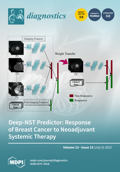
The early prediction of response to neoadjuvant systemic therapy (NST) can increase the patient’s likelihood of survival and help with decisions regarding breast-conserving surgery. An automated NST predictor that can precisely predict which patient undergoing NST will achieve a complete pathological response at an early stage is needed. We developed a multimodal spatiotemporal deep learning framework (deep-NST) by incorporating MRIs at different points in the NST regimens, the tumor’s molecular data, and the patient’s demographic data to predict the outcome of NST prior to or at an early stage of treatment. Deep-NST introduces a cross-kernel feature fusion module, which enhances the learnability of the framework by paying attention to multiple receptive fields to extract spatiotemporal features. View this paper
  • Issues are regarded as officially published after their release is announced to the table of contents alert mailing list.
  • You may sign up for e-mail alerts to receive table of contents of newly released issues.
  • PDF is the official format for papers published in both, html and pdf forms. To view the papers in pdf format, click on the "PDF Full-text" link, and use the free Adobe Reader to open them.
Order results
Result details
Section
Select all
Export citation of selected articles as:
7 pages, 212 KiB  
Editorial
Cell-Free Nucleic Acids: Physico-Chemical Properties, Analytical Considerations, and Clinical Applications
by Abel J. Bronkhorst and Stefan Holdenrieder
Diagnostics 2023, 13(13), 2312; https://doi.org/10.3390/diagnostics13132312 - 07 Jul 2023
Viewed by 741
Abstract
Human body fluids are rich sources of cell-free nuclear material, which exhibits unique characteristics [...] Full article
16 pages, 4810 KiB  
Review
Hyperpolarized Carbon-13 MRI in Breast Cancer
by Ramona Woitek and Kevin M. Brindle
Diagnostics 2023, 13(13), 2311; https://doi.org/10.3390/diagnostics13132311 - 07 Jul 2023
Cited by 2 | Viewed by 1380
Abstract
One of the hallmarks of cancer is metabolic reprogramming, including high levels of aerobic glycolysis (the Warburg effect). Pyruvate is a product of glucose metabolism, and 13C-MR imaging of the metabolism of hyperpolarized (HP) [1-13C]pyruvate (HP 13C-MRI) has been [...] Read more.
One of the hallmarks of cancer is metabolic reprogramming, including high levels of aerobic glycolysis (the Warburg effect). Pyruvate is a product of glucose metabolism, and 13C-MR imaging of the metabolism of hyperpolarized (HP) [1-13C]pyruvate (HP 13C-MRI) has been shown to be a potentially versatile tool for the clinical evaluation of tumor metabolism. Hyperpolarization of the 13C nuclear spin can increase the sensitivity of detection by 4–5 orders of magnitude. Therefore, following intravenous injection, the location of hyperpolarized 13C-labeled pyruvate in the body and its subsequent metabolism can be tracked using 13C-MRI. Hyperpolarized [13C]urea and [1,4-13C2]fumarate are also likely to translate to the clinic in the near future as tools for imaging tissue perfusion and post-treatment tumor cell death, respectively. For clinical breast imaging, HP 13C-MRI can be combined with 1H-MRI to address the need for detailed anatomical imaging combined with improved functional tumor phenotyping and very early identification of patients not responding to standard and novel neoadjuvant treatments. If the technical complexity of the hyperpolarization process and the relatively high associated costs can be reduced, then hyperpolarized 13C-MRI has the potential to become more widely available for large-scale clinical trials. Full article
(This article belongs to the Special Issue New Horizons in Breast Cancer Magnetic Resonance Imaging)
Show Figures

Figure 1

12 pages, 645 KiB  
Article
Is There a Potential Link between Gastroesophageal Reflux Disease and Recurrent Respiratory Tract Infections in Children?
by Vasile Valeriu Lupu, Gabriela Stefanescu, Ana Maria Laura Buga, Lorenza Forna, Elena Tarca, Iuliana Magdalena Starcea, Cristina Maria Mihai, Laura Florescu, Andrei Tudor Cernomaz, Adriana Mocanu, Viorel Tarca, Aye Aung Thet and Ancuta Lupu
Diagnostics 2023, 13(13), 2310; https://doi.org/10.3390/diagnostics13132310 - 07 Jul 2023
Cited by 3 | Viewed by 1298
Abstract
Background: The implications of gastroesophageal reflux disease in respiratory tract infections have been investigated over time. The aim of our study was to evaluate the relationship between these two pathologic entities and the outcome after proper antireflux treatment. Methods: A group of 53 [...] Read more.
Background: The implications of gastroesophageal reflux disease in respiratory tract infections have been investigated over time. The aim of our study was to evaluate the relationship between these two pathologic entities and the outcome after proper antireflux treatment. Methods: A group of 53 children with recurrent respiratory tract infections admitted in the gastroenterology clinic of a children’s hospital in North-East Romania was investigated for gastroesophageal reflux disease through 24 h pH-metry. Those with a Boix-Ochoa score higher than 11.99 received proton pump inhibitor treatment and were reevaluated after 2 months. Results: A total of 41 children were found with a positive Boix-Ochoa score. After 2 months of antireflux therapy, eight patients still had a positive Boix-Ochoa score. Conclusions: Recurrent respiratory tract infections with symptoms resistant to treatment should be considered a reason to investigate for gastroesophageal reflux, because the symptoms may be due to micro- or macro-aspiration of the gastric refluxate or to an esophageal-bronchial reflex mediated through the vagal nerve. Full article
(This article belongs to the Special Issue Pediatric Gastrointestinal Diseases: Diagnosis and Management)
Show Figures

Figure 1

15 pages, 2277 KiB  
Review
Diagnosis, Treatment, and Management of Otitis Media with Artificial Intelligence
by Xin Ding, Yu Huang, Xu Tian, Yang Zhao, Guodong Feng and Zhiqiang Gao
Diagnostics 2023, 13(13), 2309; https://doi.org/10.3390/diagnostics13132309 - 07 Jul 2023
Cited by 3 | Viewed by 2159
Abstract
A common infectious disease, otitis media (OM) has a low rate of early diagnosis, which significantly increases the difficulty of treating the disease and the likelihood of serious complications developing including hearing loss, speech impairment, and even intracranial infection. Several areas of healthcare [...] Read more.
A common infectious disease, otitis media (OM) has a low rate of early diagnosis, which significantly increases the difficulty of treating the disease and the likelihood of serious complications developing including hearing loss, speech impairment, and even intracranial infection. Several areas of healthcare have shown great promise in the application of artificial intelligence (AI) systems, such as the accurate detection of diseases, the automated interpretation of images, and the prediction of patient outcomes. Several articles have reported some machine learning (ML) algorithms such as ResNet, InceptionV3 and Unet, were applied to the diagnosis of OM successfully. The use of these techniques in the OM is still in its infancy, but their potential is enormous. We present in this review important concepts related to ML and AI, describe how these technologies are currently being applied to diagnosing, treating, and managing OM, and discuss the challenges associated with developing AI-assisted OM technologies in the future. Full article
(This article belongs to the Section Machine Learning and Artificial Intelligence in Diagnostics)
Show Figures

Figure 1

22 pages, 2521 KiB  
Review
Artificial Intelligence in the Advanced Diagnosis of Bladder Cancer-Comprehensive Literature Review and Future Advancement
by Matteo Ferro, Ugo Giovanni Falagario, Biagio Barone, Martina Maggi, Felice Crocetto, Gian Maria Busetto, Francesco del Giudice, Daniela Terracciano, Giuseppe Lucarelli, Francesco Lasorsa, Michele Catellani, Antonio Brescia, Francesco Alessandro Mistretta, Stefano Luzzago, Mattia Luca Piccinelli, Mihai Dorin Vartolomei, Barbara Alicja Jereczek-Fossa, Gennaro Musi, Emanuele Montanari, Ottavio de Cobelli and Octavian Sabin Tataruadd Show full author list remove Hide full author list
Diagnostics 2023, 13(13), 2308; https://doi.org/10.3390/diagnostics13132308 - 07 Jul 2023
Cited by 10 | Viewed by 1884
Abstract
Artificial intelligence is highly regarded as the most promising future technology that will have a great impact on healthcare across all specialties. Its subsets, machine learning, deep learning, and artificial neural networks, are able to automatically learn from massive amounts of data and [...] Read more.
Artificial intelligence is highly regarded as the most promising future technology that will have a great impact on healthcare across all specialties. Its subsets, machine learning, deep learning, and artificial neural networks, are able to automatically learn from massive amounts of data and can improve the prediction algorithms to enhance their performance. This area is still under development, but the latest evidence shows great potential in the diagnosis, prognosis, and treatment of urological diseases, including bladder cancer, which are currently using old prediction tools and historical nomograms. This review focuses on highly significant and comprehensive literature evidence of artificial intelligence in the management of bladder cancer and investigates the near introduction in clinical practice. Full article
(This article belongs to the Special Issue Advanced Diagnosis and Treatment Strategies for Bladder Cancer)
Show Figures

Figure 1

15 pages, 3317 KiB  
Article
Optic Nerve Sheath Meningiomas: Solving Diagnostic Challenges with 68Ga-DOTATOC PET/CT
by Tatiana Horowitz, Betty Salgues, Laetitia Padovani, Kaissar Farah, Henry Dufour, Olivier Chinot, Eric Guedj and Thomas Graillon
Diagnostics 2023, 13(13), 2307; https://doi.org/10.3390/diagnostics13132307 - 07 Jul 2023
Cited by 1 | Viewed by 1513
Abstract
68Ga-DOTATOC PET could be a noninvasive, highly sensitive, and specific technique for the challenging diagnosis of optic nerve sheath meningioma (ONSM). Our objective was to report the use and results of 68Ga-DOTATOC PET in suspected ONSM. Twelve subjects who underwent 68 [...] Read more.
68Ga-DOTATOC PET could be a noninvasive, highly sensitive, and specific technique for the challenging diagnosis of optic nerve sheath meningioma (ONSM). Our objective was to report the use and results of 68Ga-DOTATOC PET in suspected ONSM. Twelve subjects who underwent 68Ga-DOTATOC PET for suspected ONSM in our department were retrospectively included. Standardised clinical and radiological data were collected. The PET examination results were classified as positive or negative, and lesion standardised uptake values (SUVmax) were recorded. 68Ga-DOTATOC PET confirmed positive uptake in six cases (SUVmax > 5), leading to ONSM diagnoses followed by radiation therapy in patients with vision loss. Six 68Ga-DOTATOC PET scans were considered negative (SUVmax < 5); these comprised one case of neurosarcoidosis, one cavernous malformation, and four uncertain diagnoses, leading to further investigation. 68Ga-DOTATOC PET was helpful in tumour volume delineation before radiation therapy, leading to a decrease in dose exposure. Noninvasive 68Ga-DOTATOC PET should be performed before treating nonhistologically proven meningiomas with radiotherapy or stereotactic radiosurgery, particularly in cases of uncertain diagnosis with MRI, which characterises most ONSM cases. PET SUVmax thresholds to distinguish meningioma from nonspecific uptake in other lesions need to be adapted to ONSM. 68Ga-DOTATOC PET improves the intraorbital lesion diagnostic approach and therefore impacts therapeutic management. Full article
(This article belongs to the Special Issue Diagnosis and Treatment of Meningioma)
Show Figures

Graphical abstract

19 pages, 1278 KiB  
Article
Performance Evaluation of VIDAS® Diagnostic Assays Detecting Anti-Chikungunya Virus IgM and IgG Antibodies: An International Study
by Geovana M. Pereira, Erika R. Manuli, Laurie Coulon, Marina F. Côrtes, Mariana S. Ramundo, Loïc Dromenq, Audrey Larue-Triolet, Frédérique Raymond, Carole Tourneur, Carolina dos Santos Lázari, Patricia Brasil, Ana Maria Bispo de Filippis, Glaucia Paranhos-Baccalà, Alice Banz and Ester C. Sabino
Diagnostics 2023, 13(13), 2306; https://doi.org/10.3390/diagnostics13132306 - 07 Jul 2023
Viewed by 1763
Abstract
Chikungunya (CHIK) is a debilitating mosquito-borne disease with an epidemiology and early clinical symptoms similar to those of other arboviruses-triggered diseases such as dengue or Zika. Accurate and rapid diagnosis of CHIK virus (CHIKV) infection is therefore challenging. This international study evaluated the [...] Read more.
Chikungunya (CHIK) is a debilitating mosquito-borne disease with an epidemiology and early clinical symptoms similar to those of other arboviruses-triggered diseases such as dengue or Zika. Accurate and rapid diagnosis of CHIK virus (CHIKV) infection is therefore challenging. This international study evaluated the performance of the automated VIDAS® anti-CHIKV IgM and IgG assays compared to that of manual competitor IgM and IgG ELISA for the detection of anti-CHIKV IgM and IgG antibodies in 660 patients with suspected CHIKV infection. Positive and negative agreements of the VIDAS® CHIKV assays with ELISA ranged from 97.5% to 100.0%. The sensitivity of the VIDAS® CHIKV assays evaluated in patients with a proven CHIKV infection confirmed reported kinetics of anti-CHIKV IgM and IgG response, with a positive detection of 88.2–100.0% for IgM ≥ 5 days post symptom onset and of 100.0% for IgG ≥ 11 days post symptom onset. Our study also demonstrated the superiority of ELISA and VIDAS® assays over rapid diagnostic IgM/IgG tests. The analytical performance of VIDAS® anti-CHIKV IgM and IgG assays was excellent, with a high precision (coefficients of variation ≤ 7.4%) and high specificity (cross-reactivity rate ≤ 2.9%). This study demonstrates the suitability of the automated VIDAS® anti-CHIKV IgM and IgG assays to diagnose CHIKV infections and supports its applicability for epidemiological surveillance and differential diagnosis in regions endemic for CHIKV. Full article
(This article belongs to the Special Issue Laboratory Diagnosis in Microbial Diseases)
Show Figures

Figure 1

3 pages, 156 KiB  
Editorial
State-of-the-Art Research: Current Developments in CT Imaging
by Christian Booz
Diagnostics 2023, 13(13), 2305; https://doi.org/10.3390/diagnostics13132305 - 07 Jul 2023
Viewed by 977
Abstract
This Special Issue of Diagnostics entitled “Advances in CT Images” provides an interesting selection of articles on recent technical developments in CT imaging with a special focus on spectral imaging, including dual-energy CT (DECT) and photon-counting CT (PCCT) [...] Full article
(This article belongs to the Special Issue Advances in CT Images)
18 pages, 6664 KiB  
Review
Multidetector CT Imaging Biomarkers as Predictors of Prognosis in Shock: Updates and Future Directions
by Tullio Valente, Giorgio Bocchini, Candida Massimo, Gaetano Rea, Roberta Lieto, Salvatore Guarino, Emanuele Muto, Ahmad Abu-Omar, Mariano Scaglione and Giacomo Sica
Diagnostics 2023, 13(13), 2304; https://doi.org/10.3390/diagnostics13132304 - 07 Jul 2023
Viewed by 1392
Abstract
A severe mismatch between the supply and demand of oxygen is the common sequela of all types of shock, which present a mortality of up to 80%. Various organs play a protective role in shock and contribute to whole-body homeostasis. The ever-increasing number [...] Read more.
A severe mismatch between the supply and demand of oxygen is the common sequela of all types of shock, which present a mortality of up to 80%. Various organs play a protective role in shock and contribute to whole-body homeostasis. The ever-increasing number of multidetector CT examinations in severely ill and sometimes unstable patients leads to more frequently encountered findings leading to imminent death, together called “hypovolemic shock complex”. Features on CT include dense opacification of the right heart and major systemic veins, venous layering of contrast material and blood, densely opacified parenchyma in the right hepatic lobe, decreased enhancement of the abdominal organ, a dense pulmonary artery, contrast pooling in dependent lungs, and contrast stasis in pulmonary veins. These findings are biomarkers and prognostic indicators of paramount importance which stratify risk and improve patient outcomes. In this review, we illustrate the various CT patterns in shock and review the spectrum and prognostic significance of thoraco-abdominal vascular and visceral alarming signs of impending death with the intention of increasing awareness among radiologists and radiographers to prepare for immediate resuscitation when required. Full article
Show Figures

Figure 1

13 pages, 1531 KiB  
Review
Recent Advances of Artificial Intelligence Applications in Interstitial Lung Diseases
by Konstantinos P. Exarchos, Georgia Gkrepi, Konstantinos Kostikas and Athena Gogali
Diagnostics 2023, 13(13), 2303; https://doi.org/10.3390/diagnostics13132303 - 06 Jul 2023
Cited by 6 | Viewed by 2673
Abstract
Interstitial lung diseases (ILDs) comprise a rather heterogeneous group of diseases varying in pathophysiology, presentation, epidemiology, diagnosis, treatment and prognosis. Even though they have been recognized for several years, there are still areas of research debate. In the majority of ILDs, imaging modalities [...] Read more.
Interstitial lung diseases (ILDs) comprise a rather heterogeneous group of diseases varying in pathophysiology, presentation, epidemiology, diagnosis, treatment and prognosis. Even though they have been recognized for several years, there are still areas of research debate. In the majority of ILDs, imaging modalities and especially high-resolution Computed Tomography (CT) scans have been the cornerstone in patient diagnostic approach and follow-up. The intricate nature of ILDs and the accompanying data have led to an increasing adoption of artificial intelligence (AI) techniques, primarily on imaging data but also in genetic data, spirometry and lung diffusion, among others. In this literature review, we describe the most prominent applications of AI in ILDs presented approximately within the last five years. We roughly stratify these studies in three categories, namely: (i) screening, (ii) diagnosis and classification, (iii) prognosis. Full article
(This article belongs to the Special Issue Advances in the Diagnosis and Management of Interstitial Lung Disease)
Show Figures

Figure 1

14 pages, 5592 KiB  
Article
Skeleton Segmentation on Bone Scintigraphy for BSI Computation
by Po-Nien Yu, Yung-Chi Lai, Yi-You Chen and Da-Chuan Cheng
Diagnostics 2023, 13(13), 2302; https://doi.org/10.3390/diagnostics13132302 - 06 Jul 2023
Cited by 1 | Viewed by 1125
Abstract
Bone Scan Index (BSI) is an image biomarker for quantifying bone metastasis of cancers. To compute BSI, not only the hotspots (metastasis) but also the bones have to be segmented. Most related research focus on binary classification in bone scintigraphy: having metastasis or [...] Read more.
Bone Scan Index (BSI) is an image biomarker for quantifying bone metastasis of cancers. To compute BSI, not only the hotspots (metastasis) but also the bones have to be segmented. Most related research focus on binary classification in bone scintigraphy: having metastasis or none. Rare studies focus on pixel-wise segmentation. This study compares three advanced convolutional neural network (CNN) based models to explore bone segmentation on a dataset in-house. The best model is Mask R-CNN, which reaches the precision, sensitivity, and F1-score: 0.93, 0.87, 0.90 for prostate cancer patients and 0.92, 0.86, and 0.88 for breast cancer patients, respectively. The results are the average of 10-fold cross-validation, which reveals the reliability of clinical use on bone segmentation. Full article
(This article belongs to the Section Machine Learning and Artificial Intelligence in Diagnostics)
Show Figures

Figure 1

13 pages, 1485 KiB  
Systematic Review
Diagnostic Blood Biomarkers for Acute Pulmonary Embolism: A Systematic Review
by Vårin Eiriksdatter Wikan, Birgitte Gladsø Tøndel, Vânia Maris Morelli, Ellen Elisabeth Brodin, Sigrid Kufaas Brækkan and John-Bjarne Hansen
Diagnostics 2023, 13(13), 2301; https://doi.org/10.3390/diagnostics13132301 - 06 Jul 2023
Cited by 1 | Viewed by 1322
Abstract
(1) Background: The current diagnostic algorithm for acute pulmonary embolism (PE) is associated with the overuse of CT pulmonary angiography (CTPA). An additional highly specific blood test could potentially lower the proportion of patients with suspected PE that require CTPA. The aim was [...] Read more.
(1) Background: The current diagnostic algorithm for acute pulmonary embolism (PE) is associated with the overuse of CT pulmonary angiography (CTPA). An additional highly specific blood test could potentially lower the proportion of patients with suspected PE that require CTPA. The aim was to summarize the literature on the diagnostic performance of biomarkers of patients admitted to an emergency department with suspected acute PE. (2) Methods: Medline and Embase databases were searched from 1995 to the present. The study selection process, data extraction, and risk of bias assessment were conducted by two reviewers. Eligibility criteria accepted all blood biomarkers except D-dimer, and CTPA was used as the reference standard. Qualitative data synthesis was performed. (3) Results: Of the 8448 identified records, only 6 were included. Eight blood biomarkers were identified, of which, three were investigated in two separate studies. Red distribution width and mean platelet volume were reported to have a specificity of ≥ 90% in one study, although these findings were not confirmed by other studies. The majority of the studies contained a high risk of selection bias. (4) Conclusions: The modest findings and the uncertain validity of the included studies suggest that none of the biomarkers identified in this systematic review have the potential to improve the current diagnostic algorithm for acute PE by reducing the overuse of CTPA. Full article
(This article belongs to the Section Clinical Laboratory Medicine)
Show Figures

Figure 1

19 pages, 996 KiB  
Review
MRI-Based Radiomics in Bladder Cancer: A Systematic Review and Radiomics Quality Score Assessment
by Bianca Boca, Cosmin Caraiani, Teodora Telecan, Roxana Pintican, Andrei Lebovici, Iulia Andras, Nicolae Crisan, Alexandru Pavel, Laura Diosan, Zoltan Balint, Monica Lupsor-Platon and Mircea Marian Buruian
Diagnostics 2023, 13(13), 2300; https://doi.org/10.3390/diagnostics13132300 - 06 Jul 2023
Cited by 2 | Viewed by 1403
Abstract
(1): Background: With the recent introduction of vesical imaging reporting and data system (VI-RADS), magnetic resonance imaging (MRI) has become the main imaging method used for the preoperative local staging of bladder cancer (BCa). However, the VI-RADS score is subject to interobserver variability [...] Read more.
(1): Background: With the recent introduction of vesical imaging reporting and data system (VI-RADS), magnetic resonance imaging (MRI) has become the main imaging method used for the preoperative local staging of bladder cancer (BCa). However, the VI-RADS score is subject to interobserver variability and cannot provide information about tumor cellularity. These limitations may be overcome by using a quantitative approach, such as the new emerging domain of radiomics. (2) Aim: To systematically review published studies on the use of MRI-based radiomics in bladder cancer. (3) Materials and Methods: We performed literature research using the PubMed MEDLINE, Scopus, and Web of Science databases using PRISMA principles. A total of 1092 papers that addressed the use of radiomics for BC staging, grading, and treatment response were retrieved using the keywords “bladder cancer”, “magnetic resonance imaging”, “radiomics”, and “textural analysis”. (4) Results: 26 papers met the eligibility criteria and were included in the final review. The principal applications of radiomics were preoperative tumor staging (n = 13), preoperative prediction of tumor grade or molecular correlates (n = 9), and prediction of prognosis/response to neoadjuvant therapy (n = 4). Most of the developed radiomics models included second-order features mainly derived from filtered images. These models were validated in 16 studies. The average radiomics quality score was 11.7, ranging between 8.33% and 52.77%. (5) Conclusions: MRI-based radiomics holds promise as a quantitative imaging biomarker of BCa characterization and prognosis. However, there is still need for improving the standardization of image preprocessing, feature extraction, and external validation before applying radiomics models in the clinical setting. Full article
(This article belongs to the Special Issue State of the Art of Abdominal Radiology)
Show Figures

Figure 1

14 pages, 2227 KiB  
Article
Super Formula for Diagnosing Disseminated Intravascular Coagulation Using Soluble C-Type Lectin-like Receptor 2
by Akitaka Yamamoto, Hideo Wada, Masaki Tomida, Yuhuko Ichikawa, Minoru Ezaki, Katsuya Shiraki, Motomu Shimaoka, Toshiaki Iba, Katsue Suzuki-Inoue, Masahide Kawamura and Hideto Shimpo
Diagnostics 2023, 13(13), 2299; https://doi.org/10.3390/diagnostics13132299 - 06 Jul 2023
Cited by 3 | Viewed by 1114
Abstract
The scoring systems for disseminated intravascular coagulation (DIC) criteria require several adequate cutoff values, vary, and are complicated. Accordingly, a simpler and quicker diagnostic method for DIC is needed. Under such circumstances, soluble C-type lectin-like receptor 2 (sCLEC-2) received attention as a biomarker [...] Read more.
The scoring systems for disseminated intravascular coagulation (DIC) criteria require several adequate cutoff values, vary, and are complicated. Accordingly, a simpler and quicker diagnostic method for DIC is needed. Under such circumstances, soluble C-type lectin-like receptor 2 (sCLEC-2) received attention as a biomarker for platelet activation. Materials and Methods: The diagnostic usefulness of sCLEC-2 and several formulas, including sCLEC-2xD-dimer, sCLEC-2/platelet count (sCLEC-2/PLT), and sCLEC-2/PLT × D-dimer (sCLEC-2xD-dimer/PLT), were evaluated among 38 patients with DIC, 39 patients with pre-DIC and 222 patients without DIC or pre-DIC (non-DIC). Results: Although the plasma level of sCLEC-2 alone was not a strong biomarker for the diagnosis of DIC or pre-DIC, the sCLEC-2xD-dimer/PLT values in patients with DIC were significantly higher than those in patients without DIC, and in a receiver operating characteristic (ROC) analysis for the diagnosis of DIC, sCLEC-2xD-dimer/PLT showed the highest AUC, sensitivity, and odds ratio. This formula is useful for the diagnosis of both pre-DIC and DIC. sCLEC-2xD-dimer/PLT values were significantly higher in non-survivors than in survivors. Conclusion: The sCLEC-2xD-dimer/PLT formula is simple, easy, and highly useful for the diagnosis of DIC and pre-DIC without the use of a scoring system. Full article
(This article belongs to the Section Clinical Laboratory Medicine)
Show Figures

Figure 1

13 pages, 1006 KiB  
Article
Brain-Derived Neurotrophic Factor (BDNF) and Translocator Protein (TSPO) as Diagnostic Biomarkers for Acute Ischemic Stroke
by Mayuri N. Tuwar, Wei-Hung Chen, Arthur M. Chiwaya, Hsu-Ling Yeh, Minh H. Nguyen and Chyi-Huey Bai
Diagnostics 2023, 13(13), 2298; https://doi.org/10.3390/diagnostics13132298 - 06 Jul 2023
Cited by 2 | Viewed by 1449
Abstract
Brain-derived neurotrophic factor (BDNF) interacts with tropomyosin-related kinase B (TrkB) to promote neuronal growth, survival, differentiation, neurotransmitter release, and synaptic plasticity. The translocator protein (TSPO) is known to be found in arterial plaques, which are a symptom of atherosclerosis and a contributory cause [...] Read more.
Brain-derived neurotrophic factor (BDNF) interacts with tropomyosin-related kinase B (TrkB) to promote neuronal growth, survival, differentiation, neurotransmitter release, and synaptic plasticity. The translocator protein (TSPO) is known to be found in arterial plaques, which are a symptom of atherosclerosis and a contributory cause of ischemic stroke. This study aims to determine the diagnostic accuracy of plasma BDNF and TSPO levels in discriminating new-onset acute ischemic stroke (AIS) patients from individuals without acute ischemic stroke. A total of 90 AIS patients (61% male, with a mean age of 67.7 ± 12.88) were recruited consecutively in a stroke unit, and each patient was paired with two age- and gender-matched controls. The sensitivity, specificity, and area of the curve between high plasma BDNF and TSPO and having AIS was determined using receiver operating characteristic curves. Furthermore, compared to the controls, AIS patients exhibited significantly higher levels of BDNF and TSPO, blood pressure, HbA1c, and white blood cells, as well as higher creatinine levels. The plasma levels of BDNF and TSPO can significantly discriminate AIS patients from healthy individuals (AUC 0.76 and 0.89, respectively). However, combining the two biomarkers provided little improvement in AUC (0.90). It may be possible to use elevated levels of TSPO as a diagnostic biomarker in patients with acute ischemic stroke upon admission. Full article
(This article belongs to the Special Issue Advances in the Diagnosis and Management of Stroke)
Show Figures

Figure 1

13 pages, 2254 KiB  
Article
Applying Deep Learning Model to Predict Diagnosis Code of Medical Records
by Jakir Hossain Bhuiyan Masud, Chen-Cheng Kuo, Chih-Yang Yeh, Hsuan-Chia Yang and Ming-Chin Lin
Diagnostics 2023, 13(13), 2297; https://doi.org/10.3390/diagnostics13132297 - 06 Jul 2023
Cited by 3 | Viewed by 3093
Abstract
The International Classification of Diseases (ICD) code is a diagnostic classification standard that is frequently used as a referencing system in healthcare and insurance. However, it takes time and effort to find and use the right diagnosis code based on a patient’s medical [...] Read more.
The International Classification of Diseases (ICD) code is a diagnostic classification standard that is frequently used as a referencing system in healthcare and insurance. However, it takes time and effort to find and use the right diagnosis code based on a patient’s medical records. In response, deep learning (DL) methods have been developed to assist physicians in the ICD coding process. Our findings propose a deep learning model that utilized clinical notes from medical records to predict ICD-10 codes. Our research used text-based medical data from the outpatient department (OPD) of a university hospital from January to December 2016. The dataset used clinical notes from five departments, and a total of 21,953 medical records were collected. Clinical notes consisted of a subjective component, objective component, assessment, plan (SOAP) notes, diagnosis code, and a drug list. The dataset was divided into two groups: 90% for training and 10% for test cases. We applied natural language processing (NLP) technique (word embedding, Word2Vector) to process the data. A deep learning-based convolutional neural network (CNN) model was created based on the information presented above. Three metrics (precision, recall, and F-score) were used to calculate the achievement of the deep learning CNN model. Clinically acceptable results were achieved through the deep learning model for five departments (precision: 0.53–0.96; recall: 0.85–0.99; and F-score: 0.65–0.98). With a precision of 0.95, a recall of 0.99, and an F-score of 0.98, the deep learning model performed the best in the department of cardiology. Our proposed CNN model significantly improved the prediction performance for an automated ICD-10 code prediction system based on prior clinical information. This CNN model could reduce the laborious task of manual coding and could assist physicians in making a better diagnosis. Full article
(This article belongs to the Section Machine Learning and Artificial Intelligence in Diagnostics)
Show Figures

Figure 1

13 pages, 1659 KiB  
Article
Underexpression of Carbamoyl Phosphate Synthetase I as Independent Unfavorable Prognostic Factor in Intrahepatic Cholangiocarcinoma: A Potential Theranostic Biomarker
by Khaa Hoo Ong, Yao-Yu Hsieh, Ding-Ping Sun, Steven Kuan-Hua Huang, Yu-Feng Tian, Chia-Ling Chou, Yow-Ling Shiue, Keva Joseph and I-Wei Chang
Diagnostics 2023, 13(13), 2296; https://doi.org/10.3390/diagnostics13132296 - 06 Jul 2023
Viewed by 1151
Abstract
Intrahepatic cholangiocarcinoma (IHCC) is the second most common malignant neoplasm of the liver. In spite of the increasing incidence worldwide, it is relatively rare in Western countries. IHCC is relatively common in Eastern and Southeastern Asia. Patients with IHCC are usually diagnosed at [...] Read more.
Intrahepatic cholangiocarcinoma (IHCC) is the second most common malignant neoplasm of the liver. In spite of the increasing incidence worldwide, it is relatively rare in Western countries. IHCC is relatively common in Eastern and Southeastern Asia. Patients with IHCC are usually diagnosed at an advanced stage, therefore, the clinical outcome is dismal. Dysregulation of urea cycle metabolic enzyme expression is found in different types of cancers. Nevertheless, a comprehensive evaluation of genes related to the urea cycle (i.e., GO:0000050) has not been conducted in IHCC. By performing a comparative analysis of gene expression profiles, we specifically examined genes associated with the urea cycle (GO:0000050) in a publicly accessible transcriptomic dataset (GSE26566). Interestingly, CPS1 was identified as the second most prominently down-regulated gene in this context. Tumor tissues of 182 IHCC patients who underwent curative-intent hepatectomy were enrolled. The expression level of CPS1 protein in our IHCC cohort was assessed by immunohistochemical study. Subsequent to that, statistical analyses were carried out to examine the expression of CPS1 in relation to various clinicopathological factors, as well as to assess its impact on survival outcomes. We noticed that lower immunoreactivity of CPS1 in IHCC was associated with tumor progression (pT status) with statistical significance (p = 0.003). CPS1 underexpression was not only negatively correlated to overall survival (OS), disease-free survival (DFS), local recurrence-free survival (LRFS) and metastasis-free survival (MeFS) in univariate analysis but also an independent prognosticator to forecast poorer clinical outcome for all prognostic indices (OS, DFS, LRFS and MeFs) in patients with IHCC (all p ≤ 0.001). These results support that CPS1 may play a crucial role in IHCC oncogenesis and tumor progression and serve as a novel prognostic factor and a potential diagnostic and theranostic biomarker. Full article
Show Figures

Figure 1

15 pages, 1772 KiB  
Review
Advances for Pulmonary Functional Imaging: Dual-Energy Computed Tomography for Pulmonary Functional Imaging
by Yoshiyuki Ozawa, Yoshiharu Ohno, Hiroyuki Nagata, Keigo Tamokami, Keitaro Nishikimi, Yuka Oshima, Nayu Hamabuchi, Takahiro Matsuyama, Takahiro Ueda and Hiroshi Toyama
Diagnostics 2023, 13(13), 2295; https://doi.org/10.3390/diagnostics13132295 - 06 Jul 2023
Cited by 1 | Viewed by 1352
Abstract
Dual-energy computed tomography (DECT) can improve the differentiation of material by using two different X-ray energy spectra, and may provide new imaging techniques to diagnostic radiology to overcome the limitations of conventional CT in characterizing tissue. Some techniques have used dual-energy imaging, which [...] Read more.
Dual-energy computed tomography (DECT) can improve the differentiation of material by using two different X-ray energy spectra, and may provide new imaging techniques to diagnostic radiology to overcome the limitations of conventional CT in characterizing tissue. Some techniques have used dual-energy imaging, which mainly includes dual-sourced, rapid kVp switching, dual-layer detectors, and split-filter imaging. In iodine images, images of the lung’s perfused blood volume (PBV) based on DECT have been applied in patients with pulmonary embolism to obtain both images of the PE occluding the pulmonary artery and the consequent perfusion defects in the lung’s parenchyma. PBV images of the lung also have the potential to indicate the severity of PE, including chronic thromboembolic pulmonary hypertension. Virtual monochromatic imaging can improve the accuracy of diagnosing pulmonary vascular diseases by optimizing kiloelectronvolt settings for various purposes. Iodine images also could provide a new approach in the area of thoracic oncology, for example, for the characterization of pulmonary nodules and mediastinal lymph nodes. DECT-based lung ventilation imaging is also available with noble gases with high atomic numbers, such as xenon, which is similar to iodine. A ventilation map of the lung can be used to image various pulmonary diseases such as chronic obstructive pulmonary disease. Full article
(This article belongs to the Section Medical Imaging and Theranostics)
Show Figures

Figure 1

25 pages, 2078 KiB  
Review
Artificial Intelligence in Renal Cell Carcinoma Histopathology: Current Applications and Future Perspectives
by Alfredo Distante, Laura Marandino, Riccardo Bertolo, Alexandre Ingels, Nicola Pavan, Angela Pecoraro, Michele Marchioni, Umberto Carbonara, Selcuk Erdem, Daniele Amparore, Riccardo Campi, Eduard Roussel, Anna Caliò, Zhenjie Wu, Carlotta Palumbo, Leonardo D. Borregales, Peter Mulders and Constantijn H. J. Muselaers
Diagnostics 2023, 13(13), 2294; https://doi.org/10.3390/diagnostics13132294 - 06 Jul 2023
Cited by 1 | Viewed by 1931
Abstract
Renal cell carcinoma (RCC) is characterized by its diverse histopathological features, which pose possible challenges to accurate diagnosis and prognosis. A comprehensive literature review was conducted to explore recent advancements in the field of artificial intelligence (AI) in RCC pathology. The aim of [...] Read more.
Renal cell carcinoma (RCC) is characterized by its diverse histopathological features, which pose possible challenges to accurate diagnosis and prognosis. A comprehensive literature review was conducted to explore recent advancements in the field of artificial intelligence (AI) in RCC pathology. The aim of this paper is to assess whether these advancements hold promise in improving the precision, efficiency, and objectivity of histopathological analysis for RCC, while also reducing costs and interobserver variability and potentially alleviating the labor and time burden experienced by pathologists. The reviewed AI-powered approaches demonstrate effective identification and classification abilities regarding several histopathological features associated with RCC, facilitating accurate diagnosis, grading, and prognosis prediction and enabling precise and reliable assessments. Nevertheless, implementing AI in renal cell carcinoma generates challenges concerning standardization, generalizability, benchmarking performance, and integration of data into clinical workflows. Developing methodologies that enable pathologists to interpret AI decisions accurately is imperative. Moreover, establishing more robust and standardized validation workflows is crucial to instill confidence in AI-powered systems’ outcomes. These efforts are vital for advancing current state-of-the-art practices and enhancing patient care in the future. Full article
Show Figures

Figure 1

8 pages, 943 KiB  
Case Report
Utility of FDG PET/CT in Patient with Synchronous Breast and Colon Cancer
by I-Lin Su and Yen-Kung Chen
Diagnostics 2023, 13(13), 2293; https://doi.org/10.3390/diagnostics13132293 - 06 Jul 2023
Viewed by 1134
Abstract
The most common malignancy in women is breast cancer, and the second one is colon cancer. Synchronous breast and colon cancers are rare. Here, we reported a 60-year-old woman with a left breast mass for six months. Biopsy revealed an invasive ductal carcinoma. [...] Read more.
The most common malignancy in women is breast cancer, and the second one is colon cancer. Synchronous breast and colon cancers are rare. Here, we reported a 60-year-old woman with a left breast mass for six months. Biopsy revealed an invasive ductal carcinoma. She underwent 2-[Fluorine-18]fluoro-2-deoxy-D-glucose (FDG) positron emission tomography (PET)/computed tomography (CT) scan for evaluation of the extent of the disease. FDG PET/CT revealed an advanced left breast cancer with multiple metastases in both regional and distant lymph nodes (in left axilla level I/II, lower paratracheal region, and right lung hilum), bilateral lungs, and axial and proximal appendicular skeletons. An early staged synchronous colon cancer was detected incidentally on FDG PET/CT images. After endoscopic mucosal resection of colon cancer, she received palliative chemotherapy for breast cancer with a marked therapeutic response. The disease status of post-treated breast cancer remained relatively stationary for more than one year. Brain metastasis was noted afterward. Nevertheless, there was no evidence of colon cancer recurrence throughout her breast cancer disease course. Full article
(This article belongs to the Section Medical Imaging and Theranostics)
Show Figures

Figure 1

17 pages, 3618 KiB  
Article
The Role of BIA Analysis in Osteoporosis Risk Development: Hierarchical Clustering Approach
by Giacinto Angelo Sgarro, Luca Grilli, Anna Antonia Valenzano, Fiorenzo Moscatelli, Domenico Monacis, Giusi Toto, Antonella De Maria, Giovanni Messina and Rita Polito
Diagnostics 2023, 13(13), 2292; https://doi.org/10.3390/diagnostics13132292 - 06 Jul 2023
Cited by 2 | Viewed by 1077
Abstract
Osteoporosis is a common musculoskeletal disorder among the elderly and a chronic condition which, like many other chronic conditions, requires long-term clinical management. It is caused by many factors, including lifestyle and obesity. Bioelectrical impedance analysis (BIA) is a method to estimate body [...] Read more.
Osteoporosis is a common musculoskeletal disorder among the elderly and a chronic condition which, like many other chronic conditions, requires long-term clinical management. It is caused by many factors, including lifestyle and obesity. Bioelectrical impedance analysis (BIA) is a method to estimate body composition based on a weak electric current flow through the body. The measured voltage is used to calculate body bioelectrical impedance, divided into resistance and reactance, which can be used to estimate body parameters such as total body water (TBW), fat-free mass (FFM), fat mass (FM), and muscle mass (MM). This study aims to find the tendency of osteoporosis in obese subjects, presenting a method based on hierarchical clustering, which, using BIA parameters, can group patients who show homogeneous characteristics. Grouping similar patients into clusters can be helpful in the field of medicine to identify disorders, pathologies, or more generally, characteristics of significant importance. Another added value of the clustering process is the possibility to define cluster prototypes, i.e., imaginary patients who represent models of “states”, which can be used together with clustering results to identify subjects with similar characteristics in a classification context. The results show that hierarchical clustering is a method that can be used to provide the detection of states and, consequently, supply a more personalized medicine approach. In addition, this method allowed us to elect BIA as a potential prognostic and diagnostic instrument in osteoporosis risk development. Full article
(This article belongs to the Section Machine Learning and Artificial Intelligence in Diagnostics)
Show Figures

Figure 1

24 pages, 6676 KiB  
Article
A Framework for Prediction of Oncogenomic Progression Aiding Personalized Treatment of Gastric Cancer
by Fahad M. Alotaibi and Yaser Daanial Khan
Diagnostics 2023, 13(13), 2291; https://doi.org/10.3390/diagnostics13132291 - 06 Jul 2023
Viewed by 1089
Abstract
Mutations in genes can alter their DNA patterns, and by recognizing these mutations, many carcinomas can be diagnosed in the progression stages. The human body contains many hidden and enigmatic features that humankind has not yet fully understood. A total of 7539 neoplasm [...] Read more.
Mutations in genes can alter their DNA patterns, and by recognizing these mutations, many carcinomas can be diagnosed in the progression stages. The human body contains many hidden and enigmatic features that humankind has not yet fully understood. A total of 7539 neoplasm cases were reported from 1 January 2021 to 31 December 2021. Of these, 3156 were seen in males (41.9%) and 4383 (58.1%) in female patients. Several machine learning and deep learning frameworks are already implemented to detect mutations, but these techniques lack generalized datasets and need to be optimized for better results. Deep learning-based neural networks provide the computational power to calculate the complex structures of gastric carcinoma-driven gene mutations. This study proposes deep learning approaches such as long and short-term memory, gated recurrent units and bi-LSTM to help in identifying the progression of gastric carcinoma in an optimized manner. This study includes 61 carcinogenic driver genes whose mutations can cause gastric cancer. The mutation information was downloaded from intOGen.org and normal gene sequences were downloaded from asia.ensembl.org, as explained in the data collection section. The proposed deep learning models are validated using the self-consistency test (SCT), 10-fold cross-validation test (FCVT), and independent set test (IST); the IST prediction metrics of accuracy, sensitivity, specificity, MCC and AUC of LSTM, Bi-LSTM, and GRU are 97.18%, 98.35%, 96.01%, 0.94, 0.98; 99.46%, 98.93%, 100%, 0.989, 1.00; 99.46%, 98.93%, 100%, 0.989 and 1.00, respectively. Full article
(This article belongs to the Section Machine Learning and Artificial Intelligence in Diagnostics)
Show Figures

Figure 1

15 pages, 7344 KiB  
Systematic Review
Diagnostic Efficacy of Carotid Ultrasound for Predicting the Risk of Perioperative Hypotension or Fluid Responsiveness: A Meta-Analysis
by Kuo-Chuan Hung, Yen-Ta Huang, Wen-Wen Tsai, Ping-Heng Tan, Jheng-Yan Wu, Po-Yu Huang, Ting-Hui Liu, I-Wen Chen and Cheuk-Kwan Sun
Diagnostics 2023, 13(13), 2290; https://doi.org/10.3390/diagnostics13132290 - 06 Jul 2023
Viewed by 1225
Abstract
Despite the acceptance of carotid ultrasound for predicting patients’ fluid responsiveness in critical care and anesthesia, its efficacy for predicting hypotension and fluid responsiveness remains unclear in the perioperative setting. Electronic databases were searched from inception to May 2023 to identify observational studies [...] Read more.
Despite the acceptance of carotid ultrasound for predicting patients’ fluid responsiveness in critical care and anesthesia, its efficacy for predicting hypotension and fluid responsiveness remains unclear in the perioperative setting. Electronic databases were searched from inception to May 2023 to identify observational studies focusing on the use of corrected blood flow time (FTc) and respirophasic variation in carotid artery blood flow peak velocity (ΔVpeak) for assessing the risks of hypotension and fluid responsiveness. Using FTc as a predictive tool (four studies), the analysis yielded a pooled sensitivity of 0.82 (95% confidence interval (CI): 0.72 to 0.89) and specificity of 0.94 (95% CI: 0.88 to 0.97) for the risk of hypotension (area under curve (AUC): 0.95). For fluid responsiveness, the sensitivity and specificity of FTc were 0.79 (95% CI: 0.72 to 0.84) and 0.81 (95% CI: 0.75 to 0.86), respectively (AUC: 0.87). In contrast, the use of ΔVpeak to predict the risk of fluid responsiveness showed a pooled sensitivity of 0.76 (95% CI: 0.63 to 0.85) and specificity of 0.74 (95% CI: 0.66 to 0.8) (AUC: 0.79). The current meta-analysis provides robust evidence supporting the high diagnostic accuracy of FTc in predicting perioperative hypotension and fluid responsiveness, which requires further studies for verification. Full article
(This article belongs to the Special Issue Advances in Point-of-Care Ultrasonography)
Show Figures

Figure 1

17 pages, 2820 KiB  
Article
Prediction of Distant Metastases in Patients with Kidney Cancer Based on Gene Expression and Methylation Analysis
by Natalya Apanovich, Alexey Matveev, Natalia Ivanova, Alexey Burdennyy, Pavel Apanovich, Irina Pronina, Elena Filippova, Tatiana Kazubskaya, Vitaly Loginov, Eleonora Braga and Andrei Alimov
Diagnostics 2023, 13(13), 2289; https://doi.org/10.3390/diagnostics13132289 - 06 Jul 2023
Cited by 3 | Viewed by 1544
Abstract
Clear cell renal cell carcinoma (ccRCC) is the most common and aggressive histological type of cancer in this location. Distant metastases are present in approximately 30% of patients at the time of first examination. Therefore, the ability to predict the occurrence of metastases [...] Read more.
Clear cell renal cell carcinoma (ccRCC) is the most common and aggressive histological type of cancer in this location. Distant metastases are present in approximately 30% of patients at the time of first examination. Therefore, the ability to predict the occurrence of metastases in patients at early stages of the disease is an urgent task aimed at personalized treatment. Samples of tumor and paired histologically normal kidney tissue from patients with metastatic and non-metastatic ccRCC were studied. Gene expression was analyzed using real-time PCR. The level of gene methylation was evaluated using bisulfite conversion followed by quantitative methylation-specific PCR. Two groups of genes were analyzed in this study. The first group includes genes whose expression is significantly reduced during metastasis: CA9, NDUFA4L2, EGLN3, and BHLHE41 (p < 0.001, ROC analysis). The second group includes microRNA genes: MIR125B-1, MIR137, MIR375, MIR193A, and MIR34B/C, whose increased methylation levels are associated with the development of distant metastases (p = 0.002 to <0.001, ROC analysis). Based on the data obtained, a combined panel of genes was formed to identify patients whose tumors have a high metastatic potential. The panel can estimate the probability of metastasis with an accuracy of up to 92%. Full article
(This article belongs to the Section Pathology and Molecular Diagnostics)
Show Figures

Figure 1

1 pages, 763 KiB  
Correction
Correction: Kusayanagi et al. A Smartphone Application for Personalized Tooth Shade Determination. Diagnostics 2023, 13, 1969
by Tomoya Kusayanagi, Sota Maegawa, Shuya Terauchi, Wataru Hashimoto and Shohei Kaneda
Diagnostics 2023, 13(13), 2288; https://doi.org/10.3390/diagnostics13132288 - 06 Jul 2023
Viewed by 453
Abstract
In the original publication [...] Full article
(This article belongs to the Section Point-of-Care Diagnostics and Devices)
Show Figures

Figure 1

9 pages, 1024 KiB  
Article
Bronchiectasis Assessment in Primary Ciliary Dyskinesia: A Non-Invasive Approach Using Forced Oscillation Technique
by Wilfredo De Jesús-Rojas, Luis Reyes-Peña, José Muñiz-Hernández, Patricia Quiles Ruiz de Porras, Jesús Meléndez-Montañez, Marcos J. Ramos-Benitez and Ricardo A. Mosquera
Diagnostics 2023, 13(13), 2287; https://doi.org/10.3390/diagnostics13132287 - 06 Jul 2023
Viewed by 1796
Abstract
Primary ciliary dyskinesia (PCD) is an autosomal recessive disorder that results from the dysfunction of motile cilia, which can cause chronic upper and lower respiratory infections leading to bronchiectasis. However, there is a need for additional tools to monitor the progression of bronchiectasis [...] Read more.
Primary ciliary dyskinesia (PCD) is an autosomal recessive disorder that results from the dysfunction of motile cilia, which can cause chronic upper and lower respiratory infections leading to bronchiectasis. However, there is a need for additional tools to monitor the progression of bronchiectasis in PCD. The forced oscillation technique (FOT) is an effort-independent lung function test that can be used to evaluate respiratory mechanics. In this retrospective study, we aimed to describe the radiographic findings associated with respiratory impedance (resistance (Rrs) and reactance (Xrs)) measured by FOT in six adult PCD patients and one pediatric with the (RSPH4A (c.921+3_921+6delAAGT (intronic)) founder mutation. We compared the radiographic findings on a high-resolution chest computed tomography (CT) scan with the FOT results. Our findings suggest that respiratory impedance measured by FOT may be a valuable tool for detecting and monitoring the progression of bronchiectasis in PCD patients with the (RSPH4A (c.921+3_921+6delAAGT (intronic)) founder mutation. However, further research is necessary to validate these results and determine the sensitivity and specificity of bronchiectasis monitoring in PCD patients with other genetic mutations. Full article
Show Figures

Figure 1

12 pages, 2152 KiB  
Article
Bleeding-Source Exploration in Subdural Hematoma: Observational Study on the Usefulness of Postmortem Computed Tomography Angiography
by Kazuhisa Funayama, Akihide Koyama, Rieka Katsuragi-Go, Takashi Aoyama, Hiraku Watanabe, Naoya Takahashi and Hisakazu Takatsuka
Diagnostics 2023, 13(13), 2286; https://doi.org/10.3390/diagnostics13132286 - 06 Jul 2023
Viewed by 1469
Abstract
In a few cases, postmortem computed tomography angiography (PMCTA) is effective in postmortem detection of cortical artery rupture causing subdural hematoma (SDH), which is difficult to detect at autopsy. Here, we explore the usefulness and limitations of PMCTA in detecting the sites of [...] Read more.
In a few cases, postmortem computed tomography angiography (PMCTA) is effective in postmortem detection of cortical artery rupture causing subdural hematoma (SDH), which is difficult to detect at autopsy. Here, we explore the usefulness and limitations of PMCTA in detecting the sites of cortical arterial rupture for SDH. In 6 of 10 cases, extravascular leakage of contrast material at nine different places enabled PMCTA to identify cortical arterial rupture. PMCTA did not induce destructive arterial artifacts, which often occur during autopsy. We found that, although not in all cases, PMCTA could show the site of cortical arterial rupture causing subdural hematoma in some cases. This technique is beneficial for cases of SDH autopsy, as it can be performed nondestructively and before destructive artifacts from the autopsy occur. Full article
(This article belongs to the Section Medical Imaging and Theranostics)
Show Figures

Graphical abstract

13 pages, 8210 KiB  
Article
Non-Rigid Registration for High-Resolution Retinal Imaging
by Mircea Mujat, James D. Akula, Anne B. Fulton, R. Daniel Ferguson and Nicusor Iftimia
Diagnostics 2023, 13(13), 2285; https://doi.org/10.3390/diagnostics13132285 - 06 Jul 2023
Cited by 3 | Viewed by 1200
Abstract
Adaptive optics provides improved resolution in ophthalmic imaging when retinal microstructures need to be identified, counted, and mapped. In general, multiple images are averaged to improve the signal-to-noise ratio or analyzed for temporal dynamics. Image registration by cross-correlation is straightforward for small patches; [...] Read more.
Adaptive optics provides improved resolution in ophthalmic imaging when retinal microstructures need to be identified, counted, and mapped. In general, multiple images are averaged to improve the signal-to-noise ratio or analyzed for temporal dynamics. Image registration by cross-correlation is straightforward for small patches; however, larger images require more sophisticated registration techniques. Strip-based registration has been used successfully for photoreceptor mosaic alignment in small patches; however, if the deformations along strips are not simple displacements, averaging can degrade the final image. We have applied a non-rigid registration technique that improves the quality of processed images for mapping cones over large image patches. In this approach, correction of local deformations compensates for local image stretching, compressing, bending, and twisting due to a number of causes. The main result of this procedure is improved definition of retinal microstructures that can be better identified and segmented. Derived metrics such as cone density, wall-to-lumen ratio, and quantification of structural modification of blood vessel walls have diagnostic value in many retinal diseases, including diabetic retinopathy and age-related macular degeneration, and their improved evaluations may facilitate early diagnostics of retinal diseases. Full article
(This article belongs to the Section Optical Diagnostics)
Show Figures

Figure 1

28 pages, 423 KiB  
Review
Marfan Syndrome: Enhanced Diagnostic Tools and Follow-up Management Strategies
by Susan Marelli, Emanuele Micaglio, Jacopo Taurino, Paolo Salvi, Erica Rurali, Gianluca L. Perrucci, Claudia Dolci, Nathasha Samali Udugampolage, Rosario Caruso, Davide Gentilini, Giuliana Trifiro’, Edward Callus, Alessandro Frigiola, Carlo De Vincentiis, Carlo Pappone, Gianfranco Parati and Alessandro Pini
Diagnostics 2023, 13(13), 2284; https://doi.org/10.3390/diagnostics13132284 - 05 Jul 2023
Cited by 1 | Viewed by 3688
Abstract
Marfan syndrome (MFS) is a rare inherited autosomic disorder, which encompasses a variety of systemic manifestations caused by mutations in the Fibrillin-1 encoding gene (FBN1). Cardinal clinical phenotypes of MFS are highly variable in terms of severity, and commonly involve cardiovascular, [...] Read more.
Marfan syndrome (MFS) is a rare inherited autosomic disorder, which encompasses a variety of systemic manifestations caused by mutations in the Fibrillin-1 encoding gene (FBN1). Cardinal clinical phenotypes of MFS are highly variable in terms of severity, and commonly involve cardiovascular, ocular, and musculoskeletal systems with a wide range of manifestations, such as ascending aorta aneurysms and dissection, mitral valve prolapse, ectopia lentis and long bone overgrowth, respectively. Of note, an accurate and prompt diagnosis is pivotal in order to provide the best treatment to the patients as early as possible. To date, the diagnosis of the syndrome has relied upon a systemic score calculation as well as DNA mutation identification. The aim of this review is to summarize the latest MFS evidence regarding the definition, differences and similarities with other connective tissue pathologies with severe systemic phenotypes (e.g., Autosomal dominant Weill–Marchesani syndrome, Loeys–Dietz syndrome, Ehlers–Danlos syndrome) and clinical assessment. In this regard, the management of MFS requires a multidisciplinary team in order to accurately control the evolution of the most severe and potentially life-threatening complications. Based on recent findings in the literature and our clinical experience, we propose a multidisciplinary approach involving specialists in different clinical fields (i.e., cardiologists, surgeons, ophthalmologists, orthopedics, pneumologists, neurologists, endocrinologists, geneticists, and psychologists) to comprehensively characterize, treat, and manage MFS patients with a personalized medicine approach. Full article
(This article belongs to the Section Pathology and Molecular Diagnostics)
22 pages, 408 KiB  
Review
Up-to-Date Imaging and Diagnostic Techniques for Prostate Cancer: A Literature Review
by Ming Zhu, Zhen Liang, Tianrui Feng, Zhipeng Mai, Shijie Jin, Liyi Wu, Huashan Zhou, Yuliang Chen and Weigang Yan
Diagnostics 2023, 13(13), 2283; https://doi.org/10.3390/diagnostics13132283 - 05 Jul 2023
Cited by 2 | Viewed by 2871
Abstract
Prostate cancer (PCa) faces great challenges in early diagnosis, which often leads not only to unnecessary, invasive procedures, but to over-diagnosis and treatment as well, thus highlighting the need for modern PCa diagnostic techniques. The review aims to provide an up-to-date summary of [...] Read more.
Prostate cancer (PCa) faces great challenges in early diagnosis, which often leads not only to unnecessary, invasive procedures, but to over-diagnosis and treatment as well, thus highlighting the need for modern PCa diagnostic techniques. The review aims to provide an up-to-date summary of chronologically existing diagnostic approaches for PCa, as well as their potential to improve clinically significant PCa (csPCa) diagnosis and to reduce the proliferation and monitoring of PCa. Our review demonstrates the primary outcomes of the most significant studies and makes comparisons across the diagnostic efficacies of different PCa tests. Since prostate biopsy, the current mainstream PCa diagnosis, is an invasive procedure with a high risk of post-biopsy complications, it is vital we dig out specific, sensitive, and accurate diagnostic approaches in PCa and conduct more studies with milestone findings and comparable sample sizes to validate and corroborate the findings. Full article
(This article belongs to the Section Medical Imaging and Theranostics)
Previous Issue
Next Issue
Back to TopTop