Next Issue
Volume 13, August-1
Previous Issue
Volume 13, July-1


Diagnostics, Volume 13, Issue 14 (July-2 2023) – 154 articles

Cover Story (view full-size image): 
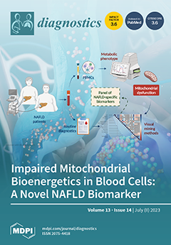
Non-alcoholic fatty liver disease (NAFLD) is a highly prevalent condition, lacking specific noninvasive diagnostic tools, whose pathogenesis comprises liver mitochondrial dysfunction. Garrafa and colleagues measured mitochondrial bioenergetics in peripheral blood mononuclear cells (PBMCs). They found significantly reduced basal respiration, ATP production, maximal respiration, and spare respiratory capacity in NAFLD compared to non-NAFLD cases. Correlation plots showed intriguing correlations between the respiratory parameters and anthropometric or biochemical parameters of interest for NAFLD diagnosis. Machine learning methods identified ATP production among the best NAFLD predictors. The authors propose blood cell respirometry as a novel tool for NAFLD diagnosis and therapeutic response monitoring. View this paper
  • Issues are regarded as officially published after their release is announced to the table of contents alert mailing list.
  • You may sign up for e-mail alerts to receive table of contents of newly released issues.
  • PDF is the official format for papers published in both, html and pdf forms. To view the papers in pdf format, click on the "PDF Full-text" link, and use the free Adobe Reader to open them.
Order results
Result details
Section
Select all
Export citation of selected articles as:
17 pages, 7824 KiB  
Review
Blue-Light Fundus Autofluorescence (BAF), an Essential Modality for the Evaluation of Inflammatory Diseases of the Photoreceptors: An Imaging Narrative
by Alessandro Mantovani, Carl P. Herbort, Jr., Alireza Hedayatfar and Ioannis Papasavvas
Diagnostics 2023, 13(14), 2466; https://doi.org/10.3390/diagnostics13142466 - 24 Jul 2023
Viewed by 1479
Abstract
Our purpose is to describe blue-light fundus autofluorescence (BAF) features of inflammatory diseases of the outer retina characterised by photoreceptor damage. BAF from patients diagnosed with secondary and primary inflammatory photoreceptor damage were retrospectively analyzed and compared to other imaging modalities including fluorescein [...] Read more.
Our purpose is to describe blue-light fundus autofluorescence (BAF) features of inflammatory diseases of the outer retina characterised by photoreceptor damage. BAF from patients diagnosed with secondary and primary inflammatory photoreceptor damage were retrospectively analyzed and compared to other imaging modalities including fluorescein angiography (FA), indocyanine green angiography (ICGA), and spectral domain optical coherence tomography (SD-OCT). Multiple evanescent white dot syndrome (MEWDS), idiopathic multifocal choroiditis (MFC), acute posterior multifocal placoid pigment epitheliopathy (APMPPE), serpiginous choroiditis (SC), and acute syphilitic posterior placoid chorioretinitis (ASPPC), all cases corresponding to secondary photoreceptor diseases caused by inflammatory choriocapillaris nonperfusion, were included and compared to primary photoreceptor disease entities, including acute zonal occult outer retinopathy (AZOOR) and cancer-associated retinopathy (CAR). Both groups showed increased BAFs of variable intensity. In severe cases of APMPPE and ASPPC, BAF also showed hypoautofluorescent areas. In group 1 (secondary diseases) BAF hyperautofluorescent areas were associated with colocalized ICGA hypofluorescent areas, indicating choriocapillaris nonperfusion; whereas in group 2 (primary diseases), no ICGA signs were detected. The associated colocalized areas of hypofluorescence on ICGA in the first group, which were absent in the second group, were crucial to allow the differentiation between primary (photoreceptoritis) and secondary (choriocapillaritis) photoreceptor diseases. BAF patterns in inflammatory diseases of the outer retina can give relevant information on the photoreceptor and RPE involvement, with ICGA being crucial to detect concurring choriocapillaris damage and differentiating the two pathologies. Full article
Show Figures

Figure 1

14 pages, 1107 KiB  
Article
A First Computational Frame for Recognizing Heparin-Binding Protein
by Wen Zhu, Shi-Shi Yuan, Jian Li, Cheng-Bing Huang, Hao Lin and Bo Liao
Diagnostics 2023, 13(14), 2465; https://doi.org/10.3390/diagnostics13142465 - 24 Jul 2023
Cited by 16 | Viewed by 928
Abstract
Heparin-binding protein (HBP) is a cationic antibacterial protein derived from multinuclear neutrophils and an important biomarker of infectious diseases. The correct identification of HBP is of great significance to the study of infectious diseases. This work provides the first HBP recognition framework based [...] Read more.
Heparin-binding protein (HBP) is a cationic antibacterial protein derived from multinuclear neutrophils and an important biomarker of infectious diseases. The correct identification of HBP is of great significance to the study of infectious diseases. This work provides the first HBP recognition framework based on machine learning to accurately identify HBP. By using four sequence descriptors, HBP and non-HBP samples were represented by discrete numbers. By inputting these features into a support vector machine (SVM) and random forest (RF) algorithm and comparing the prediction performances of these methods on training data and independent test data, it is found that the SVM-based classifier has the greatest potential to identify HBP. The model could produce an auROC of 0.981 ± 0.028 on training data using 10-fold cross-validation and an overall accuracy of 95.0% on independent test data. As the first model for HBP recognition, it will provide some help for infectious diseases and stimulate further research in related fields. Full article
Show Figures

Figure 1

35 pages, 2538 KiB  
Systematic Review
The Predictive Capabilities of Artificial Intelligence-Based OCT Analysis for Age-Related Macular Degeneration Progression—A Systematic Review
by George Adrian Muntean, Anca Marginean, Adrian Groza, Ioana Damian, Sara Alexia Roman, Mădălina Claudia Hapca, Maximilian Vlad Muntean and Simona Delia Nicoară
Diagnostics 2023, 13(14), 2464; https://doi.org/10.3390/diagnostics13142464 - 24 Jul 2023
Cited by 4 | Viewed by 1643
Abstract
The era of artificial intelligence (AI) has revolutionized our daily lives and AI has become a powerful force that is gradually transforming the field of medicine. Ophthalmology sits at the forefront of this transformation thanks to the effortless acquisition of an abundance of [...] Read more.
The era of artificial intelligence (AI) has revolutionized our daily lives and AI has become a powerful force that is gradually transforming the field of medicine. Ophthalmology sits at the forefront of this transformation thanks to the effortless acquisition of an abundance of imaging modalities. There has been tremendous work in the field of AI for retinal diseases, with age-related macular degeneration being at the top of the most studied conditions. The purpose of the current systematic review was to identify and evaluate, in terms of strengths and limitations, the articles that apply AI to optical coherence tomography (OCT) images in order to predict the future evolution of age-related macular degeneration (AMD) during its natural history and after treatment in terms of OCT morphological structure and visual function. After a thorough search through seven databases up to 1 January 2022 using the Preferred Reporting Items for Systematic Reviews and Meta-Analyses (PRISMA) guidelines, 1800 records were identified. After screening, 48 articles were selected for full-text retrieval and 19 articles were finally included. From these 19 articles, 4 articles concentrated on predicting the anti-VEGF requirement in neovascular AMD (nAMD), 4 articles focused on predicting anti-VEGF efficacy in nAMD patients, 3 articles predicted the conversion from early or intermediate AMD (iAMD) to nAMD, 1 article predicted the conversion from iAMD to geographic atrophy (GA), 1 article predicted the conversion from iAMD to both nAMD and GA, 3 articles predicted the future growth of GA and 3 articles predicted the future outcome for visual acuity (VA) after anti-VEGF treatment in nAMD patients. Since using AI methods to predict future changes in AMD is only in its initial phase, a systematic review provides the opportunity of setting the context of previous work in this area and can present a starting point for future research. Full article
(This article belongs to the Special Issue Diagnosis, Treatment and Management of Eye Diseases)
Show Figures

Figure 1

28 pages, 1004 KiB  
Article
A Deep Learning Framework for the Characterization of Thyroid Nodules from Ultrasound Images Using Improved Inception Network and Multi-Level Transfer Learning
by O. A. Ajilisa, V. P. Jagathy Raj and M. K. Sabu
Diagnostics 2023, 13(14), 2463; https://doi.org/10.3390/diagnostics13142463 - 24 Jul 2023
Cited by 1 | Viewed by 1409
Abstract
In the past few years, deep learning has gained increasingly widespread attention and has been applied to diagnosing benign and malignant thyroid nodules. It is difficult to acquire sufficient medical images, resulting in insufficient data, which hinders the development of an efficient deep-learning [...] Read more.
In the past few years, deep learning has gained increasingly widespread attention and has been applied to diagnosing benign and malignant thyroid nodules. It is difficult to acquire sufficient medical images, resulting in insufficient data, which hinders the development of an efficient deep-learning model. In this paper, we developed a deep-learning-based characterization framework to differentiate malignant and benign nodules from the thyroid ultrasound images. This approach improves the recognition accuracy of the inception network by combining squeeze and excitation networks with the inception modules. We have also integrated the concept of multi-level transfer learning using breast ultrasound images as a bridge dataset. This transfer learning approach addresses the issues regarding domain differences between natural images and ultrasound images during transfer learning. This paper aimed to investigate how the entire framework could help radiologists improve diagnostic performance and avoid unnecessary fine-needle aspiration. The proposed approach based on multi-level transfer learning and improved inception blocks achieved higher precision (0.9057 for the benign class and 0.9667 for the malignant class), recall (0.9796 for the benign class and 0.8529 for malignant), and F1-score (0.9412 for benign class and 0.9062 for malignant class). It also obtained an AUC value of 0.9537, which is higher than that of the single-level transfer learning method. The experimental results show that this model can achieve satisfactory classification accuracy comparable to experienced radiologists. Using this model, we can save time and effort as well as deliver potential clinical application value. Full article
(This article belongs to the Special Issue Artificial Intelligence in Clinical Medical Imaging Analysis)
Show Figures

Figure 1

11 pages, 703 KiB  
Article
Triiodothyronine and Protein Malnutrition Could Influence Pulse Wave Velocity in Pre-Dialysis Chronic Kidney Disease Patients
by Crina Claudia Rusu, Ina Kacso, Diana Moldovan, Alina Potra, Dacian Tirinescu, Maria Ticala, Ancuta M. Rotar, Remus Orasan, Cristian Budurea, Andrada Barar, Florin Anton, Ana Valea, Cosmina Ioana Bondor and Madalina Ticolea
Diagnostics 2023, 13(14), 2462; https://doi.org/10.3390/diagnostics13142462 - 24 Jul 2023
Cited by 1 | Viewed by 1025
Abstract
Cardiovascular diseases (CVD) are the first cause of chronic kidney disease (CKD) mortality. For personalized improved medicine, detecting correctable markers of CVD can be considered a priority. The aim of this study was the evaluation of the impact of nutritional, hormonal and inflammatory [...] Read more.
Cardiovascular diseases (CVD) are the first cause of chronic kidney disease (CKD) mortality. For personalized improved medicine, detecting correctable markers of CVD can be considered a priority. The aim of this study was the evaluation of the impact of nutritional, hormonal and inflammatory markers on brachial-ankle Pulse Wave Velocity (PWV) in pre-dialysis CKD patients. A cross-sectional observational study was conducted on 68 pre-dialysis CKD patients (median age of 69 years, 41.2% with diabetes mellitus, 52.9% male). Laboratory data were collected, including levels of prolactin, triiodothyronine, TGF α, IL-6, and IL-1β. The high values of brachial-ankle PWV were associated with reduced muscle mass (p = 0.001, r = −0.44), low levels of total cholesterol (p = 0.04, r = −0.26), triglycerides (p = 0.03, r = −0.31), triiodothyronine (p = 0.04, r = −0.24), and prolactin (p = 0.02, r = −0.27). High PWV was associated with advanced age (p < 0.001, r = 0.19). In the multivariate analysis, reduced muscle mass (p = 0.018), low levels of triiodothyronine (p = 0.002), and triglycerides (p = 0.049) were significant predictors of PWV, but age (p < 0.001) remained an important factor. In conclusion, reduced triiodothyronine together with markers of malnutrition and age were associated with PWV in pre-dialysis CKD patients. Full article
(This article belongs to the Special Issue Evaluating Novel Biomarkers for Personalized Medicine)
Show Figures

Figure 1

15 pages, 2606 KiB  
Article
Diagnostic Role of Native T1 Mapping Compared to Conventional Magnetic Resonance Techniques in Cardiac Disease in a Real-Life Cohort
by Giovanni Donato Aquaro, Silvia Monastero, Giancarlo Todiere, Andrea Barison, Carmelo De Gori, Crysanthos Grigoratos, Maria Luisa Parisella, Lorenzo Faggioni, Dania Cioni, Riccardo Lencioni and Emanuele Neri
Diagnostics 2023, 13(14), 2461; https://doi.org/10.3390/diagnostics13142461 - 24 Jul 2023
Cited by 1 | Viewed by 892
Abstract
We sought to compare native T1 mapping to conventional late gadolinium enhancement (LGE) and T2-STIR techniques in a cohort of consecutive patients undergoing cardiac MRI (CMR). CMR was performed in 323 patients, 206 males (64%), mean age 54 ± 8 years, and in [...] Read more.
We sought to compare native T1 mapping to conventional late gadolinium enhancement (LGE) and T2-STIR techniques in a cohort of consecutive patients undergoing cardiac MRI (CMR). CMR was performed in 323 patients, 206 males (64%), mean age 54 ± 8 years, and in 27 age- and sex- matched healthy controls. In T2-STIR images, myocardial hyperintensity suggesting edema was found in 41 patients (27%). LGE images were positive in 206 patients (64%). T1 mapping was abnormal in 171 (49%). In 206 patients (64%), a matching between LGE and native T1 was found. T1 was abnormal in 32 out of 41 (78%) with edema in T2-STIR images. Overall, LGE and/or T2-STIR were abnormal in 209 patients, whereas native T1 was abnormal in 154 (52%). Conventional techniques and T1 mapping were concordant in 208 patients (64%). In 39 patients, T1 mapping was positive despite negative conventional techniques (12%). T1 mapping was able in conditions with diffuse myocardial damage such as cardiac amyloidosis, scleroderma, and Fabry disease (additive role in 42%). In contrast, T1 mapping was less effective in cardiac disease with regional distribution of myocardial damage such as myocardial infarction, HCM, and myocarditis. In conclusion, conventional LGE/T2-STIR and T1 mapping are complementary techniques and should be used together in every CMR examination. Full article
(This article belongs to the Special Issue Diagnostic and Clinical Application of Magnetic Resonance Imaging)
Show Figures

Figure 1

24 pages, 11744 KiB  
Review
A Review of Machine Learning Techniques for the Classification and Detection of Breast Cancer from Medical Images
by Reem Jalloul, H. K. Chethan and Ramez Alkhatib
Diagnostics 2023, 13(14), 2460; https://doi.org/10.3390/diagnostics13142460 - 24 Jul 2023
Cited by 5 | Viewed by 3629
Abstract
Cancer is an incurable disease based on unregulated cell division. Breast cancer is the most prevalent cancer in women worldwide, and early detection can lower death rates. Medical images can be used to find important information for locating and diagnosing breast cancer. The [...] Read more.
Cancer is an incurable disease based on unregulated cell division. Breast cancer is the most prevalent cancer in women worldwide, and early detection can lower death rates. Medical images can be used to find important information for locating and diagnosing breast cancer. The best information for identifying and diagnosing breast cancer comes from medical pictures. This paper reviews the history of the discipline and examines how deep learning and machine learning are applied to detect breast cancer. The classification of breast cancer, using several medical imaging modalities, is covered in this paper. Numerous medical imaging modalities’ classification systems for tumors, non-tumors, and dense masses are thoroughly explained. The differences between various medical image types are initially examined using a variety of study datasets. Following that, numerous machine learning and deep learning methods exist for diagnosing and classifying breast cancer. Finally, this review addressed the challenges of categorization and detection and the best results of different approaches. Full article
Show Figures

Figure 1

19 pages, 7996 KiB  
Article
An Explainable Vision Transformer Model Based White Blood Cells Classification and Localization
by Oguzhan Katar and Ozal Yildirim
Diagnostics 2023, 13(14), 2459; https://doi.org/10.3390/diagnostics13142459 - 24 Jul 2023
Cited by 1 | Viewed by 3627
Abstract
White blood cells (WBCs) are crucial components of the immune system that play a vital role in defending the body against infections and diseases. The identification of WBCs subtypes is useful in the detection of various diseases, such as infections, leukemia, and other [...] Read more.
White blood cells (WBCs) are crucial components of the immune system that play a vital role in defending the body against infections and diseases. The identification of WBCs subtypes is useful in the detection of various diseases, such as infections, leukemia, and other hematological malignancies. The manual screening of blood films is time-consuming and subjective, leading to inconsistencies and errors. Convolutional neural networks (CNN)-based models can automate such classification processes, but are incapable of capturing long-range dependencies and global context. This paper proposes an explainable Vision Transformer (ViT) model for automatic WBCs detection from blood films. The proposed model uses a self-attention mechanism to extract features from input images. Our proposed model was trained and validated on a public dataset of 16,633 samples containing five different types of WBCs. As a result of experiments on the classification of five different types of WBCs, our model achieved an accuracy of 99.40%. Moreover, the model’s examination of misclassified test samples revealed a correlation between incorrect predictions and the presence or absence of granules in the cell samples. To validate this observation, we divided the dataset into two classes, Granulocytes and Agranulocytes, and conducted a secondary training process. The resulting ViT model, trained for binary classification, achieved impressive performance metrics during the test phase, including an accuracy of 99.70%, recall of 99.54%, precision of 99.32%, and F-1 score of 99.43%. To ensure the reliability of the ViT model’s, we employed the Score-CAM algorithm to visualize the pixel areas on which the model focuses during its predictions. Our proposed method is suitable for clinical use due to its explainable structure as well as its superior performance compared to similar studies in the literature. The classification and localization of WBCs with this model can facilitate the detection and reporting process for the pathologist. Full article
(This article belongs to the Special Issue Classifications of Diseases Using Machine Learning Algorithms)
Show Figures

Figure 1

12 pages, 5065 KiB  
Review
Recent Advances in Imaging Polypoidal Choroidal Vasculopathy with Swept-Source Optical Coherence Tomography Angiography
by Xingwang Gu, Xinyu Zhao, Qing Zhao, Yuelin Wang and Youxin Chen
Diagnostics 2023, 13(14), 2458; https://doi.org/10.3390/diagnostics13142458 - 24 Jul 2023
Viewed by 1143
Abstract
The gold standard for polypoidal choroidal vasculopathy (PCV) diagnosis is indocyanine green angiography (ICGA), but optical coherence tomography angiography (OCTA) has shown promise for PCV imaging in recent years. However, earlier generations of OCTA technology lacked the diagnostic efficacy to replace ICGA. Swept-source [...] Read more.
The gold standard for polypoidal choroidal vasculopathy (PCV) diagnosis is indocyanine green angiography (ICGA), but optical coherence tomography angiography (OCTA) has shown promise for PCV imaging in recent years. However, earlier generations of OCTA technology lacked the diagnostic efficacy to replace ICGA. Swept-source optical coherence tomography angiography (SS-OCTA), the latest generation of OCTA technology, has significantly improved penetrating ability, scanning speed, scanning range, and overall image quality compared with earlier generations of OCTA. SS-OCTA reveals a “tangled vasculature” pattern of polypoidal lesions (PLs), providing evidence that they are neovascular rather than aneurysmal structures. New choroidal biomarkers, such as the choriocapillaris flow void (FV), have been identified to explain the development of PCV lesions. Although no direct comparison between SS-OCTA and previous OCTA generations in terms of diagnostic capability has been performed, SS-OCTA has shown several advantages in differential diagnosis and monitoring early reactivation for PCV. These improvements make SS-OCTA a valuable tool for PCV diagnosis and follow-up, and it may become more important for this disease in the future. This review summarized recent advances in PCV morphology and structure, as well as the possible pathogenesis based on SS-OCTA findings. The value of SS-OCTA for PCV management is discussed, along with remaining issues, to provide an updated understanding of PCV and OCTA-guided management. Full article
(This article belongs to the Section Medical Imaging and Theranostics)
Show Figures

Figure 1

6 pages, 4337 KiB  
Interesting Images
Cat Scratch Disease—A Benign Disease with Thymic Hyperplasia Mimicking Lymphoma
by Ming Hui Leong, Mohd Jadi Nabillah, Iqbal Hussain Rizuana, Abdullah Asma, Thean Yean Kew and Geok Chin Tan
Diagnostics 2023, 13(14), 2457; https://doi.org/10.3390/diagnostics13142457 - 24 Jul 2023
Viewed by 1054
Abstract
Cat scratch disease (CSD) is a benign condition caused by the inoculation of Bartonella henselae. The imaging findings are non-specific, and it is difficult to diagnose the disease via imaging. However, imaging studies help exclude other differential diagnoses in diagnostic dilemmas. We encountered [...] Read more.
Cat scratch disease (CSD) is a benign condition caused by the inoculation of Bartonella henselae. The imaging findings are non-specific, and it is difficult to diagnose the disease via imaging. However, imaging studies help exclude other differential diagnoses in diagnostic dilemmas. We encountered a case of a 17-year-old adolescent who presented with painful neck swelling. CT showed multiple bilateral cervical lymphadenopathies with triangular soft tissue mass at the anterior mediastinum likely to be thymic hyperplasia, which is unusual in CSD and was mistaken for a lymphoproliferative disorder. Tissue diagnosis with a thorough clinical history yielded the diagnosis of cat scratch disease, and follow-up imaging showed resolution of the cervical lymphadenopathy and thymic hyperplasia. Full article
(This article belongs to the Section Medical Imaging and Theranostics)
Show Figures

Figure 1

10 pages, 1313 KiB  
Review
Robot-Guided Ultrasonography in Surgical Interventions
by Răzvan Alexandru Ciocan, Florin Graur, Andra Ciocan, Cosmin Andrei Cismaru, Sebastian Romeo Pintilie, Ioana Berindan-Neagoe, Nadim Al Hajjar and Claudia Diana Gherman
Diagnostics 2023, 13(14), 2456; https://doi.org/10.3390/diagnostics13142456 - 24 Jul 2023
Viewed by 991
Abstract
Introduction. The introduction of robotic-guided procedures in surgical techniques has brought an increase in the accuracy and control of resections. Surgery has evolved as a technique since the development of laparoscopy, which has added to the visualisation of the peritoneal cavity from a [...] Read more.
Introduction. The introduction of robotic-guided procedures in surgical techniques has brought an increase in the accuracy and control of resections. Surgery has evolved as a technique since the development of laparoscopy, which has added to the visualisation of the peritoneal cavity from a different perspective. Multi-armed robot associated with real-time intraoperative imaging devices brings important manoeuvrability and dexterity improvements in certain surgical fields. Materials and Methods. The present study is designed to synthesise the development of imaging techniques with a focus on ultrasonography in robotic surgery in the last ten years regarding abdominal surgical interventions. Results. All studies involved abdominal surgery. Out of the seven studies, two were performed in clinical trials. The other five studies were performed on organs or simulators and attempted to develop a hybrid surgical technique using ultrasonography and robotic surgery. Most studies aim to surgically identify both blood vessels and nerve structures through this combined technique (surgery and imaging). Conclusions. Ultrasonography is often used in minimally invasive surgical techniques. This adds to the visualisation of blood vessels, the correct identification of tumour margins, and the location of surgical instruments in the tissue. The development of ultrasound technology from 2D to 3D and 4D has brought improvements in minimally invasive and robotic surgical techniques, and it should be further studied to bring surgery to a higher level. Full article
(This article belongs to the Special Issue Cancer Diagnosis and Oncological Treatments in Romania)
Show Figures

Figure 1

5 pages, 6185 KiB  
Interesting Images
Unusual Duodenal Ulcer: Metastatic Urothelial Carcinoma of the Renal Pelvis
by Yoo Dong Won, Su Lim Lee and Kyung Jin Seo
Diagnostics 2023, 13(14), 2455; https://doi.org/10.3390/diagnostics13142455 - 24 Jul 2023
Viewed by 904
Abstract
Metastatic urothelial carcinoma of the renal pelvis (MUCP), a type of metastatic upper tract urothelial carcinoma (MUTUC), is a rare malignancy, and some patients with MUCP present with distant metastasis at the time of diagnosis. MUCP in the gastrointestinal tract is even rarer. [...] Read more.
Metastatic urothelial carcinoma of the renal pelvis (MUCP), a type of metastatic upper tract urothelial carcinoma (MUTUC), is a rare malignancy, and some patients with MUCP present with distant metastasis at the time of diagnosis. MUCP in the gastrointestinal tract is even rarer. Herein, we report a 78-year-old man with MUCP that presented as a duodenal ulcer. He complained of anorexia, dizziness, and melena for one month. Endoscopic examination at a local clinic revealed a duodenal hemorrhagic and ulcerative lesion, and the patient was referred. He noted dark-colored stools with increasing frequency, but he denied hematochezia, coffee ground emesis, weight changes, or abdominal pain. Gastroduodenoscopic examination at our hospital demonstrated an ulcerofungating lesion of the second portion of the duodenum. Colonoscopic findings showed no abnormality. Computed tomography showed a 6.7 cm sized mass abutting the inferior vena cava, second portion of the duodenum, lower pole of the right kidney, and right iliopsoas. The mass showed heterogeneous enhancement and internal hemorrhagic necrosis and infiltrated the perinephric soft tissues, the second portion of the duodenum, the right psoas muscle, the right renal vein, and the right adrenal gland. Duodenal biopsy showed moderately differentiated squamous cell carcinoma. Immunohistochemistry (IHC) showed diffuse and strong positivity for CK5/6. Tissue from the liver biopsy showed similar histopathologic features and showed GATA3 positivity on IHC. The imprint cytology smears of the liver tissue showed “cercariform” cell features. We confirmed the diagnosis as MUCP. This case illustrated a rare cause of a secondary duodenal tumor, MUCP. Full article
(This article belongs to the Special Issue Advances in the Diagnosis of Gastrointestinal Diseases—Volume 2)
Show Figures

Figure 1

21 pages, 3930 KiB  
Article
An Optimal Hierarchical Approach for Oral Cancer Diagnosis Using Rough Set Theory and an Amended Version of the Competitive Search Algorithm
by Simin Song, Xiaojing Ren, Jing He, Meng Gao, Jia’nan Wang and Bin Wang
Diagnostics 2023, 13(14), 2454; https://doi.org/10.3390/diagnostics13142454 - 24 Jul 2023
Viewed by 773
Abstract
Oral cancer is introduced as the uncontrolled cells’ growth that causes destruction and damage to nearby tissues. This occurs when a sore or lump grows in the mouth that does not disappear. Cancers of the cheeks, lips, floor of the mouth, tongue, sinuses, [...] Read more.
Oral cancer is introduced as the uncontrolled cells’ growth that causes destruction and damage to nearby tissues. This occurs when a sore or lump grows in the mouth that does not disappear. Cancers of the cheeks, lips, floor of the mouth, tongue, sinuses, hard and soft palate, and lungs (throat) are types of this cancer that will be deadly if not detected and cured in the beginning stages. The present study proposes a new pipeline procedure for providing an efficient diagnosis system for oral cancer images. In this procedure, after preprocessing and segmenting the area of interest of the inputted images, the useful characteristics are achieved. Then, some number of useful features are selected, and the others are removed to simplify the method complexity. Finally, the selected features move into a support vector machine (SVM) to classify the images by selected characteristics. The feature selection and classification steps are optimized by an amended version of the competitive search optimizer. The technique is finally implemented on the Oral Cancer (Lips and Tongue) images (OCI) dataset, and its achievements are confirmed by the comparison of it with some other latest techniques, which are weight balancing, a support vector machine, a gray-level co-occurrence matrix (GLCM), the deep method, transfer learning, mobile microscopy, and quadratic discriminant analysis. The simulation results were authenticated by four indicators and indicated the suggested method’s efficiency in relation to the others in diagnosing the oral cancer cases. Full article
(This article belongs to the Section Machine Learning and Artificial Intelligence in Diagnostics)
Show Figures

Figure 1

13 pages, 1120 KiB  
Article
Electric Bioimpedance Sensing for the Detection of Head and Neck Squamous Cell Carcinoma
by Andrea Luigi Camillo Carobbio, Zhuoqi Cheng, Tomaso Gianiorio, Francesco Missale, Stefano Africano, Alessandro Ascoli, Marco Fragale, Marta Filauro, Filippo Marchi, Luca Guastini, Francesco Mora, Giampiero Parrinello, Frank Rikki Mauritz Canevari, Giorgio Peretti and Leonardo S. Mattos
Diagnostics 2023, 13(14), 2453; https://doi.org/10.3390/diagnostics13142453 - 24 Jul 2023
Viewed by 1066
Abstract
The early detection of head and neck squamous cell carcinoma (HNSCC) is essential to improve patient prognosis and enable organ and function preservation treatments. The objective of this study is to assess the feasibility of using electrical bioimpedance (EBI) sensing technology to detect [...] Read more.
The early detection of head and neck squamous cell carcinoma (HNSCC) is essential to improve patient prognosis and enable organ and function preservation treatments. The objective of this study is to assess the feasibility of using electrical bioimpedance (EBI) sensing technology to detect HNSCC tissue. A prospective study was carried out analyzing tissue from 46 patients undergoing surgery for HNSCC. The goal was the correct identification of pathologic tissue using a novel needle-based EBI sensing device and AI-based classifiers. Considering the data from the overall patient cohort, the system achieved accuracies between 0.67 and 0.93 when tested on tissues from the mucosa, skin, muscle, lymph node, and cartilage. Furthermore, when considering a patient-specific setting, the accuracy range increased to values between 0.82 and 0.95. This indicates that more reliable results may be achieved when considering a tissue-specific and patient-specific tissue assessment approach. Overall, this study shows that EBI sensing may be a reliable technology to distinguish pathologic from healthy tissue in the head and neck region. This observation supports the continuation of this research on the clinical use of EBI-based devices for early detection and margin assessment of HNSCC. Full article
(This article belongs to the Section Point-of-Care Diagnostics and Devices)
Show Figures

Figure 1

12 pages, 846 KiB  
Review
Robotic Colonoscopy and Beyond: Insights into Modern Lower Gastrointestinal Endoscopy
by Emanuele Tumino, Pierfrancesco Visaggi, Valeria Bolognesi, Linda Ceccarelli, Christian Lambiase, Sergio Coda, Purushothaman Premchand, Massimo Bellini, Nicola de Bortoli and Emanuele Marciano
Diagnostics 2023, 13(14), 2452; https://doi.org/10.3390/diagnostics13142452 - 23 Jul 2023
Cited by 2 | Viewed by 1650
Abstract
Lower gastrointestinal endoscopy is considered the gold standard for the diagnosis and removal of colonic polyps. Delays in colonoscopy following a positive fecal immunochemical test increase the likelihood of advanced adenomas and colorectal cancer (CRC) occurrence. However, patients may refuse to undergo conventional [...] Read more.
Lower gastrointestinal endoscopy is considered the gold standard for the diagnosis and removal of colonic polyps. Delays in colonoscopy following a positive fecal immunochemical test increase the likelihood of advanced adenomas and colorectal cancer (CRC) occurrence. However, patients may refuse to undergo conventional colonoscopy (CC) due to fear of possible risks and pain or discomfort. In this regard, patients undergoing CC frequently require sedation to better tolerate the procedure, increasing the risk of deep sedation or other complications related to sedation. Accordingly, the use of CC as a first-line screening strategy for CRC is hampered by patients’ reluctance due to its invasiveness and anxiety about possible discomfort. To overcome the limitations of CC and improve patients’ compliance, several studies have investigated the use of robotic colonoscopy (RC) both in experimental models and in vivo. Self-propelling robotic colonoscopes have proven to be promising thanks to their peculiar dexterity and adaptability to the shape of the lower gastrointestinal tract, allowing a virtually painless examination of the colon. In some instances, when alternatives to CC and RC are required, barium enema (BE), computed tomographic colonography (CTC), and colon capsule endoscopy (CCE) may be options. However, BE and CTC are limited by the need for subsequent investigations whenever suspicious lesions are found. In this narrative review, we discussed the current clinical applications of RC, CTC, and CCE, as well as the advantages and disadvantages of different endoscopic procedures, with a particular focus on RC. Full article
(This article belongs to the Special Issue Advances in Endoscopic Diagnosis and Tissue Resection Techniques)
Show Figures

Figure 1

13 pages, 1376 KiB  
Article
Artificial Intelligence-Assisted Processing of Anterior Segment OCT Images in the Diagnosis of Vitreoretinal Lymphoma
by Fabrizio Gozzi, Marco Bertolini, Pietro Gentile, Laura Verzellesi, Valeria Trojani, Luca De Simone, Elena Bolletta, Valentina Mastrofilippo, Enrico Farnetti, Davide Nicoli, Stefania Croci, Lucia Belloni, Alessandro Zerbini, Chantal Adani, Michele De Maria, Areti Kosmarikou, Marco Vecchi, Alessandro Invernizzi, Fiorella Ilariucci, Magda Zanelli, Mauro Iori and Luca Ciminoadd Show full author list remove Hide full author list
Diagnostics 2023, 13(14), 2451; https://doi.org/10.3390/diagnostics13142451 - 23 Jul 2023
Cited by 3 | Viewed by 1147
Abstract
Anterior segment optical coherence tomography (AS-OCT) allows the explore not only the anterior chamber but also the front part of the vitreous cavity. Our cross-sectional single-centre study investigated whether AS-OCT can distinguish between vitreous involvement due to vitreoretinal lymphoma (VRL) and vitritis in [...] Read more.
Anterior segment optical coherence tomography (AS-OCT) allows the explore not only the anterior chamber but also the front part of the vitreous cavity. Our cross-sectional single-centre study investigated whether AS-OCT can distinguish between vitreous involvement due to vitreoretinal lymphoma (VRL) and vitritis in uveitis. We studied AS-OCT images from 28 patients (11 with biopsy-proven VRL and 17 with differential diagnosis uveitis) using publicly available radiomics software written in MATLAB. Patients were divided into two balanced groups: training and testing. Overall, 3260/3705 (88%) AS-OCT images met our defined quality criteria, making them eligible for analysis. We studied five different sets of grey-level samplings (16, 32, 64, 128, and 256 levels), finding that 128 grey levels performed the best. We selected the five most effective radiomic features ranked by the ability to predict the class (VRL or uveitis). We built a classification model using the xgboost python function; through our model, 87% of eyes were correctly diagnosed as VRL or uveitis, regardless of exam technique or lens status. Areas under the receiver operating characteristic curves (AUC) in the 128 grey-level model were 0.95 [CI 0.94, 0.96] and 0.84 for training and testing datasets, respectively. This preliminary retrospective study highlights how AS-OCT can support ophthalmologists when there is clinical suspicion of VRL. Full article
Show Figures

Figure 1

14 pages, 3453 KiB  
Article
The Clinical and Microbiological Effects of LANAP Compared to Scaling and Root Planing Alone in the Management of Periodontal Conditions
by Edwin Sever Bechir
Diagnostics 2023, 13(14), 2450; https://doi.org/10.3390/diagnostics13142450 - 22 Jul 2023
Cited by 1 | Viewed by 1441
Abstract
The purpose of this study was to evaluate the efficiency of two therapeutic procedures clinically and microbiologically in the management of periodontally affected teeth: scaling and root planing alone and the laser-assisted new attachment procedure (LANAP). Molecular biological determinations of bacterial markers through [...] Read more.
The purpose of this study was to evaluate the efficiency of two therapeutic procedures clinically and microbiologically in the management of periodontally affected teeth: scaling and root planing alone and the laser-assisted new attachment procedure (LANAP). Molecular biological determinations of bacterial markers through the polymerase chain reaction (real-time PCR) method with standard PET tests (species-specific DNA probes at a time) were used for the quantification of three of the most important periodontal pathogens (Aggregatibacter actinomycetemcomitans, Porphyromonas gingivalis, and Treponema denticola). Both nonsurgical periodontal therapies were proven effective in patients with chronic periodontal disease; however, LANAP was associated with a greater reduction in pocket depth and improved clinical outcomes, associated with a significant decrease in the amount of Porphyromonas gingivalis. The clinical results included a decrease in periodontal pocket depth, bleeding on probing, and dental plaque, with LANAP having better overall outcomes than SRP alone. The use of Nd:YAG lasers in LANAP therapy is a safe and effective procedure that is well accepted by patients. Full article
(This article belongs to the Special Issue Detection, Diagnosis and Management of Periodontal Conditions)
Show Figures

Figure 1

13 pages, 694 KiB  
Review
Biofluid Biomarkers in the Prognosis of Chronic Subdural Hematoma: A Systematic Scoping Review
by Georgios Georgountzos, Ioannis Gkalonakis, Lykourgos Anastasopoulos, George Stranjalis and Theodosis Κalamatianos
Diagnostics 2023, 13(14), 2449; https://doi.org/10.3390/diagnostics13142449 - 22 Jul 2023
Viewed by 1139
Abstract
The present systematic scoping review aimed at mapping and analyzing the available literature on biological fluid (biofluid) biomarkers showing promise in the prediction of chronic subdural hematoma (cSDH) recurrence and the prognosis of neurological/functional patient outcome. Twenty-three studies published between 2003 and 2023 [...] Read more.
The present systematic scoping review aimed at mapping and analyzing the available literature on biological fluid (biofluid) biomarkers showing promise in the prediction of chronic subdural hematoma (cSDH) recurrence and the prognosis of neurological/functional patient outcome. Twenty-three studies published between 2003 and 2023 investigating a diverse range of biomarkers in hematoma fluid and/or the circulation in 3749 patients were included. Immune cell populations and inflammatory/anti-inflammatory cytokines comprised the most studied category of biomarkers displaying significant findings. A notable time trend in biomarker studies was a recent shift in research focus towards the analysis of circulating biomarkers. Several biomarkers were indicated as independent predictors of cSDH recurrence and/or functional/neurological outcome, including circulating fibrinogen degradation products (FDP), brain natriuretic peptide (BNP-1) and high-density lipoprotein (HDL), as well as blood urea nitrogen (BUN) and the ratios of blood neutrophil to lymphocyte (NLR) or red blood cell distribution width to platelet count (RPR). While studies on cSDH prognostic biomarkers have gained, in recent years, momentum, additional multicenter prospective studies are warranted to confirm and extend their findings. The identification of prognostic biofluid biomarkers in cSDH is an active field of research that may provide future tools, guiding clinical decisions and allowing for the design of treatments based on risk stratification. Full article
(This article belongs to the Special Issue Clinical Prognostic and Predictive Biomarkers)
Show Figures

Figure 1

10 pages, 1021 KiB  
Article
Heterogeneity of Glycolytic Phenotype Determined by 18F-FDG PET/CT Using Coefficient of Variation in Patients with Advanced Non-Small Cell Lung Cancer
by Sara Pellegrino, Rosa Fonti, Armin Hakkak Moghadam Torbati, Roberto Bologna, Rocco Morra, Vincenzo Damiano, Elide Matano, Sabino De Placido and Silvana Del Vecchio
Diagnostics 2023, 13(14), 2448; https://doi.org/10.3390/diagnostics13142448 - 22 Jul 2023
Cited by 2 | Viewed by 1220
Abstract
We investigated the role of Coefficient of Variation (CoV), a first-order texture parameter derived from 18F-FDG PET/CT, in the prognosis of Non-Small Cell Lung Cancer (NSCLC) patients. Eighty-four patients with advanced NSCLC who underwent 18F-FDG PET/CT before therapy were retrospectively studied. [...] Read more.
We investigated the role of Coefficient of Variation (CoV), a first-order texture parameter derived from 18F-FDG PET/CT, in the prognosis of Non-Small Cell Lung Cancer (NSCLC) patients. Eighty-four patients with advanced NSCLC who underwent 18F-FDG PET/CT before therapy were retrospectively studied. SUVmax, SUVmean, CoV, total Metabolic Tumor Volume (MTVTOT) and whole-body Total Lesion Glycolysis (TLGWB) were determined by an automated contouring program (SUV threshold at 2.5). We analyzed 194 lesions: primary tumors (n = 84), regional (n = 48) and non-regional (n = 17) lymph nodes and metastases in liver (n = 9), bone (n = 23) and other sites (n = 13); average CoVs were 0.36 ± 0.13, 0.36 ± 0.14, 0.42 ± 0.18, 0.30 ± 0.14, 0.37 ± 0.17, 0.34 ± 0.13, respectively. No significant differences were found between the CoV values among the different lesion categories. Survival analysis included age, gender, histology, stage, MTVTOT, TLGWB and imaging parameters derived from primary tumors. At univariate analysis, CoV (p = 0.0184), MTVTOT (p = 0.0050), TLGWB (p = 0.0108) and stage (p = 0.0041) predicted Overall Survival (OS). At multivariate analysis, age, CoV, MTVTOT and stage were retained in the model (p = 0.0001). Patients with CoV > 0.38 had significantly better OS than those with CoV ≤ 0.38 (p = 0.0143). Patients with MTVTOT ≤ 89.5 mL had higher OS than those with MTVTOT > 89.5 mL (p = 0.0063). Combining CoV and MTVTOT, patients with CoV ≤ 0.38 and MTVTOT > 89.5 mL had the worst prognosis. CoV, by reflecting the heterogeneity of glycolytic phenotype, can predict clinical outcomes in NSCLC patients. Full article
(This article belongs to the Section Medical Imaging and Theranostics)
Show Figures

Figure 1

13 pages, 1517 KiB  
Article
Reagent Effects on the Activated Partial Thromboplastin Time Clot Waveform Analysis: A Multi-Centre Study
by Wan Hui Wong, Chuen Wen Tan, Nabeelah Binti Abdul Khalid, Nadjwa Zamalek Dalimoenthe, Christina Yip, Chaicharoen Tantanate, Rodelio D. Lim, Ji Hyun Kim and Heng Joo Ng
Diagnostics 2023, 13(14), 2447; https://doi.org/10.3390/diagnostics13142447 - 22 Jul 2023
Viewed by 1135
Abstract
(1) Background: The activated partial thromboplastin time (APTT)- based clot waveform analysis (CWA) quantitatively extends information obtained from the APTT waveform through its derivatives. However, pre-analytical variables including reagent effects on the CWA parameters are poorly understood and must be standardized as a [...] Read more.
(1) Background: The activated partial thromboplastin time (APTT)- based clot waveform analysis (CWA) quantitatively extends information obtained from the APTT waveform through its derivatives. However, pre-analytical variables including reagent effects on the CWA parameters are poorly understood and must be standardized as a potential diagnostic assay. (2) Methods: CWA was first analysed with patient samples to understand reagent lot variation in three common APTT reagents: Pathromtin SL, Actin FS, and Actin FSL. A total of 1055 healthy volunteers were also recruited from seven institutions across the Asia-Pacific region and CWA data were collected with the Sysmex CS analysers. (3) Results: CWA parameters varied less than 10% between lots and the linear mixed model analysis showed few site-specific effects within the same reagent group. However, the CWA parameters were significantly different amongst all reagent groups and thus reagent-specific 95% reference intervals could be calculated using the nonparametric method. Post-hoc analysis showed some degree of influence by age and gender with weak correlation to the CWA (r < 0.3). (4) Conclusions: Reagent type significantly affects APTT-based CWA with minimal inter-laboratory variations with the same coagulometer series that allow for data pooling across laboratories with more evidence required for age- and gender-partitioning. Full article
(This article belongs to the Special Issue Haematology: Diagnosis and Management)
Show Figures

Figure 1

21 pages, 3308 KiB  
Article
The Impact of the COVID-19 Pandemic on Outcomes in Acute Pancreatitis: A Propensity Score Matched Study Comparing before and during the Pandemic
by Patricia Mihaela Rădulescu, Elena Irina Căluianu, Emil Tiberius Traşcă, Dorin Mercuţ, Ion Georgescu, Eugen Florin Georgescu, Eleonora Daniela Ciupeanu-Călugăru, Maria Filoftea Mercuţ, Răzvan Mercuţ, Vlad Padureanu, Costin Teodor Streba, Cristina Călăraşu and Dumitru Rădulescu
Diagnostics 2023, 13(14), 2446; https://doi.org/10.3390/diagnostics13142446 - 22 Jul 2023
Cited by 4 | Viewed by 1791
Abstract
We aimed to evaluate the outcomes and survival of patients with acute pancreatitis who shared the same clinical form, age, and sex before the pandemic, during the pandemic, and among those with confirmed COVID-19 infection upon hospital admission. This consideration used the sparse [...] Read more.
We aimed to evaluate the outcomes and survival of patients with acute pancreatitis who shared the same clinical form, age, and sex before the pandemic, during the pandemic, and among those with confirmed COVID-19 infection upon hospital admission. This consideration used the sparse data in the existing literature on the influence of the pandemic and COVID-19 infection on patients with acute pancreatitis. To accomplish this, we conducted a multicentric, retrospective case–control study using propensity score matching with a 2:1 match of 28 patients with SARS-CoV-2 infection and acute pancreatitis, with 56 patients with acute pancreatitis pre-pandemic, and 56 patients with acute pancreatitis during the pandemic. The study outcome demonstrated a six-fold relative risk of death in patients with acute pancreatitis and SARS-CoV-2 infection compared to those with acute pancreatitis before the pandemic. Furthermore, restrictive measures implemented during the pandemic period led to a partial delay in the care of patients with acute pancreatitis, which likely resulted in an impairment of their immune state. This, in certain circumstances, resulted in a restriction of surgical treatment indications, leading to a three-fold relative risk of death in patients with acute pancreatitis during the pandemic compared to those with acute pancreatitis before the pandemic. Full article
(This article belongs to the Special Issue Advances in the Diagnosis of Gastrointestinal Diseases—Volume 2)
Show Figures

Figure 1

11 pages, 1195 KiB  
Article
Exploring Extended White Blood Cell Parameters for the Evaluation of Sepsis among Patients Admitted to Intensive Care Units
by Sook Fong Ho, Swee Jin Tan, Mohd Zulfakar Mazlan, Salfarina Iberahim, Ying Xian Lee and Rosline Hassan
Diagnostics 2023, 13(14), 2445; https://doi.org/10.3390/diagnostics13142445 - 21 Jul 2023
Cited by 2 | Viewed by 2046
Abstract
Sepsis is a major cause of mortality and morbidity in intensive care units. This case–control study aimed to investigate the haematology cell population data and extended inflammatory parameters for sepsis management. The study included three groups of patients: sepsis, non-sepsis, and healthy controls. [...] Read more.
Sepsis is a major cause of mortality and morbidity in intensive care units. This case–control study aimed to investigate the haematology cell population data and extended inflammatory parameters for sepsis management. The study included three groups of patients: sepsis, non-sepsis, and healthy controls. Patients suspected of having sepsis underwent a Sequential Organ Failure Assessment (SOFA) evaluation and had blood drawn for blood cultures, complete peripheral blood counts (CBC), and measurements of various markers such as C-reactive protein (CRP), procalcitonin (PCT), and interleukin-6 (IL-6). We observed significant changes in numerous CBC parameters and extended inflammation parameters (EIPs), in addition to significant biochemical analysis markers CRP and IL-6 in sepsis cohorts. Multiple logistic regression analyses showed that combining different CBC parameters and EIPs were effective to profile these patients. Two different models have been developed using white blood cell counts and their extended parameters. Our findings indicate that the absolute counts of white blood cells, and the EIPs which reflect their activation states, are important for the prediction and assessment of sepsis, as the body responds to an insult that triggers an immune response. In an emergency situation, having timely updates on patient conditions becomes crucial for guiding the management process. Identifying trends in these specific patient groups will aid early diagnosis, complementing clinical signs and symptoms, especially as CBC is the most commonly ordered test in a diagnostic workup. Full article
(This article belongs to the Special Issue Haematology: Diagnosis and Management)
Show Figures

Figure 1

21 pages, 1208 KiB  
Review
Early Optical Coherence Tomography Biomarkers for Selected Retinal Diseases—A Review
by Ewa Goździewska, Małgorzata Wichrowska and Jarosław Kocięcki
Diagnostics 2023, 13(14), 2444; https://doi.org/10.3390/diagnostics13142444 - 21 Jul 2023
Viewed by 1313
Abstract
Optical coherence tomography (OCT) is a non-invasive, easily accessible imaging technique that enables diagnosing several retinal diseases at various stages of development. This review discusses early OCT findings as non-invasive imaging biomarkers for predicting the future development of selected retinal diseases, with emphasis [...] Read more.
Optical coherence tomography (OCT) is a non-invasive, easily accessible imaging technique that enables diagnosing several retinal diseases at various stages of development. This review discusses early OCT findings as non-invasive imaging biomarkers for predicting the future development of selected retinal diseases, with emphasis on age-related macular degeneration, macular telangiectasia, and drug-induced maculopathies. Practitioners, by being able to predict the development of many conditions and start treatment at the earliest stage, may thus achieve better treatment outcomes. Full article
Show Figures

Figure 1

17 pages, 2144 KiB  
Article
From Euglycemia to Recent Onset of Type 2 Diabetes Mellitus: A Proof-of-Concept Study on Circulating microRNA Profiling Reveals Distinct, and Early microRNA Signatures
by Marta Greco, Maria Mirabelli, Alessandro Salatino, Francesca Accattato, Vincenzo Aiello, Francesco S. Brunetti, Eusebio Chiefari, Salvatore A. Pullano, Antonino S. Fiorillo, Daniela P. Foti and Antonio Brunetti
Diagnostics 2023, 13(14), 2443; https://doi.org/10.3390/diagnostics13142443 - 21 Jul 2023
Cited by 4 | Viewed by 1112
Abstract
Background and aim—Alterations in circulating microRNA (miRNA) expression patterns are thought to be involved in the early stages of prediabetes, as well as in the progression to overt type 2 diabetes mellitus (T2D) and its vascular complications. However, most research findings are conflicting, [...] Read more.
Background and aim—Alterations in circulating microRNA (miRNA) expression patterns are thought to be involved in the early stages of prediabetes, as well as in the progression to overt type 2 diabetes mellitus (T2D) and its vascular complications. However, most research findings are conflicting, in part due to differences in miRNA extraction and normalization methods, and in part due to differences in the study populations and their selection. This cross-sectional study seeks to find new potentially useful biomarkers to predict and/or diagnose T2D by investigating the differential expression patterns of circulating miRNAs in the serum of patients with impaired fasting glucose (IFG) and new-onset T2D, with respect to euglycemic controls, using a high-throughput 384-well array and real-time PCR. Methods—Thirty subjects, aged 45–65 years, classified into three matched groups (of 10 participants each) according to their glycometabolic status, namely (1) healthy euglycemic controls, (2) patients with IFG and (3) patients with new-onset, uncomplicated T2D (<2 years since diagnosis) were enrolled. Circulating miRNAs were extracted from blood serum and profiled through real-time PCR on a commercial 384 well-array, containing spotted forward primers for 372 miRNAs. Data analysis was performed by using the online data analysis software GeneGlobe and normalized by the global Ct mean method. Results—Of the 372 analyzed miRNAs, 33 showed a considerably different expression in IFG and new-onset T2D compared to healthy euglycemic controls, with 2 of them down-regulated and 31 up-regulated. Stringent analysis conditions, using a differential fold regulation threshold ≥ 10, revealed that nine miRNAs (hsa-miR-3610, hsa-miR-3200-5p, hsa-miR-4651, hsa-miR-3135b, hsa-miR-1281, hsa-miR-4301, hsa-miR-195-5p, hsa-miR-523-5p and hsa-let-7a-5p) showed a specific increase in new-onset T2D patients compared to IFG patients, suggesting their possible role as early biomarkers of progression from prediabetes to T2D. Moreover, by conventional fold regulation thresholds of ±2, hsa-miR-146a-5p was down-regulated and miR-1225-3p up-regulated in new-onset T2D patients only. Whereas hsa-miR-146a-5p has a well-known role in glucose metabolism, insulin resistance and T2D complications, no association between hsa-miR-1225-3p and T2D has been previously reported. Bioinformatic and computational analysis predict a role of hsa-miR-1225-3p in the pathogenesis of T2D through the interaction with MAP3K1 and HMGA1. Conclusions—The outcomes of this study could aid in the identification and characterization of circulating miRNAs as potential novel biomarkers for the early diagnosis of T2D and may serve as a proof-of-concept for future mechanistic investigations. Full article
(This article belongs to the Section Pathology and Molecular Diagnostics)
Show Figures

Figure 1

25 pages, 1277 KiB  
Article
A Deep Learning Approach for Atrial Fibrillation Classification Using Multi-Feature Time Series Data from ECG and PPG
by Bader Aldughayfiq, Farzeen Ashfaq, N. Z. Jhanjhi and Mamoona Humayun
Diagnostics 2023, 13(14), 2442; https://doi.org/10.3390/diagnostics13142442 - 21 Jul 2023
Cited by 7 | Viewed by 2392
Abstract
Atrial fibrillation is a prevalent cardiac arrhythmia that poses significant health risks to patients. The use of non-invasive methods for AF detection, such as Electrocardiogram and Photoplethysmogram, has gained attention due to their accessibility and ease of use. However, there are challenges associated [...] Read more.
Atrial fibrillation is a prevalent cardiac arrhythmia that poses significant health risks to patients. The use of non-invasive methods for AF detection, such as Electrocardiogram and Photoplethysmogram, has gained attention due to their accessibility and ease of use. However, there are challenges associated with ECG-based AF detection, and the significance of PPG signals in this context has been increasingly recognized. The limitations of ECG and the untapped potential of PPG are taken into account as this work attempts to classify AF and non-AF using PPG time series data and deep learning. In this work, we emploted a hybrid deep neural network comprising of 1D CNN and BiLSTM for the task of AF classification. We addressed the under-researched area of applying deep learning methods to transmissive PPG signals by proposing a novel approach. Our approach involved integrating ECG and PPG signals as multi-featured time series data and training deep learning models for AF classification. Our hybrid 1D CNN and BiLSTM model achieved an accuracy of 95% on test data in identifying atrial fibrillation, showcasing its strong performance and reliable predictive capabilities. Furthermore, we evaluated the performance of our model using additional metrics. The precision of our classification model was measured at 0.88, indicating its ability to accurately identify true positive cases of AF. The recall, or sensitivity, was measured at 0.85, illustrating the model’s capacity to detect a high proportion of actual AF cases. Additionally, the F1 score, which combines both precision and recall, was calculated at 0.84, highlighting the overall effectiveness of our model in classifying AF and non-AF cases. Full article
(This article belongs to the Section Machine Learning and Artificial Intelligence in Diagnostics)
Show Figures

Figure 1

15 pages, 1858 KiB  
Review
Detecting Diabetic Ketoacidosis with Infection: Combating a Life-Threatening Emergency with Practical Diagnostic Tools
by Rahnuma Ahmad, Mahendra Narwaria, Arya Singh, Santosh Kumar and Mainul Haque
Diagnostics 2023, 13(14), 2441; https://doi.org/10.3390/diagnostics13142441 - 21 Jul 2023
Cited by 1 | Viewed by 3836
Abstract
Background: Diabetic ketoacidosis (DKA) is a life-threatening acute complication of diabetes mellitus and can lead to patient demise if not immediately treated. From the recent literature, the diabetic ketoacidosis mortality rate, depending on age, is 2–5%. Insulin discontinuation and infection remain the two [...] Read more.
Background: Diabetic ketoacidosis (DKA) is a life-threatening acute complication of diabetes mellitus and can lead to patient demise if not immediately treated. From the recent literature, the diabetic ketoacidosis mortality rate, depending on age, is 2–5%. Insulin discontinuation and infection remain the two most common triggers for diabetic ketoacidosis. About 50% of cases of ketoacidosis result from bacterial infections like urinary tract infections and pneumonia. It is also important to diagnose the presence of infection in diabetic ketoacidosis patients to prevent the excessive use of antibiotics, which may lead to antibiotic resistance. Although performing bacterial culture is confirmatory for the presence or absence of bacterial infection, the time required to obtain the result is long. At the same time, emergency treatment needs to be started as early as possible. Methods: This narrative review examines various septic markers to identify the appropriate tools for diagnosis and to distinguish between diabetic ketoacidosis with and without infection. Electronic databases were searched using the Google engine with the keywords “Diabetes Mellitus”, “Diabetic Ketoacidosis”, “Infection with Diabetic Ketoacidosis”, “biomarkers for infection in Diabetic Ketoacidosis”, “Procalcitonin”, “Inflammatory cytokines in DKA”, “Lactic acidosis in DKA”, and “White blood cell in infection in DKA”. Results: This narrative review article presents the options for diagnosis and also aims to create awareness regarding the gravity of diabetic ketoacidosis with infection and emphasizes the importance of early diagnosis for appropriate management. Diabetes mellitus is a clinical condition that may lead to several acute and chronic complications. Acute diabetic ketoacidosis is a life-threatening condition in which an excess production of ketone bodies results in acidosis and hypovolemia. Infection is one of the most common triggers of diabetic ketoacidosis. When bacterial infection is present along with diabetic ketoacidosis, the mortality rate is even higher than for patients with diabetic ketoacidosis without infection. The symptoms and biomarkers of diabetic ketoacidosis are similar to that of infection, like fever, C reactive protein, and white blood cell count, since both create an environment of systemic inflammation. It is also essential to distinguish between the presence and absence of bacterial infection to ensure the appropriate use of antibiotics and prevent antimicrobial resistance. A bacterial culture report is confirmatory for the existence of bacterial infection, but this may take up to 24 h. Diagnosis needs to be performed approximately in the emergency room upon admission since there is a need for immediate management. Therefore, researching the possible diagnostic tools for the presence of infection in diabetic ketoacidosis patients is of great importance. Several of such biomarkers have been discussed in this research work. Full article
(This article belongs to the Special Issue Diagnostics in Medical/Surgical Emergency)
Show Figures

Figure 1

29 pages, 10232 KiB  
Review
Thoracic Diseases: Technique and Applications of Dual-Energy CT
by Armando Perrella, Giulio Bagnacci, Nunzia Di Meglio, Vito Di Martino and Maria Antonietta Mazzei
Diagnostics 2023, 13(14), 2440; https://doi.org/10.3390/diagnostics13142440 - 21 Jul 2023
Cited by 3 | Viewed by 1681
Abstract
Dual-energy computed tomography (DECT) is one of the most promising technological innovations made in the field of imaging in recent years. Thanks to its ability to provide quantitative and reproducible data, and to improve radiologists’ confidence, especially in the less experienced, its applications [...] Read more.
Dual-energy computed tomography (DECT) is one of the most promising technological innovations made in the field of imaging in recent years. Thanks to its ability to provide quantitative and reproducible data, and to improve radiologists’ confidence, especially in the less experienced, its applications are increasing in number and variety. In thoracic diseases, DECT is able to provide well-known benefits, although many recent articles have sought to investigate new perspectives. This narrative review aims to provide the reader with an overview of the applications and advantages of DECT in thoracic diseases, focusing on the most recent innovations. The research process was conducted on the databases of Pubmed and Cochrane. The article is organized according to the anatomical district: the review will focus on pleural, lung parenchymal, breast, mediastinal, lymph nodes, vascular and skeletal applications of DECT. In conclusion, considering the new potential applications and the evidence reported in the latest papers, DECT is progressively entering the daily practice of radiologists, and by reading this simple narrative review, every radiologist will know the state of the art of DECT in thoracic diseases. Full article
Show Figures

Figure 1

24 pages, 9451 KiB  
Review
Medical Radiology: Current Progress
by Alessia Pepe, Filippo Crimì, Federica Vernuccio, Giulio Cabrelle, Amalia Lupi, Chiara Zanon, Sebastiano Gambato, Anna Perazzolo and Emilio Quaia
Diagnostics 2023, 13(14), 2439; https://doi.org/10.3390/diagnostics13142439 - 21 Jul 2023
Cited by 4 | Viewed by 1856
Abstract
Recently, medical radiology has undergone significant improvements in patient management due to advancements in image acquisition by the last generation of machines, data processing, and the integration of artificial intelligence. In this way, cardiovascular imaging is one of the fastest-growing radiological subspecialties. In [...] Read more.
Recently, medical radiology has undergone significant improvements in patient management due to advancements in image acquisition by the last generation of machines, data processing, and the integration of artificial intelligence. In this way, cardiovascular imaging is one of the fastest-growing radiological subspecialties. In this study, a compressive review was focused on addressing how and why CT and MR have gained a I class indication in most cardiovascular diseases, and the potential impact of tissue and functional characterization by CT photon counting, quantitative MR mapping, and 4-D flow. Regarding rectal imaging, advances in cancer imaging using diffusion-weighted MRI sequences for identifying residual disease after neoadjuvant chemoradiotherapy and [18F] FDG PET/MRI were provided for high-resolution anatomical and functional data in oncological patients. The results present a large overview of the approach to the imaging of diffuse and focal liver diseases by US elastography, contrast-enhanced US, quantitative MRI, and CT for patient risk stratification. Italy is currently riding the wave of these improvements. The development of large networks will be crucial to create high-quality databases for patient-centered precision medicine using artificial intelligence. Dedicated radiologists with specific training and a close relationship with the referring clinicians will be essential human factors. Full article
(This article belongs to the Special Issue Medical Radiology in Italy: Current Progress)
Show Figures

Figure 1

14 pages, 4840 KiB  
Article
Deep Learning Convolutional Neural Network Reconstruction and Radial k-Space Acquisition MR Technique for Enhanced Detection of Retropatellar Cartilage Lesions of the Knee Joint
by Malwina Kaniewska, Eva Deininger-Czermak, Maelene Lohezic, Falko Ensle and Roman Guggenberger
Diagnostics 2023, 13(14), 2438; https://doi.org/10.3390/diagnostics13142438 - 21 Jul 2023
Cited by 1 | Viewed by 1052
Abstract
Objectives: To assess diagnostic performance of standard radial k-space (PROPELLER) MRI sequences and compare with accelerated acquisitions combined with a deep learning-based convolutional neural network (DL-CNN) reconstruction for evaluation of the knee joint. Methods: Thirty-five patients undergoing MR imaging of the knee at [...] Read more.
Objectives: To assess diagnostic performance of standard radial k-space (PROPELLER) MRI sequences and compare with accelerated acquisitions combined with a deep learning-based convolutional neural network (DL-CNN) reconstruction for evaluation of the knee joint. Methods: Thirty-five patients undergoing MR imaging of the knee at 1.5 T were prospectively included. Two readers evaluated image quality and diagnostic confidence of standard and DL-CNN accelerated PROPELLER MR sequences using a four-point Likert scale. Pathological findings of bone, cartilage, cruciate and collateral ligaments, menisci, and joint space were analyzed. Inter-reader agreement (IRA) for image quality and diagnostic confidence was assessed using intraclass coefficients (ICC). Cohen’s Kappa method was used for evaluation of IRA and consensus between sequences in assessing different structures. In addition, image quality was quantitatively evaluated by signal-to-noise ratio (SNR) and contrast-to-noise ratio (CNR) measurements. Results: Mean acquisition time of standard vs. DL-CNN sequences was 10 min 3 s vs. 4 min 45 s. DL-CNN sequences showed significantly superior image quality and diagnostic confidence compared to standard MR sequences. There was moderate and good IRA for assessment of image quality in standard and DL-CNN sequences with ICC of 0.524 and 0.830, respectively. Pathological findings of the knee joint could be equally well detected in both sequences (κ-value of 0.8). Retropatellar cartilage could be significantly better assessed on DL-CNN sequences. SNR and CNR was significantly higher for DL-CNN sequences (both p < 0.05). Conclusions: In MR imaging of the knee, DL-CNN sequences showed significantly higher image quality and diagnostic confidence compared to standard PROPELLER sequences, while reducing acquisition time substantially. Both sequences perform comparably in the detection of knee-joint pathologies, while DL-CNN sequences are superior for evaluation of retropatellar cartilage lesions. Full article
(This article belongs to the Special Issue What's New in Diagnostic Radiological Imaging?)
Show Figures

Figure 1

17 pages, 319 KiB  
Review
The Evolving Concept of the Multidisciplinary Approach in the Diagnosis and Management of Interstitial Lung Diseases
by Stefano Sanduzzi Zamparelli, Alessandro Sanduzzi Zamparelli and Marialuisa Bocchino
Diagnostics 2023, 13(14), 2437; https://doi.org/10.3390/diagnostics13142437 - 21 Jul 2023
Cited by 5 | Viewed by 1515
Abstract
Background: Interstitial lung diseases (ILDs) are a group of heterogeneous diseases characterized by inflammation and/or fibrosis of the lung interstitium, leading to a wide range of clinical manifestations and outcomes. Over the years, the literature has demonstrated the increased diagnostic accuracy and confidence [...] Read more.
Background: Interstitial lung diseases (ILDs) are a group of heterogeneous diseases characterized by inflammation and/or fibrosis of the lung interstitium, leading to a wide range of clinical manifestations and outcomes. Over the years, the literature has demonstrated the increased diagnostic accuracy and confidence associated with a multidisciplinary approach (MDA) in assessing diseases involving lung parenchyma. This approach was recently emphasized by the latest guidelines from the American Thoracic Society, European Respiratory Society, Japanese Respiratory Society, and Latin American Thoracic Association for the diagnosis of ILDs. Methods: In this review, we will discuss the role, composition, and timing of multidisciplinary diagnosis (MDD) concerning idiopathic pulmonary fibrosis, connective tissue disease associated with ILDs, hypersensitive pneumonia, and idiopathic pneumonia with autoimmune features, based on the latest recommendations for their diagnosis. Results: The integration of clinical, radiological, histopathological, and, often, serological data is crucial in the early identification and management of ILDs, improving patient outcomes. Based on the recent endorsement of transbronchial cryo-biopsy in idiopathic pulmonary fibrosis guidelines, an MDA helps guide the choice of the sampling technique, obtaining the maximum diagnostic performance, and avoiding the execution of more invasive procedures such as a surgical lung biopsy. A multidisciplinary team should include pulmonologists, radiologists, pathologists, and, often, rheumatologists, being assembled regularly to achieve a consensus diagnosis and to review cases in light of new features. Conclusions: The literature highlighted that an MDA is essential to improve the accuracy and reliability of ILD diagnosis, allowing for the early optimization of therapy and reducing the need for invasive procedures. The multidisciplinary diagnosis of ILDs is an ongoing and dynamic process, often referred to as a “working diagnosis”, involving the progressive integration and re-evaluation of clinical, radiological, and histological features. Full article
(This article belongs to the Special Issue Advances in Diagnostics and Management of Respiratory System)
Previous Issue
Next Issue
Back to TopTop