Article

Prediction of Distant Metastases in Patients with Kidney Cancer Based on Gene Expression and Methylation Analysis

1
Research Centre for Medical Genetics, 1 Moskvorechye St., Moscow 115522, Russia
2
Federal State Budgetary Institution (N.N. Blokhin National Medical Research Center of Oncology) of the Ministry of Health of the Russian Federation, 24 Kashirskoe Shosse, Moscow 115478, Russia
3
Institute of General Pathology and Pathophysiology, Baltijskaya St. 8, Moscow 125315, Russia
*
Author to whom correspondence should be addressed.
Diagnostics 2023, 13(13), 2289; https://doi.org/10.3390/diagnostics13132289
Submission received: 9 June 2023 / Revised: 28 June 2023 / Accepted: 4 July 2023 / Published: 6 July 2023
(This article belongs to the Section Pathology and Molecular Diagnostics)

Abstract

:
Clear cell renal cell carcinoma (ccRCC) is the most common and aggressive histological type of cancer in this location. Distant metastases are present in approximately 30% of patients at the time of first examination. Therefore, the ability to predict the occurrence of metastases in patients at early stages of the disease is an urgent task aimed at personalized treatment. Samples of tumor and paired histologically normal kidney tissue from patients with metastatic and non-metastatic ccRCC were studied. Gene expression was analyzed using real-time PCR. The level of gene methylation was evaluated using bisulfite conversion followed by quantitative methylation-specific PCR. Two groups of genes were analyzed in this study. The first group includes genes whose expression is significantly reduced during metastasis: CA9, NDUFA4L2, EGLN3, and BHLHE41 (p < 0.001, ROC analysis). The second group includes microRNA genes: MIR125B-1, MIR137, MIR375, MIR193A, and MIR34B/C, whose increased methylation levels are associated with the development of distant metastases (p = 0.002 to <0.001, ROC analysis). Based on the data obtained, a combined panel of genes was formed to identify patients whose tumors have a high metastatic potential. The panel can estimate the probability of metastasis with an accuracy of up to 92%.

1. Introduction

The most common type of kidney cancer is a renal cell carcinoma (RCC) (approximately 90% of cases), of which 80% are clear cell renal cell carcinoma (ccRCC) [1,2]. Men are affected about twice as often as women, and a higher incidence occurs in the elderly population [3]. The disease progresses asymptomatically until late stages and is characterized by a high frequency of lethal outcomes, especially when metastasis develops. The mortality rate in the first year of observation is approximately 20% [4]. Approximately 30% of patients with localized disease develop distant metastases during follow-up [5]. The 5-year survival rate for ccRCC patients with metastases is 12% [1]. Significant progress in the treatment of metastatic ccRCC has been achieved in some patients due to the introduction of targeted immunotherapy, including the use of regimens aimed at suppressing the immune checkpoint [6]. Nevertheless, the problem of early detection of tumors with high metastatic potential remains relevant, and prognostic markers of metastasis need to be developed.
Currently, a large number of research groups are focused on studying the molecular mechanisms of metastasis and searching for molecular markers to predict its aggressiveness. The study of gene expression associated with kidney cancer metastasis is one such direction for identifying potential expression-based prognostic markers [7,8,9]. Recently, the level of post-transcriptional regulation, a significant part of which is carried out by microRNA (miRNA) molecules, has been actively investigated. In particular, such studies have included an analysis of gene methylation patterns involved in the carcinogenesis process [10,11]. Hypermethylation leading to inactivation of suppressor miRNA genes has been identified in various types of cancer, including ccRCC, and can be used as a biomarker to predict metastasis [12,13].
Despite the advances in this field, there are still not enough tests based on molecular markers in clinical practice to predict the risk of developing a metastasis [2]. The identification of such markers will create the opportunity to personalize approaches in treatment by forming groups for dynamic monitoring and timely detection of emerging metastases.
In this study, levels of expression of four protein-encoding genes and levels of methylation of nine miRNA genes were investigated in the same tumor samples, and their association with metastasis in ccRCC was analyzed. This made it possible to create a new panel of genes to assess the metastatic potential of kidney cancer tumors.

2. Materials and Methods

Samples of ccRCC tumor and normal tissue from the same organ obtained during surgical procedures were collected and clinically characterized at the N.N. Blokhin National Medical Research Centre of Oncology. After collection, the tissue was immediately frozen and stored at −70 °C. Only tumors from untreated patients were collected. The mean age of the patients was 60.5 ± 8.4 years at the time of diagnosis. Males prevailed among the patients—58.8%. All collected tumor samples were of the ccRCC subtype. The study used 80 paired samples of tumor and normal tissue. Distant metastases were found in 31 cases out of 80 at the time of surgical treatment. The criterion for inclusion in the study was the histological report of an examination of kidney tumor.
Isolation of high molecular weight DNA. DNA was isolated from tissue by phenol–chloroform extraction according to the standard protocol [14]. The quality and concentration of DNA was determined by optical density on a Nanodrop 1000 spectrophotometer (Thermo Fisher Scientific, Wilmington, DE, USA).
Total RNA isolation. RNeasy Mini Kit (QIAGEN, Germantown, MD, USA) was used for RNA isolation. Isolation was carried out according to the instructions for the kit. The quality of the isolated RNA was assessed by electrophoretic separation on a 1.8% agarose gel. The concentration of RNA in the aqueous solution was determined on a Nanodrop 1000 spectrophotometer (Thermo Fisher Scientific, Wilmington, DE, USA).
Gene expression analysis. The reverse transcription reaction was carried out using the ImProm-II™ Reverse Transcription System kit (Madison, WI, USA). Real-time polymerase chain reaction (PCR) was performed using Applied Biosystems (Foster City, CA, USA) USA kits: TaqMan® Gene Expression Master Mix and TaqMan® Gene Expression Assay, specifically designed for each analyzed gene (GAPDH, CA9, NDUFA4L2, EGLN3, and BHLHE41). Assay ID, respectively: Hs02758991_g1, Hs00154208_m1, Hs00220041_m1, Hs00222966_m1, and Hs00229146_m1. The GAPDH gene was used as an endogenous control. The relative mRNA expression level of the gene in tumor tissue compared to the expression level of the same gene in normal tissue of the same kidney was calculated using the ∆∆Ct (RQ) method with QuantStudio™ Design and Analysis Software v1.5.2.
Analysis of miRNA gene methylation. The level of miRNA gene methylation was analyzed using quantitative methylation-specific PCR with real-time detection (qMS-PCR) according to the method published in [15]. In brief, 1 µg DNA from the tumor and normal samples was used in bisulfite conversion. DNA conversion completeness was determined using the control locus ACTB, using oligonucleotides specific to the unconverted template. The commercial purified human genomic DNA #G1471 (Promega, Madison, WI, USA) was applied as a control for unmethylated alleles. The commercial enzymatically methylated human genomic DNA #SD1131 (Thermo Fisher Scientific, Waltham, MA, USA) was used as a positive control for 100% methylation. The set of miRNA gene specific primers has been described previously and applied in this study [16,17]. Primer sequences are as follows: mMIR125B-1F 5′-CGTTTTTTATTGAAATATTTCGTTTAG-3′, mMIR125B-1R 5′-CGAAACCCGCACAACCTCCT-3′, uMIR125B-1F 5′-TTTTTGTTTTTTGTTTTTTATTGAAATA-3′, uMIR125B-1R 5′-CAAAACCCACACAACCTCCTATAAC-3′, mMIR137F 5′-GGTTTTTTGATTTTTTTCGGTGACG-3′, mMIR137R 5′-CCGCTAATACTCTCCTCGACTACGC-3′, uMIR137F 5′-GGTTTTTTGATTTTTTTTGGTGATGG-3′, uMIR137R 5′-CCCCCCTACCACTAATACTCTCCTCAA-3′, mMIR375F 5′-CGTCGTTATCGTTATCGTTATTTTAATC-3′, mMIR375R 5′-AATTTCTATTCTAAACCACGACCCC-3′, uMIR375F 5′-TTGTGTGTTGTTTTAGGGGAGATTTG-3′, uMIR375R 5′-ATAACTTACCCAAAACCATAAAAATCA-3′, mMIR193AF: 5′-GAGGTATTTGGTCGGAGCGTAC MR-3′, mMIR193AR 5′-GACCCCCGAAACCAACG-3′, uMIR193A F 5′-ATTGATTTATATTTTTGAGAGTGTTG-3′, uMIR193AR 5′-TCCCAAACTAACATACACTCCA-3′, mMIR34B/CF 5′-TTTAGTTACGCGTGTTGTGC-3′, mMIR34B/CR 5′-ACTACAACTCCCGAACGATC-3′, uMIR34B/CF 5′-TGGTTTAGTTATGTGTGTTGTGT-3′, uMIR34B/CR 5′-CAACTACAACTCCCAAACAATCC-3′, mMIR1258F 5′-AGGTCGTGGAAGTTATAGGC-3′, mMIR1258R 5′-CGAACCTACACCTAAACGC-3′, uMIR1258F 5′-ATTAGGTTGTGGAAGTTATAGGT-3′, uMIR1258R 5′-AACAAACCTACACCTAAACACA-3′, mMIR107F 5′-TGTGTAGTAGTTCGTTTATAGC-3′, mMIR107R 5′-GACTCTACGACTACTAAATCG-3′, uMIR107F 5′-TGTGTAGTAGTTTGTTTATAGTG-3′, uMIR107R 5′-CCAACTCTACAACTACTAAATC-3′, mMIR132F 5′-GCGTCGGCGTCGTTCG-3′ mMIR132R 5′-CGCCCCCGCCTCCTTCTA-3′, uMIR132F 5′-GTGTGTGTGTTGTTTG-3′ uMIR132R 5′-ACCCCCACCTCCTTCTAC-3′, mMIR203AF 5′-TTTCGGGTCGTGGAGGATTAGTC-3′, mMIR203AR 5′-ACTCCGAACGACGATAACCAACG-3′, uMIR203AF 5′-GTGGAGGATTAGTTGTGGGATTTAT-3′, uMIR203AR 5′-CCAACACAACAACACCTTTTATACAA-3′.
DNA amplification was performed using the qPCRmix-HS SYBR kit according to the manufacturer’s instructions on a Bio-Rad CFX96 Real-Time PCR Detection System (Bio-Rad, Hercules, CA, USA) according to the instructions provided with the device. The collected data were processed using the Precision Melt Analysis Software v 2.3 (Bio-Rad). The data were evaluated using a methylation index (MI) calculated for each sample based on the notion that MI is a continuous value ranging from 0 to 100% and can be interpreted as the percentage of methylation. MI = 0 indicates a complete absence of modification and MI = 100% indicates a complete methylation of the gene [18].
Statistical data processing was performed using Statistica 10.0 (StatSoft Inc., Tulsa, OK, USA). Differences were considered significant at p < 0.05. The Mann–Whitney U-test was used to search for correlations with metastasis. The Receiver Operator Characteristic (ROC) analysis was performed using the MedCalc software v 15.8 (MedCalc software Ltd., Ostend, Belgium). The cutoff values were used to determine the optimal marker system. An online calculator [19] was used to calculate the characteristics of the marker panel. Marker sets were evaluated by sensitivity, specificity, and the area under the curve (AUC). AUC values >0.7 were considered acceptable.
Since we conducted a study on the association between metastasis and the simultaneous expression of several genes, we applied the correction for the multiplicity of comparisons using the false discovery rate method (FDR) [20]. The application of this amendment avoids false “discoveries” that might arise for statistical reasons in multiple comparisons. The significance level was equal to or less than 0.05.

3. Results

A total of 80 paired tissue samples from patients with ccRCC were analyzed. Clinical and morphological characteristics of tumors in patients with ccRCC are presented in Table 1.
In all paired samples from patients with metastatic and non-metastatic ccRCC, levels of expression of protein-coding genes CA9, NDUFA4L2, EGLN3, and BHLHE41 were determined. These genes were selected based on data from our previous study on the gene expression profiling of ccRCC [21]. Additionally, a group of miRNA genes (MIR1258, MIR34B/C, MIR107, MIR132, MIR125B-1, MIR137, MIR375, MIR193A, and MIR203A) was selected for analysis of the methylation levels and their association with ccRCC metastasis. The selection of these genes was based on previously published data [16]. The results of the analysis of CA9, NDUFA4L2, EGLN3, and BHLHE41 expression and MIR1258, MIR34B/C, MIR107, MIR132, MIR125B-1, MIR137, MIR375, MIR193A, and MIR203A methylation levels are presented in Figure 1 and Table 2.
The study results showed that the reduction in expression levels of all four investigated genes, CA9, NDUFA4L2, EGLN3, and BHLHE41, is associated with tumor metastasis (Table 2). The relationship between methylation levels of miRNA genes and metastasis was selective. wherein an increase in methylation levels of all significantly different genes indicates the metastatic potential of the tumor. The best values were obtained for five out of nine genes, MIR125B-1, MIR137, MIR375, MIR193A, and MIR34B/C.
According to the results obtained, an analysis of the possibility of using the identified genes as potential markers of metastasis was made.
For further study, the following genes were selected: CA9, NDUFA4L2, EGLN3, BHLHE41, MIR125B-1, MIR137, MIR375, MIR193A, MIR34B/C, and MIR1258. Calculated U-test values and the results of logistic regression analysis were applied as selection criteria.
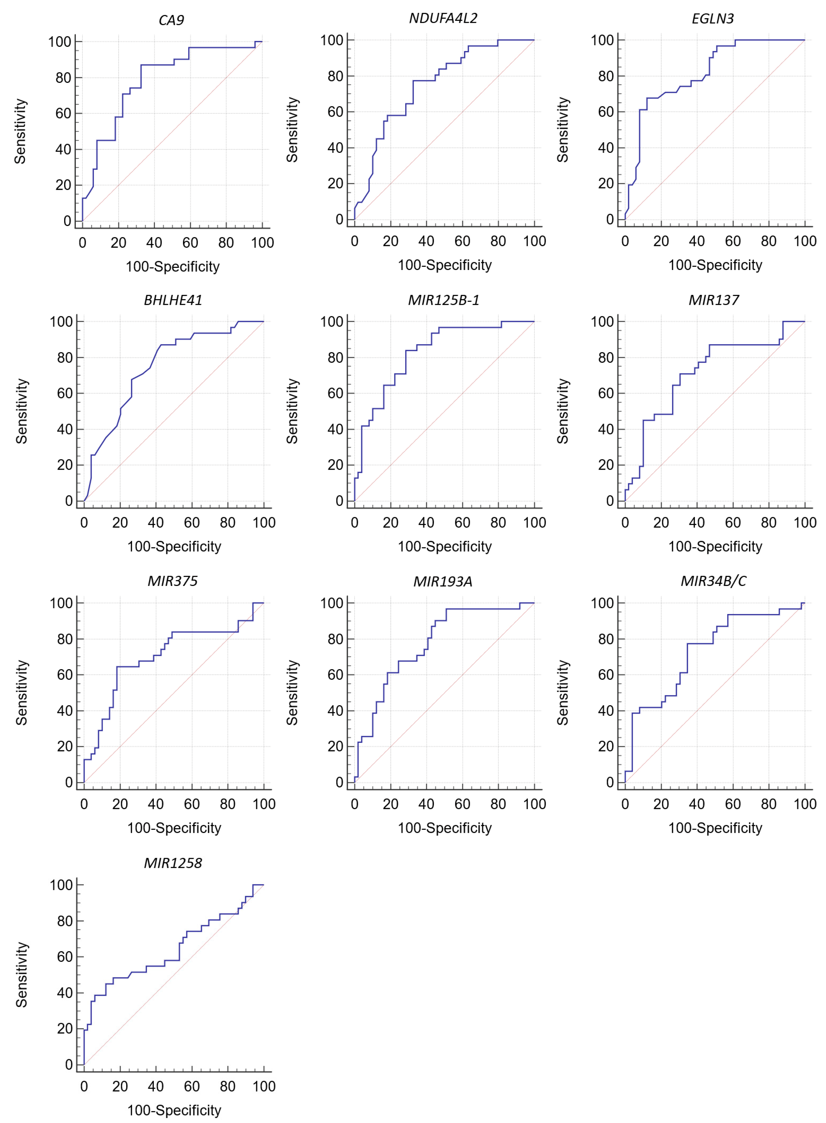
ROC analysis was used to analyze the particularities of the relationship between gene expression and methylation levels with the metastatic potential of the tumor (Table 3, Figure 2).
The CA9, NDUFA4L2, EGLN3, and BHLHE41 genes were found to be the most relevant to the panel for predicting metastasis.
Among miRNA genes, MIR125B-1, MIR137, MIR375, MIR193A, and MIR34B/C have the best classifier qualities (Table 2 and Table 3, Figure 2).
An unfavorable prognosis for the development of metastases is a decrease in the expression level of CA9, NDUFA4L2, EGLN3, and BHLHE41 and an increase in the level of methylation of miRNA genes MIR125B-1, MIR137, MIR375, MIR193A, and MIR34B/C.
The values of prognostic sensitivity and specificity for these genes ranged from 65% to 97% and from 49% to 88%, respectively (Table 3).
Consequently, the differences in median values, logistic regression method, and ROC analysis indicate an association between the nine genes studied and RCC metastasis.
Based on AUC values >0.7, nine genes were identified as the strongest candidates for biomarkers of metastasis.
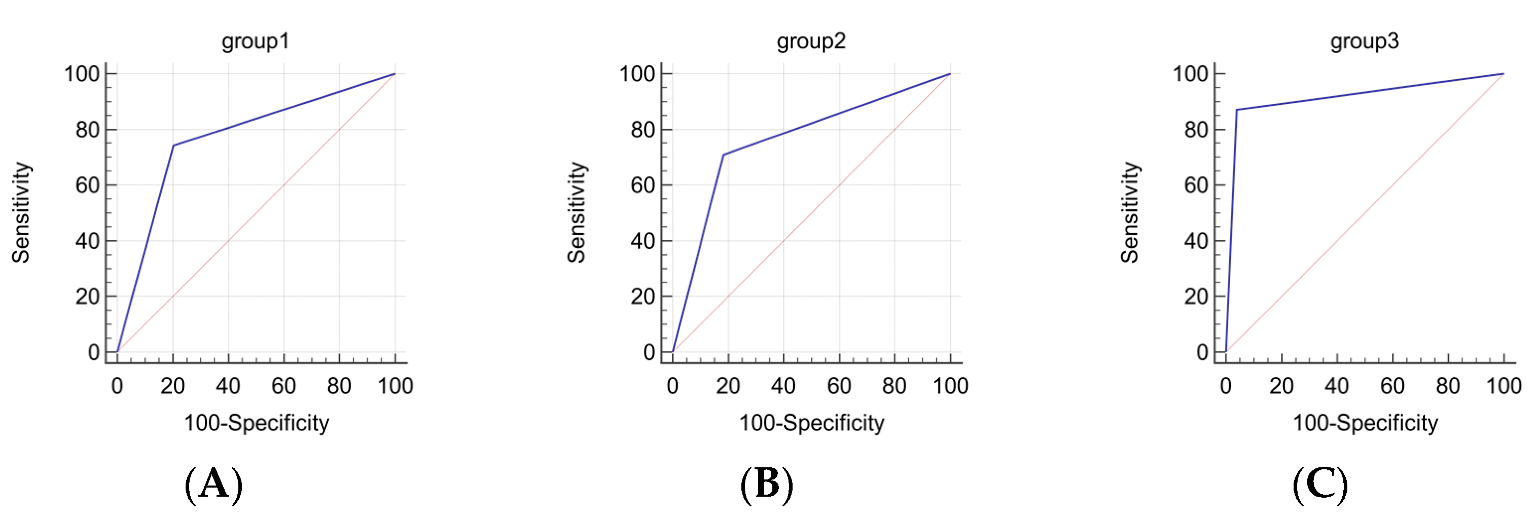
To improve the sensitivity and specificity indices, and to increase the practical significance of the obtained data, three sets of markers were compiled, with each analyzed as a separate group of genes (Table 4). The prognostic significance of these marker sets was evaluated for future use in molecular genetic testing of tissue samples of clear cell renal cell carcinoma (Table 4).
Figure 3 shows the graphical result of the ROC analysis.
Simultaneous analysis of mRNA expression levels of CA9, NDUF4L2, EGLN3, and BHLHE41 genes and/or methylation levels of miRNA genes (MIR125B-1, MIR137, MIR375, MIR193A, and MIR34B/C) increases the reliability of the method. The decrease in the probability of random association of the marker with ccRCC metastasis indicates an improvement in the reliability of the method in this case.
In the practical use of a panel consisting only of protein-coding genes, a recorded decrease in the expression of three out of four of them indicates a high probability of developing metastases. When testing a panel consisting of only five miRNA genes, the recorded increased methylation of four of them indicates a high probability of metastasis. Both judgments are based on the limit value determined by ROC analysis for each gene. When testing a combined panel of genes, any combination of the six or more recorded events listed above corresponds to a high metastatic potential of the tumor.
The corresponding calculation based on the obtained data shows that the combined panel based on simultaneous measurement of gene expression levels and methylation levels has the best sensitivity (87%) and specificity (95%), making it the most reliable predictive panel. However, in case of limited resources, performing either only gene expression level measurements or only methylation level measurements also shows fairly good sensitivity and specificity.
The presented data suggest that the developed combined panel has high sensitivity and specificity. Our determined AUC level of 0.915 indicates high accuracy in predicting metastasis development, which is another important argument in favor of using the combined panel in molecular genetic testing. It should also be noted that the probability of absence of metastasis in case of a negative test is 92%.

4. Discussion

The prediction of metastasis in patients with localized and locally advanced clear cell kidney cancer can be of great practical importance, since it can affect both the volume of surgical intervention, the need for lymphadenectomy, and the need for adjuvant immunotherapy. In cases where renal tumors do not have metastases, the standard scope of surgical intervention is nephrectomy without regional lymph node dissection. Conducting a tumor biopsy prior to surgery or using non-invasive methods of obtaining biological material in the future, such as liquid biopsy, may make it possible to identify a group of patients with a high risk of developing metastases based on the results of gene expression analysis and methylation. In these patients, lymphadenectomy can improve long-term treatment outcomes and provide better staging. Furthermore, prediction of metastasis may be the key to deciding whether adjuvant therapy is necessary. In particular, the randomized phase 3 study KEYNOTE-564 [22] showed a benefit of pembrolizumab (PDL-1 inhibitor) in the adjuvant regimen in patients at intermediate/high risk of progression in terms of recurrence-free survival. It was shown that pembrolizumab reduced the risk of metastasis by 32% with the adjuvant regimen. The two-year disease-free survival rate was 77.3% in the pembrolizumab group and 68.1% in the placebo group (hazard ratio for recurrence or death 0.68; 95% CI 0.53–0.87, p = 0.002). More accurate prediction of metastases based not only on clinical signs, but also on the expression and methylation levels of the genes studied, will help to more selectively identify patients with a high risk of progression and, as a result, to more effectively prescribe adjuvant therapy. To meet this challenge, our study identified a set of genes that predict the development of metastases in ccRCC through the assessment of mRNA expression levels of CA9, NDUF4L2, EGLN3, and BHLHE41, and/or methylation levels of miRNA genes (MIR125B-1, MIR137, MIR375, MIR193A, and MIR34B/C) in the tumor.
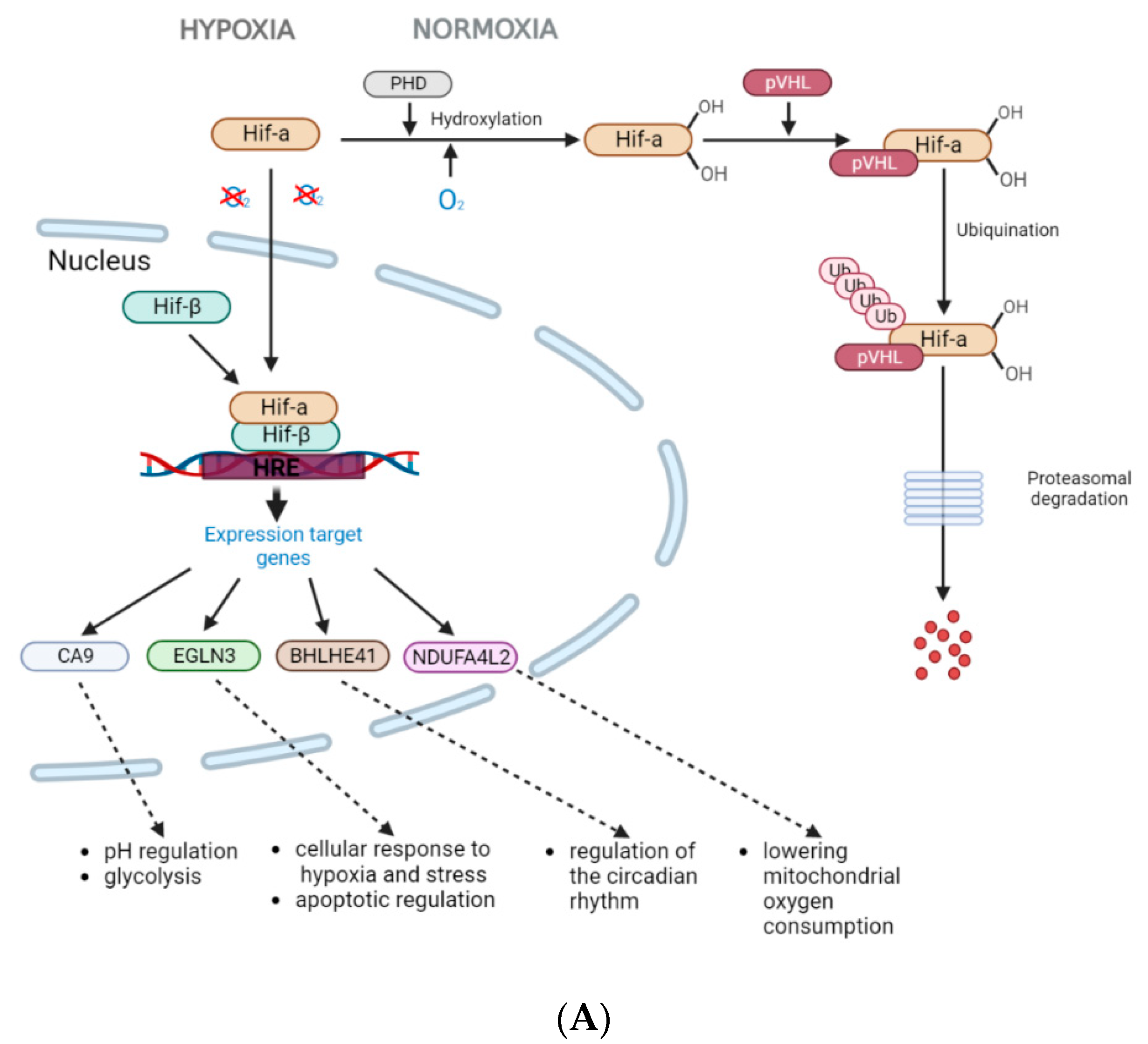
All four protein-encoding genes (CA9, NDUF4L2, EGLN3, and BHLHE41) are direct targets of HIF1 (hypoxia-inducible factor 1) [23,24,25,26]. Under conditions of normal oxygenation and in the absence of mutation in the VHL gene, the product of this gene is a part of the E3 ubiquitin ligase complex, which promotes the attachment of ubiquitin to hydroxylated transcription factors HIFs, leading to their degradation by the proteasome pathway. Under hypoxic conditions, HIF1α translocates to the nucleus where it dimerizes with the constitutively expressed HIF1b, forming the HIF1 complex. Accumulation of HIF1α also occurs due to inactivation of the VHL gene. Under hypoxia and/or absence of functional pVHL, the VHL complex does not bind to the non-hydroxylated transcription factors HIFs, leading to their accumulation in cells and the formation of the HIF1 complex [27,28]. HIF1 activates the transcription of a number of target genes by binding to the hypoxia response element (HRE) located in the regulatory region of the target gene. Induction of the expression of genes activated by hypoxia is necessary for cellular adaptation to a microenvironment with low oxygen levels, for example, during the growth of solid tumors and the transition of homeostatic regulation to a new level (Figure 4A) [29].
The increase in expression levels of the genes we have identified as associated with metastasis in ccRCC is apparently mediated by HIF1 processes of adaptation to a microenvironment with low oxygen levels, as well as by the accumulation of HIF1α caused by the inactivation of the VHL gene. However, during tumor development, their expression levels decrease. It can be assumed that as the kidney tumor progresses, processes related to a strong initial response to hypoxia, regulated by the action of HIF1α, diminish. Meanwhile, other processes such as inflammation develop.
CA9 (carbonic anhydrase 9) is a transmembrane glycoprotein. Its function is to regulate the pH of the cell and maintain the acid–base balance in the body. CA9 catalyzes the reversible hydration of carbon dioxide to bicarbonate, allowing tumor cells to maintain a neutral pH level inside the cell, while acidifying the extracellular microenvironment [30]. CA9 expression has been studied in some detail in ccRCC, and it is molecularly linked to pVHL and regulated by the transcription factor HIF1α [31,32]. Decreased CA9 immunohistochemical staining intensity in metastases compared to corresponding primary tumor samples was demonstrated by Bui et al. [33]. It was also shown that decreased CA9 expression occurs in tumors with the highest malignant potential [33]. A meta-analysis conducted by Zhao Z. et al. [34] showed by qPCR that low CA9 expression levels correlate with a number of clinical characteristics, such as a high degree of differentiation, presence of distant metastases and metastases to lymph nodes. A correlation with decreased expression level and low disease-specific, progression-free, and overall survival was also demonstrated [34]. Some researchers associate decreased CA9 expression in patients with poor prognosis with the activation of AKT and mTOR pathways, which makes further tumor growth less dependent on hypoxia and shifts it to an alternative pathway [32].
The decrease in CA9 expression levels, according to our results, indicates an increased metastatic potential of ccRCC consistent with the published data.
The product of the NDUFA4L2 gene is a subunit of NADH dehydrogenase, which is a component of the mitochondrial respiratory chain. NDUFA4L2 has a pronounced dependence on hypoxia and is a direct target of HIF1α. The functions of NDUFA4L2 are poorly understood, but likely involve the regulation of oxidative phosphorylation through interaction with subunits within complex I of the mitochondrial respiratory chain. Activated under hypoxic conditions, NDUFA4L2 reduces mitochondrial oxygen consumption by inhibiting complex I activity, limiting intracellular ATP production under low oxygen conditions [35]. Decreased expression of the NDUFA4L2 gene is associated with a disruption of the molecular mechanism of cell transition to anaerobic glycolysis, which prevents the reduction of reactive oxygen species (ROS) production and promotes further progression [36]. The significance of NDUFA4L2 expression for the development of ccRCC was first shown by us [37], and confirmed later in other studies [21,38,39,40]. In the study by Meng et al., NDUFA4L2 was found to be overexpressed in non-small cell lung cancer (NSCLC) tissue and cell lines under hypoxic conditions. Activated by HIF1a, NDUFA4L2 suppressed mitochondrial ROS production in NSCLC cells. Knockdown of NDUFA4L2 promoted increased ROS production, apoptosis, and promotion of the epithelial–mesenchymal transition (EMT) of NSCLC cell lines [41]. It is likely that a decrease in the expression level of NDUFA4L2 leading to the activation of EMT is also the mechanism of its effect on metastasis in ccRCC.
EGLN3 (PHD3) is a prolyl hydroxylase 3 whose expression is HIF1-dependent and contributes to preventing apoptosis in cells under hypoxic stress conditions. There is also feedback in this signaling pathway. Under normal conditions, PHD3 hydroxylates HIF1a, which then binds to VHL, becomes ubiquitinated, and degraded via the proteasome. PHD3 also hydroxylates and activates the HIF1a coactivator PKM2 [42]. Another important function of PHD3 is its involvement in regulating glucose metabolism. It has been shown that PHD3 regulates key glycolytic enzymes, including PFKP, TPI1, ENO1, PGAM1, and LDHA, together with the glucose transporter GLUT1. LDHA catalyzes the conversion of pyruvate to lactate. Thus, PHD3 maintains a high rate of glycolysis and lactate production in cancer cells [43]. LDHA is overexpressed in many types of cancer and plays a crucial role in tumor proliferation, invasion, and metastasis [44]. A decreased level of EGLN3 expression leads to a disruption of the hydroxylation of extracellular signal-regulated kinase 3 (Erk3)—one of the key players in regulating tumor progression [45]. The expression of this gene has also been found to be associated with overall survival [45] and progression [46] in kidney cancer. Screening studies have shown differential expression of EGLN3 in cancer cells [47,48].
BHLHE41 (DEC2) is a basic helix–loop–helix (bHLH) transcription factor that is activated by hypoxia [26]. Under hypoxic conditions, HIF-1α induces the transcription of BHLHE41. BHLHE41 can in turn block the hypoxic response by promoting proteasomal degradation of HIF-1α [44]. Among the many functions of BHLHE41 are participation in cell differentiation, immune response, regulation of molecular clocks, and carcinogenesis [49]. Significant upregulation of BHLHE41 expression in kidney cancer was observed in TCGA analysis [50]. The importance of increased expression of this gene for disease progression is currently not well understood, but it has been shown that its suppression in cultured cells by siRNA leads to inhibition of proliferation [51]. In another study, transfection of BHLHE41 into cultured cancer cells resulted in an increase in the proportion of cells in S and G2 phases compared to those in the G1 phase [52]. Therefore, increased expression of BHLHE41 can enhance the proliferative properties of tumor cells at the early stage of ccRCC. At the same time, increased expression of BHLHE41 can inhibit tumor development by suppressing invasion, as it is demonstrated in various types of cancer [49,53,54,55]. We found that the known mechanisms of action of the BHLHE41 correspond to the relationship between the expression level of this gene and ccRCC metastasis.
Thus, from a theoretical point of view, our findings suggest that the association between the reduced expression of studied genes and metastasis allows us to judge the degree of activation of molecular mechanisms of tumor progression in the cell.
DNA methylation is necessary for normal development and transcriptional regulation, but in cancer, it is usually altered towards hypermethylation. In our study, we identified five miRNA genes (MIR125B-1, MIR137, MIR375, MIR193A, and MIR34B/C) whose hypermethylation is associated with metastasis in ccRCC. Increased frequency of methylation of these genes may indicate their tumor suppressor function or the activation of expression of target protein-encoding genes (oncogenes).
The role of miR-125b in cancer progression is contradictory, as it can act both as a tumor suppressor and as an oncogene in different tumor types. MiR-125b originates from miR-125b-1 and miR-125b-2 and leads to degradation of target mRNA or inhibition of translation by binding to the 3’-untranslated regions (3’-UTR) of target mRNA. MiR-125b can promote signal transduction in various signaling pathways [56]. MiR-125b may exert its tumor suppressor function by directly interacting with the delta-catalytic subunit of phosphoinositide 3-kinase (PIK3CD) and reducing its expression, as shown in the anaplastic thyroid cancer cell line (ATC). Overexpression of PIK3CD leads to increased migration and invasion of ATC cells. In addition, exogenous miR-125b reduced the expression of PI3K, p-Akt, and p-mTOR in ATC cells [57]. In hepatocellular carcinoma, miR-125b has an inhibitory effect on EMT and EMT-related features through SMAD2 and SMAD4, which are signal transducers of the TGFβ signaling pathway [58]. Increased expression of miR-125b inhibited the invasion and migration of ovarian cancer cells and was associated with decreased expression of EIF4EBP1 (eukaryotic translation initiation factor 4E-binding protein 1). EIF4EBP1 is an oncogene that plays a critical role in controlling protein synthesis, cell growth, and survival, thereby promoting oncogenesis [59]. Direct correlation between decreased expression levels of miR-125b and increased gene MIR125B-1 methylation levels have been shown in various types of cancer [60,61,62]. Our results indicate that hypermethylation of MIR125B-1 in ccRCC samples with metastasis suggests tumor suppressor properties of this miRNA in kidney cancer.
Overexpression of miR-137 in RCC cells significantly suppressed cell proliferation, migration, and invasion, and induced apoptosis in vitro, as well as inhibited tumor growth in vivo. Additionally, miR-137 was shown to inhibit the activation of the PI3K/AKT signaling pathway in renal cancer cell lines [63]. The direct target of miR-137 in RCC cells is PIK3R3, which is involved in regulating the AKT/mTOR signaling pathway. By suppressing the expression of PIK3R3, miR-137 inhibited cell migration and invasion in RCC [64]. Similar results were shown in gastric cancer, where miR-137 suppressed the activation of the PI3K/AKT signaling pathway by targeting cyclooxygenase-2 (Cox-2), both in vitro and in vivo [65]. Hypermethylation of the MIR137 promoter was shown in gastric cancer tissue, and its suppression induced activation of its target, Cdc42, which is associated with cancer initiation and progression [66]. MiR-137 was shown to directly target Snail and inhibit EMT in ovarian cancer, which is an early and critical stage of metastasis [67]. MiR-137 expression was suppressed and correlated with hypermethylation in renal cancer [68]. Thus, our data on the relationship between hypermethylation of MIR137 and tumor metastasis are consistent with the existing published data.
Overexpression of miR-375 suppressed migration and invasion and inhibited proliferation by inducing apoptosis in renal cancer cell lines. Its suppressive effect was achieved by inhibiting PDK1 and preventing AKT phosphorylation in renal cancer cells [69]. Similar results were shown in pancreatic cancer cells, where miR-375 suppressed cell growth and induced apoptosis by negatively regulating the expression of 3-phosphoinositide-dependent protein kinase 1 (PDK1) [70]. Activation of miR-375 inhibited migration and invasion of human non-small cell lung carcinoma (NSCLC) cells by directly targeting the human epidermal growth factor receptor 2 (HER-2) [71]. Suppression of HER-2 expression by miR-375 has also been found in gastric cancer [72]. It has also been demonstrated that miR-375 suppresses growth, metastasis, and drug sensitivity of ovarian cancer cells [73]. The expression of miR-375 was decreased in samples with altered methylation in ccRCC [68].
Increased expression of miR-193a-3p/5p inhibited migration, invasion, and EMT of (NSCLC) cells in vitro, as well as lung metastasis formation in vivo. Furthermore, ERBB4 and S6K2 were identified as direct targets of miR-193a-3p, while PIK3R3 and mTOR were direct targets of miR-193a-5p in NRML cells. It was also shown that miR-193a-3p/5p can inactivate the AKT/mTOR signaling pathway [74]. The expression of miR-193a inhibited growth, oncogenicity, and radiation sensitivity of medulloblastoma cells with increased expression of MYC. MAX, DCAF7, and STMN1 were identified as novel targets of miR-193a, which may contribute to its anti-tumor effect. The expression of miR-193a in medulloblastoma cells leads to widespread repression of gene expression, including genes involved in WNT signaling, NOTCH signaling, cell cycle regulators, and DNA replication, as well as chromatin organization and modification [75]. It has been shown that the product of the MIR193A gene is one of the key post-transcriptional regulators of protein-7 expression, an adaptor protein of the growth factor receptor, which is one of the key mediators involved in receptor tyrosine kinase signaling. Aberrant elevation of GRB7 levels is often associated with the progression of human cancer. Reduction of miR-193a-3p expression due to DNA hypermethylation is a dynamic process of ovarian cancer progression [76]. Serum levels of miR-193a-3p were significantly elevated in stage I pancreatic cancer patients compared to cancer-free controls (p < 0.01) [77].
Members of the miR-34 family have been described as tumor suppressors in various types of cancer. In cervical cancer, the expression of miR-34b was decreased, and its expression level was associated with increased malignant potential. Overexpression of miR-34b strongly suppressed cell proliferation and induced apoptosis in cervical cancer cell lines [78]. The expression of miR-34b in colorectal adenocarcinoma tissue was negatively correlated with the expression of p-PI3K, p-AKT, and mTOR proteins. MiR-34b can inhibit colorectal adenocarcinoma [79]. It has also been shown that miR-34b/c suppresses CDK4/6 expression in breast cancer cell lines [80]. CpG hypermethylation of MIR34B suppresses miR-34b in prostate cancer. The anti-proliferative and anti-migratory/invasive effects of miR-34b were partially due to the suppression of the AKT pathway and EMT markers. It has also been shown that miR-34b inhibits oncogenicity both in vitro and in vivo [81]. MiR-34b/c may be suppressed in gastric cancer by hypermethylation of MIR34B/C. On the other hand, miR-34b/c regulates the expression of genes involved in the p53-mediated signaling network, suppressing cell proliferation, migration, and metastasis [82]. MiR-34b/c enhanced the attachment of cancer cells and suppressed cell growth and invasion in a mouse model of lung cancer. Moreover, patients with lung adenocarcinoma had better survival rates with higher levels of miR-34a/b/c than those with lower levels [83].
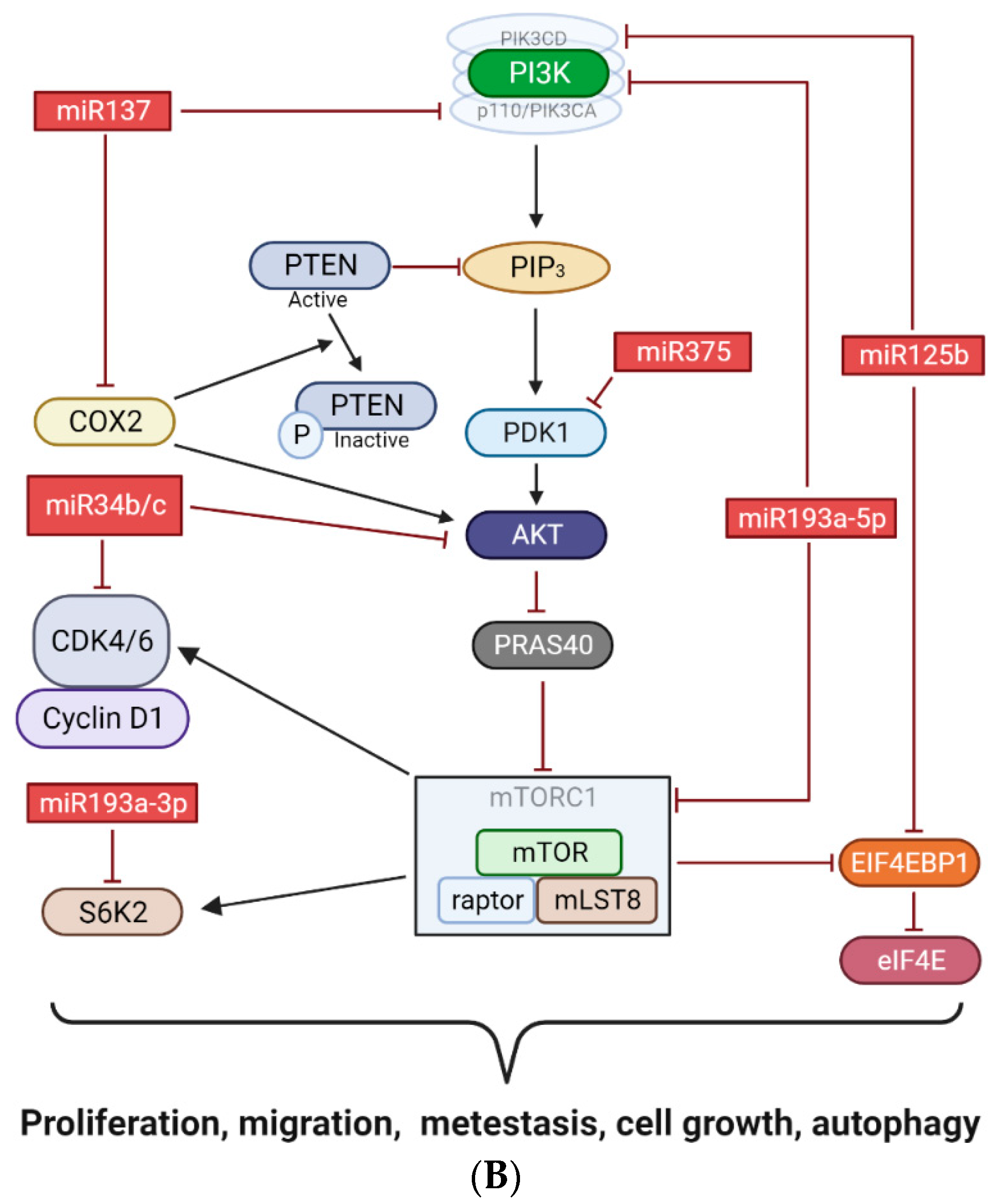
According to our results, hypermethylation of the miRNA genes MIR125B-1, MIR137, MIR375, MIR193A, and MIR34B/C is associated with the metastatic potential of tumors. From the published data, it has been shown that methylation of these genes is associated with decreased expression of the corresponding miRNAs. Taken together, these data may indicate a suppressive function of miR-125b-1, miR-137, miR-375, miR-193a, and miR-34b/c in RCC. From the data described in publications, the studied miRNAs are involved in many signaling pathways and some of them exert their regulatory functions, both as suppressors and as oncogenes. The common feature of these five miRNAs is their involvement in the regulation of the PI3K/AKT/mTOR signaling pathway. As is known, the PI3K/AKT/mTOR signaling pathway is often activated in kidney cancer and leads to increased cell growth, proliferation, and metastasis [84]. This can explain the unidirectional effect of hypermethylation of the identified miRNA genes on the progression of RCC (Figure 4B).
A distinctive feature of our proposed approach for predicting metastasis in kidney cancer patients is the combination of analyzing the expression and methylation of two different groups of genes, protein-coding and miRNA, in one study. Currently, there are no prognostic gene panels constructed based on this principle, highlighting the originality of our proposed approach. By using this methodology, gene groups that function independently in two different pathways are involved, which is registered as a realization of decreased expression or increased methylation in individual samples, complementing each other as markers, thereby contributing to increased prognostic accuracy during testing.
Most of the nomograms used in the clinic are based on the use of indirect signs related to disease progression. The main factors for the prognosis of metastasis that are currently used are TNM classification, G grade of tumor differentiation, C reactive protein, neutrophil-to-lymphocyte ratio, and several other clinical signs in different prognostic models [2]. To determine the independent value of our gene panel for improving existing prognostic models, a multivariate analysis is planned, taking into account the above clinical data on a large cohort of patients with a long follow-up period. We believe that our proposed approach, based on direct registration of genetic events associated with metastases, can improve the accuracy and quality of prognosis because it is based on direct assessment.

5. Conclusions

Determining significant markers of metastasis in ccRCC is a priority area of research, as there are currently no confirmed markers. All studies on this issue are exploratory and represent separate results that have not yet been integrated into a system. The data presented above are conceptual in nature and expand existing ideas about methodological approaches for creating new prognostic gene panels for assessing the metastatic potential of a kidney tumor.
The proposed gene panel with high probability allows the prediction of the development of metastases based on the analysis of the expression levels of CA9, NDUFA4L2, EGLN3, and BHLHE41 genes, and the methylation of miRNA genes MIR125B-1, MIR137, MIR375, MIR193A, and MIR34B/C. We believe that the suggested set of markers is useful in planning an extended analysis of a small amount of tumor tissue in order to improve molecular testing for predicting the development of renal metastases.

Author Contributions

Conceptualization, E.B., V.L. and A.A.; methodology, A.M., N.A., A.B. and V.L.; software, E.F.; validation, N.I., E.F., I.P. and A.B.; formal analysis, P.A.; investigation, A.M., N.I. and T.K.; resources, A.M.; data curation, N.A.; writing—original draft preparation, N.A.; writing—review and editing, A.A., I.P., A.B. and E.B.; visualization, P.A.; supervision, A.A. All authors have read and agreed to the published version of the manuscript.

Funding

This research was carried out within the state assignment of the Ministry of Science and Higher Education of the Russian Federation for Research Centre for Medical Genetics and for the Institute of General Pathology and Pathophysiology (state task NFGFU-2022-0007).

Institutional Review Board Statement

This study was conducted in accordance with the Declaration of Helsinki, and the protocol was approved by the Ethics Committee of the Federal State Budgetary Institution “Research Centre for Medical Genetics” (approval number 2017-4/2).

Informed Consent Statement

Informed consent was obtained from all subjects involved in this study.

Data Availability Statement

The data presented in this study are available on request from the corresponding author.

Acknowledgments

The authors are grateful to the staff of the N.N. Blokhin National Medical Research Center of Oncology for the collection and clinical and histological characterization of the kidney cancer specimens. The authors are grateful to Alexander Vasilievich Karpukhin for scientific and methodological advice in performing this study.

Conflicts of Interest

The authors declare no conflict of interest.

References

  1. Makino, T.; Kadomoto, S.; Izumi, K.; Mizokami, A. Epidemiology and Prevention of Renal Cell Carcinoma. Cancers 2022, 14, 4059. [Google Scholar] [CrossRef] [PubMed]
  2. Ljungberg, B.; Albiges, L.; Bedke, J.; Bex, A.; Capitanio, U.; Giles, R.H.; Hora, M.; Klatte, T.; Marconi, L.; Powles, T.; et al. EAU Guidelines on Renal Cell Carcinoma; EAU Annual Congress Milan; 2023; ISBN 978-94-92671-19-6. Available online: https://d56bochluxqnz.cloudfront.net/documents/full-guideline/EAU-Guidelines-on-Renal-Cell-Carcinoma-2023.pdf (accessed on 1 March 2023).
  3. Kaprin, A.; Starinsky, V.; Shakhzadova, A. Malignant Neoplasms in Russia in 2020 (Morbidity and Mortality); Moscow; Russia; 2021; ISBN 978-5-85502-268-1. Available online: https://oncology-association.ru/wp-content/uploads/2021/11/zis-2020-elektronnaya-versiya.pdf (accessed on 1 March 2021).
  4. Merabishvili, V.M.; Poltorackiy, A.N.; Nosov, A.K.; Artem’Eva, A.S.; Merabishvili, E.N. The State of Oncology Care in Russia. Kidney Cancer (Morbidity, Mortality, Index of Accuracy, One-Year and Year-by-Year Mortality, Histological Structure). Part 1. Onkourologiya 2021, 17, 182–194. [Google Scholar] [CrossRef]
  5. Athanazio, D.A.; Amorim, L.S.; da Cunha, I.W.; Leite, K.R.M.; da Paz, A.R.; de Paula Xavier Gomes, R.; Tavora, F.R.F.; Faraj, S.F.; Cavalcanti, M.S.; Bezerra, S.M. Classification of Renal Cell Tumors—Current concepts and use of ancillary tests: Recommendations of the Brazilian Society of Pathology. Surg. Exp. Pathol. 2021, 4, 4. [Google Scholar] [CrossRef]
  6. Mollica, V.; Di Nunno, V.; Gatto, L.; Santoni, M.; Scarpelli, M.; Cimadamore, A.; Lopez-Beltran, A.; Cheng, L.; Battelli, N.; Montironi, R.; et al. Resistance to Systemic Agents in Renal Cell Carcinoma Predict and Overcome Genomic Strategies Adopted by Tumor. Cancers 2019, 11, 830. [Google Scholar] [CrossRef] [Green Version]
  7. Ma, C.G.; Xu, W.H.; Xu, Y.; Wang, J.; Liu, W.R.; Cao, D.L.; Wang, H.K.; Shi, G.H.; Zhu, Y.P.; Qu, Y.Y.; et al. Identification and validation of novel metastasis-related signatures of clear cell renal cell carcinoma using gene expression databases. Am. J. Transl. Res. 2020, 12, 4108–4126. [Google Scholar] [PubMed]
  8. Wan, B.; Yang, Y.; Zhang, Z. Identification of Differentially Methylated Genes Associated with Clear Cell Renal Cell Carcinoma and Their Prognostic Values. J. Environ. Public Health 2023, 2023, 8405945. [Google Scholar] [CrossRef] [PubMed]
  9. Zhong, T.; Jiang, Z.; Wang, X.; Wang, H.; Song, M.; Chen, W.; Yang, S. Key genes associated with prognosis and metastasis of clear cell renal cell carcinoma. PeerJ 2022, 10, e12493. [Google Scholar] [CrossRef]
  10. Outeiro-Pinho, G.; Barros-Silva, D.; Aznar, E.; Sousa, A.-I.; Vieira-Coimbra, M.; Oliveira, J.; Gonçalves, C.S.; Costa, B.M.; Junker, K.; Henrique, R.; et al. MicroRNA-30a-5pme: A novel diagnostic and prognostic biomarker for clear cell renal cell carcinoma in tissue and urine samples. J. Exp. Clin. Cancer Res. 2020, 39, 98. [Google Scholar] [CrossRef]
  11. Grammatikaki, S.; Katifelis, H.; Farooqi, A.A.; Stravodimos, K.; Karamouzis, M.V.; Souliotis, K.; Varvaras, D.; Gazouli, M. An Overview of Epigenetics in Clear Cell Renal Cell Carcinoma. In Vivo 2023, 37, 1–10. [Google Scholar] [CrossRef]
  12. Patil, N.; Abba, M.L.; Zhou, C.; Chang, S.; Gaiser, T.; Leupold, J.H.; Allgayer, H. Changes in Methylation across Structural and MicroRNA Genes Relevant for Progression and Metastasis in Colorectal Cancer. Cancers 2021, 13, 5951. [Google Scholar] [CrossRef]
  13. Loginov, V.I.; Beresneva, E.V.; Kazubskaya, T.R.; Braga, E.A.; Karpukhin, A.V. Methylation of 10 miRNA genes in clear cell renal cell carcinoma and their diagnostic value. Onkourologiya 2017, 13, 27–33. [Google Scholar] [CrossRef] [Green Version]
  14. Grimberg, J.; Nawoschik, S.; Belluscio, L.; McKee, R.; Turck, A.; Eisenberg, A. A simple and efficient non-organic procedure for the isolation of genomic DNA from blood. Nucleic Acids Res. 1989, 17, 8390. [Google Scholar] [CrossRef]
  15. Hattermann, K.; Mehdorn, H.M.; Mentlein, R.; Schultka, S.; Held-Feindt, J. A methylation-specific and SYBR-green-based quantitative polymerase chain reaction technique for O6-methylguanine DNA methyltransferase promoter methylation analysis. Anal. Biochem. 2008, 377, 62–71. [Google Scholar] [CrossRef] [PubMed]
  16. Loginov, V.I.; Burdennyy, A.M.; Filippova, E.A.; Pronina, I.V.; Lukina, S.S.; Kazubskaya, T.P.; Karpukhin, A.V.; Khodyrev, D.S.; Braga, E.A. Aberrant Methylation of 21 MicroRNA Genes in Breast Cancer: Sets of Genes Associated with Progression and a System of Markers for Predicting Metastasis. Bull. Exp. Biol. Med. 2021, 172, 67–71. [Google Scholar] [CrossRef] [PubMed]
  17. Loginov, V.I.; Burdennyy, A.M.; Filippova, E.A.; Pronina, I.V.; Kazubskaya, T.P.; Kushlinsky, D.N.; Ermilova, V.D.; Rykov, S.V.; Khodyrev, D.S.; Braga, E.A. Hypermethylation of miR-107, miR-130b, miR-203a, miR-1258 Genes Associated with Ovarian Cancer Development and Metastasis. Mol. Biol. 2018, 52, 693–700. [Google Scholar] [CrossRef]
  18. van Hoesel, A.Q.; Sato, Y.; Elashoff, D.A.; Turner, R.R.; Giuliano, A.E.; Shamonki, J.M.; Kuppen, P.J.K.; van de Velde, C.J.H.; Hoon, D.S.B. Assessment of DNA methylation status in early stages of breast cancer development. Br. J. Cancer 2013, 108, 2033–2038. [Google Scholar] [CrossRef] [Green Version]
  19. MedCalc Statistical Software; Ostend, Belgium; 2023. Available online: https://www.medcalc.org/calc/ (accessed on 1 March 2023).
  20. Benjamini, Y.; Hochberg, Y. Controlling the False Discovery Rate: A Practical and Powerful Approach to Multiple Testing. J. R. Stat. Soc. Ser. B Methodol. 1995, 57, 289–300. [Google Scholar] [CrossRef]
  21. Apanovich, N.; Peters, M.; Apanovich, P.; Mansorunov, D.; Markova, A.; Matveev, V.; Karpukhin, A. The Genes—Candidates for Prognostic Markers of Metastasis by Expression Level in Clear Cell Renal Cell Cancer. Diagnostics 2020, 10, 30. [Google Scholar] [CrossRef] [Green Version]
  22. Powles, T.; Tomczak, P.; Park, S.H.; Venugopal, B.; Ferguson, T.; Symeonides, S.N.; Hajek, J.; Gurney, H.; Chang, Y.-H.; Lee, J.L.; et al. Pembrolizumab versus placebo as post-nephrectomy adjuvant therapy for clear cell renal cell carcinoma (KEYNOTE-564): 30-month follow-up analysis of a multicentre, randomised, double-blind, placebo-controlled, phase 3 trial. Lancet Oncol. 2022, 23, 1133–1144. [Google Scholar] [CrossRef]
  23. Zatovicova, M.; Jelenska, L.; Hulikova, A.; Csaderova, L.; Ditte, Z.; Ditte, P.; Goliasova, T.; Pastorek, J.; Pastorekova, S. Carbonic Anhydrase IX as an Anticancer Therapy Target: Preclinical Evaluation of Internalizing Monoclonal Antibody Directed to Catalytic Domain. Curr. Pharm. Des. 2010, 16, 3255–3263. [Google Scholar] [CrossRef]
  24. Fredlund, E.; Ovenberger, M.; Borg, K.; Påhlman, S. Transcriptional adaptation of neuroblastoma cells to hypoxia. Biochem. Biophys. Res. Commun. 2008, 366, 1054–1060. [Google Scholar] [CrossRef]
  25. Pescador, N.; Cuevas, Y.; Naranjo, S.; Alcaide, M.; Villar, D.; Landázuri, M.O.; del Peso, L. Identification of a functional hypoxia-responsive element that regulates the expression of the egl nine homologue 3 (egln3/phd3) gene. Biochem. J. 2005, 390, 189–197. [Google Scholar] [CrossRef] [PubMed] [Green Version]
  26. Miyazaki, K.; Kawamoto, T.; Tanimoto, K.; Nishiyama, M.; Honda, H.; Kato, Y. Identification of Functional Hypoxia Response Elements in the Promoter Region of the DEC1 and DEC2 Genes. J. Biol. Chem. 2002, 277, 47014–47021. [Google Scholar] [CrossRef] [PubMed] [Green Version]
  27. Semenza, G.L. Targeting HIF-1 for cancer therapy. Nat. Rev. Cancer 2003, 3, 721–732. [Google Scholar] [CrossRef] [PubMed]
  28. Linehan, W.M.; Ricketts, C.J. The Cancer Genome Atlas of renal cell carcinoma: Findings and clinical implications. Nat. Rev. Urol. 2019, 16, 539–552. [Google Scholar] [CrossRef] [PubMed]
  29. Semenza, G.L. HIF-1 mediates metabolic responses to intratumoral hypoxia and oncogenic mutations. J. Clin. Investig. 2013, 123, 3664–3671. [Google Scholar] [CrossRef] [Green Version]
  30. Tostain, J.; Li, G.; Gentil-Perret, A.; Gigante, M. Carbonic anhydrase 9 in clear cell renal cell carcinoma: A marker for diagnosis, prognosis and treatment. Eur. J. Cancer 2010, 46, 3141–3148. [Google Scholar] [CrossRef]
  31. Wykoff, C.C.; Beasley, N.J.; Watson, P.H.; Turner, K.J.; Pastorek, J.; Sibtain, A.; Wilson, G.D.; Turley, H.; Talks, K.L.; Maxwell, P.; et al. Hypoxia-inducible expression of tumor-associated carbonic anhydrases. Cancer Res. 2000, 60, 7075–7083. [Google Scholar]
  32. Courcier, J.; De La Taille, A.; Nourieh, M.; Leguerney, I.; Lassau, N.; Ingels, A. Carbonic Anhydrase IX in Renal Cell Carcinoma, Implications for Disease Management. Int. J. Mol. Sci. 2020, 21, 7146. [Google Scholar] [CrossRef]
  33. Bui, M.H.T.; Seligson, D.; Han, K.-R.; Pantuck, A.J.; Dorey, F.J.; Huang, Y.; Horvath, S.; Leibovich, B.C.; Chopra, S.; Liao, S.-Y.; et al. Carbonic anhydrase IX is an independent predictor of survival in advanced renal clear cell carcinoma: Implications for prognosis and therapy. Clin. Cancer Res. 2003, 9, 802–811. [Google Scholar]
  34. Zhao, Z.; Liao, G.; Li, Y.; Zhou, S.; Zou, H.; Fernando, S. Prognostic Value of Carbonic Anhydrase IX Immunohistochemical Expression in Renal Cell Carcinoma: A Meta-Analysis of the Literature. PLoS ONE 2014, 9, e114096. [Google Scholar] [CrossRef]
  35. Tello, D.; Balsa, E.; Acosta-Iborra, B.; Fuertes-Yebra, E.; Elorza, A.; Ordóñez, A.; Corral-Escariz, M.; Soro, I.; López-Bernardo, E.; Perales-Clemente, E.; et al. Induction of the Mitochondrial NDUFA4L2 Protein by HIF-1α Decreases Oxygen Consumption by Inhibiting Complex I Activity. Cell Metab. 2011, 14, 768–779. [Google Scholar] [CrossRef] [PubMed] [Green Version]
  36. Brown, J.A.; Bourke, E.; Eriksson, L.A.; Kerin, M.J. Targeting cancer using KAT inhibitors to mimic lethal knockouts. Biochem. Soc. Trans. 2016, 44, 979–986. [Google Scholar] [CrossRef] [PubMed] [Green Version]
  37. Apanovich, N.V.; Poyarkov, S.V.; Peters, M.V.; Korotaeva, A.A.; Markova, A.S.; Kamolov, B.S.; Pronina, I.V.; Braga, E.A.; Matveev, V.B.; Karpukhin, A. The Differential Gene Expression in Clear Cell Renal Cell Carcinoma and Biomarker Development. Eur. J. Hum.Gen. 2015, 23, 446. [Google Scholar]
  38. Apanovich, N.V.; Peters, M.V.; Korotaeva, A.A.; Apanovich, P.V.; Markova, A.S.; Kamolov, B.S.; Matveev, V.B.; Karpukhin, A.V. Molecular genetic diagnostics of clear cell renal cell carcinoma. Onkourologiya 2016, 12, 16–20. [Google Scholar] [CrossRef] [Green Version]
  39. Minton, D.R.; Fu, L.; Mongan, N.P.; Shevchuk, M.M.; Nanus, D.M.; Gudas, L.J. Role of NADH Dehydrogenase (Ubiquinone) 1 Alpha Subcomplex 4-Like 2 in Clear Cell Renal Cell Carcinoma. Clin. Cancer Res. 2016, 22, 2791–2801. [Google Scholar] [CrossRef] [PubMed] [Green Version]
  40. Liu, L.; Lan, G.; Peng, L.; Xie, X.; Peng, F.; Yu, S.; Wang, Y.; Tang, X. NDUFA4L2 expression predicts poor prognosis in clear cell renal cell carcinoma patients. Ren. Fail. 2016, 38, 8. [Google Scholar] [CrossRef] [Green Version]
  41. Meng, L.; Yang, X.; Xie, X.; Wang, M. Mitochondrial NDUFA4L2 protein promotes the vitality of lung cancer cells by repressing oxidative stress. Thorac. Cancer 2019, 10, 676–685. [Google Scholar] [CrossRef] [Green Version]
  42. Luo, W.; Hu, H.; Chang, R.; Zhong, J.; Knabel, M.; O’Meally, R.; Cole, R.N.; Pandey, A.; Semenza, G.L. Pyruvate Kinase M2 Is a PHD3-Stimulated Coactivator for Hypoxia-Inducible Factor 1. Cell 2011, 145, 732–744. [Google Scholar] [CrossRef] [Green Version]
  43. Miikkulainen, P.; Högel, H.; Rantanen, K.; Suomi, T.; Kouvonen, P.; Elo, L.L.; Jaakkola, P.M. HIF prolyl hydroxylase PHD3 regulates translational machinery and glucose metabolism in clear cell renal cell carcinoma. Cancer Metab. 2017, 5, 5. [Google Scholar] [CrossRef]
  44. Doherty, J.R.; Cleveland, J.L. Targeting lactate metabolism for cancer therapeutics. J. Clin. Investig. 2013, 123, 3685–3692. [Google Scholar] [CrossRef]
  45. Tamukong, P.K.; Kuhlmann, P.; You, S.; Su, S.; Wang, Y.; Yoon, S.; Gong, J.; Figlin, R.A.; Janes, J.L.; Freedland, S.J.; et al. Hypoxia-inducible factor pathway genes predict survival in metastatic clear cell renal cell carcinoma. Urol. Oncol. Semin. Orig. Investig. 2022, 40, 495.e1–495.e10. [Google Scholar] [CrossRef]
  46. Lin, L.; Cai, J. Circular RNA circ-EGLN3 promotes renal cell carcinoma proliferation and aggressiveness via miR-1299-mediated IRF7 activation. J. Cell Biochem. 2020, 121, 4377–4385. [Google Scholar] [CrossRef]
  47. Zaravinos, A.; Pieri, M.; Mourmouras, N.; Anastasiadou, N.; Zouvani, I.; Delakas, D.; Deltas, C. Altered metabolic pathways in clear cell renal cell carcinoma: A meta-analysis and validation study focused on the deregulated genes and their associated networks. Oncoscience 2014, 1, 117–131. [Google Scholar] [CrossRef] [PubMed] [Green Version]
  48. Zodro, E.; Jaroszewski, M.; Ida, A.; Wrzesinski, T.; Kwias, Z.; Bluyssen, H.; Wesoly, J. FUT11 as a potential biomarker of clear cell renal cell carcinoma progression based on meta-analysis of gene expression data. Tumor Biol. 2014, 35, 2607–2617. [Google Scholar] [CrossRef] [PubMed] [Green Version]
  49. Li, P.; Jia, Y.F.; Ma, X.L.; Zheng, Y.; Kong, Y.; Zhang, Y.; Zong, S.; Chen, Z.T.; Wang, Y.S. DEC2 Suppresses Tumor Proliferation and Metastasis by Regulating ERK/NF-ΚB Pathway in Gastric Cancer. Am. J. Cancer Res. 2016, 6, 1741–1757. [Google Scholar] [CrossRef]
  50. Bigot, P.; Colli, L.M.; Machiela, M.J.; Jessop, L.; Myers, T.A.; Carrouget, J.; Wagner, S.; Roberson, D.; Eymerit, C.; Henrion, D.; et al. Functional characterization of the 12p12.1 renal cancer-susceptibility locus implicates BHLHE41. Nat. Commun. 2016, 7, 12098. [Google Scholar] [CrossRef] [Green Version]
  51. Shen, Z.; Zhu, L.; Zhang, C.; Cui, X.; Lu, J. Overexpression of BHLHE41, correlated with DNA hypomethylation in 3’UTR region, promotes the growth of human clear cell renal cell carcinoma. Oncol. Rep. 2019, 41, 2137–2147. [Google Scholar] [CrossRef] [Green Version]
  52. Wang, C.; Zhao, N.; Zheng, Q.; Zhang, D.; Liu, Y. BHLHE41 promotes U87 and U251 cell proliferation via ERK/cyclinD1 signaling pathway. Cancer Manag. Res. 2019, 11, 7657–7672. [Google Scholar] [CrossRef] [PubMed] [Green Version]
  53. Montagner, M.; Enzo, E.; Forcato, M.; Zanconato, F.; Parenti, A.; Rampazzo, E.; Basso, G.; Leo, G.; Rosato, A.; Bicciato, S.; et al. SHARP1 suppresses breast cancer metastasis by promoting degradation of hypoxia-inducible factors. Nature 2012, 487, 380–384. [Google Scholar] [CrossRef]
  54. Asanoma, K.; Liu, G.; Yamane, T.; Miyanari, Y.; Takao, T.; Yagi, H.; Ohgami, T.; Ichinoe, A.; Sonoda, K.; Wake, N.; et al. Regulation of the Mechanism of TWIST1 Transcription by BHLHE40 and BHLHE41 in Cancer Cells. Mol. Cell Biol. 2015, 35, 4096–4109. [Google Scholar] [CrossRef] [PubMed] [Green Version]
  55. Sato, F.; Kawamura, H.; Wu, Y.; Sato, H.; Jin, D.; Bhawal, U.K.; Kawamoto, T.; Fujimoto, K.; Noshiro, M.; Seino, H.; et al. The basic helix-loop-helix transcription factor DEC2 inhibits TGF-β-induced tumor progression in human pancreatic cancer BxPC-3 cells. Int. J. Mol. Med. 2012, 30, 495–501. [Google Scholar] [CrossRef] [PubMed] [Green Version]
  56. Wang, Y.; Zeng, G.; Jiang, Y. The Emerging Roles of miR-125b in Cancers. Cancer Manag. Res. 2020, 12, 1079–1088. [Google Scholar] [CrossRef] [PubMed] [Green Version]
  57. Bu, Q.; You, F.; Pan, G.; Yuan, Q.; Cui, T.; Hao, L.; Zhang, J. MiR-125b inhibits anaplastic thyroid cancer cell migration and invasion by targeting PIK3CD. Biomed. Pharmacother. 2017, 88, 443–448. [Google Scholar] [CrossRef]
  58. Zhou, J.-N.; Zeng, Q.; Wang, H.-Y.; Zhang, B.; Li, S.-T.; Nan, X.; Cao, N.; Fu, C.-J.; Yan, X.-L.; Jia, Y.-L.; et al. MicroRNA-125b attenuates epithelial-mesenchymal transitions and targets stem-like liver cancer cells through small mothers against decapentaplegic 2 and 4. Hepatology 2015, 62, 801–815. [Google Scholar] [CrossRef]
  59. Lee, M.; Kim, E.J.; Jeon, M.J. MicroRNAs 125a and 125b inhibit ovarian cancer cells through post-transcriptional inactivation of EIF4EBP1. Oncotarget 2016, 7, 8726–8742. [Google Scholar] [CrossRef] [Green Version]
  60. Chen, H.; Xu, Z. Hypermethylation-Associated Silencing of miR-125a and miR-125b: A Potential Marker in Colorectal Cancer. Dis. Markers 2015, 2015, 345080. [Google Scholar] [CrossRef] [Green Version]
  61. Zhang, Y.; Yan, L.-X.; Wu, Q.-N.; Du, Z.-M.; Chen, J.; Liao, D.-Z.; Huang, M.-Y.; Hou, J.-H.; Wu, Q.-L.; Zeng, M.-S.; et al. miR-125b Is Methylated and Functions as a Tumor Suppressor by Regulating the ETS1 Proto-oncogene in Human Invasive Breast Cancer. Cancer Res 2011, 71, 3552–3562. [Google Scholar] [CrossRef] [Green Version]
  62. Ferracin, M.; Bassi, C.; Pedriali, M.; Pagotto, S.; D’abundo, L.; Zagatti, B.; Corrà, F.; Musa, G.; Callegari, E.; Lupini, L.; et al. miR-125b targets erythropoietin and its receptor and their expression correlates with metastatic potential and ERBB2/HER2 expression. Mol. Cancer 2013, 12, 130. [Google Scholar] [CrossRef] [Green Version]
  63. Zhang, H.; Li, H. miR-137 inhibits renal cell carcinoma growth in vitro and in vivo. Oncol. Lett. 2016, 12, 715–720. [Google Scholar] [CrossRef] [Green Version]
  64. Sang, L.; Liu, T.; Liu, H.; Li, H.; Zhang, H.; Jiang, F. MicroRNA-137 Suppresses Cell Migration and Invasion in Renal Cell Carcinoma by Targeting PIK3R3. Int. J. Clin. Exp. Med. 2016, 9, 7160–7167. [Google Scholar]
  65. Cheng, Y.; Li, Y.; Liu, D.; Zhang, R.; Zhang, J. miR-137 effects on gastric carcinogenesis are mediated by targeting Cox-2-activated PI3K/AKT signaling pathway. FEBS Lett. 2014, 588, 3274–3281. [Google Scholar] [CrossRef] [Green Version]
  66. Chen, Q.; Chen, X.; Zhang, M.; Fan, Q.; Luo, S.; Cao, X. miR-137 Is Frequently Down-Regulated in Gastric Cancer and Is a Negative Regulator of Cdc42. Dig. Dis. Sci. 2011, 56, 2009–2016. [Google Scholar] [CrossRef] [PubMed]
  67. Dong, P.; Xiong, Y.; Watari, H.; Hanley, S.J.B.; Konno, Y.; Ihira, K.; Yamada, T.; Kudo, M.; Yue, J.; Sakuragi, N. MiR-137 and miR-34a directly target Snail and inhibit EMT, invasion and sphere-forming ability of ovarian cancer cells. J. Exp. Clin. Cancer Res. 2016, 35, 132. [Google Scholar] [CrossRef] [PubMed]
  68. Varachev, V.; Loginov, V.; Pronina, I.; Khodyrev, D.; Kazubskaya, T.; Morozov, A. Hypermethylated Tumor Suppressor MicroRNAs as Novel Markers of Clear Cell Renal Cell Carcinoma. FEBS Open Bio 2018, 8, 304–305. [Google Scholar]
  69. Wang, J.; Sun, X. MicroRNA-375 inhibits the proliferation, migration and invasion of kidney cancer cells by triggering apoptosis and modulation of PDK1 expression. Environ. Toxicol. Pharmacol. 2018, 62, 227–233. [Google Scholar] [CrossRef]
  70. Zhou, J.; Song, S.; He, S.; Zhu, X.; Zhang, Y.; Yi, B.; Zhang, B.; Qin, G.; Li, D. MicroRNA-375 targets PDK1 in pancreatic carcinoma and suppresses cell growth through the Akt signaling pathway. Int. J. Mol. Med. 2014, 33, 950–956. [Google Scholar] [CrossRef] [Green Version]
  71. Cheng, L.; Zhan, B.; Luo, P.; Wang, B. miRNA-375 regulates the cell survival and apoptosis of human non-small cell carcinoma by targeting HER2. Mol. Med. Rep. 2017, 15, 1387–1392. [Google Scholar] [CrossRef] [PubMed] [Green Version]
  72. Shen, Z.-Y.; Zhang, Z.-Z.; Liu, H.; Zhao, E.-H.; Cao, H. miR-375 inhibits the proliferation of gastric cancer cells by repressing ERBB2 expression. Exp. Ther. Med. 2014, 7, 1757–1761. [Google Scholar] [CrossRef] [Green Version]
  73. Yang, S.; Yang, R.; Lin, R.; Si, L. MicroRNA-375 inhibits the growth, drug sensitivity and metastasis of human ovarian cancer cells by targeting PAX2. J. Buon 2020, 24, 2341–2346. [Google Scholar]
  74. Yu, T.; Li, J.; Yan, M.; Liu, L.; Lin, H.; Zhao, F.; Sun, L.; Zhang, Y.; Cui, Y.; Zhang, F.; et al. MicroRNA-193a-3p and -5p suppress the metastasis of human non-small-cell lung cancer by downregulating the ERBB4/PIK3R3/mTOR/S6K2 signaling pathway. Oncogene 2015, 34, 413–423. [Google Scholar] [CrossRef]
  75. Bharambe, H.S.; Joshi, A.; Yogi, K.; Kazi, S.; Shirsat, N.V. Restoration of miR-193a expression is tumor-suppressive in MYC amplified Group 3 medulloblastoma. Acta Neuropathol. Commun. 2020, 8, 70. [Google Scholar] [CrossRef] [PubMed]
  76. Chen, K.; Liu, M.X.; Mak, C.S.-L.; Yung, M.M.-H.; Leung, T.H.-Y.; Xu, D.; Ngu, S.-F.; Chan, K.K.-L.; Yang, H.; Ngan, H.Y.-S.; et al. Methylation-associated silencing of miR-193a-3p promotes ovarian cancer aggressiveness by targeting GRB7 and MAPK/ERK pathways. Theranostics 2018, 8, 423–436. [Google Scholar] [CrossRef] [PubMed]
  77. Wang, C.; Hu, J.; Lu, M.; Gu, H.; Zhou, X.; Chen, X.; Zen, K.; Zhang, C.-Y.; Zhang, T.; Ge, J.; et al. A panel of five serum miRNAs as a potential diagnostic tool for early-stage renal cell carcinoma. Sci. Rep. 2015, 5, 7610. [Google Scholar] [CrossRef] [PubMed] [Green Version]
  78. Cao, Z.; Zhang, G.; Xie, C.; Zhou, Y. MiR-34b regulates cervical cancer cell proliferation and apoptosis. Artif. Cells Nanomed. Biotechnol. 2019, 47, 2042–2047. [Google Scholar] [CrossRef] [Green Version]
  79. Li, Y.; Jin, L.; Rong, W.; Meng, F.; Wang, X.; Wang, S.; Yu, X. Expression and significance of miR-34 with PI3K, AKT and mTOR proteins in colorectal adenocarcinoma tissues. Cell Mol. Biol. 2022, 68, 57–62. [Google Scholar] [CrossRef]
  80. Liu, X.; Feng, J.; Tang, L.; Liao, L.; Xu, Q.; Zhu, S. The Regulation and Function of miR-21-FOXO3a-miR-34b/c Signaling in Breast Cancer. Int. J. Mol. Sci. 2015, 16, 3148–3162. [Google Scholar] [CrossRef] [Green Version]
  81. Majid, S.; Dar, A.A.; Saini, S.; Shahryari, V.; Arora, S.; Zaman, M.S.; Chang, I.; Yamamura, S.; Tanaka, Y.; Chiyomaru, T.; et al. miRNA-34b Inhibits Prostate Cancer through Demethylation, Active Chromatin Modifications, and AKT Pathways. Clin. Cancer Res. 2013, 19, 73–84. [Google Scholar] [CrossRef] [Green Version]
  82. Xiong, S.; Hu, M.; Li, C.; Zhou, X.; Chen, H. Role of miR-34 in gastric cancer: From bench to bedside (Review). Oncol. Rep. 2019, 42, 1635–1646. [Google Scholar] [CrossRef]
  83. Kim, J.S.; Kim, E.J.; Lee, S.; Tan, X.; Liu, X.; Park, S.; Kang, K.; Yoon, J.-S.; Ko, Y.H.; Kurie, J.M.; et al. MiR-34a and miR-34b/c have distinct effects on the suppression of lung adenocarcinomas. Exp. Mol. Med. 2019, 51, 1–10. [Google Scholar] [CrossRef] [Green Version]
  84. Pantuck, A.J.; Seligson, D.B.; Klatte, T.; Yu, H.; Leppert, J.T.; Moore, L.; O’Toole, T.; Gibbons, J.; Belldegrun, A.S.; Figlin, R.A. Prognostic relevance of the mTOR pathway in renal cell carcinoma: Implications for Molecular Patient Selection for Targeted Therapy. Cancer 2007, 109, 2257–2267. [Google Scholar] [CrossRef] [PubMed]
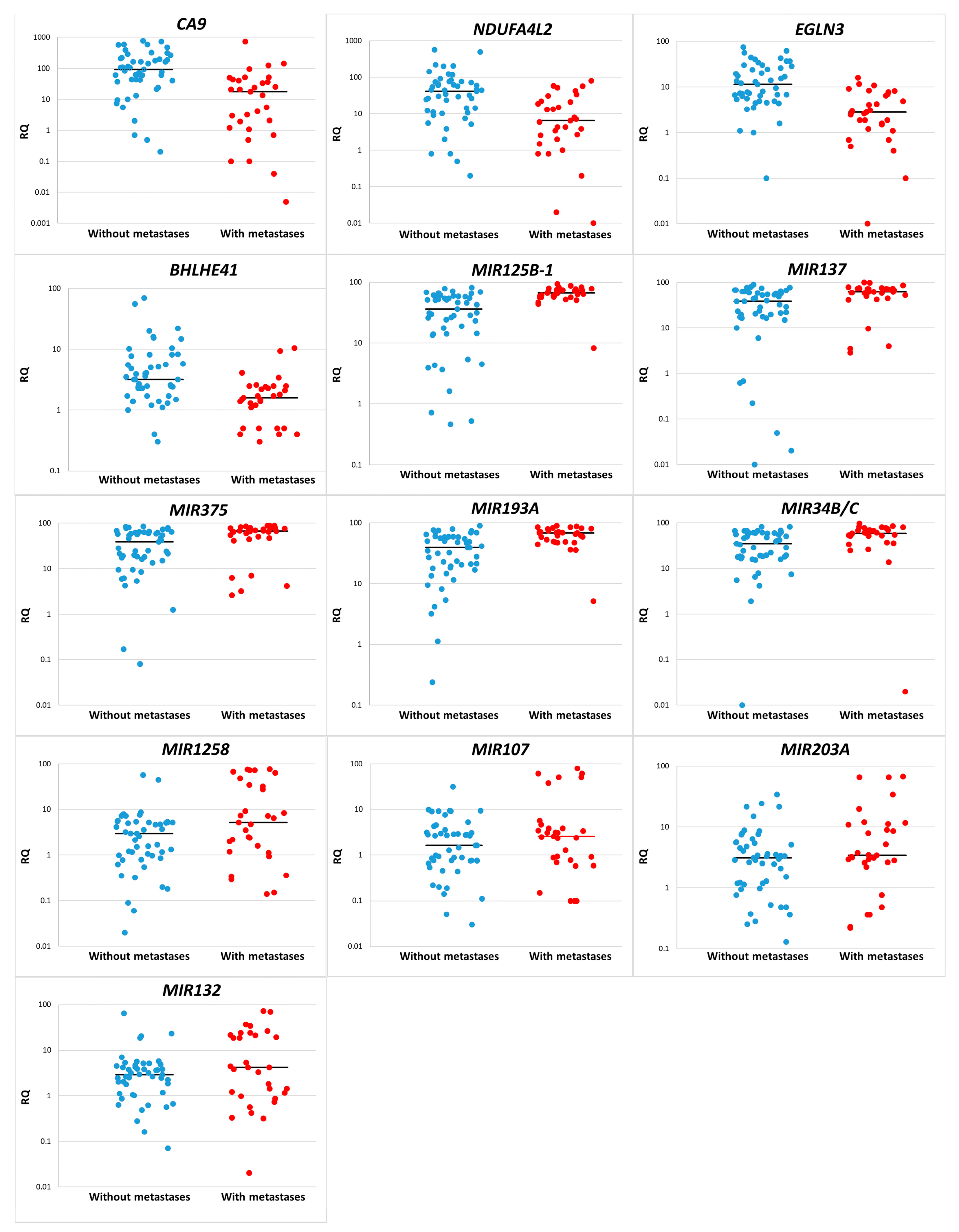
Figure 1. Relative gene expression and methylation levels (RQ) in groups without metastasis () and with metastasis (). Gene expression values and methylation levels are presented on a logarithmic scale. The median is indicated by a line on the graph.
Diagnostics 13 02289 g001
Figure 2. ROC curve analysis to test the association of gene expression and methylation levels with ccRCC metastasis.
Diagnostics 13 02289 g002
Figure 3. ROC analysis of marker panels: (A) CA9, NDUFA4L2, EGLN3, and BHLHE41; (B) MIR125B-1, MIR137, MIR375, MIR193A, and MIR34B/C; (C) CA9, NDUFA4L2, EGLN3, BHLHE41, MIR125B-1, MIR137, MIR375, MIR193A, and MIR34B/C.
Diagnostics 13 02289 g003
Figure 4. Schematic representation of gene interaction pathways and their regulation in cancer. (A). HIF1 regulation of gene expression under hypoxia. (B). miRNAs regulating the PI3K/AKT signaling pathway in cancer.
Diagnostics 13 02289 g004aDiagnostics 13 02289 g004b
Table 1. Clinical and morphological characteristics of tumors in patients with ccRCC.
CharacteristicNumber of SamplesAgeGender M/F (%M/%F)
TNM stageI2060.7 ± 11.110/10 (50/50)
II659.3 ± 8.25/1 (83.3/16.7)
III2362.3 ± 6.916/7 (69.6/30.4)
IV3159.3 ± 7.716/15 (51.6/48.4)
The presence of metastasesWith distant metastases3159.3 ± 7.716/15 (51.6/48.4)
No metastases4961.3 ± 8.931/18 (63.3/36.7)
Localization of distant metastasesLungs1960.4 ± 8.39/10 (47.4/52.6)
Adrenal956.2 ± 8.15/4 (55.6/44.4)
Bones361.0 ± 6.61/2 (33.3/66.7)
Other457.3 ± 7.04/0 (100/0)
Table 2. Median values of gene expression (A) and methylation (B) levels, and significance of differences in ccRCC tumor groups.
GeneThe Median Value (Mann–Whitney U-Test), p =Logistic Regression, p =
In the Non-Metastasis GroupIn the Metastasis Group
(A)
CA992.717.8<0.0010.022
NDUFA4L241.16.5<0.0010.007
EGLN311.42.8<0.0010.004
BHLHE413.21.6<0.0010.018
(B)
MIR125B-136.2766.34<0.0010.001
MIR13738.1061.840.0020.006
MIR37538.9966.190.0030.007
MIR193A38.9967.58<0.0010.001
MIR34B/C35.2659.230.0010.004
MIR12582.975.190.0400.010
MIR1071.622.570.2520.036
MIR203A3.113.420.2350.061
MIR1322.94.170.2520.036
Note: significance levels (p) are presented taking into account the correction of the Benjamini–Hochberg method.
Table 3. The association gene expression and methylation levels with metastasis in ccRCC.
GeneArea under ROC Curve (AUC)95% CICutoff ValueSignificance
Level, p
(Area = 0.5)
SensitivitySpecificity
CA90.7890.684–0.873≤51.3 *<0.00187.1067.35
NDUFA4L20.7530.644–0.842≤22 *<0.00177.4267.35
EGLN30.8180.716–0.895≤4.2 *<0.00167.7487.76
BHLHE410.7510.642–0.841≤2.6 *<0.00187.1057.14
MIR125B-10.8270.726–0.902>55.18 **<0.00183.8771.43
MIR1370.7160.604–0.811>57.62 **0.00170.9769.39
MIR3750.7060.593–0.802>64.29 **0.00164.5281.63
MIR193A0.7760.668–0.861>34.65 **<0.00196.7748.98
MIR34B/C0.7320.621–0.825>50.35 **<0.00177.4265.31
MIR12580.6440.529–0.748>7.15 **0.03345.1687.76
Note: *—expression level; **—methylation level; significance levels (p) are presented taking into account the correction of the Benjamini–Hochberg method.
Table 4. Characterization of combined prognostic panels based on protein-coding genes and miRNA genes.
Gene GroupSensitivity/
Specificity
Area under ROC Curve (AUC)Significance
Level, p
(Area = 0.5)
Negative Predictive Value % (95% CI)Positive Predictive Value % (95% CI)
CA9
NDUFA4L2
EGLN3
BHLHE41
74.19/79.590.769<0.000182.98
(69.19–92.35)
69.70
(51.29–84.41)
MIR125B-1
MIR137
MIR375
MIR193A
MIR34B/C
70.97/81.630.763<0.000181.63
(67.98–91.24)
70.97
(51.96–85.78)
CA9
NDUFA4L2
EGLN3
BHLHE41
MIR125B-1
MIR137
MIR375
MIR193A
MIR34B/C
87.10/95.920.915<0.000192.16
(81.12–97.82)
93.10
(77.23–99.15)
Disclaimer/Publisher’s Note: The statements, opinions and data contained in all publications are solely those of the individual author(s) and contributor(s) and not of MDPI and/or the editor(s). MDPI and/or the editor(s) disclaim responsibility for any injury to people or property resulting from any ideas, methods, instructions or products referred to in the content.