


Brain Sci. 2026, 16(7), 742; https://doi.org/10.3390/brainsci16070742 (registering DOI) - 13 Jul 2026
Abstract
Background: A systematic review and meta-analysis of polysomnographic sleep parameters in bipolar disorder was conducted using articles identified through searches of major databases from inception to May 5, 2026. Methods: One hundred and seven studies were identified in the systematic review.
[...] Read more.
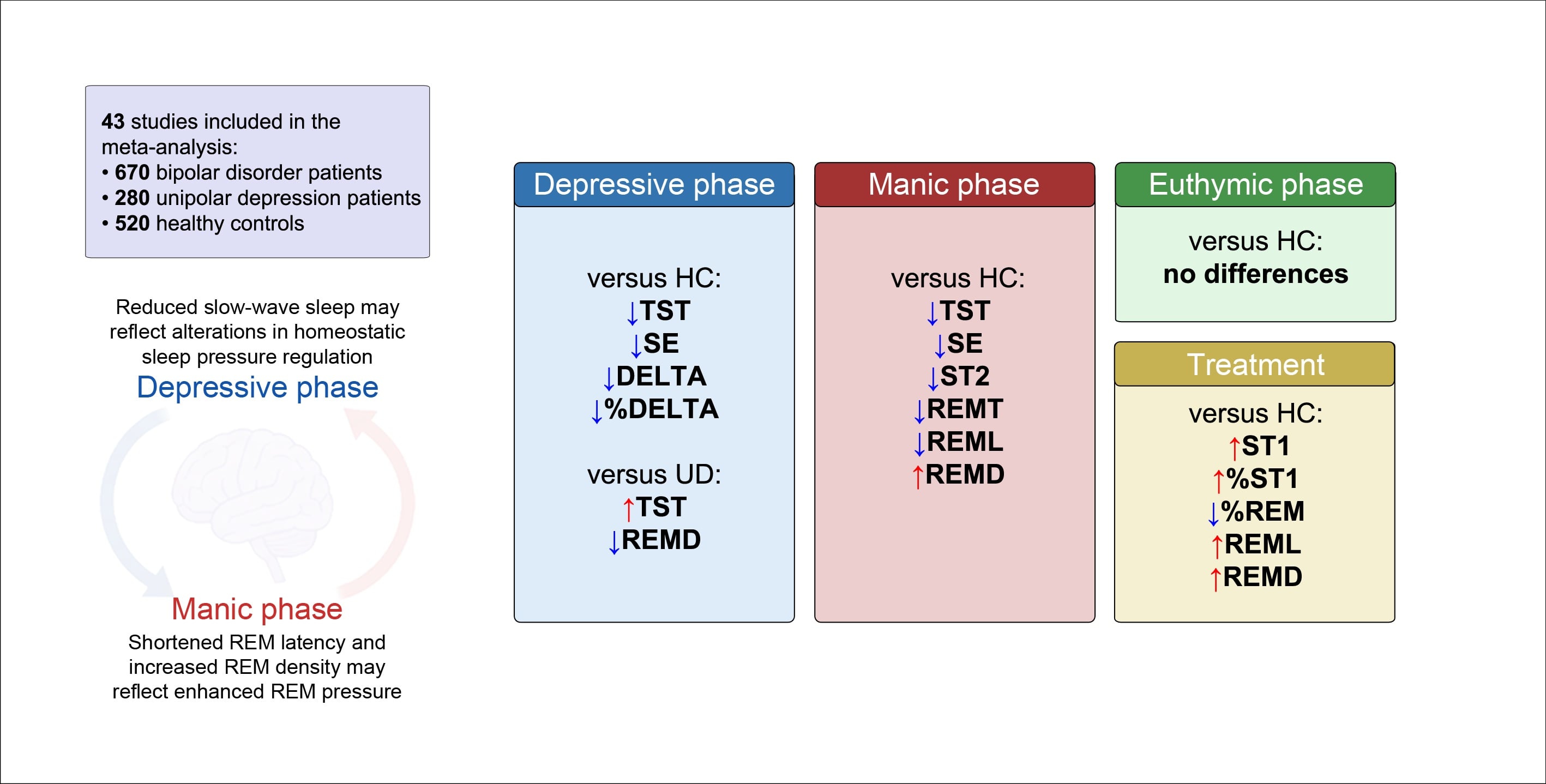
Background: A systematic review and meta-analysis of polysomnographic sleep parameters in bipolar disorder was conducted using articles identified through searches of major databases from inception to May 5, 2026. Methods: One hundred and seven studies were identified in the systematic review. Forty-three case-control studies with 670 bipolar patients, 520 healthy controls and 280 patients with unipolar depression were eligible for the meta-analyses. Total sleep time, sleep onset latency, sleep efficiency, wake after sleep onset, REM time and percentage, REM latency, REM density, stage 1, 2, time and percentage, slow wave sleep (DELTA) time and percentage of drug-free patients with bipolar disorder were compared with case-control data of healthy controls and drug-free patients with unipolar depression. The primary outcome was the standard mean difference. Data were fitted with a random effects model. Publication bias assessment was checked by Egger’s regression and funnel plot asymmetry Results: Total sleep time and sleep efficiency were reduced in both manic and depressive drug-free bipolar patients compared with healthy controls. Delta sleep time and percentage were reduced only in the depressive patients, whereas the manic patients showed decreased stage 2 sleep time, reduced REM sleep time, shortened REM latency and increased REM density. Drug-free patients with unipolar depression showed reduced total sleep time and increased REM density compared with drug-free patients with bipolar depression. Drug-treated bipolar patients showed no differences compared with healthy controls, except for reduced % REM, increased REM latency and increased REM density. Conclusions: The results confirm the presence of sleep alterations in bipolar disorder. Although sleep duration is reduced in both manic and depressive patients, reduced delta sleep in depressive patients and increased activity/pressure of REM in manic patients appear to characterize the two phases of the illness. Altered monoaminergic activity during the depressive phase and increased cholinergic activity during the manic phase might possibly be linked to the sleep alterations also contributing to the mood changes and switch mechanisms.
Full article
(This article belongs to the Special Issue Advances in Circadian Rhythms, Sleep, and Cognitive Function: Translational and Technological Perspectives)
►
Show Figures
Graphical abstract


