


Biomolecules 2026, 16(5), 713; https://doi.org/10.3390/biom16050713 (registering DOI) - 12 May 2026
Abstract
Microglia are brain immune cells that phagocytose cell debris and beta-amyloid plaques in patients with Alzheimer’s disease. They develop from round amoeboid cells into ramified microglia or large macrophages, which can be studied in three-dimensional organotypic mouse brain slices. In a recent publication,
[...] Read more.
Microglia are brain immune cells that phagocytose cell debris and beta-amyloid plaques in patients with Alzheimer’s disease. They develop from round amoeboid cells into ramified microglia or large macrophages, which can be studied in three-dimensional organotypic mouse brain slices. In a recent publication, we showed for the first time that we can track GFAP+ astrocytes and laminin+ vessels in organotypic brain slices using live-cell imaging . The aim of the present study was to use microcontact printing on organotypic brain slices to label microglia with Iba1 and CD11b antibodies and visualise them through live-cell imaging. We show that microglia can be easily labelled with antibodies and tracked via live-cell fluorescence microscopy for up to 20 days. Incubation in lipopolysaccharide (LPS) or granulocyte–macrophage colony-stimulating factor (GM-CSF) stimulates the migration of round amoeboid microglia, whereas interleukin-10 induces their differentiation into ramified forms. Taken together, we show the first-time live cell imaging of microglia in organotypic mouse brain slices using microcontact printing.
Full article
(This article belongs to the Section Cellular Biochemistry)
►
Show Figures
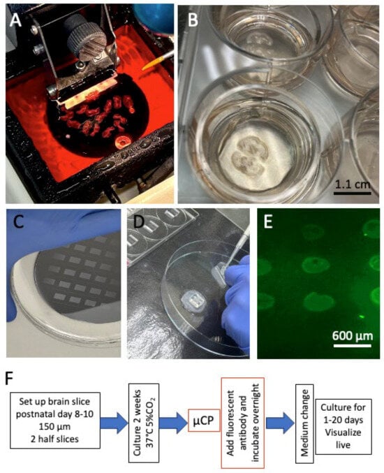
Figure 1


