Next Issue
Volume 13, November-1
Previous Issue
Volume 13, October-1


Diagnostics, Volume 13, Issue 20 (October-2 2023) – 131 articles

Cover Story (view full-size image): 
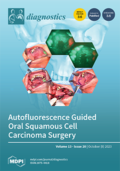
Oral squamous cell carcinoma (OSCC) is a life-threatening disease for which primary surgical treatment currently has the best curative results. Incomplete tumor removal is a strong negative prognostic factor, and therefore new ways to ensure complete tumor removal are being investigated. We test our hypothesis that the use of autofluorescence could increase the success of surgical treatment in OSCC in a group of 122 patients enrolled in our study after meeting the inclusion criteria. The histopathological results after surgical treatment, i.e., the margin status, were then compared. This study demonstrated that preoperative autofluorescence assessment of the mucosal surroundings of OSCC increased the ability to achieve tumor free margin resection by 4.8 times in terms of lateral margins. View this paper
  • Issues are regarded as officially published after their release is announced to the table of contents alert mailing list.
  • You may sign up for e-mail alerts to receive table of contents of newly released issues.
  • PDF is the official format for papers published in both, html and pdf forms. To view the papers in pdf format, click on the "PDF Full-text" link, and use the free Adobe Reader to open them.
Order results
Result details
Section
Select all
Export citation of selected articles as:
12 pages, 1034 KiB  
Article
Correlation between Statin Solubility and Mortality in Patients on Chronic Hemodialysis
by Seok-Hui Kang, Gui-Ok Kim, Bo-Yeon Kim, Eun-Jung Son and Jun-Young Do
Diagnostics 2023, 13(20), 3290; https://doi.org/10.3390/diagnostics13203290 - 23 Oct 2023
Viewed by 830
Abstract
This study aimed to evaluate the effect of statin solubility on the survival of patients undergoing hemodialysis (HD). This retrospective study used laboratory and clinical data from a national HD quality assessment program and claims data (n = 53,345). The use of [...] Read more.
This study aimed to evaluate the effect of statin solubility on the survival of patients undergoing hemodialysis (HD). This retrospective study used laboratory and clinical data from a national HD quality assessment program and claims data (n = 53,345). The use of statins was defined as prescription ≥30 days during 6 months of each HD quality assessment period. We divided the patients into three groups based on the use and solubility of statins: No group, patients without a prescription of statins (n = 37,944); Hydro group, patients with a prescription of hydrophilic statins (n = 2823); and Lipo group, patients with a prescription of lipophilic statins (n = 12,578). The 5-year survival rates in the No, Hydro, and Lipo groups were 69.6%, 67.9%, and 67.9%, respectively (p < 0.001 for the trend). Multivariable Cox regression analyses showed that the Lipo group had better patient survival than the No group. However, multivariable analyses did not show statistical significance between the Hydro and No or Lipo groups. In all subgroups based on sex, age, presence of diabetes mellitus, and heart disease, the Lipo group had better patient survival than the No group. We identified no significant association between hydrophilic and lipophilic statins and patient survival. However, patients taking lipophilic statins had a modest survival benefit compared with those who did not receive statins. Full article
(This article belongs to the Section Clinical Laboratory Medicine)
Show Figures

Figure 1

38 pages, 3291 KiB  
Article
Exploration and Enhancement of Classifiers in the Detection of Lung Cancer from Histopathological Images
by Karthikeyan Shanmugam and Harikumar Rajaguru
Diagnostics 2023, 13(20), 3289; https://doi.org/10.3390/diagnostics13203289 - 23 Oct 2023
Cited by 3 | Viewed by 959
Abstract
Lung cancer is a prevalent malignancy that impacts individuals of all genders and is often diagnosed late due to delayed symptoms. To catch it early, researchers are developing algorithms to study lung cancer images. The primary objective of this work is to propose [...] Read more.
Lung cancer is a prevalent malignancy that impacts individuals of all genders and is often diagnosed late due to delayed symptoms. To catch it early, researchers are developing algorithms to study lung cancer images. The primary objective of this work is to propose a novel approach for the detection of lung cancer using histopathological images. In this work, the histopathological images underwent preprocessing, followed by segmentation using a modified approach of KFCM-based segmentation and the segmented image intensity values were dimensionally reduced using Particle Swarm Optimization (PSO) and Grey Wolf Optimization (GWO). Algorithms such as KL Divergence and Invasive Weed Optimization (IWO) are used for feature selection. Seven different classifiers such as SVM, KNN, Random Forest, Decision Tree, Softmax Discriminant, Multilayer Perceptron, and BLDC were used to analyze and classify the images as benign or malignant. Results were compared using standard metrics, and kappa analysis assessed classifier agreement. The Decision Tree Classifier with GWO feature extraction achieved good accuracy of 85.01% without feature selection and hyperparameter tuning approaches. Furthermore, we present a methodology to enhance the accuracy of the classifiers by employing hyperparameter tuning algorithms based on Adam and RAdam. By combining features from GWO and IWO, and using the RAdam algorithm, the Decision Tree classifier achieves the commendable accuracy of 91.57%. Full article
Show Figures

Figure 1

19 pages, 5687 KiB  
Article
The Clinical Value of Multimodal Ultrasound for the Differential Diagnosis of Hepatocellular Carcinoma from Other Liver Tumors in Relation to Histopathology
by Marinela-Cristiana Urhuț, Larisa Daniela Săndulescu, Adriana Ciocâlteu, Sergiu Marian Cazacu and Suzana Dănoiu
Diagnostics 2023, 13(20), 3288; https://doi.org/10.3390/diagnostics13203288 - 23 Oct 2023
Viewed by 965
Abstract
Recent advances in the field of ultrasonography offer promising tools for the evaluation of liver tumors. We aim to assess the value of multimodal ultrasound in differentiating hepatocellular carcinomas (HCCs) from other liver lesions. We prospectively included 66 patients with 72 liver tumors. [...] Read more.
Recent advances in the field of ultrasonography offer promising tools for the evaluation of liver tumors. We aim to assess the value of multimodal ultrasound in differentiating hepatocellular carcinomas (HCCs) from other liver lesions. We prospectively included 66 patients with 72 liver tumors. The histological analysis was the reference standard for the diagnosis of malignant liver lesions, and partially for benign tumors. All liver lesions were assessed by multiparametric ultrasound: standard ultrasound, contrast-enhanced ultrasound (CEUS), the point shear wave elastography (pSWE) using shear wave measurement (SWM) method and real-time tissue elastography (RTE). To diagnose HCCs, CEUS achieved a sensitivity, specificity, accuracy and positive predictive value (PPV) of 69.05%, 92.86%, 78.57% and 93.55%, respectively. The mean shear-wave velocity (Vs) value in HCCs was 1.59 ± 0.29 m/s, which was lower than non-HCC malignancies (p < 0.05). Using a cut-off value of 1.58 m/s, SWM achieved a sensitivity of 54.76%, and 82.35% specificity, for differentiating HCCs from other malignant lesions. The combination of SWM and CEUS showed higher sensitivity (79.55%) compared with each technique alone, while maintaining a high specificity (89.29%). In RTE, most HCCs (61.53%) had a mosaic pattern with dominant blue areas corresponding to type “c” elasticity. Elasticity type “c” was 70.59% predictive for HCCs. In conclusion, combining B-mode ultrasound, CEUS, pSWE and RTE can provide complementary diagnostic information and potentially decrease the requirements for other imaging modalities. Full article
(This article belongs to the Special Issue Diagnostic Imaging of Liver Diseases)
Show Figures

Figure 1

17 pages, 450 KiB  
Article
Role of SIRT1 Gene Polymorphisms and Serum Levels in Patients with Multiple Sclerosis
by Kriste Kaikaryte, Greta Gedvilaite, Renata Balnyte, Ingrida Uloziene and Rasa Liutkeviciene
Diagnostics 2023, 13(20), 3287; https://doi.org/10.3390/diagnostics13203287 - 23 Oct 2023
Viewed by 821
Abstract
Aim: The purpose of this work was to investigate the prevalence of SIRT1 rs3818292, rs3758391, and rs7895833 single nucleotide polymorphisms and SIRT1 serum levels associated with multiple sclerosis (MS) in the Lithuanian population. Methods: A total of 250 MS patients and 250 healthy [...] Read more.
Aim: The purpose of this work was to investigate the prevalence of SIRT1 rs3818292, rs3758391, and rs7895833 single nucleotide polymorphisms and SIRT1 serum levels associated with multiple sclerosis (MS) in the Lithuanian population. Methods: A total of 250 MS patients and 250 healthy controls were included in the study. Genotyping was performed using the RT-PCR method. Statistical analysis was performed using “IBM SPSS version 29.0”. The serum SIRT1 level was determined by the ELISA method. Results: We found that rs3818292 was associated with increased odds of developing MS under the dominant (p = 0.007) and allelic genetic (p = 0.004) models. rs3758391 was associated with increased odds of developing under the co-dominant (p < 0.001), overdominant (p < 0.001), dominant (p < 0.001), and allelic (p = 0.002) genetic models. rs7895833 was associated with increased odds of developing MS under co-dominant (p < 0.001), overdominant (p < 0.001), dominant (p < 0.001), and allelic (p < 0.001) genetic models. Additional sex-differentiated analysis within females revealed that the rs3758391 was associated with an increased odds ratio for the occurrence of MS among the co-dominant (p = 0.006), dominant (p = 0.002), and allelic (p = 0.001). rs7895833 was associated with an increased odds ratio for the development of MS under the co-dominant (p < 0.001), overdominant (p < 0.001), dominant (p < 0.001), and allelic (p < 0.001) genetic models. Age-differentiated analysis showed that rs3758391 was associated with an increased odds ratio for the development of MS in younger patients under the codominant (p = 0.002), overdominant (p = 0.003), and dominant (p = 0.004) genetic models. rs7895833 was associated with an increased odds ratio for the occurrence of MS under the overdominant genetic model (p = 0.013). In elderly patients, rs3818292 was associated with an increased odds ratio for the occurrence of MS under the dominant (p = 0.008) and allelic (p = 0.009) genetic models. rs7895833 was associated with an increased odds ratio for the occurrence of MS under the codominant (p = 0.011 and p = 0.012), dominant (p = 0.001), and allelic (p < 0.001) genetic models. We also found that serum SIRT1 levels were statistically significantly different between MS patients and control group subjects (p < 0.001). In addition, comparison of SIRT1 levels between study groups and genotypes showed that rs3818292 AA (p = 0.001), rs3758391 CT (p < 0.001), and rs7895833 AA (p = 0.002) and AG (p = 0.004) had higher SIRT1 levels in the control group than in the MS group. All results were provided after strict Bonferroni correction. Conclusions: Genetic variations in SIRT1 rs3818292, rs3758391, and rs7895833 are associated with multiple sclerosis, with possible differences in gender and age, as well as lower serum SIRT1 levels. Full article
Show Figures

Figure 1

11 pages, 2134 KiB  
Article
Characterization of New Alpha Zero (α0) Thalassaemia Deletion (−−GB) among Malays in Malaysian Population
by Norafiza Mohd Yasin, Faidatul Syazlin Abdul Hamid, Syahzuwan Hassan, Yuslina Mat Yusoff, Ermi Neiza Mohd Sahid and Ezalia Esa
Diagnostics 2023, 13(20), 3286; https://doi.org/10.3390/diagnostics13203286 - 23 Oct 2023
Viewed by 922
Abstract
Malaysia is a multicultural and multiethnic country comprising numerous ethnic groups. From the total population of 32.7 million, Malays form the bulk of the Bumiputera in Malaysia comprise about 69.9%, followed by Chinese 22.8%, Indian 6.6%, and others 0.7%. The heterogeneous population and [...] Read more.
Malaysia is a multicultural and multiethnic country comprising numerous ethnic groups. From the total population of 32.7 million, Malays form the bulk of the Bumiputera in Malaysia comprise about 69.9%, followed by Chinese 22.8%, Indian 6.6%, and others 0.7%. The heterogeneous population and increasing numbers of non-citizens in this country affects the heterogeneity of genetic diseases, diversity, and heterogeneity of thalassaemia mutations. Alpha (α)-thalassaemia is an inherited haemoglobin disorder characterized by hypochromic microcytic anaemia caused by a quantitative reduction in the α-globin chain. A majority of the α-thalassaemia are caused by deletions in the α-globin gene cluster. Among Malays, the most common deletional alpha thalassaemia is −α3.7 deletion followed by −−SEA deletion. We described the molecular characterization of a new −−GB deletion in our population, involving both alpha genes in cis. Interestingly, we found that this mutation is unique among Malay ethnicities. It is important to diagnose this deletion because of the 25% risk of Hb Bart’s with hydrops fetalis in the offspring when in combination with another α0- thalassaemia allele. MLPA is a suitable method to detect unknown and uncommon deletions and to characterize those cases which remain unresolved after a standard diagnostic approach. Full article
(This article belongs to the Section Pathology and Molecular Diagnostics)
Show Figures

Figure 1

12 pages, 5525 KiB  
Article
Ask Doctor Smartphone! An App to Help Physicians Manage Foreign Body Ingestions in Children
by Marco Di Mitri, Giovanni Parente, Cristian Bisanti, Eduje Thomas, Sara Maria Cravano, Chiara Cordola, Marzia Vastano, Edoardo Collautti, Annalisa Di Carmine, Michela Maffi, Simone D’Antonio, Michele Libri, Tommaso Gargano and Mario Lima
Diagnostics 2023, 13(20), 3285; https://doi.org/10.3390/diagnostics13203285 - 23 Oct 2023
Viewed by 631
Abstract
Background: Foreign body ingestion (FBI) represents the most common cause of emergent gastrointestinal endoscopy in children. FBI’s management can be quite challenging for physicians because of the variability of the clinical presentation, and the decision tree becomes even more intricate because of patient-specific [...] Read more.
Background: Foreign body ingestion (FBI) represents the most common cause of emergent gastrointestinal endoscopy in children. FBI’s management can be quite challenging for physicians because of the variability of the clinical presentation, and the decision tree becomes even more intricate because of patient-specific variables that must be considered in the pediatric age range (e.g., age of patients and neuropsychiatric disorders) in addition to the mere characteristics of the foreign body. We present an application for smartphones designed for pediatricians and pediatric surgeons based on the latest guidelines from the official pediatric societies. The app aims to help physicians manage FBI quickly and properly in children. Materials and methods: The latest pediatric FBI management guidelines were reviewed and summarized. The flow chart we obtained guided the development of a smartphone application. A questionnaire was administered to all pediatric surgeon trainees at our institute to test the feasibility and helpfulness of the application. Results: An app for smartphones was obtained and shared for free on the Google Play Store and Apple Store. The app guides the physician step by step in the diagnostic process, analyzing all patient- and foreign body-specific characteristics. The app consultation ends with a suggestion of the most proper decision to make in terms of further radiological investigations and the indication and timing of endoscopy. A questionnaire administered to trainees proved the app to be useful and easy to use. Conclusion: We developed an app able to help pediatricians and pediatric surgeons manage FBI in children, providing standardized and updated recommendations in a smart and easily available way. Full article
(This article belongs to the Special Issue Diagnosis and Treatment of Pediatric Emergencies)
Show Figures

Figure 1

13 pages, 8156 KiB  
Article
Comparison of Enamel Surface Integrity after De-Bracketing as Affected by Seven Different Orthodontic Residual Cement Removal Systems
by Abdullazez Almudhi, Arwa Aldeeri, Abdullah Abdulrahman A. Aloraini, Ahmed Ibrahim M. Alomar, Meshari Saad M. Alqudairi, Osama Abdullah A. Alzahrani, Elzahraa Eldwakhly and Sarah AlMugairin
Diagnostics 2023, 13(20), 3284; https://doi.org/10.3390/diagnostics13203284 - 23 Oct 2023
Viewed by 987
Abstract
This study used seven different adhesive removal systems to evaluate and compare enamel surface integrity, heat generation, and time consumed during residual cement removal after de-bracketing. The sample size was 140 human premolars. Teeth were cleaned, mounted, and prepared for orthodontic bracket bonding. [...] Read more.
This study used seven different adhesive removal systems to evaluate and compare enamel surface integrity, heat generation, and time consumed during residual cement removal after de-bracketing. The sample size was 140 human premolars. Teeth were cleaned, mounted, and prepared for orthodontic bracket bonding. Brackets were then debonded using bracket-removing pliers. Teeth were randomly assigned to seven groups based on the residual cement removal system: Group 1: Stainbuster bur, Group 2: Renew diamond bur #129, Group 3: Renew carbide bur, Group 4: OneGloss Complete system, Group 5: Sof-Lex system, Group 6: Enhance Finishing and PoGo Polishing complete kit, and Group 7: Renew friction grip points. The enamel surface was evaluated for roughness before bracketing and after residual cement removal using surface profilometry. The time taken for cement removal was recorded using a digital timer, and heat generation was measured using a laser thermometer before and after cement removal. One-way ANOVA compared the pre- and post-values for enamel surface roughness, temperature, and time consumed. When comparing the difference between the post- and pre-finishing roughness using one-way ANOVA, the Renew diamond bur produced the roughest enamel surface post-removal with a mean of 4.716 μm, while the Sof-Lex recorded the lowest at 0.760 μm. The highest mean temperature was recorded with the Stainbuster bur at 5.545 °C, and the lowest temperature was recorded with the Enhance bur at 2.260 °C. The time for cement removal was the shortest with the Enhance bur at 12.2 s, whereas the time was the longest with the Renew diamond bur at 30.4 s. In conclusion, all the residual cement removal systems used in this clinically simulated study were not able to restore the original enamel surface smoothness. However, the 3M Sof-Lex produced the lowest enamel roughness but with more time consumption and heat generation. When selecting the best residual cement removal system to be used, clinicians should weigh the merits and demerits of each system based on the clinical judgement of the operator. Full article
(This article belongs to the Special Issue New Insights into Diagnosis of Orthodontics)
Show Figures

Figure 1

17 pages, 17363 KiB  
Review
Spectrum of Imaging Patterns of Lung Cancer following Radiation Therapy
by Taylor A. Strange, Lauren T. Erasmus, Jitesh Ahuja, Rishi Agrawal, Girish S. Shroff, Mylene T. Truong and Chad D. Strange
Diagnostics 2023, 13(20), 3283; https://doi.org/10.3390/diagnostics13203283 - 23 Oct 2023
Viewed by 1475
Abstract
Radiation therapy using conventional or newer high-precision dose techniques, including three-dimensional conformal radiotherapy, intensity-modulated radiation therapy, stereotactic body radiation therapy, four-dimensional conformational radiotherapy, and proton therapy, is an important component of treating patients with lung cancer. Knowledge of the radiation technique used and [...] Read more.
Radiation therapy using conventional or newer high-precision dose techniques, including three-dimensional conformal radiotherapy, intensity-modulated radiation therapy, stereotactic body radiation therapy, four-dimensional conformational radiotherapy, and proton therapy, is an important component of treating patients with lung cancer. Knowledge of the radiation technique used and the expected temporal evolution of radiation-induced lung injury, as well as patient-specific parameters such as previous radiotherapy, concurrent chemoradiotherapy, or immunotherapy, is important in image interpretation. This review discusses factors that affect the development and severity of radiation-induced lung injury and its radiological manifestations, as well as the differences between conventional and high-precision dose radiotherapy techniques. Full article
(This article belongs to the Special Issue Selected Topics in Thoracic Pathology)
Show Figures

Figure 1

19 pages, 3487 KiB  
Systematic Review
Prevalence of the Bifid Mandibular Condyle and Its Relationship with Pathologies of the Temporomandibular Joint: A Systematic Review and Meta-Analysis
by Juan José Valenzuela-Fuenzalida, Kora-lle Keller Navarro, Pia Urbina, Martin Trujillo-Riveros, Pablo Nova-Baeza, Mathias Orellana-Donoso, Macarena Rodriguez-Luengo, Alvaro Beccerra Farfan and Juan A. Sanchis-Gimeno
Diagnostics 2023, 13(20), 3282; https://doi.org/10.3390/diagnostics13203282 - 23 Oct 2023
Viewed by 1213
Abstract
Objective: The aim of this study was to describe the prevalence of anatomical variants in the bifid mandibular condyle (BMC) and report its association with temporomandibular joint (TMJ) pathology. Methods: We searched the Medline, Scopus, Web of Science, Google Scholar, CINAHL, and LILACS [...] Read more.
Objective: The aim of this study was to describe the prevalence of anatomical variants in the bifid mandibular condyle (BMC) and report its association with temporomandibular joint (TMJ) pathology. Methods: We searched the Medline, Scopus, Web of Science, Google Scholar, CINAHL, and LILACS databases from their inception up to September 2023. Two authors independently performed the search, study selection, and data extraction, and they also assessed the methodological quality with an assurance tool for anatomical studies (AQUA). Finally, the pooled prevalence was estimated using a random effects model. Results: A total of 50 studies met the eligibility criteria. Twenty studies, with a total of 88,625 subjects, were included in the meta-analysis. The overall prevalence of the bifid mandibular condyle (BMC) variant was 1% (95% CI = 1% to 2%). Conclusions: The correlation between the BMC and TMJ pathologies has a relatively low prevalence in studies that present a considerable number of subjects. From a clinical point of view, a direct association cannot be made between the presence of the BMC and TMJ pathologies or symptoms. Full article
(This article belongs to the Special Issue New Insights into Diagnosis of Orthodontics)
Show Figures

Figure 1

14 pages, 2435 KiB  
Article
Using Visual Patient to Show Vital Sign Predictions, a Computer-Based Mixed Quantitative and Qualitative Simulation Study
by Amos Malorgio, David Henckert, Giovanna Schweiger, Julia Braun, Kai Zacharowski, Florian J. Raimann, Florian Piekarski, Patrick Meybohm, Sebastian Hottenrott, Corinna Froehlich, Donat R. Spahn, Christoph B. Noethiger, David W. Tscholl and Tadzio R. Roche
Diagnostics 2023, 13(20), 3281; https://doi.org/10.3390/diagnostics13203281 - 23 Oct 2023
Viewed by 736
Abstract
Background: Machine learning can analyze vast amounts of data and make predictions for events in the future. Our group created machine learning models for vital sign predictions. To transport the information of these predictions without numbers and numerical values and make them easily [...] Read more.
Background: Machine learning can analyze vast amounts of data and make predictions for events in the future. Our group created machine learning models for vital sign predictions. To transport the information of these predictions without numbers and numerical values and make them easily usable for human caregivers, we aimed to integrate them into the Philips Visual-Patient-avatar, an avatar-based visualization of patient monitoring. Methods: We conducted a computer-based simulation study with 70 participants in 3 European university hospitals. We validated the vital sign prediction visualizations by testing their identification by anesthesiologists and intensivists. Each prediction visualization consisted of a condition (e.g., low blood pressure) and an urgency (a visual indication of the timespan in which the condition is expected to occur). To obtain qualitative user feedback, we also conducted standardized interviews and derived statements that participants later rated in an online survey. Results: The mixed logistic regression model showed 77.9% (95% CI 73.2–82.0%) correct identification of prediction visualizations (i.e., condition and urgency both correctly identified) and 93.8% (95% CI 93.7–93.8%) for conditions only (i.e., without considering urgencies). A total of 49 out of 70 participants completed the online survey. The online survey participants agreed that the prediction visualizations were fun to use (32/49, 65.3%), and that they could imagine working with them in the future (30/49, 61.2%). They also agreed that identifying the urgencies was difficult (32/49, 65.3%). Conclusions: This study found that care providers correctly identified >90% of the conditions (i.e., without considering urgencies). The accuracy of identification decreased when considering urgencies in addition to conditions. Therefore, in future development of the technology, we will focus on either only displaying conditions (without urgencies) or improving the visualizations of urgency to enhance usability for human users. Full article
(This article belongs to the Special Issue Point-of-Care Diagnostics Technology and Applications)
Show Figures

Figure 1

14 pages, 1912 KiB  
Systematic Review
The Potential of Ultrasound Radiomics in Carpal Tunnel Syndrome Diagnosis: A Systematic Review and Meta-Analysis
by Wei-Ting Wu, Che-Yu Lin, Yi-Chung Shu, Peng-Chieh Shen, Ting-Yu Lin, Ke-Vin Chang and Levent Özçakar
Diagnostics 2023, 13(20), 3280; https://doi.org/10.3390/diagnostics13203280 - 23 Oct 2023
Viewed by 1140
Abstract
Background: Carpal tunnel syndrome (CTS) is the most common entrapment neuropathy for which ultrasound imaging has recently emerged as a valuable diagnostic tool. This meta-analysis aims to investigate the role of ultrasound radiomics in the diagnosis of CTS and compare it with [...] Read more.
Background: Carpal tunnel syndrome (CTS) is the most common entrapment neuropathy for which ultrasound imaging has recently emerged as a valuable diagnostic tool. This meta-analysis aims to investigate the role of ultrasound radiomics in the diagnosis of CTS and compare it with other diagnostic approaches. Methods: We conducted a comprehensive search of electronic databases from inception to September 2023. The included studies were assessed for quality using the Quality Assessment Tool for Diagnostic Accuracy Studies. The primary outcome was the diagnostic performance of ultrasound radiomics compared to radiologist evaluation for diagnosing CTS. Results: Our meta-analysis included five observational studies comprising 840 participants. In the context of radiologist evaluation, the combined statistics for sensitivity, specificity, and diagnostic odds ratio were 0.78 (95% confidence interval (CI), 0.71 to 0.83), 0.72 (95% CI, 0.59 to 0.81), and 9 (95% CI, 5 to 15), respectively. In contrast, the ultrasound radiomics training mode yielded a combined sensitivity of 0.88 (95% CI, 0.85 to 0.91), a specificity of 0.88 (95% CI, 0.84 to 0.92), and a diagnostic odds ratio of 58 (95% CI, 38 to 87). Similarly, the ultrasound radiomics testing mode demonstrated an aggregated sensitivity of 0.85 (95% CI, 0.78 to 0.89), a specificity of 0.80 (95% CI, 0.73 to 0.85), and a diagnostic odds ratio of 22 (95% CI, 12 to 41). Conclusions: In contrast to assessments by radiologists, ultrasound radiomics exhibited superior diagnostic performance in detecting CTS. Furthermore, there was minimal variability in the diagnostic accuracy between the training and testing sets of ultrasound radiomics, highlighting its potential as a robust diagnostic tool in CTS. Full article
(This article belongs to the Section Pathology and Molecular Diagnostics)
Show Figures

Figure 1

11 pages, 713 KiB  
Article
Association between Red Cell Distribution Width and Outcomes of Nonagenarians Admitted to the Intensive Care Unit—A Retrospective Cohort Study
by Pauline Theile, Jakob Müller, Rikus Daniels, Stefan Kluge and Kevin Roedl
Diagnostics 2023, 13(20), 3279; https://doi.org/10.3390/diagnostics13203279 - 23 Oct 2023
Viewed by 608
Abstract
The red cell distribution width (RDW) measures the heterogeneity of the erythrocyte volume. Different clinical conditions are associated with increased RDW, and high levels (>14.5%) have been described as a predictive marker for unfavorable outcomes and mortality in critically ill patients. However, there [...] Read more.
The red cell distribution width (RDW) measures the heterogeneity of the erythrocyte volume. Different clinical conditions are associated with increased RDW, and high levels (>14.5%) have been described as a predictive marker for unfavorable outcomes and mortality in critically ill patients. However, there is a lack of data on very elderly critically ill patients. Therefore, we aimed to investigate the association of RDW with outcomes in critically ill patients ≥ 90 years. A retrospective analysis was conducted for all consecutive critically ill patients ≥ 90 years who were admitted to the Department of Intensive Care Medicine of the Medical University Centre Hamburg-Eppendorf (Hamburg, Germany) with available RDW on admission. Clinical course and laboratory were analyzed for all patients with eligible RDW. High RDW was defined as (>14.5%). We clinically assessed factors associated with mortality. Univariable and multivariable Cox regression analysis was performed to determine the prognostic impact of RDW on 28-day mortality. During a 12-year period, we identified 863 critically ill patients ≥ 90 years old with valid RDW values and complete clinical data. In total, 32% (n = 275) died within 28 days, and 68% (n = 579) survived for 28 days. Median RDW levels on ICU admission were significantly higher in non-survivors compared with survivors (15.6% vs. 14.8%, p < 0.001). Overall, 38% (n = 327) had low, and 62% (n = 536) had high RDW. The proportion of high RDW (>14.5%) was significantly higher in non-survivors (73% vs. 57%, p < 0.001). Patients with low RDW presented with a lower Charlson Comorbidity Index (p = 0.014), and their severity of illness on admission was lower (SAPS II: 35 vs. 38 points, p < 0.001). In total, 32% (n = 104) in the low and 35% (n = 190) in the high RDW group were mechanically ventilated (p = 0.273). The use of vasopressors (35% vs. 49%, p < 0.001) and renal replacement therapy (1% vs. 5%, p = 0.007) was significantly higher in the high RDW group. Cox regression analysis demonstrated that high RDW was significantly associated with 28-day mortality [crude HR 1.768, 95% CI (1.355–2.305); p < 0.001]. This association remained significant after adjusting for multiple confounders [adjusted HR 1.372, 95% CI (1.045–1.802); p = 0.023]. High RDW was significantly associated with mortality in critically ill patients ≥ 90 years. RDW is a useful simple parameter for risk stratification and may aid guidance for the therapy in very elderly critically ill patients. Full article
Show Figures

Figure 1

11 pages, 2651 KiB  
Article
Is Marfan Syndrome Associated with Primary Structural Changes in the Left Atrium?
by Kun Zhang, Lucas Ernst, Isabel Schobert, Karla Philipp, Georg Böning, Frank R. Heinzel, Leif-Hendrik Boldt and Petra Gehle
Diagnostics 2023, 13(20), 3278; https://doi.org/10.3390/diagnostics13203278 - 23 Oct 2023
Viewed by 948
Abstract
Marfan syndrome (MFS) is an autosomal-dominant multisystem connective tissue disorder that is based on mutations in the FBN1 gene and variably affects different organs, including the heart. In this study, we investigated cardiac function with a focus on the left atrium (LA) in [...] Read more.
Marfan syndrome (MFS) is an autosomal-dominant multisystem connective tissue disorder that is based on mutations in the FBN1 gene and variably affects different organs, including the heart. In this study, we investigated cardiac function with a focus on the left atrium (LA) in a relatively large cohort of patients with MFS. After screening of 1165 patients that had been examined in our center between 2016 and 2020, 231 adult MFS patients with and without aortic operation were included in our study and compared to a healthy control group (n = 106). Cardiac function was assessed by transthoracic echocardiography and NT-proBNP was used as a secretory marker. Most (94.8%) of the patients received genetic testing. Left ventricular function was within normal ranges and not impaired. Interestingly, we found that LA size and secretory activity were increased in MFS patients, despite normal left ventricular filling pressures. This finding was even more pronounced in MFS patients with prior aortic surgery. A correlation between LA size or NT-proBNP levels and the type of pathogenic FBN1 variant could not be identified. Right ventricular function and right atrial size were increased only in MFS patients that had undergone aortic surgery. In conclusion, these findings suggest that MFS leads to structural changes in the LA that are not solely resulting from left ventricular dysfunction, but probably can be considered a primary pathology of MFS. Full article
(This article belongs to the Section Pathology and Molecular Diagnostics)
Show Figures

Figure 1

16 pages, 1219 KiB  
Article
Recombinant TBEV Protein E of the Siberian Subtype Is a Candidate Antigen in the ELISA Test System for Differential Diagnosis
by Victoria Baryshnikova, Yuriy Turchenko, Ksenia Tuchynskaya, Ilmira Belyaletdinova, Alexander Butenko, Alena Dereventsova, Georgy Ignatiev, Ivan Kholodilov, Victor Larichev, Ekaterina Lyapeykova, Anastasiya Rogova, Armen Shakaryan, Anna Shishova, Anatoly Gmyl and Galina Karganova
Diagnostics 2023, 13(20), 3277; https://doi.org/10.3390/diagnostics13203277 - 23 Oct 2023
Cited by 1 | Viewed by 1037
Abstract
The tick-borne encephalitis virus (TBEV) is one of the most common members of the Orthoflavivirus genus, which comprises the causative agents of severe diseases in humans and animals. Due to the expanding areas of orthoflavivirus infection, its differential diagnosis is highly demanded. Commercial [...] Read more.
The tick-borne encephalitis virus (TBEV) is one of the most common members of the Orthoflavivirus genus, which comprises the causative agents of severe diseases in humans and animals. Due to the expanding areas of orthoflavivirus infection, its differential diagnosis is highly demanded. Commercial test kits based on inactivated TBEV may not provide reliable differentiation between flaviviruses because of serological crossover in this genus. Application of recombinant domains (sE and dIII) of the TBEV Sukhar-strain protein E as antigens in an ELISA test system allowed us to identify a wide range of antibodies specific to different TBEV strains. We tested 53 sera from human patients with confirmed TBE diagnosis (the efficacy of our test system based on sE protein was 98%) and 56 sera from patients with other orthoflavivirus infections in which no positive ones were detected using our ELISA test system, thus being indicative of its 100% specificity. We also tested mouse and rabbit sera containing antibodies specific to 17 TBEV strains belonging to different subtypes; this assay exhibited high efficacy and differentiation ability in detecting antibodies against TBEV from other orthoflaviviruses such as Omsk hemorrhagic fever, Powassan, yellow fever, dengue, West Nile, Zika, and Japanese encephalitis viruses. Full article
(This article belongs to the Special Issue Diagnosis of Dengue and Other Flaviviral Infections)
Show Figures

Figure 1

14 pages, 2891 KiB  
Article
Brochoscopic Airway Clearance Therapy vs. Conventional Sputum Aspiration: The Future of Flexible Brochoscopes in Intensive Care Units?
by Anjie Yao, Zixuan Liu, Wenni He, Hanyu Rao, Changhui Wang and Shuanshuan Xie
Diagnostics 2023, 13(20), 3276; https://doi.org/10.3390/diagnostics13203276 - 22 Oct 2023
Viewed by 877
Abstract
(1) Background: The aim of our study is to investigate the effectiveness of bronchoscopic airway clearance therapy (B-ACT) on severe pneumonia (SP) patients with invasive mechanical ventilation (IMV) in the intensive care unit (ICU). (2) Methods: Our study retrospectively enrolled 49 patients with [...] Read more.
(1) Background: The aim of our study is to investigate the effectiveness of bronchoscopic airway clearance therapy (B-ACT) on severe pneumonia (SP) patients with invasive mechanical ventilation (IMV) in the intensive care unit (ICU). (2) Methods: Our study retrospectively enrolled 49 patients with sputum aspiration and 99 patients with B-ACT, and the latter were divided into the ≤once every 3 days group (n = 50) and >once every 3 days group (n = 49). (3) Results: We found most laboratory blood results were significantly improved in the B-ACT group as compared with those in sputum aspiration group (p < 0.05). Patients in the B-ACT group and those in ≤once every 3 days group also had significantly better survival to hospital discharge than those in their counterpart groups (Logrank p < 0.001). In patients with cardiopulmonary diseases or positive cultures for bacteria, the B-ACT group and those in the ≤once every 3 days group had significantly better survival outcomes to discharge than those in their counterpart groups (Logrank p < 0.001). B-ACT and the average frequency of ≤once every 3 days had significantly better impact on survival outcomes than their counterpart groups (HR: 0.444, 95% CI: 0.238–0.829, p = 0.011; HR: 0.285, 95% CI: 0163–0.498, p < 0.001). (4) Conclusions: In the future, flexible bronchoscopes may paly an important role in ACT for SP patients with IMV. Full article
(This article belongs to the Section Medical Imaging and Theranostics)
Show Figures

Figure 1

12 pages, 509 KiB  
Review
Investigation and Causes of Spontaneous (Non-Diabetic) Hypoglycaemia in Adults: Pitfalls to Avoid
by Maiar Elghobashy, Rousseau Gama and Raashda Ainuddin Sulaiman
Diagnostics 2023, 13(20), 3275; https://doi.org/10.3390/diagnostics13203275 - 22 Oct 2023
Viewed by 1547
Abstract
Although adult spontaneous (non-diabetic) hypoglycaemia is rare, its recognition is important for the preventative or curative treatment of the underlying cause. Establishing Whipple’s triad—low blood glucose, neuroglycopaenia and resolution of neuroglycopaenia on increasing blood glucose levels to normal or above—is essential to verify [...] Read more.
Although adult spontaneous (non-diabetic) hypoglycaemia is rare, its recognition is important for the preventative or curative treatment of the underlying cause. Establishing Whipple’s triad—low blood glucose, neuroglycopaenia and resolution of neuroglycopaenia on increasing blood glucose levels to normal or above—is essential to verify hypoglycaemia. Awareness that hypoglycaemia may occur in severely ill patients is important for its prevention. Further investigation in such cases is unnecessary unless another cause of hypoglycaemia is suspected. Patients are often asymptomatic and normoglycaemic at review. Their history of medication, self-medication, access to hypoglycaemic drugs, alcohol use and comorbidity may provide aetiological clues. The investigation involves obtaining blood samples during symptoms for laboratory glucose measurement or provoking fasting or postprandial hypoglycaemia as directed by symptoms. If confirmed, insulin, c-peptide, proinsulin and beta-hydroxybutyrate are analysed in hypoglycaemic samples. These will classify hypoglycaemia due to non-ketotic hyperinsulinaemia, non-ketotic hypoinsulinaemia and ketotic hypoinsulinaemia, and direct investigations to identify the underlying cause. There are, however, many pitfalls that may mislabel healthy individuals as “hypoglycaemic” or misdiagnose treatable or preventable causes of spontaneous hypoglycaemia. Clinical acumen and appropriate investigations will mostly identify hypoglycaemia and its cause. Full article
Show Figures

Figure 1

14 pages, 2209 KiB  
Article
Feasibility of Ultrasound-Guided Axillary Vein Puncture under Valsalva Maneuver for Diagnostic and Cardiovascular Interventional Purposes: Pacemaker and Cardioverter-Defibrillator Implantation
by Biagio Sassone, Enrico Bertagnin, Santo Virzì, Giuseppe Simeti and Paolo Tolomeo
Diagnostics 2023, 13(20), 3274; https://doi.org/10.3390/diagnostics13203274 - 21 Oct 2023
Viewed by 846
Abstract
Although ultrasound-guided axillary vein access (USGAVA) has proven to be a highly effective and safe method for cardiac electronic implantable device (CIED) lead placement, the collapsibility of the axillary vein (AV) during tidal breathing can lead to narrowing or complete collapse, posing a [...] Read more.
Although ultrasound-guided axillary vein access (USGAVA) has proven to be a highly effective and safe method for cardiac electronic implantable device (CIED) lead placement, the collapsibility of the axillary vein (AV) during tidal breathing can lead to narrowing or complete collapse, posing a challenge for successful vein puncture and cannulation. We investigated the potential of the Valsalva maneuver (Vm) as a facilitating technique for USGAVA in this context. Out of 148 patients undergoing CIED implantation via USGAVA, 41 were asked to perform the Vm, because they were considered unsuitable for venipuncture due to a narrower AV diameter, as assessed by ultrasound (2.7 ± 1.7 mm vs. 9.1 ± 3.3 mm, p < 0.0001). Among them, 37 patients were able to perform the Vm correctly. Overall, the Vm resulted in an average increase in the AV diameter of 4.9 ± 3.4 mm (p < 0.001). USGAVA performed during the Vm was successful in 30 patients (81%), and no Vm-related complications were observed during the 30-day follow-up. In patients with unsuccessful USGAVA, the Vm resulted in a notably smaller increase in AV diameter (0.5 ± 0.3 mm vs. 6.0 ± 2.8 mm, p < 0.0001) compared to patients who achieved successful USGAVA, while performing the Vm. Therefore, the Vm is a feasible maneuver to enhance AV diameter and the success rate of USGAVA in most patients undergoing CIED implantation while maintaining safety. Full article
(This article belongs to the Collection Vascular Diseases Diagnostics)
Show Figures

Figure 1

5 pages, 1953 KiB  
Interesting Images
Use of Transradial Access to Install Two Sequential Stents for Pseudoaneurysms along the Celiac Artery and Common Hepatic Artery Axes
by Abheek Ghosh, Sean Lee, Christina Lim, Tanvir Agnihotri and Nabeel Akhter
Diagnostics 2023, 13(20), 3273; https://doi.org/10.3390/diagnostics13203273 - 21 Oct 2023
Cited by 1 | Viewed by 676
Abstract
Transfemoral access is the most common method for stenting visceral aneurysms. Over the years, transradial access has gained tremendous traction in interventional procedures due to many reported benefits, including increased patient comfort, decreased procedural cost, and reduced rates of procedural complications, among others. [...] Read more.
Transfemoral access is the most common method for stenting visceral aneurysms. Over the years, transradial access has gained tremendous traction in interventional procedures due to many reported benefits, including increased patient comfort, decreased procedural cost, and reduced rates of procedural complications, among others. Moreover, transradial access can serve as a valuable alternative when transfemoral access may be contraindicated. Here, we successfully utilized transradial access to sequentially place two stents for pseudoaneurysms in the celiac artery and common hepatic artery. Full article
(This article belongs to the Special Issue Advances in Diagnostic and Interventional Radiology)
Show Figures

Figure 1

13 pages, 1989 KiB  
Article
Digital Transformation in Musculoskeletal Ultrasound: Acceptability of Blended Learning
by Andreas Michael Weimer, Rainer Berthold, Christian Schamberger, Thomas Vieth, Gerd Balser, Svenja Berthold, Stephan Stein, Lukas Müller, Daniel Merkel, Florian Recker, Gerhard Schmidmaier, Maximilian Rink, Julian Künzel, Roman Kloeckner and Johannes Weimer
Diagnostics 2023, 13(20), 3272; https://doi.org/10.3390/diagnostics13203272 - 20 Oct 2023
Viewed by 874
Abstract
Background: ultrasound diagnostics have a broad spectrum of applications, including among diseases of the musculoskeletal system. Accordingly, it is important for the users to have a well-founded and up-to-date education in this dynamic examination method. The right balance between online and in-class teaching [...] Read more.
Background: ultrasound diagnostics have a broad spectrum of applications, including among diseases of the musculoskeletal system. Accordingly, it is important for the users to have a well-founded and up-to-date education in this dynamic examination method. The right balance between online and in-class teaching still needs to be explored in this context. Certifying institutions are currently testing digitally transformed teaching concepts to provide more evidence. Methods: this study compared two musculoskeletal ultrasound blended learning models. Model A was more traditional, with a focus on in-person teaching, while Model B was more digitally oriented with compulsory webinar. Both used e-learning for preparation. Participants completed evaluations using a seven-point Likert scale, later converted to a 0–1 scale. Digital teaching media (e-learning) were used for preparation in both courses. Results: the analysis included n = 41 evaluations for Model A and n = 30 for Model B. Model B received a better overall assessment (median: 0.73 vs. 0.69, p = 0.05). Model B also excelled in “course preparation” (p = 0.02), “webinar quality” (p = 0.04), and “course concept” (p = 0.04). The “gain of competence” (p = 0.82), “learning materials” (p = 0.30), and “tutor quality” (p = 0.28) showed no significant differences. Conclusion: participants favorably assessed blended learning in ultrasound teaching. Certifying institutions should consider accrediting models that combine digital methods (e.g., internet lectures/webinars) and materials (e.g., e-learning) with hands-on ultrasound training. Further research is needed to validate these subjective findings for a stronger evidential basis. Full article
(This article belongs to the Special Issue Current Challenges and Perspectives of Ultrasound)
Show Figures

Figure 1

9 pages, 641 KiB  
Article
Detection of Plasmodium falciparum in Saliva and Stool Samples from Children Living in Franceville, a Highly Endemic Region of Gabon
by Roméo Karl Imboumy-Limoukou, Jean-Claude Biteghe-Bi-Essone, Judicael Boris Lendongo Wombo, Sonia Etenna Lekana-Douki, Virginie Rougeron, Steede-Seinnat Ontoua, Lydie Sandrine Oyegue-Liabagui, Cherone Nancy Mbani Mpega Ntigui, Lady Charlène Kouna and Jean-Bernard Lekana-Douki
Diagnostics 2023, 13(20), 3271; https://doi.org/10.3390/diagnostics13203271 - 20 Oct 2023
Cited by 1 | Viewed by 893
Abstract
Due to the difficulty of obtaining blood samples, which is the invasive method that is currently used for the detection of Plasmodium spp., alternative diagnostic sampling methods that are effective and non-invasive are needed, particularly for long-term studies. Saliva and stool samples from [...] Read more.
Due to the difficulty of obtaining blood samples, which is the invasive method that is currently used for the detection of Plasmodium spp., alternative diagnostic sampling methods that are effective and non-invasive are needed, particularly for long-term studies. Saliva and stool samples from malaria-infected individuals contain trace amounts of Plasmodium DNA and therefore could be used as alternatives. Malaria was screened using rapid diagnosis tests and confirmed via microscopy. Nested PCR tests targeting the Plasmodium falciparum-specific STEVOR gene were performed for blood, saliva and stool samples that were positive for malaria. Three hundred sixty-seven (367) children were enrolled and eighty (22.22%) were confirmed to be positive for malaria. Matched blood, saliva and stool samples were available for 35 children. By using blood smears as the gold standard for the diagnosis of malaria, our study indicates that Plasmodium DNA was more detectable in blood (100%) than in saliva (22.86%) and stools (14.29%). Applying qPCR to the STEVOR gene to detect Plasmodium falciparum DNA in saliva and stool samples cannot be considered as an alternative to the current malaria detection processes using blood specimens. Full article
(This article belongs to the Special Issue Advances in the Diagnosis of Infectious Diseases and Microorganisms)
Show Figures

Figure 1

16 pages, 5398 KiB  
Article
Pancreatic Ductal Adenocarcinoma: The Characteristics of Contrast-Enhanced Ultrasound Are Correlated with the Hypoxic Microenvironment
by Lan Wang, Ming Li, Tiantian Dong, Yuanyuan Li, Ci Yin and Fang Nie
Diagnostics 2023, 13(20), 3270; https://doi.org/10.3390/diagnostics13203270 - 20 Oct 2023
Viewed by 875
Abstract
A hypoxic microenvironment is associated with an increased risk of metastasis, treatment resistance and poor prognosis of pancreatic ductal adenocarcinoma (PDAC). This study aimed to identify contrast-enhanced ultrasound (CEUS) characteristics that could predict the hypoxic microenvironment of PDAC. A total of 102 patients [...] Read more.
A hypoxic microenvironment is associated with an increased risk of metastasis, treatment resistance and poor prognosis of pancreatic ductal adenocarcinoma (PDAC). This study aimed to identify contrast-enhanced ultrasound (CEUS) characteristics that could predict the hypoxic microenvironment of PDAC. A total of 102 patients with surgically resected PDAC who underwent CEUS were included. CEUS qualitative and quantitative characteristics were analyzed. The expression of hypoxia-inducible factor-1α (HIF-1) and glucose transporter-1 (GLUT1) were demonstrated by immunohistochemistry. The associations between CEUS characteristics and the HIF-1α and GLUT1 expression of PDACs were evaluated. We found that HIF-1α-high PDACs and GLUT1-high PDACs had a larger tumor size and were more prone to lymph node metastasis. There was a significant positive linear correlation between the expression of HIF-1α and GLUT1. CEUS qualitative characteristics including completeness of enhancement and peak enhancement degree (PED) were related to the expression of HIF-1α and GLUT1. A logistic regression analysis showed that tumor size, lymph node metastasis, incomplete enhancement and iso-enhancement of PED were independent predictors for HIF-1α-high PDACs and GLUT1-high PDACs. As for quantitative characteristics, HIF-1α-high PDACs and GLUT1-high PDACs showed higher peak enhancement (PE) and wash-in rate (WIR). CEUS can effectively reflect the hypoxia microenvironment of PDAC, which may become a noninvasive imaging biomarker for prognosis prediction and individualized treatment. Full article
(This article belongs to the Special Issue Ultrasound Imaging in Digestive and Kidney Diseases)
Show Figures

Figure 1

10 pages, 2087 KiB  
Article
Optimal Bowel Preparation Method to Visualize the Distal Ileum via Small Bowel Capsule Endoscopy
by Daisuke Kametaka, Mamoru Ito, Seiji Kawano, Shuhei Ishiyama, Akiko Fujiwara, Junichirou Nasu, Masao Yoshioka, Junji Shiode, Kazuhide Yamamoto, Masaya Iwamuro, Yoshiro Kawahara, Hiroyuki Okada and Motoyuki Otsuka
Diagnostics 2023, 13(20), 3269; https://doi.org/10.3390/diagnostics13203269 - 20 Oct 2023
Viewed by 915
Abstract
Small bowel capsule endoscopy (SBCE) is a convenient and minimally invasive method widely used to evaluate the small intestine. However, especially in the distal ileum, visualization of the intestinal mucosa is frequently hampered by the remaining intestinal contents, making it difficult to detect [...] Read more.
Small bowel capsule endoscopy (SBCE) is a convenient and minimally invasive method widely used to evaluate the small intestine. However, especially in the distal ileum, visualization of the intestinal mucosa is frequently hampered by the remaining intestinal contents, making it difficult to detect critical lesions. Although several studies have reported on the efficacy of bowel preparation before SBCE, no standardized protocol has been established. Herein, we determined the optimal preparation method for better visualization of the distal ileum using SBCE. We retrospectively analyzed 259 consecutive patients who had undergone SBCE between July 2009 and December 2019, divided into three groups: Group A (no preparation except overnight fasting), Group B (ingestion of 1–2 L polyethylene glycol 4 h before colonoscopy after overnight fasting and performing SBCE immediately after colonoscopy), and Group C (ingestion of 0.9 L magnesium citrate [MC] before SBCE after overnight fasting). The visibility of the intestinal mucosa in the first 10 min and at the last 10 min during the period of observation of the distal ileum was examined using a scoring system and compared. The visibility of the images captured by SBCE was assessed based on the scoring of the degree of bile/chyme staining, residual fluid and debris, brightness, bubble reduction, and visualized mucosa. The status of intestinal collapse was also assessed. In the first 10 min of observation of the distal ileum, no significant differences were detected among the groups. In the last 10 min, significantly better images were acquired in Group C in terms of bile/chyme staining, brightness, bubble reduction, and visualized mucosa. Bowel preparation using a low-dose MC solution 2 h before SBCE provided significantly higher-quality images of the distal ileum. Further optimization, such as the timing of initiating the preparation, is necessary to determine the optimal regimen for bowel preparation prior to SBCE. Full article
(This article belongs to the Special Issue Capsule Endoscopy: A New Era)
Show Figures

Figure 1

11 pages, 1622 KiB  
Article
Metabolic Dysfunction-Associated Fatty Liver Disease in Taiwanese Patients with Inflammatory Bowel Disease: A Study in Patients with Clinical Remission
by Shun-Wen Hsiao, Ting-Chun Chen, Pei-Yuan Su, Chen-Ta Yang, Siou-Ping Huang, Yang-Yuan Chen and Hsu-Heng Yen
Diagnostics 2023, 13(20), 3268; https://doi.org/10.3390/diagnostics13203268 - 20 Oct 2023
Viewed by 1156
Abstract
The prevalence of inflammatory bowel disease (IBD) has increased worldwide. The prevalence of metabolic dysfunction associated fatty liver disease (MAFLD) has also risen. However, there is limited research on the connection between MAFLD and IBD in the Asian population. This study aims to [...] Read more.
The prevalence of inflammatory bowel disease (IBD) has increased worldwide. The prevalence of metabolic dysfunction associated fatty liver disease (MAFLD) has also risen. However, there is limited research on the connection between MAFLD and IBD in the Asian population. This study aims to analyze the prevalence and clinical significance of MAFLD in Taiwanese IBD patients with clinical remission. We retrospectively analyzed IBD patients who received transient elastography for liver fibrosis and controlled attenuation parameter evaluation for liver steatosis. This study enrolled 120 patients with IBD, including 45 Crohn’s disease (CD) and 75 ulcerative colitis (UC). MAFLD prevalence in IBD was 29.2%. Patients with MAFLD had a shorter disease duration (2.8 years vs. 5.3 years, p = 0.017), higher alanine aminotransferase levels (24 U/L vs. 17 U/L, p = 0.003), a lower estimated glomerular filtration rate (91.37 mL/min/1.73 m2 vs. 103.92 mL/min/1.73 m2, p = 0.004), and higher γ-glutamyl transferase (γ-GT) (24 mg/dL vs. 13 mg/dL, p < 0.001). The prevalence of significant fibrosis in IBD with MAFLD was 17.1%. Significant fibrosis was found in older age (58.5 years vs. 40 years, p = 0.004) and the high type 2 diabetes mellitus proportion (50.0% vs. 10.3%, p = 0.049). A trend of longer disease duration was found in significant fibrosis (4.9 years vs. 1.6 years, p = 0.051). The prevalence of MALFD in IBD was 29.2%. and 17.1% of them had significant fibrosis. In addition to the intestinal manifestation, the study findings remind clinicians that they should be aware of the possibility of hepatic complications for IBD patients. Full article
(This article belongs to the Special Issue Diagnosis and Prognosis of Gastrointestinal Diseases)
Show Figures

Figure 1

14 pages, 1261 KiB  
Systematic Review
The Role of Artificial Intelligence in Prospective Real-Time Histological Prediction of Colorectal Lesions during Colonoscopy: A Systematic Review and Meta-Analysis
by Bhamini Vadhwana, Munir Tarazi and Vanash Patel
Diagnostics 2023, 13(20), 3267; https://doi.org/10.3390/diagnostics13203267 - 20 Oct 2023
Cited by 1 | Viewed by 1072
Abstract
Artificial intelligence (AI) presents a novel platform for improving disease diagnosis. However, the clinical utility of AI remains limited to discovery studies, with poor translation to clinical practice. Current data suggests that 26% of diminutive pre-malignant lesions and 3.5% of colorectal cancers are [...] Read more.
Artificial intelligence (AI) presents a novel platform for improving disease diagnosis. However, the clinical utility of AI remains limited to discovery studies, with poor translation to clinical practice. Current data suggests that 26% of diminutive pre-malignant lesions and 3.5% of colorectal cancers are missed during colonoscopies. The primary aim of this study was to explore the role of artificial intelligence in real-time histological prediction of colorectal lesions during colonoscopy. A systematic search using MeSH headings relating to “AI”, “machine learning”, “computer-aided”, “colonoscopy”, and “colon/rectum/colorectal” identified 2290 studies. Thirteen studies reporting real-time analysis were included. A total of 2958 patients with 5908 colorectal lesions were included. A meta-analysis of six studies reporting sensitivities (95% CI) demonstrated that endoscopist diagnosis was superior to a computer-assisted detection platform, although no statistical significance was reached (p = 0.43). AI applications have shown encouraging results in differentiating neoplastic and non-neoplastic lesions using narrow-band imaging, white light imaging, and blue light imaging. Other modalities include autofluorescence imaging and elastic scattering microscopy. The current literature demonstrates that despite the promise of new endoscopic AI models, they remain inferior to expert endoscopist diagnosis. There is a need to focus developments on real-time histological predictions prior to clinical translation to demonstrate improved diagnostic capabilities and time efficiency. Full article
(This article belongs to the Special Issue Diagnosis and Management in Digestive Surgery)
Show Figures

Figure 1

15 pages, 3013 KiB  
Article
Deep Features from Pretrained Networks Do Not Outperform Hand-Crafted Features in Radiomics
by Aydin Demircioğlu
Diagnostics 2023, 13(20), 3266; https://doi.org/10.3390/diagnostics13203266 - 20 Oct 2023
Viewed by 1184
Abstract
In radiomics, utilizing features extracted from pretrained deep networks could result in models with a higher predictive performance than those relying on hand-crafted features. This study compared the predictive performance of models trained with either deep features, hand-crafted features, or a combination of [...] Read more.
In radiomics, utilizing features extracted from pretrained deep networks could result in models with a higher predictive performance than those relying on hand-crafted features. This study compared the predictive performance of models trained with either deep features, hand-crafted features, or a combination of these features in terms of the area under the receiver-operating characteristic curve (AUC) and other metrics. We trained models on ten radiological datasets using five feature selection methods and three classifiers. Our results indicate that models based on deep features did not show an improved AUC compared to those utilizing hand-crafted features (deep: AUC 0.775, hand-crafted: AUC 0.789; p = 0.28). Including morphological features alongside deep features led to overall improvements in prediction performance for all models (+0.02 gain in AUC; p < 0.001); however, the best model did not benefit from this (+0.003 gain in AUC; p = 0.57). Using all hand-crafted features in addition to the deep features resulted in a further overall improvement (+0.034 in AUC; p < 0.001), but only a minor improvement could be observed for the best model (deep: AUC 0.798, hand-crafted: AUC 0.789; p = 0.92). Furthermore, our results show that models based on deep features extracted from networks pretrained on medical data have no advantage in predictive performance over models relying on features extracted from networks pretrained on ImageNet data. Our study contributes a benchmarking analysis of models trained on hand-crafted and deep features from pretrained networks across multiple datasets. It also provides a comprehensive understanding of their applicability and limitations in radiomics. Our study shows, in conclusion, that models based on features extracted from pretrained deep networks do not outperform models trained on hand-crafted ones. Full article
(This article belongs to the Section Medical Imaging and Theranostics)
Show Figures

Figure 1

13 pages, 4448 KiB  
Review
The Endoscopic Retrograde Cholangiopancreatography and Endoscopic Ultrasound Connection: Unity Is Strength, or the Endoscopic Ultrasonography Retrograde Cholangiopancreatography Concept
by Claudio Giovanni De Angelis, Eleonora Dall’Amico, Maria Teresa Staiano, Marcantonio Gesualdo, Mauro Bruno, Silvia Gaia, Marco Sacco, Federica Fimiano, Anna Mauriello, Simone Dibitetto, Chiara Canalis, Rosa Claudia Stasio, Alessandro Caneglias, Federica Mediati and Rodolfo Rocca
Diagnostics 2023, 13(20), 3265; https://doi.org/10.3390/diagnostics13203265 - 20 Oct 2023
Viewed by 944
Abstract
Endoscopic ultrasound (EUS) and endoscopic retrograde cholangiopancreatography (ERCP) are both crucial for the endoscopic management of biliopancreatic diseases: the combination of their diagnostic and therapeutic potential is useful in many clinical scenarios, such as indeterminate biliary stenosis, biliary stones, chronic pancreatitis and biliary [...] Read more.
Endoscopic ultrasound (EUS) and endoscopic retrograde cholangiopancreatography (ERCP) are both crucial for the endoscopic management of biliopancreatic diseases: the combination of their diagnostic and therapeutic potential is useful in many clinical scenarios, such as indeterminate biliary stenosis, biliary stones, chronic pancreatitis and biliary and pancreatic malignancies. This natural and evident convergence between EUS and ERCP, which by 2006 we were calling the “Endoscopic ultrasonography retrograde colangiopancreatography (EURCP) concept”, has become a hot topic in the last years, together with the implementation of the therapeutic possibilities of EUS (from EUS-guided necrosectomy to gastro-entero anastomoses) and with the return of ERCP to its original diagnostic purpose thanks to ancillary techniques (extraductal ultrasound (EDUS), intraductal ultrasound (IDUS), cholangiopancreatoscopy with biopsies and probe-based confocal laser endomicroscopy (pCLE)). In this literary review, we retraced the recent history of EUS and ERCP, reported examples of the clinical applicability of the EURCP concept and explored the option of performing the two procedures in only one endoscopic session, with its positive implications for the patient, the endoscopist and the health care system. In the last few years, we also evaluated the possibility of combining EUS and ERCP into a single endoscopic instrument in a single step, but certain obstacles surrounding this approach remain. Full article
Show Figures

Figure 1

13 pages, 3442 KiB  
Article
The Value of Ultrasound Diagnostic Imaging of Meniscal Knee Injuries Verified by Experimental and Arthroscopic Investigations
by Cezary Wasilczyk
Diagnostics 2023, 13(20), 3264; https://doi.org/10.3390/diagnostics13203264 - 20 Oct 2023
Cited by 1 | Viewed by 1256
Abstract
(1) Introduction: Meniscal knee injuries may develop as the result of trauma or overloading. Ultrasound imaging is an observer-dependent modality, meaning that the assessment of structural damage depends on the investigator’s experience.. None of the published papers provides a standardized method for ultrasound [...] Read more.
(1) Introduction: Meniscal knee injuries may develop as the result of trauma or overloading. Ultrasound imaging is an observer-dependent modality, meaning that the assessment of structural damage depends on the investigator’s experience.. None of the published papers provides a standardized method for ultrasound examination of knee menisci. The main goal of this study is to realize and standardize ultrasound imaging diagnostics of meniscal knee injuries based on individual features of ultrasound presentation and to evaluate the applicability of this modality in clinical practice. (2) Material and methods: This study consisted of two anatomical parts, including a clinical part that started with clinical examination of 50 patients with suspected meniscal knee injuries. After this we performed ultrasound examinations in patients with positive clinical test results, using sonographic confirmation for inclusion in the next stage. Finally, knee arthroscopy by two physicians in an operating room was performed, with procedures documented through photographs and video recordings, and analytic material obtained from patients in the control group documented similarly. (3) Results: In the clinical part of the study, arthroscopic examination revealed 13 longitudinal injuries (corresponding to 36% of all injuries in the group), 14 multidirectional injuries (corresponding to 28% of all injuries), 3 radial injuries (corresponding to 6% of all injuries), and 20 oblique injuries (corresponding to 40% of all injuries). The analysis of the sensitivity and specificity of the diagnostic test in terms of recognizing actual meniscal injuries on the basis of full-thickness or partial-thickness delamination, meniscal cyst oedema, and articular space stenosis revealed that the presence of at least two of these three characteristics was associated with the sensitivity of 88% and the specificity of 86% relative to the number of actual meniscal injuries as seen in arthroscopic examination. (4) Conclusions: Research results confirm that clinical examination combined with ultrasound imaging is a very efficient tool for evaluation of meniscal injuries. Full article
(This article belongs to the Section Medical Imaging and Theranostics)
Show Figures

Figure 1

14 pages, 1478 KiB  
Review
A Narrative Review on Non-Cirrohotic Portal Hypertension: Not All Portal Hypertensions Mean Cirrhosis
by Michele Fiordaliso, Giuseppe Marincola, Barbara Pala, Raffaella Muraro, Mariangela Mazzone, Maria Carmela Di Marcantonio and Gabriella Mincione
Diagnostics 2023, 13(20), 3263; https://doi.org/10.3390/diagnostics13203263 - 20 Oct 2023
Viewed by 2578
Abstract
Non-cirrhotic portal hypertension (NCPH), also known as idiopathic non-cirrhotic portal hypertension (INCPH) and porto-sinusoidal vascular disorder (PSVD), is a rare disease characterized by intrahepatic portal hypertension (IPH) in the absence of cirrhosis. The precise etiopathogenesis of IPH is an area of ongoing research. [...] Read more.
Non-cirrhotic portal hypertension (NCPH), also known as idiopathic non-cirrhotic portal hypertension (INCPH) and porto-sinusoidal vascular disorder (PSVD), is a rare disease characterized by intrahepatic portal hypertension (IPH) in the absence of cirrhosis. The precise etiopathogenesis of IPH is an area of ongoing research. NCPH diagnosis is challenging, as there are no specific tests available to confirm the disease, and a high-quality liver biopsy, detailed clinical information, and an expert pathologist are necessary for diagnosis. Currently, the treatment of NCPH relies on the prevention of complications related to portal hypertension, following current guidelines of cirrhotic portal hypertension. No treatment has been studied that aimed to modify the natural history of the disease; however, transjugular intrahepatic porto-systemic shunt (TIPS) placement, shunt and liver transplantation are considerable symptomatic options. In this review, we discuss the heterogeneity of NCPH as well as its etiopathogenesis, clinical presentation and management issues. Starting from the assumption that portal hypertension does not always mean cirrhosis, cooperative studies are probably needed to clarify the issues of etiology and the possible genetic background of this rare disease. This knowledge might lead to better treatment and perhaps better prevention. Full article
(This article belongs to the Special Issue Diagnosis and Management of Hepatic Vascular Disease)
Show Figures

Figure 1

18 pages, 4688 KiB  
Review
The Value of Micro-CT in the Diagnosis of Lung Carcinoma: A Radio-Histopathological Perspective
by Serpil Dizbay Sak, Selim Sevim, Arda Buyuksungur, Ayten Kayı Cangır and Kaan Orhan
Diagnostics 2023, 13(20), 3262; https://doi.org/10.3390/diagnostics13203262 - 20 Oct 2023
Cited by 1 | Viewed by 1296
Abstract
Micro-computed tomography (micro-CT) is a relatively new imaging modality and the three-dimensional (3D) images obtained via micro-CT allow researchers to collect both quantitative and qualitative information on various types of samples. Micro-CT could potentially be used to examine human diseases and several studies [...] Read more.
Micro-computed tomography (micro-CT) is a relatively new imaging modality and the three-dimensional (3D) images obtained via micro-CT allow researchers to collect both quantitative and qualitative information on various types of samples. Micro-CT could potentially be used to examine human diseases and several studies have been published on this topic in the last decade. In this study, the potential uses of micro-CT in understanding and evaluating lung carcinoma and the relevant studies conducted on lung and other tumors are summarized. Currently, the resolution of benchtop laboratory micro-CT units has not reached the levels that can be obtained with light microscopy, and it is not possible to detect the histopathological features (e.g., tumor type, adenocarcinoma pattern, spread through air spaces) required for lung cancer management. However, its ability to provide 3D images in any plane of section, without disturbing the integrity of the specimen, suggests that it can be used as an auxiliary technique, especially in surgical margin examination, the evaluation of tumor invasion in the entire specimen, and calculation of primary and metastatic tumor volume. Along with future developments in micro-CT technology, it can be expected that the image resolution will gradually improve, the examination time will decrease, and the relevant software will be more user friendly. As a result of these developments, micro-CT may enter pathology laboratories as an auxiliary method in the pathological evaluation of lung tumors. However, the safety, performance, and cost effectiveness of micro-CT in the areas of possible clinical application should be investigated. If micro-CT passes all these tests, it may lead to the convergence of radiology and pathology applications performed independently in separate units today, and the birth of a new type of diagnostician who has equal knowledge of the histological and radiological features of tumors. Full article
(This article belongs to the Special Issue Diagnosis and Management of Lung Cancer)
Show Figures

Figure 1

16 pages, 7221 KiB  
Article
Respiratory Diaphragm Motion-Based Asynchronization and Limitation Evaluation on Chronic Obstructive Pulmonary Disease
by Xingyu Zhou, Chen Ye, Yuma Iwao, Takayuki Okamoto, Naoko Kawata, Ayako Shimada and Hideaki Haneishi
Diagnostics 2023, 13(20), 3261; https://doi.org/10.3390/diagnostics13203261 - 20 Oct 2023
Viewed by 807
Abstract
Background: Chronic obstructive pulmonary disease (COPD) typically causes airflow blockage and breathing difficulties, which may result in the abnormal morphology and motion of the lungs or diaphragm. Purpose: This study aims to quantitatively evaluate respiratory diaphragm motion using a thoracic sagittal magnetic resonance [...] Read more.
Background: Chronic obstructive pulmonary disease (COPD) typically causes airflow blockage and breathing difficulties, which may result in the abnormal morphology and motion of the lungs or diaphragm. Purpose: This study aims to quantitatively evaluate respiratory diaphragm motion using a thoracic sagittal magnetic resonance imaging (MRI) series, including motion asynchronization and limitations. Method: First, the diaphragm profile is extracted using a deep-learning-based field segmentation approach. Next, by measuring the motion waveforms of each position in the extracted diaphragm profile, obvious differences in the independent respiration cycles, such as the period and peak amplitude, are verified. Finally, focusing on multiple breathing cycles, the similarity and amplitude of the motion waveforms are evaluated using the normalized correlation coefficient (NCC) and absolute amplitude. Results and Contributions: Compared with normal subjects, patients with severe COPD tend to have lower NCC and absolute amplitude values, suggesting motion asynchronization and limitation of their diaphragms. Our proposed diaphragmatic motion evaluation method may assist in the diagnosis and therapeutic planning of COPD. Full article
Show Figures

Figure 1

Previous Issue
Back to TopTop