Next Issue
Volume 15, September-2
Previous Issue
Volume 15, August-2


Diagnostics, Volume 15, Issue 17 (September-1 2025) – 166 articles

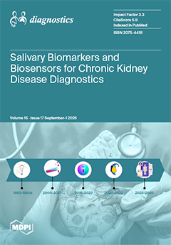
Cover Story (view full-size image): Chronic kidney disease is often detected late, when intervention options narrow. Saliva offers a painless, non-invasive alternative to blood and urine testing. In this scoping review, evidence from 29 studies on the diagnostic performance of salivary biomarkers against reference standards was synthesized, and enabling technologies were surveyed. Creatinine and urea correlate strongly with serum measures (AUCs up to 1.00; sensitivity/specificity often >85%), while emerging markers, such as TMAO, cystatin C, and amino acids, may improve risk stratification and earlier detection. We also highlight rapid point-of-care approaches, including electrochemical biosensors and ATR-FTIR spectroscopy, and outline the path to adoption: protocol standardization, validation in early-stage CKD, and integration into primary- and dental-care screening. View this paper
  • Issues are regarded as officially published after their release is announced to the table of contents alert mailing list.
  • You may sign up for e-mail alerts to receive table of contents of newly released issues.
  • PDF is the official format for papers published in both, html and pdf forms. To view the papers in pdf format, click on the "PDF Full-text" link, and use the free Adobe Reader to open them.
Order results
Result details
Section
Select all
Export citation of selected articles as:
11 pages, 208 KB  
Article
Three Year Follow-Up of Reduced Dose of Direct Oral Anticoagulants for Extended Treatment of Venous Thromboembolism: An Ambispective Cohort Study
by Emanuele Valeriani, Arianna Pannunzio, Tommaso Brogi, Ilaria Maria Palumbo, Danilo Menichelli, Silvia Marucci, Luca Tretola, Claudio Maria Mastroianni, Daniele Pastori and Pasquale Pignatelli
Diagnostics 2025, 15(17), 2283; https://doi.org/10.3390/diagnostics15172283 - 8 Sep 2025
Viewed by 990
Abstract
Background: Few data are available on the outcomes of patients with venous thromboembolism (VTE) on long-term reduced dose of direct oral anticoagulants (DOACs). We evaluated the effectiveness and safety of reduced dose of DOACs for the extended treatment of VTE. Methods: In this [...] Read more.
Background: Few data are available on the outcomes of patients with venous thromboembolism (VTE) on long-term reduced dose of direct oral anticoagulants (DOACs). We evaluated the effectiveness and safety of reduced dose of DOACs for the extended treatment of VTE. Methods: In this monocenter, ambispective cohort study, 140 patients receiving a reduced dose of DOACs for VTE were included. The primary outcomes were recurrent VTE, major bleeding and clinically relevant non-major bleeding. The secondary outcomes were arterial events and minor bleedings. The incidence of the primary outcomes was calculated. The rate for secondary outcomes was descriptively reported. Results: The mean age of the overall cohort was 72 years. Half of the patients were female, 51.4% had a persistent risk factor, 40.0% an unprovoked VTE, and 8.6% a minor transient risk factor. Most patients had lower extremity deep vein thrombosis with or without pulmonary embolism (55.0%) and received apixaban (73.6%) or rivaroxaban (14.3%) for a mean duration of 2.7 years. Regarding the primary outcomes, there was one recurrent VTE (0.7%), four major bleedings (2.9%) and two clinically relevant non-major bleedings (1.4%). Regarding the secondary outcomes, there were four acute ischemic strokes (2.9%) and two minor bleedings (1.4%). Conclusions: Reduced dose of DOACs was associated with a low rate of recurrent VTE and an acceptably low rate of bleeding complications. The rate of arterial events during follow-up suggests the need for an assessment of cardiovascular risk factors in this study population. Full article
12 pages, 2862 KB  
Article
Diagnostic Clinical Predictors of Early Recovery from Stone-Induced Systemic Inflammatory Response Syndrome After Urgent Decompression
by Sungbin Yoon, Yeonuk Jung, Han Kyu Chae, Wook Nam, Hoon Yu, Youngjong Cho and Sung Jin Kim
Diagnostics 2025, 15(17), 2282; https://doi.org/10.3390/diagnostics15172282 - 8 Sep 2025
Viewed by 238
Abstract
Background: To identify clinical predictors of early recovery in patients with stone-induced systemic inflammatory response syndrome (SIRS) undergoing emergency decompression and compare the short-term inflammatory and renal function outcomes between retrograde ureteral stenting (RUS) and percutaneous nephrostomy (PCN). Method: We retrospectively [...] Read more.
Background: To identify clinical predictors of early recovery in patients with stone-induced systemic inflammatory response syndrome (SIRS) undergoing emergency decompression and compare the short-term inflammatory and renal function outcomes between retrograde ureteral stenting (RUS) and percutaneous nephrostomy (PCN). Method: We retrospectively evaluated data from 178 patients with stone-induced SIRS who were treated with RUS (n = 98) or PCN (n = 80) between 2011 and 2020. Early recovery was defined as readiness for discharge or no fever relapse within 3 days after drainage. Results: Univariate and multivariate logistic regression analyses identified significant predictors, and clinical outcomes were compared based on drainage methods. Univariate analysis showed that diabetes mellitus (p = 0.009), mid (p = 0.014) and upper (p = 0.017) stone locations, stone size of 10–20 mm, and renal stones were associated with early recovery, whereas female sex (p = 0.01) predicted poorer outcomes. In multivariate analysis, diabetes mellitus (p = 0.031), as well as mid (p = 0.007) and upper (p = 0.026) stone locations, remained favorable predictors, and female sex (p = 0.036) remained a negative predictor. PCN was associated with a transient increase in leukocyte count but facilitated earlier creatinine recovery compared with RUS. Conclusions: Female sex was an independent predictor of failure to achieve early recovery after urgent decompression, whereas diabetes mellitus and proximal ureteral stone location were independent predictors of early recovery. Baseline clinical factors were the main determinants of early recovery, supporting management tailored to these factors. Full article
(This article belongs to the Section Diagnostic Microbiology and Infectious Disease)
Show Figures

Graphical abstract

28 pages, 2689 KB  
Review
Diagnostic Criteria and Technical Evaluation of Complex Regional Pain Syndrome: A Narrative Review
by Shahnaz Fooladi, Jamal Hasoon, Alan D. Kaye and Alaa Abd-Elsayed
Diagnostics 2025, 15(17), 2281; https://doi.org/10.3390/diagnostics15172281 - 8 Sep 2025
Viewed by 186
Abstract
Complex Regional Pain Syndrome (CRPS) is a chronic pain disorder with several sensory, autonomic, motor, and trophic symptoms. Diagnosis is based on clinical criteria like the Budapest Criteria, but there are limitations to those criteria, especially for pediatric cases and different clinical presentations. [...] Read more.
Complex Regional Pain Syndrome (CRPS) is a chronic pain disorder with several sensory, autonomic, motor, and trophic symptoms. Diagnosis is based on clinical criteria like the Budapest Criteria, but there are limitations to those criteria, especially for pediatric cases and different clinical presentations. Technical testing—including laboratory tests, electrophysiological studies, sensory and autonomic function tests, and more advanced imaging—provides supportive, but not definitive, evidence. Biomarkers such as certain microRNAs, inflammatory mediators, and autoantibodies may offer the potential for improved diagnostic accuracy, although they have not yet been adequately validated. New imaging techniques, including ultrasound elastography and neuroimaging, have identified both peripheral and central pathophysiological changes in CRPS. We can improve our diagnosis of CRPS by integrating standardized clinical criteria with technical evaluations and biomarker improvements; this should serve to make diagnosis earlier, reduce diagnostic delay, and promote individualized treatment. Full article
(This article belongs to the Collection Clinical Guidelines/Expert Consensus on Diagnostics)
Show Figures

Figure 1

14 pages, 622 KB  
Article
Ultra-Short-Term Corneal Changes to Nd:YAG Laser Capsulotomy: Energy-Dependent Changes Assessed by Specular Microscopy and Topographic Analysis
by Çağrı Mutaf, Ali Hakim Reyhan, Mübeccel Bulut and Funda Yüksekyayla
Diagnostics 2025, 15(17), 2280; https://doi.org/10.3390/diagnostics15172280 - 8 Sep 2025
Viewed by 188
Abstract
Background: This prospective observational study was conducted to systematically assess immediate changes occurring (within one hour) in corneal endothelial cell morphology and anterior segment parameters following Nd:YAG laser posterior capsulotomy in pseudophakic patients and to analyze the correlation between these changes and laser [...] Read more.
Background: This prospective observational study was conducted to systematically assess immediate changes occurring (within one hour) in corneal endothelial cell morphology and anterior segment parameters following Nd:YAG laser posterior capsulotomy in pseudophakic patients and to analyze the correlation between these changes and laser energy parameters. Methods: A single-arm, within-subject pre–post design was employed to evaluate corneal endothelial morphology (cell density, count, area, coefficient of variation and hexagonal percentage) and anterior chamber parameters (depth, angle, volume) before and one hour after the procedure using specular microscopy and Pentacam analysis. Patient demographics (age), clinical parameters (best corrected visual acuity and intraocular pressure), postoperative-YAG laser interval, and laser energy parameters (energy per shot, pulse count, and total applied energy) were also documented. Results: Thirty-two pseudophakic patients (mean age 56.3 ± 19.2 years) underwent Nd:YAG laser posterior capsulotomy with mean energy per shot of 3.15 ± 1.07 mJ and pulse count of 34.3 ± 20.4. Specular microscopy revealed significant post-procedural decreases in endothelial cell density (2184.05 to 2057.2 cells/mm2; p = 0.006) and increases in average cell area (529.25 ± 242.72 to 587.75 ± 281.09 µm2; p = 0.004) and minimum cell area (199.3 ± 170.62 to 248.35 ± 202.7 µm2; p = 0.035). Corneal topography also decreased significantly in the anterior chamber angle (40.07 ± 10.34 to 35.42 ± 6.78 degrees; p = 0.048), with positive correlations between energy per shot and endothelial cell density (r = 0.557; p = 0.011) and average cell area (r = 0.544; p = 0.013). Conclusions: This study demonstrates that Nd:YAG laser capsulotomy causes immediate, energy-dependent alterations in corneal endothelial density and anterior chamber parameters within one hour post-procedurally. The identification of energy per shot as a key determinant represents a preliminary observation for optimizing laser parameters and reducing potential complications in pseudophakic patients. Full article
(This article belongs to the Section Clinical Diagnosis and Prognosis)
Show Figures

Figure 1

14 pages, 3809 KB  
Article
Nested Melanoma and Superficial Spreading Melanoma with Prominent Nests—A Retrospective Study on Clinical Characteristics and PRAME Expression
by Daniela Lenders, Valentin Aebischer, Maximilian Gassenmaier, Matthias Hahn, Gisela Metzler and Stephan Forchhammer
Diagnostics 2025, 15(17), 2279; https://doi.org/10.3390/diagnostics15172279 - 8 Sep 2025
Viewed by 167
Abstract
Background: Nested melanoma is a rare subtype of superficial spreading melanoma. Due to its typical histology, characterized by the predominance of large melanocytic nests in an extensive horizontal spread, it is challenging to distinguish it from other benign nested melanocytic lesions. There is [...] Read more.
Background: Nested melanoma is a rare subtype of superficial spreading melanoma. Due to its typical histology, characterized by the predominance of large melanocytic nests in an extensive horizontal spread, it is challenging to distinguish it from other benign nested melanocytic lesions. There is a need to identify additional histopathological parameters that can support the diagnosis of nested melanoma. Methods: In this retrospective case-control study, we analyzed immunohistochemical staining for PRAME in 10 cases of superficial spreading melanoma with prominent nests and 26 nested melanomas. Dysplastic melanocytic nevi were used as the control group. Results: We found a diffuse PRAME positivity (>75% of melanocytes) in 60% of superficial spreading melanoma with prominent nests and 19% of nested melanoma cases, whereas the control group showed no diffuse PRAME positivity. Furthermore, using Melan A immunohistochemistry, we found an absence of pagetoid spread in 31% of nested melanoma and single cells in suprabasal epidermal layers in 69% of cases. All cases with no pagetoid spread were PRAME negative, whereas 28% of cases with a mild pagetoid spread demonstrated diffuse PRAME positivity. Conclusions: We found lower PRAME positivity in nested melanoma compared to superficial spreading melanoma with prominent nests. Particularly in cases without pagetoid intraepidermal spread of melanocytes, negative PRAME staining does not rule out the possibility of nested melanoma. The diagnosis should be made based on typical histomorphological and clinical criteria. Full article
(This article belongs to the Section Pathology and Molecular Diagnostics)
Show Figures

Figure 1

13 pages, 468 KB  
Article
Age-Based Oocyte Yield in Elective Oocyte Cryopreservation: A Retrospective Cohort Study
by Ronit Machtinger, Atalia Tuval, Ariel Hammerman, Ettie Maman, Ravit Nahum, Raoul Orvieto, Meirav Noah Hirsh, Adva Aizer and Tomer Ziv Baran
Diagnostics 2025, 15(17), 2278; https://doi.org/10.3390/diagnostics15172278 - 8 Sep 2025
Viewed by 228
Abstract
Background: Demand for elective oocyte cryopreservation (OC) among healthy women delaying childbearing is rising worldwide. Yet, clinicians and patients often rely on limited or indirect evidence to predict age-specific mature oocyte yield. Robust, real-world benchmarks are needed to guide expectations, estimate live birth [...] Read more.
Background: Demand for elective oocyte cryopreservation (OC) among healthy women delaying childbearing is rising worldwide. Yet, clinicians and patients often rely on limited or indirect evidence to predict age-specific mature oocyte yield. Robust, real-world benchmarks are needed to guide expectations, estimate live birth potential, and optimize treatment planning. Methods: We retrospectively analyzed 400 healthy women aged 30–41 undergoing their first elective OC cycle between 2019 and 2023 at a large, university-affiliated fertility center. Exclusion criteria included infertility, polycystic ovary syndrome, prior ovarian surgery, and other medical indications for OC. All cycles used a standardized GnRH antagonist protocol with an initial gonadotropin dose of 300 IU/day. Only mature (metaphase II) oocytes were cryopreserved. Age-specific percentiles for total and mature oocyte yield were modeled using the General Additive Model for Location, Scale, and Shape (GAMLSS), and nomograms were developed. Results: Mean age was 35.7 years (SD 2.3). Median total and mature oocytes retrieved were 13 (IQR 9–19) and 10 (IQR 7–15), respectively. At the 50th percentile, women aged 30, 35, and 40 yielded 20, 14, and 9 total oocytes, with 15, 11, and 6 mature oocytes cryopreserved. Nomograms across percentiles illustrated a consistent, progressive decline in yield with advancing age. Conclusions: Age-based nomograms derived from real-world data can offer a clinically relevant tool to estimate the likely oocyte yield per cycle. They can help set realistic expectations, guide the number of cycles needed to meet fertility goals, and support evidence-based, shared decision-making in elective OC. Full article
(This article belongs to the Section Clinical Diagnosis and Prognosis)
Show Figures

Figure 1

16 pages, 1046 KB  
Review
How Can Technology Improve Burn Wound Care: A Review of Wound Imaging Technologies and Their Application in Burns—UK Experience
by Nawras Farhan, Zakariya Hassan, Mohammad Al Mahdi Ali, Zaid Alqalaf, Roeya E. Rasul and Steven Jeffery
Diagnostics 2025, 15(17), 2277; https://doi.org/10.3390/diagnostics15172277 - 8 Sep 2025
Viewed by 240
Abstract
Burn wounds are complex injuries that require timely and accurate assessment to guide treatment decisions and improve healing outcomes. Traditional clinical evaluations are largely subjective, often leading to delays in intervention and increased risk of complications. Imaging technologies have emerged as valuable tools [...] Read more.
Burn wounds are complex injuries that require timely and accurate assessment to guide treatment decisions and improve healing outcomes. Traditional clinical evaluations are largely subjective, often leading to delays in intervention and increased risk of complications. Imaging technologies have emerged as valuable tools that enhance diagnostic accuracy and enable objective, real-time assessment of wound characteristics. This review aims to evaluate the range of imaging modalities currently applied in burn wound care and assess their clinical relevance, diagnostic accuracy, and cost-effectiveness. It explores how these technologies address key challenges in wound evaluation, particularly related to burn depth, perfusion status, bacterial burden, and healing potential. A comprehensive narrative review was conducted, drawing on peer-reviewed journal articles, NICE innovation briefings, and clinical trial data. The databases searched included PubMed, Ovid MEDLINE, and the Cochrane Library. Imaging modalities examined include Laser Doppler Imaging (LDI), Fluorescence Imaging (FI), Near-Infrared Spectroscopy (NIR), Hyperspectral Imaging, Spatial Frequency Domain Imaging (SFDI), and digital wound measurement systems. The clinical application and integration of these modalities in UK clinical practice were also explored. Each modality demonstrated unique clinical benefits. LDI was effective in assessing burn depth and perfusion, improving surgical planning, and reducing unnecessary procedures. FI, particularly the MolecuLight i:X device (MolecuLight Inc., Toronto, ON, Canada), accurately identified bacterial burden and guided targeted interventions. NIR and Hyperspectral Imaging provided insights into tissue oxygenation and viability, while SFDI enabled early detection of infection and vascular compromise. Digital measurement tools offered accurate, non-contact assessment and supported telemedicine use. NICE recognized both LDI and MolecuLight as valuable tools with the potential to improve outcomes and reduce healthcare costs. Imaging technologies significantly improve the precision and efficiency of burn wound care. Their ability to offer objective, non-invasive diagnostics enhances clinical decision-making. Future research should focus on broader validation and integration into clinical guidelines to ensure widespread adoption. Full article
(This article belongs to the Special Issue Diagnostics in the Emergency and Critical Care Medicine)
Show Figures

Figure 1

24 pages, 292 KB  
Article
Development and Proof-of-Concept Evaluation of a Structured Reporting Template for Emergency Radiology Using Synthetic Cases
by Betül Tiryaki Baştuğ
Diagnostics 2025, 15(17), 2276; https://doi.org/10.3390/diagnostics15172276 - 8 Sep 2025
Viewed by 191
Abstract
Background: Structured reporting is increasingly recognized as a transformative tool in radiology, offering clarity, uniformity, and enhanced clinical communication. Yet, evidence from controlled methodological studies remains limited, and the pathway from conceptual design to clinical practice is not fully mapped. Methods: [...] Read more.
Background: Structured reporting is increasingly recognized as a transformative tool in radiology, offering clarity, uniformity, and enhanced clinical communication. Yet, evidence from controlled methodological studies remains limited, and the pathway from conceptual design to clinical practice is not fully mapped. Methods: In this methodological proof-of-concept study, 40 synthetic emergency radiology cases were created to compare structured and free-text reporting across four domains: neurological, thoracic, abdominal/pelvic, and vascular/other acute pathologies. Reports were assessed for diagnostic accuracy, completeness, clarity of expression, and clinical relevance using descriptive analysis. Results: Structured reports outperformed free-text formats in completeness, clarity, and clinical relevance, reducing ambiguity and ensuring systematic inclusion of key diagnostic elements. Diagnostic accuracy was comparable overall, with structured formats offering greater consistency and protection against omission. These findings, while derived from synthetic cases, highlight the tangible advantages of structured reporting in enhancing radiological communication and decision support. Conclusions: Structured reporting represents a pivotal advancement in radiology, capable of delivering standardized, accurate, and clinically actionable outputs. This proof-of-concept study provides foundational evidence for its feasibility and value. Importantly, it sets the stage for the next phase, namely clinical validation and real-world integration, where structured reporting—augmented by artificial intelligence—may redefine diagnostic efficiency and interdisciplinary collaboration. Full article
(This article belongs to the Section Medical Imaging and Theranostics)
17 pages, 1314 KB  
Case Report
Isolated Non-Progressive Hemidystonia in a Patient Homozygous for H63D Variant of Hereditary Hemochromatosis: A Case Report and Systematic Literature Review of Movement Disorders in Hereditary Hemochromatosis
by Stefania Kalampokini, Andreas Plaitakis, Cleanthe Spanaki and Georgia Xiromerisiou
Diagnostics 2025, 15(17), 2275; https://doi.org/10.3390/diagnostics15172275 - 8 Sep 2025
Viewed by 209
Abstract
Background: Hereditary hemochromatosis (HH) is a genetic disorder of iron metabolism, characterized by progressive iron accumulation. Neurological involvement, which can manifest with various symptoms, including movement disorders, is uncommon. Methods: We describe a case of a 50-year-old male patient homozygous for [...] Read more.
Background: Hereditary hemochromatosis (HH) is a genetic disorder of iron metabolism, characterized by progressive iron accumulation. Neurological involvement, which can manifest with various symptoms, including movement disorders, is uncommon. Methods: We describe a case of a 50-year-old male patient homozygous for the H63D variant of the HFE gene (encoding the human homeostatic iron regulator protein), who also carried the c.340+4T>C polymorphism in the same gene and has been affected since the age of 13 years by hemidystonia involving primarily his right upper extremity. His brain MRI, obtained approximately 35 years after initial symptoms, revealed iron deposition predominantly in the contralateral pallidum. The patient has shown no progression of his neurologic syndrome and no systemic manifestations over the 35 years of follow-up. Moreover, we conducted a comprehensive literature search in Pubmed and Web of Science in English of all previously reported cases of movement disorders due to HH. Results: We found 19 studies including 69 patients with movement disorders. Movement disorders associated with HH were, in most cases, hypokinetic and less commonly hyperkinetic. The most common movement disorders were tremor, parkinsonism, ataxia, and less frequent dystonia, chorea, and myoclonus. Movement disorders could either precede the diagnosis of HH, or they could occur with a variable latency ranging from a few months up to 12 years after disease onset. Iron deposition on brain MRI in the basal ganglia or cerebellum was found in few of those cases. Conclusions: The association between hemochromatosis and movement disorders is rare. Blood analysis, including serum iron, ferritin, and transferrin saturation levels, should be investigated in patients with movement disorders of unknown etiology or with iron deposition on neuroimaging. A better understanding of genotype-phenotype correlations would facilitate the early diagnosis of HH. Full article
Show Figures

Figure 1

10 pages, 387 KB  
Article
Prognostic Significance of High-Sensitivity Troponin-T and Hematological Biomarkers in Spontaneous Intracranial Hemorrhage Patients Undergoing Surgery
by Akın Öztürk, Suna Dilbaz, Kadir Çakaroğlu, Abdurrahim Tekin, Engin Can, Evren Sönmez, Lokman Ayhan, Enes Özlük, Nuri Serdar Baş and Serdar Çevik
Diagnostics 2025, 15(17), 2274; https://doi.org/10.3390/diagnostics15172274 - 8 Sep 2025
Viewed by 207
Abstract
Background/Objectives: Spontaneous intracranial hemorrhage (sICH) is a life-threatening condition with high in-hospital mortality rates. Prognostic evaluation remains challenging, and biomarkers such as neutrophil-to-lymphocyte ratio (NLR), platelet-to-lymphocyte ratio (PLR), lymphocyte-to-monocyte ratio (LMR), systemic immune-inflammation index (SII), glucose-to-lymphocyte ratio (GLR), and high-sensitivity troponin-I (hs-cTn-I) have [...] Read more.
Background/Objectives: Spontaneous intracranial hemorrhage (sICH) is a life-threatening condition with high in-hospital mortality rates. Prognostic evaluation remains challenging, and biomarkers such as neutrophil-to-lymphocyte ratio (NLR), platelet-to-lymphocyte ratio (PLR), lymphocyte-to-monocyte ratio (LMR), systemic immune-inflammation index (SII), glucose-to-lymphocyte ratio (GLR), and high-sensitivity troponin-I (hs-cTn-I) have been studied for their potential prognostic significance. This study aimed to evaluate the role of hs-cTn-I, NLR, PLR, LMR, and hematoma volume in predicting prognosis in sICH patients. Methods: This retrospective study included 49 adult patients (>18 years) admitted between January 2021 and January 2024 with sICH and hematoma volume >30 mL. All patients underwent surgery within 24 h of admission. Laboratory data, including hs-cTn-I levels and hematological indices, were collected. The hematoma volume was measured using the ABC/2 method. Patients were divided into survival and mortality groups. Statistical analyses were performed using SPSS, with p < 0.05 considered significant. Results: Of the 49 patients, 24 (49%) died. Admission hs-cTn-I levels showed no significant difference between groups, but levels on days 7 and 30 were significantly higher in the mortality group (p < 0.001). ROC analysis revealed hs-cTn-I levels on day 30 had better prognostic performance (AUC: 0.89, cut-off: 46 ng/mL, sensitivity: 76%, specificity: 88%). The hematoma volume and admission hematological indices (NLR, PLR, LMR, SII, and GLR) were not significantly associated with prognosis. Conclusions: Elevated hs-cTn-I levels, particularly on days 7 and 30, were significant predictors of in-hospital mortality in sICH patients. While admission hematological indices and hematoma volume lacked prognostic value, hs-cTn-I may serve as a valuable biomarker for risk stratification in clinical practice. Further multicenter studies with larger cohorts and multivariate analyses are needed to validate these findings. Full article
(This article belongs to the Section Clinical Diagnosis and Prognosis)
Show Figures

Figure 1

11 pages, 715 KB  
Article
Ultrasound Detection of Below-the-Knee Medial Arterial Calcifications in Asymptomatic Patients Is an Early Negative Predictor of Major Adverse Cardiovascular Events
by Giulia Baldazzi, Nicola Lamberti, Martina Saladini, Maria Cristina Taddia, Valentina Ficarra, Fabio Manfredini and Aaron Thomas Fargion
Diagnostics 2025, 15(17), 2273; https://doi.org/10.3390/diagnostics15172273 - 8 Sep 2025
Viewed by 195
Abstract
Background: Medial arterial calcification (MAC) is a vascular disorder that affects the arterial media layer. It represents a predictor of major adverse limb events in patients affected by diabetes mellitus (DM). This single-center retrospective observational study investigates whether ultrasound (US) detection of MAC [...] Read more.
Background: Medial arterial calcification (MAC) is a vascular disorder that affects the arterial media layer. It represents a predictor of major adverse limb events in patients affected by diabetes mellitus (DM). This single-center retrospective observational study investigates whether ultrasound (US) detection of MAC in below-the-knee (BTK) vessels represents a negative predictor of major adverse cardiovascular events (MACE) in asymptomatic patients. Methods: In 2019, 584 patients, referred to the Vascular Surgery Unit for lower limb US, were examined by the same operator, who assessed the presence of BTK MAC. The primary outcome was the rate of MACE during a 5-year follow-up period. The secondary outcomes included the development of peripheral arterial disease (PAD), the overall survival rates, lower limb revascularizations, and major amputations. Results: MAC in BTK vessels was highlighted in 239 patients (MAC+) who exhibited a younger age (p < 0.001), DM (p < 0.001), and chronic kidney disease (CKD) (p = 0.048). The 345 subjects without MAC (MAC−) showed prior myocardial infarction (p < 0.001), stroke (p = 0.034), and smoking habits (p < 0.001). After propensity score matching, the MAC+ group presented a higher risk of MACE (HR: 1.84; CI: 1.01–3.38; p = 0.047) during a median follow-up of 57 months. Age (HR: 1.06; CI: 1.01–1.12) and MAC (HR: 1.22; CI: 1.06–1.57) were independently associated with MACE. New diagnoses of PAD mainly occurred in the MAC− group (p < 0.001). No differences were observed in major amputations, revascularization procedures, or overall survival rates. Conclusions: Ultrasound detection of BTK MAC was associated with the presence of DM and CKD and with a 1.8-fold increased risk of developing a MACE within 5 years in asymptomatic patients. Full article
(This article belongs to the Collection Vascular Diseases Diagnostics)
Show Figures

Figure 1

19 pages, 705 KB  
Systematic Review
Unilateral Lung Agenesis: A Systematic Review of Prevalence, Anatomical Variants, and Clinical Implications
by Mathias Orellana-Donoso, Mariano Barrenechea-Salvador, Joaquín Caro-Navarro, Matías Cervela-Díaz, Cristian Chacón-Ortiz, Nicolás Claudet-Córdoba, Juan Sanchis-Gimeno, Pablo Nova-Baeza, Juan José Valenzuela-Fuenzalida, Alejandra Suazo-Santibañez, Iván Valdes-Orrego, Gloria Cifuentes-Suazo and Jose E. Leon-Rojas
Diagnostics 2025, 15(17), 2272; https://doi.org/10.3390/diagnostics15172272 - 8 Sep 2025
Viewed by 262
Abstract
Background: Unilateral lung agenesis (ULA) is a rare congenital anomaly characterized by the complete absence of one lung, often accompanied by cardiovascular, skeletal, or gastrointestinal malformations. Despite its clinical significance, evidence of prevalence, anatomical variants, and outcomes remain fragmented. This systematic review aimed [...] Read more.
Background: Unilateral lung agenesis (ULA) is a rare congenital anomaly characterized by the complete absence of one lung, often accompanied by cardiovascular, skeletal, or gastrointestinal malformations. Despite its clinical significance, evidence of prevalence, anatomical variants, and outcomes remain fragmented. This systematic review aimed to synthesize existing data on ULA’s prevalence, anatomical classifications, diagnostic approaches, and clinical implications. Methods: Following PRISMA 2020 guidelines, five databases (MEDLINE, Web of Science, CINAHL, Scopus, and EMBASE) were searched from inception to January 2024. Inclusion criteria encompassed case reports, case series, and observational studies on ULA in humans. Risk of bias was assessed using the Joanna Briggs Institute (JBI) checklist. Narrative synthesis was performed due to methodological heterogeneity. Results: Thirty-two studies (137 participants) were included. Right-sided ULA predominated (58%), with poorer prognoses due to mediastinal distortion. Cardiovascular anomalies (40%) were the most common comorbidity. Diagnostic modalities included chest radiography (85%), CT (70%), and bronchoscopy (25%). Schneider-Boyden scale was used to classify the included studies. Risk of bias assessment revealed 65% of studies as low risk, 28% as moderate, and 7% as high risk. Conclusions: ULA necessitates multidisciplinary management, particularly in cases with associated anomalies. Left-sided ULA correlates with better outcomes, emphasizing the role of early imaging. Limitations include reliance on case reports and inconsistent reporting of anatomical variants. Future research should adopt standardized classifications and longitudinal designs to improve evidence quality. Open science framework (OSF): 10.17605/OSF.IO/XVQSP. Full article
(This article belongs to the Section Clinical Diagnosis and Prognosis)
Show Figures

Figure 1

14 pages, 1137 KB  
Article
Long-Term Oral Budesonide Use in Inflammatory Bowel Disease: Effects on Bone Mineral Density and Late-Onset Adverse Events
by Tugce Eskazan, Oguz Kagan Bakkaloglu, Emre Durcan, Atilla Akpinar, Enes Ali Kurt, Ugur Onal, Yusuf Ziya Erzin, Ali Ibrahim Hatemi and Aykut Ferhat Celik
Diagnostics 2025, 15(17), 2271; https://doi.org/10.3390/diagnostics15172271 - 8 Sep 2025
Viewed by 231
Abstract
Background/Objectives: Budesonide is a corticosteroid with low systemic bioavailability, commonly used for localized treatment in inflammatory bowel disease (IBD). While its short-term safety is well established, data on long-term effects—particularly regarding bone mineral density (BMD)—are limited. This study assessed the impact of prolonged [...] Read more.
Background/Objectives: Budesonide is a corticosteroid with low systemic bioavailability, commonly used for localized treatment in inflammatory bowel disease (IBD). While its short-term safety is well established, data on long-term effects—particularly regarding bone mineral density (BMD)—are limited. This study assessed the impact of prolonged oral budesonide use on BMD and related adverse events (AEs) in IBD patients. Methods: We retrospectively reviewed IBD patients treated with budesonide for ≥24 months who underwent baseline and follow-up DEXA scans. A matched control group with no history of budesonide use was selected. Clinical and biochemical data, along with DEXA scans, were collected. Changes in BMD of the femur and lumbar spine and BMD status (osteoporosis/osteopenia) were compared between the groups. Results: A total of 52 budesonide-treated patients (6 with ulcerative colitis and 46 with Crohn’s disease) and 52 matched controls were included. The mean disease duration of the budesonide group and the control group was 8.8 years and 9 years, respectively. Mean budesonide treatment duration was 46.1 ± 15.4 months (range: 25–94). No significant differences were observed between the control and treatment groups when the last BMD status was compared with the initial assessment. While femoral BMD remained stable in the budesonide group, it significantly declined in the control group (p = 0.019). L1-L4 BMD improved in the budesonide group (p = 0.002). The osteoporosis rate remained unchanged (OR: 0.136, 95% CI: 0.007–2.73, p = 0.19), while osteopenia decreased, favoring the budesonide group (OR: 0.197, 95% CI: 0.038–1.018, p = 0.05). No fragility fractures or systemic AEs occurred during follow-up. Conclusions: Long-term oral budesonide use in IBD appears safe with respect to BMD and is not associated with an increased risk of osteoporosis, osteopenia, or previously unrecognized AEs, even with treatment durations of up to four years. The slightly favorable outcome of BMD in IBD patients treated with budesonide needs further verification. Full article
(This article belongs to the Section Clinical Diagnosis and Prognosis)
Show Figures

Figure 1

14 pages, 1091 KB  
Article
Using Multivariate Adaptive Regression Splines to Estimate Summed Stress Score on Myocardial Perfusion Scintigraphy in Chinese Women with Type 2 Diabetes: A Comparative Study with Multiple Linear Regression
by Chien-Han Yuan, Po-Chun Lee, Sheng-Tang Wu, Chung-Chi Yang, Ta-Wei Chu and Dong-Feng Yeih
Diagnostics 2025, 15(17), 2270; https://doi.org/10.3390/diagnostics15172270 - 8 Sep 2025
Viewed by 249
Abstract
Background: Myocardial perfusion scintigraphy (MPS) is an important tool for evaluating ischemia in diabetic populations. However, applications of advanced predictive models like multivariate adaptive regression splines (MARS) to estimate summed stress scores (SSS) are lacking. Methods: In this study, 1028 diabetic women undergoing [...] Read more.
Background: Myocardial perfusion scintigraphy (MPS) is an important tool for evaluating ischemia in diabetic populations. However, applications of advanced predictive models like multivariate adaptive regression splines (MARS) to estimate summed stress scores (SSS) are lacking. Methods: In this study, 1028 diabetic women undergoing Thallium-201 MPS were analyzed. The dataset was split into training (80%) and testing (20%) subsets. MARS and multiple linear regression (MLR) models were constructed to predict SSS, and their performance was evaluated using root mean square error (RMSE), relative absolute error (RAE), root relative squared error (RRSE), Mean Absolute Percentage Error (MAPE), and Symmetric Mean Absolute Percentage Error (SMAPE). Results: On the testing dataset, the MARS model outperformed the MLR model across all metrics, with an RMSE of 3.25 compared to 3.89 for MLR, an RAE of 0.52 vs. 0.64, and an RRSE of 0.53 vs. 0.67. Similar trends were observed in MAPE (18.7% vs. 22.1%) and SMAPE (17.3% vs. 20.5%). Conclusions: The superior predictive accuracy of the MARS model suggests its potential to enhance non-invasive myocardial risk stratification in diabetic women. Full article
(This article belongs to the Special Issue Metabolic Diseases: Diagnosis, Management, and Pathogenesis)
Show Figures

Figure 1

14 pages, 957 KB  
Article
Improving Management of Viral Febrile Illness and Reducing the Need for Empiric Antibiotics Using VIDAS® Immunoassay for Dengue and Chikungunya: A West African Multicentric Study
by Fanette Ravel, Solenne Robert, Diakourga Arthur Djibougou, Kigninlman Horo, Aristophane Tanon, Privat Ango, Palpouguini Félix Lompo, Faustine Meynier, Ludovic Brossault, Umit Guler, Jacques Simpore and Potiandi Serge Diagbouga
Diagnostics 2025, 15(17), 2269; https://doi.org/10.3390/diagnostics15172269 - 8 Sep 2025
Viewed by 232
Abstract
Background: Dengue and chikungunya are endemic in West Africa, posing significant public health issues. The aim of this study was to evaluate the impact of differential and systematic diagnosis of dengue and chikungunya on patient management and on antibiotic use in Burkina Faso [...] Read more.
Background: Dengue and chikungunya are endemic in West Africa, posing significant public health issues. The aim of this study was to evaluate the impact of differential and systematic diagnosis of dengue and chikungunya on patient management and on antibiotic use in Burkina Faso and Ivory Coast. Methods: A multicenter prospective cohort study was conducted in both countries involving patients with suspected dengue and/or chikungunya viremia. VIDAS® diagnostic tests (bioMérieux SA, Marcy-l’Étoile, France) were provided to the intervention sites, while the control sites initially followed standard of care before testing at the end of the study. The primary outcome was defined as antibiotic prescription or non-initiation/discontinuation, and the secondary endpoints included hospital resource use, patient satisfaction, and health-related quality of life (HRQoL), analyzed through Chi-square and logistic regression using SAS software v9.4. Results: Out of 775 enrolled patients, 767 had corresponding VIDAS® Dengue and VIDAS® Chikungunya results, with 570 having recorded antibiotic therapy (initiated, non-initiated or discontinued). Both Burkina Faso and Ivory Coast observed an increase in antibiotic discontinuation (or non-initiation) rates at the intervention sites compared to control sites: increased from 60% to 78% in Burkina Faso and from 36% to 83% in Ivory Coast. Hospitalization rates within seven days following inclusion were also lower in intervention sites than in the control sites: Burkina Faso 41% as compared with 97% and Ivory Coast 24% as compared with 98%. Patient-reported antibiotic use within seven days post-inclusion was also significantly lower in intervention sites. Conclusions: The results showed a reduction in potential antibiotic overuse and hospital admissions (i.e., hospitalization rates within seven days) in both the Burkina Faso and Ivory Coast interventions sites. These findings emphasize the importance of enhanced diagnostic strategies for the improvement of patient outcomes and the fight against antibiotic resistance. This study also highlights the need for implementing systematic and differential diagnosis of dengue and chikungunya in West Africa where febrile infections are endemic. Further studies are warranted to explore the economic benefits of these diagnostic strategies. Full article
Show Figures

Figure 1

12 pages, 825 KB  
Article
High-Resolution Genetic Profiling of Hb J-Meerut and Other Hemoglobin Variants in the Tharu Population via HPLC and DNA Sequencing
by Nitu Nigam, Rashmi Kushwaha, Arti Gupta, M. L. B. Bhatt, Bhupendra Singh, Sanjay Nigam, Kirti Upadhyay, Amro Amara and Sumit Rungta
Diagnostics 2025, 15(17), 2268; https://doi.org/10.3390/diagnostics15172268 - 8 Sep 2025
Viewed by 218
Abstract
Background/Objectives: Hemoglobinopathies, including thalassemia and sickle cell disease, are among the most common inherited disorders worldwide. This study aimed to profile hemoglobin variants in the Tharu community of Lakhimpur Kheri, Uttar Pradesh, with particular focus on the rare variant Hb J-Meerut [α 120 [...] Read more.
Background/Objectives: Hemoglobinopathies, including thalassemia and sickle cell disease, are among the most common inherited disorders worldwide. This study aimed to profile hemoglobin variants in the Tharu community of Lakhimpur Kheri, Uttar Pradesh, with particular focus on the rare variant Hb J-Meerut [α 120 (H3) Ala→Glu (α1)]. Methods: A cross-sectional study was conducted during a community health camp in February 2024. Peripheral blood samples were collected from 505 individuals, of which 445 were analyzed using complete blood count (CBC) and high-performance liquid chromatography (HPLC). Suspected variants were confirmed by Sanger sequencing. Results: Hemoglobinopathies were identified in nearly one-fifth of participants. The major variants detected were sickle cell trait, β-thalassemia trait, and Hb J-Meerut. Sequencing confirmed Hb J-Meerut in the majority of suspected cases. HPLC profiles showed clear differences between groups, supporting its role as a reliable screening tool. Conclusions: Community-based screening combining HPLC and sequencing provides an effective approach for identifying both common and rare hemoglobin variants. Early detection of silent carriers such as Hb J-Meerut is essential for targeted genetic counseling and preventive strategies in high-risk populations. Full article
Show Figures

Figure 1

11 pages, 4902 KB  
Case Report
Large Emphysematous Bulla After IQOS Use: A Case-Based Literature Review
by Luiza Elena Corneanu, Diana Dumitrița Alupoae, Ștefan Valentin Creangă, Andreea Nicoleta Catană, Alexandra-Diana Diaconu, Ovidiu Rusalim Petris, Laurențiu Șorodoc and Cătălina Lionte
Diagnostics 2025, 15(17), 2267; https://doi.org/10.3390/diagnostics15172267 - 8 Sep 2025
Viewed by 224
Abstract
Background and Clinical Significance: Heated tobacco products (HTPs) are a re-emerging class of tobacco products that present themselves as alternatives to conventional cigarettes with reduced risks. However, recent evidence has shown potential association with lung injury. We present a case of a [...] Read more.
Background and Clinical Significance: Heated tobacco products (HTPs) are a re-emerging class of tobacco products that present themselves as alternatives to conventional cigarettes with reduced risks. However, recent evidence has shown potential association with lung injury. We present a case of a pulmonary complication associated with use of IQOS, a popular HTP, contributing to the growing evidence of its risks. Case Presentation: A 22-year-old man presented with sharp right posterior thoracic pain, antalgic dyspnea, chills which developed suddenly in the morning, and fever. He had no past medical history. He had been a conventional smoker for 2 years (1 pack-year) but had switched to IQOS for the previous 4 years. A thoracic X-ray examination showed a big emphysematous bulla, about 84/60 mm, located in the right middle pulmonary lobe. A thoracic CT scan described a cyst of 77/84/62 mm with hydroaeric level in the right lobe and another emphysema bulla of 11 mm in the inferior right lobe. A differential diagnosis was performed, and autoimmune diseases, tuberculosis and viral infections were excluded. Alpha-1 antitrypsin level was normal. Blood culture was positive for Pseudomonas aeruginosa. After 4 weeks of antibiotic therapy, the infection was cured. Surgery was necessary for pleuro-pulmonary release with division of adhesions of the giant bulla. Conclusions: Case reports of pulmonary injury associated with IQOS use need to be published, contributing to a better understanding of the product’s toxicity and health impact. Full article
(This article belongs to the Section Clinical Diagnosis and Prognosis)
Show Figures

Figure 1

16 pages, 2417 KB  
Article
EGFR Amplification in Diffuse Glioma and Its Correlation to Language Tract Integrity
by Alim Emre Basaran, Alonso Barrantes-Freer, Max Braune, Gordian Prasse, Paul-Philipp Jacobs, Johannes Wach, Martin Vychopen, Erdem Güresir and Tim Wende
Diagnostics 2025, 15(17), 2266; https://doi.org/10.3390/diagnostics15172266 - 8 Sep 2025
Viewed by 195
Abstract
Background: The epidermal growth factor receptor (EGFR) is an important factor in the behavior of diffuse glioma, serving as a potential biomarker for tumor aggressiveness and a therapeutic target. Diffusion tensor imaging (DTI) provides insights into the microstructural integrity of brain tissues, [...] Read more.
Background: The epidermal growth factor receptor (EGFR) is an important factor in the behavior of diffuse glioma, serving as a potential biomarker for tumor aggressiveness and a therapeutic target. Diffusion tensor imaging (DTI) provides insights into the microstructural integrity of brain tissues, allowing for detailed visualization of tumor-induced changes in white matter tracts. This imaging technique can complement molecular pathology by correlating imaging findings with molecular markers and genetic profiles, potentially enhancing the understanding of tumor behavior and aiding in the formulation of targeted therapeutic strategies. The present study aimed to investigate the molecular properties of diffuse glioma based on DTI sequences. Methods: A total of 27 patients with diffuse glioma (in accordance with the WHO 2021 classification) were investigated using preoperative DTI sequences. The study was conducted using the tractography software DSI Studio (Hou versions 2025.04.16). Following the preprocessing of the raw data, volumes of the arcuate fasciculus (AF), frontal aslant tract (FAT), inferior fronto-occipital fasciculus (IFOF), superior longitudinal fasciculus (SLF), and uncinate fasciculus (UF) were reconstructed, and fractional anisotropy (FA) was derived. Molecular pathological examination was conducted to assess the presence of EGFR amplifications. Results: The mean age of patients was 56 ± 13 years, with 33% females. EGFR amplification was observed in 8/27 (29.6%) of cases. Following correction for multiple comparisons, FA in the left AF (p = 0.025) and in the left FAT (p = 0.020) was found to be significantly lowered in EGFR amplified glioma. In the right language network, however, no statistically significant changes were observed. Conclusions: EGFR amplification may be associated with lower white matter integrity of left hemispheric language tracts, possibly impairing neurological function and impacting surgical outcomes. The underlying molecular and cellular mechanisms driving this association require further investigation. Full article
(This article belongs to the Special Issue Advanced Brain Tumor Imaging)
Show Figures

Figure 1

11 pages, 1326 KB  
Article
Differentiating True Occlusion from Pseudo-Occlusion: The Role of Extended Multiphase Computed Tomography Angiography Scan Range in Internal Carotid Artery Occlusion
by Hsin-Fan Chiang, Cheng-Chih Hsieh, Shih-Yang Wei, An-Bang Zeng, Ching-Chia Huang, Cheng-Han Chan, Chao-Yang Zheng and Chun-Chao Huang
Diagnostics 2025, 15(17), 2265; https://doi.org/10.3390/diagnostics15172265 - 7 Sep 2025
Viewed by 278
Abstract
Background: Accurate localization of internal carotid artery (ICA) occlusion is critical for optimizing endovascular thrombectomy (EVT) strategies. Conventional multiphase CT angiography (mCTA) often omits the carotid bifurcation in delayed phases, limiting differentiation between true cervical ICA occlusion and pseudo-occlusion. Methods: We [...] Read more.
Background: Accurate localization of internal carotid artery (ICA) occlusion is critical for optimizing endovascular thrombectomy (EVT) strategies. Conventional multiphase CT angiography (mCTA) often omits the carotid bifurcation in delayed phases, limiting differentiation between true cervical ICA occlusion and pseudo-occlusion. Methods: We retrospectively analyzed 56 acute ischemic stroke patients with ICA occlusion who underwent EVT and extended-range mCTA between 2016 and 2020. The scan range of the second and third arterial phases was modified to include the carotid bifurcation. Imaging patterns were evaluated to distinguish bifurcation stenosis with superimposed occlusion from proximal ICA occlusion, and to infer thrombus location by comparing arterial opacification levels across phases. Results: Extended mCTA significantly improved visualization of ICA enhancement patterns in delayed phases (p < 0.001). Cases with bifurcation stenosis showed consistently lower and stable opacification levels across phases, whereas proximal ICA occlusion demonstrated progressive contrast advancement. Distal occlusion, particularly beyond the ophthalmic artery, showed higher opacification. Including the carotid bifurcation increased scan length by ~10%, with acceptable radiation exposure. Conclusions: Incorporating the carotid bifurcation into delayed mCTA phases enhances the ability to differentiate occlusion subtypes and estimate thrombus location. This refined imaging approach enables better EVT planning, including device selection and procedural timing, thereby improving patient outcomes in acute stroke care. Full article
(This article belongs to the Section Medical Imaging and Theranostics)
Show Figures

Figure 1

14 pages, 737 KB  
Article
Diagnostic Evaluation of an Increased Risk of Developing Small Intestinal Bacterial Overgrowth Associated with Glucagon-like Peptide-1 (GLP-1) Receptor Agonists and Dual GLP-1/GIP Receptor Agonists: A Global Retrospective Multicenter Cohort Analysis
by Yan Sun, Donovan Veccia, Benjamin Douglas Xun Liu, William Tse, Ronnie Fass and Gengqing Song
Diagnostics 2025, 15(17), 2264; https://doi.org/10.3390/diagnostics15172264 - 7 Sep 2025
Viewed by 493
Abstract
Background/Objectives: Glucagon-like peptide-1 receptor agonists (GLP-1 RAs) effectively manage type 2 diabetes mellitus (T2DM) but may impair gastrointestinal motility, increasing the risk of small intestinal bacterial overgrowth (SIBO). Diagnostic evaluation of SIBO commonly involves breath testing and clinical assessment. This study aimed to [...] Read more.
Background/Objectives: Glucagon-like peptide-1 receptor agonists (GLP-1 RAs) effectively manage type 2 diabetes mellitus (T2DM) but may impair gastrointestinal motility, increasing the risk of small intestinal bacterial overgrowth (SIBO). Diagnostic evaluation of SIBO commonly involves breath testing and clinical assessment. This study aimed to assess the association between GLP-1 RAs or dual GLP-1/glucose-dependent insulinotropic polypeptide (GIP) RAs are associated with incident SIBO. Methods: We conducted a retrospective cohort study using the TriNetX global database, identifying adult T2DM patients initiating GLP-1 RA or dual GLP-1/GIP RA therapy versus other second-line T2DM agents (OSLT2DM) from 1 January 2006 to 2 December 2024. Patients with major abdominal surgery, connective tissue disorders, gastroparesis, or other high-risk conditions for SIBO were excluded. 1:1 Propensity score matching was applied. Short-term (<1 year) and long-term (up to 5 years) risks were evaluated with Kaplan–Meier curves and univariable Cox models. Results: After matching, 216,173 patients per cohort were analyzed. Short-term analysis demonstrated a higher incidence of diagnostically confirmed SIBO in patients treated with GLP-1 RA/GIP (0.177 per 1000 patient-years) compared to OSLT2DM (0.083 per 1000 patient-years; HR 2.14, 95% CI 1.13–4.07; p = 0.0491). Long-term analysis indicated a non-significant trend toward increased risk in the GLP-1 RA/GIP group (HR 2.02, 95% CI 0.98–4.12), though Kaplan–Meier analysis revealed a sustained divergence (p = 0.017). Conclusions: GLP-1 RA and dual GLP-1/GIP RA therapy are associated with increased short-term SIBO risk. Symptom-driven SIBO breath-test evaluation may be warranted in patients initiating these agents. Full article
(This article belongs to the Special Issue Advances in Diagnosis of Digestive Diseases)
Show Figures

Figure 1

28 pages, 16728 KB  
Article
Deep Learning-Based DNA Methylation Detection in Cervical Cancer Using the One-Hot Character Representation Technique
by Apoorva, Vikas Handa, Shalini Batra and Vinay Arora
Diagnostics 2025, 15(17), 2263; https://doi.org/10.3390/diagnostics15172263 - 7 Sep 2025
Viewed by 309
Abstract
Background: Cervical cancer is among the most prevalent malignancies in women worldwide, and early detection of epigenetic alterations such as Deoxyribose Nucleic Acid (DNA) methylation is of utmost significance for improving clinical results. This study introduces a novel deep learning-based framework for [...] Read more.
Background: Cervical cancer is among the most prevalent malignancies in women worldwide, and early detection of epigenetic alterations such as Deoxyribose Nucleic Acid (DNA) methylation is of utmost significance for improving clinical results. This study introduces a novel deep learning-based framework for predicting DNA methylation in cervical cancer, utilizing a UNet architecture integrated with an innovative one-hot character encoding technique. Methods: Two encoding strategies, monomer and dimer, were systematically evaluated for their ability to capture discriminative features from DNA sequences. Experiments were conducted on Cytosine–Guanine (CG) sites using varying sequence window sizes of 100 bp, 200 bp, and 300 bp, and sample sizes of 5000, 10,000, and 20,000. Model validation was performed on promoter regions of five cervical cancer-associated genes: miR-100, miR-138, miR-484, hTERT, and ERVH48-1. Results: The dimer encoding strategy, combined with a 300-base pair window and 5000 CG sites, emerged as the optimal configuration. The proposed framework demonstrated better predictive performance, with an accuracy of 91.60%, sensitivity of 96.71%, specificity of 87.32%, and an Area Under the Receiver Operating Characteristic (AUROC) score of 96.53, significantly outperforming benchmark deep learning models, including Convolutional Neural Networks and MobileNet. Validation on promoter regions further confirmed the robustness of the model, as it accurately identified 86.27% of methylated CG sites and maintained a strong AUROC of 83.99, demonstrating its precision–recall balance and practical relevance during validation in promoter-region genes. Conclusions: These findings establish the potential of the proposed UNet-based approach as a reliable and scalable tool for early detection of epigenetic modifications. Thus, the work contributes significantly to improving biomarker discovery and diagnostics in cervical cancer research. Full article
(This article belongs to the Special Issue Diagnosis and Management of Gynecological Cancers: Third Edition)
Show Figures

Figure 1

20 pages, 595 KB  
Review
Liquid Biopsy’s Role in Head and Neck Tumors: Changing Paradigms in the Era of Precision Medicine
by Rudy N. Zalzal, Najla Fakhruddin and Rami Mahfouz
Diagnostics 2025, 15(17), 2262; https://doi.org/10.3390/diagnostics15172262 - 7 Sep 2025
Viewed by 296
Abstract
In the era of precision medicine, liquid biopsy has become an indispensable tool in research and clinical diagnostics. This innovative molecular approach offers significant benefits in managing cancers, such as melanoma, colorectal cancer, lung cancer, and, now, head and neck tumors. By addressing [...] Read more.
In the era of precision medicine, liquid biopsy has become an indispensable tool in research and clinical diagnostics. This innovative molecular approach offers significant benefits in managing cancers, such as melanoma, colorectal cancer, lung cancer, and, now, head and neck tumors. By addressing the challenges posed by tumor heterogeneity, liquid biopsy aligns seamlessly with precision medicine strategies, providing more accessible methods to detect and monitor genetic alterations in tumors. While biomarkers for head and neck tumors have been identified, integrating these markers into diagnostic workflows remains a challenge due to the complexities of current sampling techniques. Could liquid biopsy pave the way for a breakthrough in the diagnosis, monitoring, and treatment of patients with head and neck cancer? This review explores the expanding role of liquid biopsy in oncology, with a focus on its potential to revolutionize the management of head and neck malignancies. Liquid biopsy has become an integral part of diagnosis, classification, and prognostication of numerous cancers, namely, head and neck tumors. Liquid biopsy should work in concert with histological testing, imaging, surgery, and other diagnostic and therapeutic approaches, as each offers distinct advantages that together contribute to achieving the best possible patient outcomes. Full article
(This article belongs to the Section Pathology and Molecular Diagnostics)
Show Figures

Figure 1

9 pages, 208 KB  
Article
The “Grey Zone” in Blood Donor Screening: A Retrospective Study and Proposal for Donor Re-Entry
by Wajnat A. Tounsi, Nora Y. Hakami, Seraj O. Alamoudi, Wejdan A. Altayeb, Shahad H. Aljuhani, Afnan J. Al-Sulami, Osama A. Alzahrani, Raed M. Garout, Taghreed S. Almansouri, Waleed M. Bawazir, Aisha Qattan and Maha A. Badawi
Diagnostics 2025, 15(17), 2261; https://doi.org/10.3390/diagnostics15172261 - 7 Sep 2025
Viewed by 185
Abstract
Background/Objectives: Grey zone serologic results in blood donor screening pose challenges for transfusion safety, donor management, and blood supply sustainability. In Saudi Arabia, standardized national protocols for managing grey zone outcomes remain lacking. This study aimed to evaluate the prevalence and follow-up [...] Read more.
Background/Objectives: Grey zone serologic results in blood donor screening pose challenges for transfusion safety, donor management, and blood supply sustainability. In Saudi Arabia, standardized national protocols for managing grey zone outcomes remain lacking. This study aimed to evaluate the prevalence and follow-up outcomes of grey zone serologic results among blood donors at a Saudi hospital over a five-year period. Methods: Serological screening results of six transfusion-transmissible infections (TTIs) markers were extracted alongside nucleic acid testing (NAT) results for HBV, HCV, and HIV. The grey zone was defined as a signal-to-cutoff (S/CO) of 0.90–0.99. Repeat and follow-up results, including subsequent donations, were assessed for seroconversion. Results: A total of 48,241 donations from 38,524 donors were analyzed. Anti-HBc showed the highest reactivity (n = 2312; 4.8%), followed by HbsAg (n = 2292; 0.31%) and syphilis (n = 218; 0.5%). Grey zone results were rare, and most frequent in anti-HBc (n = 76; 0.16%), HCV (n = 39; 0.08%), and HBsAg (n = 28; 0.06%). Grey zone-to-reactive conversion upon subsequent donation was rare. Three donors who initially tested in the grey zone for anti-HBc later tested reactive in subsequent donations, but their HBV NAT remained negative. Conclusions: While grey zone outcomes were infrequent, a subset involving HBV markers showed low-level reactivity on repeat testing. For other TTIs markers, grey zone results likely reflected assay variability rather than true infection. We propose a six-month temporary deferral with follow-up serologic and NAT testing, allowing conditional re-entry for donors with consistently non-reactive results, supporting both transfusion safety and a more sustainable donor pool. Full article
(This article belongs to the Section Clinical Laboratory Medicine)
12 pages, 2315 KB  
Article
Susceptibility-Weighted Breast MRI Differentiates Abscesses from Necrotic Tumors: A Prospective Evaluation
by Fadime Güven and Muhammed Halid Yener
Diagnostics 2025, 15(17), 2260; https://doi.org/10.3390/diagnostics15172260 - 7 Sep 2025
Viewed by 267
Abstract
Background/Objectives: Breast abscesses and necrotic masses often show similar peripheral enhancement and a fluid-containing appearance on breast MRI, leading to diagnostic confusion. Accurate differentiation is critical because biopsies that fail to sample the lesion wall may yield false-negative results, may be misinterpreted [...] Read more.
Background/Objectives: Breast abscesses and necrotic masses often show similar peripheral enhancement and a fluid-containing appearance on breast MRI, leading to diagnostic confusion. Accurate differentiation is critical because biopsies that fail to sample the lesion wall may yield false-negative results, may be misinterpreted as an infectious process, and delay diagnosis. Incorporating SWI into the protocol can provide additional clues to malignancy and, when warranted, prompt a second wall-targeted biopsy, thus reducing the risk of delayed cancer diagnosis. Methods: This single-center prospective diagnostic accuracy study included 42 female patients diagnosed between 2022 and 2025 with either necrotic breast tumors or abscesses, confirmed by histopathology. SWI-based Intralesional Susceptibility Score (ILSS), rim morphology, and mean ADC values were evaluated. Statistical analyses included the Mann–Whitney U test, chi-square test, ROC analysis, DeLong test for comparison of AUCs, and Cohen’s kappa for interobserver agreement. Results: SWI-based ILSS values were significantly higher in necrotic tumors compared to abscesses (mean ILSS: 2.28 vs. 0.85; 95% CI: 1.0–2.0; p < 0.001). Smooth hypointense rims were predominantly observed in abscesses (Sensitivity: 63.1%, 95% CI: 0.38–0.83; Specificity: 88.9%, 95% CI: 0.65–0.98; p = 0.001). Incomplete rim morphology was more frequent in tumors (Sensitivity: 78.9%, 95% CI: 0.54–0.93; Specificity: 77.8%, 95% CI: 0.52–0.93; p < 0.001). The double rim sign was highly specific for abscesses (Specificity: 95.2%, 95% CI: 0.76–0.99 p = 0.002). Conclusions: SWI provides valuable morphological information in differentiating abscesses from necrotic tumors on breast MRI. When used in combination with ADC values, it can enhance diagnostic accuracy. Full article
(This article belongs to the Special Issue Advances in Breast Radiology)
Show Figures

Figure 1

19 pages, 912 KB  
Article
Exploring IL-10 and NOS3 Genetic Variants as a Risk Factor for Neonatal Respiratory Distress Syndrome and Its Outcome
by Mădălina Anciuc-Crauciuc, George-Andrei Crauciuc, Florin Tripon, Marta Simon, Manuela Camelia Cucerea and Claudia Violeta Bănescu
Diagnostics 2025, 15(17), 2259; https://doi.org/10.3390/diagnostics15172259 - 6 Sep 2025
Viewed by 1221
Abstract
Background/Objective: Neonatal respiratory distress syndrome (RDS) is a leading cause of morbidity and mortality in preterm infants. Interleukin-10 (IL-10) and endothelial nitric oxide synthase (eNOS, also known as NOS3) regulate inflammation and vascular tone, and genetic variants may influence the risk of [...] Read more.
Background/Objective: Neonatal respiratory distress syndrome (RDS) is a leading cause of morbidity and mortality in preterm infants. Interleukin-10 (IL-10) and endothelial nitric oxide synthase (eNOS, also known as NOS3) regulate inflammation and vascular tone, and genetic variants may influence the risk of RDS. To investigate the association between IL-10 rs1800872 (c.-149+1984T>G), IL-10 rs1800896 (c.-149+2474T>C), and NOS3 rs2070744 (c.-149+1691C>T), NOS3 rs1799983 (c.894T>G) variants and the risk of RDS in a Romanian cohort of preterm neonates. Methods: This case–control study included 340 preterm neonates (113 with RDS, 227 controls) born at <36 weeks of gestation. Genotyping was performed using TaqMan SNP assays. Logistic regression adjusted for gestational age and sex estimated odds ratios (ORs) and 95% confidence intervals (CIs). ROC analyses evaluated predictive performance. Results: No significant differences in genotype or allele distributions were observed between RDS and control groups for any variant. Haplotype analysis also revealed no association with RDS susceptibility or severity. NOS3:c.894T>G variant was associated with reduced risk of severe RDS after correction (adjusted p = 0.009), though survival analysis showed no significant genotype-specific effects. Epistatic genotype interaction was observed for the IL-10 T/G + T/C, present only in RDS (p = 0.0026). ROC analysis revealed a clinical prediction of RDS (AUC = 0.996), while the addition of genetic variants improved discrimination for severity (AUC = 0.865; 95% CI: 0.773–0.957) and mortality (AUC = 0.913; 95% CI: 0.791–1.000). Conclusions: IL-10 and NOS3 variants were not individually associated with overall RDS susceptibility. The observed epistatic interactions and the potential protective effect of NOS3:c.894T>G against severe forms can suggest modulatory roles in disease progression. Larger, ethnically homogeneous cohorts are needed to confirm these findings and assess their potential for informing personalized care for neonates. Full article
(This article belongs to the Section Pathology and Molecular Diagnostics)
Show Figures

Figure 1

29 pages, 2487 KB  
Article
A Novel Knowledge Fusion Ensemble for Diagnostic Differentiation of Pediatric Pneumonia and Acute Bronchitis
by Elif Dabakoğlu, Öyküm Esra Yiğit and Yaşar Topal
Diagnostics 2025, 15(17), 2258; https://doi.org/10.3390/diagnostics15172258 - 6 Sep 2025
Viewed by 388
Abstract
Background: Differentiating pediatric pneumonia from acute bronchitis remains a persistent clinical challenge due to overlapping symptoms, often leading to diagnostic uncertainty and inappropriate antibiotic use. Methods: This study introduces DAPLEX, a structured ensemble learning framework designed to enhance diagnostic accuracy and reliability. A [...] Read more.
Background: Differentiating pediatric pneumonia from acute bronchitis remains a persistent clinical challenge due to overlapping symptoms, often leading to diagnostic uncertainty and inappropriate antibiotic use. Methods: This study introduces DAPLEX, a structured ensemble learning framework designed to enhance diagnostic accuracy and reliability. A retrospective cohort of 868 pediatric patients was analyzed. DAPLEX was developed in three phases: (i) deployment of diverse base learners from multiple learning paradigms; (ii) multi-criteria evaluation and pruning based on generalization stability to retain a subset of well-generalized and stable learners; and (iii) complementarity-driven knowledge fusion. In the final phase, out-of-fold predicted probabilities from the retained base learners were combined with a consensus-based feature importance profile to construct a hybrid meta-input for a Multilayer Perceptron (MLP) meta-learner. Results: DAPLEX achieved a balanced accuracy of 95.3%, an F1-score of ~0.96, and a ROC-AUC of ~0.99 on an independent holdout test. Compared to the range of performance from the weakest to the strongest base learner, DAPLEX improved balanced accuracy by 3.5–5.2%, enhanced the F1-score by 4.4–5.6%, and increased sensitivity by a substantial 8.2–13.6%. Crucially, DAPLEX’s performance remained robust and consistent across all evaluated demographic subgroups, confirming its fairness and potential for broad clinical. Conclusions: The DAPLEX framework offers a robust and transparent pipeline for diagnostic decision support. By systematically integrating diverse predictive models and synthesizing both outcome predictions and key feature insights, DAPLEX substantially reduces diagnostic uncertainty in differentiating pediatric pneumonia and acute bronchitis and demonstrates strong potential for clinical application. Full article
Show Figures

Figure 1

11 pages, 594 KB  
Article
Nanopore 16S-Full Length and ITS Sequencing for Microbiota Identification in Intra-Abdominal Infections
by Jian-Jhou Liao, Yong-Sian Chen, Hui-Chen Lin, Yi-Ju Chen, Kuo-Lung Lai, Yan-Chiao Mao, Po-Yu Liu and Han-Ni Chuang
Diagnostics 2025, 15(17), 2257; https://doi.org/10.3390/diagnostics15172257 - 6 Sep 2025
Viewed by 288
Abstract
Background/Objectives: Intra-abdominal infections (IAIs) constitute significant clinical challenges that can rapidly progress to life-threatening conditions if not promptly diagnosed and treated. Traditional pathogen identification methodologies, predominantly culture-based, frequently necessitate extended turnaround times (TATs) and exhibit limitations in detecting polymicrobial or anaerobic infections. [...] Read more.
Background/Objectives: Intra-abdominal infections (IAIs) constitute significant clinical challenges that can rapidly progress to life-threatening conditions if not promptly diagnosed and treated. Traditional pathogen identification methodologies, predominantly culture-based, frequently necessitate extended turnaround times (TATs) and exhibit limitations in detecting polymicrobial or anaerobic infections. Methods: We implemented Oxford Nanopore Technology (ONT) sequencing to analyze the microbiota in patients with IAIs at Taichung Veterans General Hospital. The study cohort comprised sixteen patients with IAIs. Following specimen collection, DNA extraction was performed, and then full-length 16S rRNA and ITS region amplification and subsequent ONT sequencing were conducted. Results: Conventional clinical culture-based methodologies detected pathogens in 13 patients. Among the 14 successfully sequenced specimens, ONT sequencing elucidated a diverse spectrum of bacteria and fungi, with read counts ranging from 375 to 19,716. Polymicrobial and anaerobe-enriched communities were predominantly observed in lower gastrointestinal tract infections, specifically colonic or small bowel perforations, whereas upper gastrointestinal perforations, including those of the stomach or duodenum, were frequently dominated by Streptococcus, Granulicatella, or Candida species. The sequencing identified pathogens concordant with culture results, including Escherichia coli, Enterococcus, and Candida albicans. In addition, anaerobic or low-abundance taxa were exclusively identifiable through sequencing methodologies. Conclusions: ONT sequencing facilitated results within up to 24 h and successfully detected pathogens in culture-negative cases. These findings underscore the utility of ONT sequencing as an expeditious and comprehensive diagnostic modality for IAIs. Full article
(This article belongs to the Section Diagnostic Microbiology and Infectious Disease)
Show Figures

Figure 1

9 pages, 854 KB  
Brief Report
The Effects of Age on the Human Tear Film Assessed with a Novel Imaging Device
by Alice Verticchio Vercellin, Lauren J. Isserow, Richard B. Rosen, Paul A. Sidoti, Brent A. Siesky, Keren Wood, Nathan Schanzer, Francesco Oddone, Carmela Carnevale, Tak Yee Tania Tai, Masako Chen, Kira Manusis, Katy Tai, David J. Brenner, Norman J. Kleiman, Samuel Potash, George J. Eckert and Gal Antman
Diagnostics 2025, 15(17), 2256; https://doi.org/10.3390/diagnostics15172256 - 6 Sep 2025
Viewed by 242
Abstract
Purpose: We aimed to analyze the effects of age on human tear film (TF) using a novel nanometer resolution TF imaging device (Tear Film Imager, TFI, AdOM, Israel). Methods: 44 healthy adult subjects (≥18 years of age) without ocular or systemic diseases or [...] Read more.
Purpose: We aimed to analyze the effects of age on human tear film (TF) using a novel nanometer resolution TF imaging device (Tear Film Imager, TFI, AdOM, Israel). Methods: 44 healthy adult subjects (≥18 years of age) without ocular or systemic diseases or prior eye treatments with ages spanning seven decades were enrolled in this prospective cross-sectional study. Subjects underwent a comprehensive ophthalmic examination and completed the Ocular Surface Disease Index questionnaire (OSDI). All study participants underwent TF imaging using the TFI, including assessment of muco-aqueous layer thickness (MALT), lipid-layer thickness (LLT), inter-blink interval, and lipid map uniformity. Associations between TFI parameters and age were tested using linear regression (accounting for multiple eyes). Results: A total of 80 eyes (44 subjects) were imaged: 19 eyes from 10 subjects in the 3rd decade of life (aged 20–29); 10 eyes from 5 subjects in the 4th decade of life (aged 30–39); 5 eyes from 3 subjects in the 5th decade of life (40–49); 12 eyes from 7 subjects in the 6th decade of life (50–59), 19 eyes from 11 subjects in the 7th decade of life (60–69); 11 eyes from 6 subjects in the 8th decade of life (70–79); and 4 eyes from 2 subjects in the 9th decade of life (80–89). With increasing age, MALT significantly decreased (p = 0.024), and LLT significantly increased (p = 0.001). No statistically significant linear age effects were found for the other TFI parameters (p > 0.05) or the OSDI scores of study participants of different ages (p = 0.786). Conclusions: Quantitative TF biomarkers varied significantly with advancing age in healthy individuals, highlighting the importance of accounting for age in TF assessments. Full article
(This article belongs to the Section Clinical Diagnosis and Prognosis)
Show Figures

Figure 1

11 pages, 949 KB  
Article
Patient-Reported Outcomes of Microfracture, Nanofracture, and K-Wire Drilling in Talus Osteochondral Lesions
by Ahmet Görkem Kasapoğlu, Mehmet Arıcan, Yıldıray Tekçe, Giray Tekçe and İlyas Kaban
Diagnostics 2025, 15(17), 2255; https://doi.org/10.3390/diagnostics15172255 - 6 Sep 2025
Viewed by 378
Abstract
Background/Objectives: Different patient-reported outcomes and radiological results are reported depending on whether microfracture, drilling, or nanofracture is utilized in the arthroscopic treatment of talus osteochondral lesions, but the first-line treatment is still controversial. The aim of this study is to evaluate the early [...] Read more.
Background/Objectives: Different patient-reported outcomes and radiological results are reported depending on whether microfracture, drilling, or nanofracture is utilized in the arthroscopic treatment of talus osteochondral lesions, but the first-line treatment is still controversial. The aim of this study is to evaluate the early patient-reported outcomes of microfracture, nanofracture, and antegrade drilling methods in talus anteromedial osteochondral lesions. Methods: A total of 77 patients who presented with ankle pain between October 2016 and June 2022, were diagnosed with talus osteochondral lesions, and underwent microfracture (n: 27), nanofracture (n: 25), and K-wire drilling (n: 25) were included. Demographic data of the patients were evaluated, such as age, gender, lesion side, dominant extremity, body mass index (BMI), smoking status, smoking (pack/day-year), and symptom duration. Patient-reported outcomes of the patients were evaluated with VAS (visual analog scale) and AOFAS (American Orthopedic Foot & Ankle Society) scores measured before surgery and at 6 and 12 months after surgery. The results were evaluated at the significance level of p < 0.05. Results: There were no statistically significant differences among the microfracture, nanofracture, and drilling groups in terms of age, gender, lesion side, dominant extremity, BMI, smoking, or daily cigarette use (p = 0.121, p = 0.852, p = 0.956, p = 0.731, p = 0.881, p = 0.769, p = 0.124). Similarly, the mean duration of symptoms did not differ significantly between the groups (p = 0.336). Although AOFAS and VAS scores significantly improved in all groups (p = 0.0001), there were no statistically significant differences between the microfracture, nanofracture, and drilling groups at preoperative, 6th-, and 12th-month measuring points. The microfracture group showed a significantly higher AOFAS improvement from preop to 6 months compared to the other groups (p = 0.012), though no differences were found between nanofracture and drilling or in 12-month changes. VAS percentage changes showed no significant differences among groups at either time point. Conclusions: All treatment groups had similar baseline characteristics and outcomes, with the microfracture group showing a greater functional improvement at 6 months. Full article
(This article belongs to the Special Issue Recent Advances in Bone and Joint Imaging—3rd Edition)
Show Figures

Figure 1

4 pages, 659 KB  
Interesting Images
A Patient Presenting with Joint Deformities and ST-Elevation Myocardial Infarction
by Emil J. Dąbrowski, Wiktoria U. Kozłowska, Patrycja O. Lipska, Marcin Kożuch and Sławomir Dobrzycki
Diagnostics 2025, 15(17), 2254; https://doi.org/10.3390/diagnostics15172254 - 5 Sep 2025
Viewed by 339
Abstract
A 62-year-old man presented with ST-elevation myocardial infarction and advanced tophaceous gout, despite long-term urate-lowering therapy. His history included chronic kidney disease, hypertension, heart failure, and atrial fibrillation. Examination revealed severe joint deformities with multiple tophi. Coronary angiography showed multivessel disease with critical [...] Read more.
A 62-year-old man presented with ST-elevation myocardial infarction and advanced tophaceous gout, despite long-term urate-lowering therapy. His history included chronic kidney disease, hypertension, heart failure, and atrial fibrillation. Examination revealed severe joint deformities with multiple tophi. Coronary angiography showed multivessel disease with critical right coronary artery stenosis, treated with primary percutaneous coronary intervention. Following a Heart Team consultation, the patient was bridged with cangrelor and underwent urgent hybrid coronary artery bypass grafting and left atrial appendage occlusion. This case highlights the systemic burden of treatment-refractory gout, with progressive cardiovascular and renal complications. Tophaceous gout represents a distinct, high-risk phenotype associated with increased mortality and reduced quality of life. Despite standard therapies, this patient experienced continued disease progression, prompting referral for advanced treatment with pegloticase and canakinumab. Multidisciplinary care and personalized strategies are essential in managing severe, refractory gout with multi-organ involvement. Full article
(This article belongs to the Special Issue Clinical Diagnosis and Management in Cardiology)
Show Figures

Figure 1

Previous Issue
Back to TopTop