Next Issue
Volume 13, August-2
Previous Issue
Volume 13, July-2


Diagnostics, Volume 13, Issue 15 (August-1 2023) – 150 articles

Cover Story (view full-size image): 
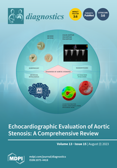
Aortic stenosis is the most common valvular heart disease and is expected to increase in prevalence due to the aging population. It can be promptly detected using echocardiography, which is the main diagnostic tool in the evaluation of patients undergoing surgical or transcatheter aortic valve replacement, both during the periprocedural phase and during the long-term follow-up. Aortic stenosis affects the entire cardiovascular system: echocardiography plays a key role in the identification of the related cardiac structural changes, which have prognostic implications. This review aims to provide an overview of the latest technical tools in echocardiography and the recent algorithms improving the diagnostic accuracy of aortic stenosis. View this paper
  • Issues are regarded as officially published after their release is announced to the table of contents alert mailing list.
  • You may sign up for e-mail alerts to receive table of contents of newly released issues.
  • PDF is the official format for papers published in both, html and pdf forms. To view the papers in pdf format, click on the "PDF Full-text" link, and use the free Adobe Reader to open them.
Order results
Result details
Section
Select all
Export citation of selected articles as:
12 pages, 14785 KiB  
Article
Diagnostic Performance of the Darth Vader Sign for the Diagnosis of Lumbar Spondylolysis in Routinely Acquired Abdominal CT
by Florian A. Huber, Cynthia S. Schmidt and Hatem Alkadhi
Diagnostics 2023, 13(15), 2616; https://doi.org/10.3390/diagnostics13152616 - 07 Aug 2023
Viewed by 1085
Abstract
Spondylolysis is underdiagnosed and often missed in non-musculoskeletal abdominal CT imaging. Our aim was to assess the inter-reader agreement and diagnostic performance of a novel “Darth Vader sign” for the detection of spondylolysis in routine axial images. We performed a retrospective search in [...] Read more.
Spondylolysis is underdiagnosed and often missed in non-musculoskeletal abdominal CT imaging. Our aim was to assess the inter-reader agreement and diagnostic performance of a novel “Darth Vader sign” for the detection of spondylolysis in routine axial images. We performed a retrospective search in the institutional report archives through keyword strings for lumbar spondylolysis and spondylolisthesis. Abdominal CTs from 53 spondylolysis cases (41% female) and from controls (n = 6) without spine abnormalities were identified. A total of 139 single axial slices covering the lumbar spine (86 normal images, 40 with spondylolysis, 13 with degenerative spondylolisthesis without spondylolysis) were exported. Two radiology residents rated all images for the presence or absence of the “Darth Vader sign”. The diagnostic accuracy for both readers, as well as the inter-reader agreement, was calculated. The “Darth Vader sign” showed an inter-reader agreement of 0.77. Using the “Darth Vader sign”, spondylolysis was detected with a sensitivity and specificity of 65.0–88.2% and 96.2–99.0%, respectively. The “Darth Vader sign” shows excellent diagnostic performance at a substantial inter-reader agreement for the detection of spondylolysis. Using the “Darth Vader sign” in the CT reading routine may be an easy yet effective tool to improve the detection rate of spondylolysis in non-musculoskeletal cases and hence improve patient care. Full article
(This article belongs to the Section Medical Imaging and Theranostics)
Show Figures

Figure 1

13 pages, 10106 KiB  
Article
Anterior Incisura Fibularis Corner Landmarks Can Safely Validate the Optimal Distal Tibiofibular Reduction in Malleolar Fractures—Prospective CT Study
by Meletis Rozis, Dimitrios Zachariou, Michalis Vavourakis, Elias Vasiliadis and John Vlamis
Diagnostics 2023, 13(15), 2615; https://doi.org/10.3390/diagnostics13152615 - 07 Aug 2023
Cited by 1 | Viewed by 737
Abstract
Background: Distal tibiofibular injuries are common in patients with malleolar fractures. Malreduction is frequently reported in the literature and is mainly caused by insufficient intraoperative radiological evaluation. In this direction, we performed a prospective observational study to validate the efficacy of the anatomical [...] Read more.
Background: Distal tibiofibular injuries are common in patients with malleolar fractures. Malreduction is frequently reported in the literature and is mainly caused by insufficient intraoperative radiological evaluation. In this direction, we performed a prospective observational study to validate the efficacy of the anatomical landmarks of the anterior incisura corner. Methods: Patients with malleolar fractures and syndesmotic instability were reduced according to specific anatomic landmarks and had a postoperative bilateral ankle CT. The quality of the reduction was compared to the healthy ankles. Results: None of the controlled parameters differed significantly between the operated and healthy ankles. Minor deviations were correlated to the normal incisura morphology rather than the reduction technique. Conclusions: The anterior incisura anatomical landmarks can be an efficient way of reducing the distal tibiofibular joint without the need for intraoperative radiological evaluation. Full article
(This article belongs to the Section Pathology and Molecular Diagnostics)
Show Figures

Figure 1

33 pages, 5252 KiB  
Review
Ultrasound-Based Image Analysis for Predicting Carotid Artery Stenosis Risk: A Comprehensive Review of the Problem, Techniques, Datasets, and Future Directions
by Najmath Ottakath, Somaya Al-Maadeed, Susu M. Zughaier, Omar Elharrouss, Hanadi Hassen Mohammed, Muhammad E. H. Chowdhury and Ahmed Bouridane
Diagnostics 2023, 13(15), 2614; https://doi.org/10.3390/diagnostics13152614 - 07 Aug 2023
Cited by 1 | Viewed by 2691
Abstract
The carotid artery is a major blood vessel that supplies blood to the brain. Plaque buildup in the arteries can lead to cardiovascular diseases such as atherosclerosis, stroke, ruptured arteries, and even death. Both invasive and non-invasive methods are used to detect plaque [...] Read more.
The carotid artery is a major blood vessel that supplies blood to the brain. Plaque buildup in the arteries can lead to cardiovascular diseases such as atherosclerosis, stroke, ruptured arteries, and even death. Both invasive and non-invasive methods are used to detect plaque buildup in the arteries, with ultrasound imaging being the first line of diagnosis. This paper presents a comprehensive review of the existing literature on ultrasound image analysis methods for detecting and characterizing plaque buildup in the carotid artery. The review includes an in-depth analysis of datasets; image segmentation techniques for the carotid artery plaque area, lumen area, and intima–media thickness (IMT); and plaque measurement, characterization, classification, and stenosis grading using deep learning and machine learning. Additionally, the paper provides an overview of the performance of these methods, including challenges in analysis, and future directions for research. Full article
(This article belongs to the Special Issue 2nd Edition: AI/ML-Based Medical Image Processing and Analysis)
Show Figures

Figure 1

12 pages, 1248 KiB  
Systematic Review
[18F]-FDHT PET for the Imaging of Androgen Receptor in Prostate and Breast Cancer: A Systematic Review
by Luca Filippi, Luca Urso, Orazio Schillaci and Laura Evangelista
Diagnostics 2023, 13(15), 2613; https://doi.org/10.3390/diagnostics13152613 - 07 Aug 2023
Cited by 2 | Viewed by 1194
Abstract
The aim of this systematic review is to provide a comprehensive overview of the role of fluoro-5α-dihydrotestosterone ([18F]-FDHT) for the in vivo imaging of androgen receptors (AR) through positron emission tomography (PET) in metastatic breast (mBC) and metastatic castration-resistant prostate cancer [...] Read more.
The aim of this systematic review is to provide a comprehensive overview of the role of fluoro-5α-dihydrotestosterone ([18F]-FDHT) for the in vivo imaging of androgen receptors (AR) through positron emission tomography (PET) in metastatic breast (mBC) and metastatic castration-resistant prostate cancer (mCRPC). Relevant studies published from 2013 up to May 2023 were selected by searching Scopus, PubMed and Web of Science. The selected imaging studies were analyzed using a modified version of the critical Appraisal Skills Programme (CASP). Eleven studies encompassing 321 patients were selected. Seven of the eleven selected papers included 266 subjects (82.2%) affected by mCRPC, while four encompassed 55 (17.2%) patients affected by mBC. [18F]-FDHT PET showed a satisfying test/retest reproducibility, and when compared to a histochemical analysis, it provided encouraging results for in vivo AR quantification both in mCRPC and mBC. [18F]-FDHT PET had a prognostic relevance in mCRPC patients submitted to AR-targeted therapy, while a clear association between [18F]-FDHT uptake and the bicalutamide response was not observed in women affected by AR-positive mBC. Further studies are needed to better define the role of [18F]-FDHT PET, alone or in combination with other tracers (i.e., [18F]-FDG/[18F]-FES), for patients’ selection and monitoring during AR-targeted therapy, especially in the case of mBC. Full article
Show Figures

Figure 1

15 pages, 2303 KiB  
Article
Fractional Flow Reserve-Guided Stent Optimisation in Focal and Diffuse Coronary Artery Disease
by Hirofumi Ohashi, Damien Collison, Takuya Mizukami, Matthaios Didagelos, Koshiro Sakai, Muhammad Aetesam-ur-Rahman, Daniel Munhoz, Peter McCartney, Thomas J. Ford, Mitchell Lindsay, Aadil Shaukat, Paul Rocchiccioli, Richard Brogan, Stuart Watkins, Margaret McEntegart, Richard Good, Keith Robertson, Patrick O’Boyle, Andrew Davie, Adnan Khan, Stuart Hood, Hany Eteiba, Tetsuya Amano, Jeroen Sonck, Colin Berry, Bernard De Bruyne, Keith G. Oldroyd and Carlos Colletadd Show full author list remove Hide full author list
Diagnostics 2023, 13(15), 2612; https://doi.org/10.3390/diagnostics13152612 - 07 Aug 2023
Viewed by 1294
Abstract
Assessing coronary physiology after stent implantation facilitates the optimisation of percutaneous coronary intervention (PCI). Coronary artery disease (CAD) patterns can be characterised by the pullback pressure gradient (PPG) index. The impact of focal vs. diffuse disease on physiology-guided incremental optimisation strategy (PIOS) is [...] Read more.
Assessing coronary physiology after stent implantation facilitates the optimisation of percutaneous coronary intervention (PCI). Coronary artery disease (CAD) patterns can be characterised by the pullback pressure gradient (PPG) index. The impact of focal vs. diffuse disease on physiology-guided incremental optimisation strategy (PIOS) is unknown. This is a sub-study of the TARGET-FFR randomized clinical trial (NCT03259815). The study protocol directed that optimisation be attempted for patients in the PIOS arm when post-PCI FFR was <0.90. Overall, 114 patients (n = 61 PIOS and 53 controls) with both pre-PCI fractional flow reserve (FFR) pullbacks and post-PCI FFR were included. A PPG ≥ 0.74 defined focal CAD. The PPG correlated significantly with post-PCI FFR (r = 0.43; 95% CI 0.26 to 0.57; p-value < 0.001) and normalised delta FFR (r = 0.49; 95% CI 0.34 to 0.62; p-value < 0.001). PIOS was more frequently applied to vessels with diffuse CAD (6% focal vs. 42% diffuse; p-value = 0.006). In patients randomized to PIOS, those with focal disease achieved higher post-PCI FFR than patients with diffuse CAD (0.93 ± 0.05 vs. 0.83 ± 0.07, p < 0.001). There was a significant interaction between CAD patterns and the randomisation arm for post-PCI FFR (p-value for interaction = 0.004). Physiology-guided stent optimisation was applied more frequently to vessels with diffuse disease; however, patients with focal CAD at baseline achieved higher post-PCI FFR. Full article
Show Figures

Figure 1

26 pages, 4083 KiB  
Review
Advanced Cardiac Imaging and Women’s Chest Pain: A Question of Gender
by Federica Dell’Aversana, Carlo Tedeschi, Rosita Comune, Luigi Gallo, Giovanni Ferrandino, Emilia Basco, Stefania Tamburrini, Giacomo Sica, Salvatore Masala, Mariano Scaglione and Carlo Liguori
Diagnostics 2023, 13(15), 2611; https://doi.org/10.3390/diagnostics13152611 - 07 Aug 2023
Viewed by 1163
Abstract
Awareness of gender differences in cardiovascular disease (CVD) has increased: both the different impact of traditional cardiovascular risk factors on women and the existence of sex-specific risk factors have been demonstrated. Therefore, it is essential to recognize typical aspects of ischemic heart disease [...] Read more.
Awareness of gender differences in cardiovascular disease (CVD) has increased: both the different impact of traditional cardiovascular risk factors on women and the existence of sex-specific risk factors have been demonstrated. Therefore, it is essential to recognize typical aspects of ischemic heart disease (IHD) in women, who usually show a lower prevalence of obstructive coronary artery disease (CAD) as a cause of acute coronary syndrome (ACS). It is also important to know how to recognize pathologies that can cause acute chest pain with a higher incidence in women, such as spontaneous coronary artery dissection (SCAD) and myocardial infarction with non-obstructive coronary arteries (MINOCA). Coronary computed tomography angiography (CCTA) and cardiac magnetic resonance imaging (CMR) gained a pivotal role in the context of cardiac emergencies. Thus, the aim of our review is to investigate the most frequent scenarios in women with acute chest pain and how advanced cardiac imaging can help in the management and diagnosis of ACS. Full article
(This article belongs to the Section Medical Imaging and Theranostics)
Show Figures

Figure 1

15 pages, 4458 KiB  
Article
Hybrid Majority Voting: Prediction and Classification Model for Obesity
by Dahlak Daniel Solomon, Shakir Khan, Sonia Garg, Gaurav Gupta, Abrar Almjally, Bayan Ibrahimm Alabduallah, Hatoon S. Alsagri, Mandour Mohamed Ibrahim and Alsadig Mohammed Adam Abdallah
Diagnostics 2023, 13(15), 2610; https://doi.org/10.3390/diagnostics13152610 - 07 Aug 2023
Cited by 3 | Viewed by 1772
Abstract
Because it is associated with most multifactorial inherited diseases like heart disease, hypertension, diabetes, and other serious medical conditions, obesity is a major global health concern. Obesity is caused by hereditary, physiological, and environmental factors, as well as poor nutrition and a lack [...] Read more.
Because it is associated with most multifactorial inherited diseases like heart disease, hypertension, diabetes, and other serious medical conditions, obesity is a major global health concern. Obesity is caused by hereditary, physiological, and environmental factors, as well as poor nutrition and a lack of exercise. Weight loss can be difficult for various reasons, and it is diagnosed via BMI, which is used to estimate body fat for most people. Muscular athletes, for example, may have a BMI in the obesity range even when they are not obese. Researchers from a variety of backgrounds and institutions devised different hypotheses and models for the prediction and classification of obesity using different approaches and various machine learning techniques. In this study, a majority voting-based hybrid modeling approach using a gradient boosting classifier, extreme gradient boosting, and a multilayer perceptron was developed. Seven distinct machine learning algorithms were used on open datasets from the UCI machine learning repository, and their respective accuracy levels were compared before the combined approaches were chosen. The proposed majority voting-based hybrid model for prediction and classification of obesity that was achieved has an accuracy of 97.16%, which is greater than both the individual models and the other hybrid models that have been developed. Full article
(This article belongs to the Special Issue Predictive Modelling in Healthcare)
Show Figures

Figure 1

10 pages, 638 KiB  
Review
The Role of Ultrasonography in Hip Impingement Syndromes: A Narrative Review
by Panagiotis Karampinas, Athanasios Galanis, John Vlamis, Michail Vavourakis, Eftychios Papagrigorakis, Evangelos Sakellariou, Dimitrios Zachariou, Spyridon Karampitianis, Elias Vasiliadis and Spiros Pneumaticos
Diagnostics 2023, 13(15), 2609; https://doi.org/10.3390/diagnostics13152609 - 07 Aug 2023
Cited by 1 | Viewed by 1811
Abstract
Hip pain is indubitably a frequent clinical problem deriving from copious etiologies. Hip impingement syndromes are one of the most prevalent causes of persistent groin pain, especially in young and active patients. Diligent imaging of the hip region is indispensable to discern femoroacetabular [...] Read more.
Hip pain is indubitably a frequent clinical problem deriving from copious etiologies. Hip impingement syndromes are one of the most prevalent causes of persistent groin pain, especially in young and active patients. Diligent imaging of the hip region is indispensable to discern femoroacetabular impingement, as the differential diagnosis of hip pain can be exceedingly arduous. Despite hip radiography being plain and broadly attainable, it offers narrow information concerning soft tissue pathologies around the hip joint (extra-articular hip impingement syndromes). Magnetic resonance imaging and arthrography remain the gold standard examination for detecting intra-articular pathologies; however, they are widely considered expensive, time-consuming and characterized by confined. Consequently, ultrasonography has emerged as an alternative valuable diagnostic tool for distinguishing the underlying abnormalities that trigger femoroacetabular impingement. Proper hip ultrasound examination provides dynamic assessment, while also beneficial for guided intervention around the hip joint. Ultrasound hip examination is exacting due to its complex regional anatomy and deep location. It is capable of providing detailed information about various hip quadrants. An adept operator can identify both intra-articular and extra-articular pathologies. In addition, with ultrasonography, hip injections have been rendered relatively undemanding, aiding in therapeutic and diagnostic purposes. This paper aims to provide a succinct and compendious review of the existing literature, accentuating the crucial role of ultrasonography in diagnosing hip impingement syndromes and determining whether an additional examination is required regarding distinguishing between intra-articular and extra-articular syndromes. Full article
(This article belongs to the Special Issue Clinical Diagnosis of Joints and Ligamentous Injury)
Show Figures

Figure 1

11 pages, 1398 KiB  
Article
Perilesional Targeted Biopsy Combined with MRI-TRUS Image Fusion-Guided Targeted Prostate Biopsy: An Analysis According to PI-RADS Scores
by Masayuki Tomioka, Kensaku Seike, Hiromi Uno, Nami Asano, Haruo Watanabe, Risa Tomioka-Inagawa, Makoto Kawase, Daiki Kato, Manabu Takai, Koji Iinuma, Yuki Tobisawa, Keita Nakane, Kunihiro Tsuchiya, Takayasu Ito and Takuya Koie
Diagnostics 2023, 13(15), 2608; https://doi.org/10.3390/diagnostics13152608 - 07 Aug 2023
Viewed by 883
Abstract
A prostate-targeted biopsy (TB) core is usually collected from a site where magnetic resonance imaging (MRI) indicates possible cancer. However, the extent of the lesion is difficult to accurately predict using MRI or TB alone. Therefore, we performed several biopsies around the TB [...] Read more.
A prostate-targeted biopsy (TB) core is usually collected from a site where magnetic resonance imaging (MRI) indicates possible cancer. However, the extent of the lesion is difficult to accurately predict using MRI or TB alone. Therefore, we performed several biopsies around the TB site (perilesional [p] TB) and analyzed the association between the positive cores obtained using TB and pTB and the Prostate Imaging Reporting and Data System (PI-RADS) scores. This retrospective study included patients who underwent prostate biopsies. The extent of pTB was defined as the area within 10 mm of a TB site. A total of 162 eligible patients were enrolled. Prostate cancer (PCa) was diagnosed in 75.2% of patients undergoing TB, with a positivity rate of 50.7% for a PI-RADS score of 3, 95.8% for a PI-RADS score of 4, and 100% for a PI-RADS score of 5. Patients diagnosed with PCa according to both TB and pTB had significantly higher positivity rates for PI-RADS scores of 4 and 5 than for a PI-RADS score of 3 (p < 0.0001 and p = 0.0009, respectively). Additional pTB may be performed in patients with PI-RADS ≥ 4 regions of interest for assessing PCa malignancy. Full article
(This article belongs to the Special Issue Imaging-Based Diagnosis of Prostate Cancer: State of the Art)
Show Figures

Figure 1

12 pages, 2875 KiB  
Article
Optical Coherence Tomography Angiography Is a Useful Tool for Distinguishing Primary Raynaud’s Phenomenon from Systemic Sclerosis and/or Very Early Disease of Systemic Sclerosis
by Adem Erturk, Ozgur Erogul and Murat Kasikci
Diagnostics 2023, 13(15), 2607; https://doi.org/10.3390/diagnostics13152607 - 05 Aug 2023
Cited by 2 | Viewed by 1822
Abstract
This cross-sectional study aimed to compare optical coherence tomography angiography (OCT-A) findings in patients with primary Raynaud’s phenomenon (PRP; n = 22), very early disease of systemic sclerosis (VEDOSS; n = 19), and systemic sclerosis (SSc; 25 patients with limited cutaneous SSc (lcSSc) [...] Read more.
This cross-sectional study aimed to compare optical coherence tomography angiography (OCT-A) findings in patients with primary Raynaud’s phenomenon (PRP; n = 22), very early disease of systemic sclerosis (VEDOSS; n = 19), and systemic sclerosis (SSc; 25 patients with limited cutaneous SSc (lcSSc) and 13 patients with diffuse cutaneous SSc (dcSSc)). Whole, parafoveal, and perifoveal superficial capillary plexus (SCP) vessel densities (VDs), deep capillary plexus VDs, and whole, inside, and peripapillary VDs were significantly higher in the PRP group (p < 0.001). In the lcSSc group, the FAZ perimeter was significantly higher than that in the VEDOSS group (p = 0.017). Retinal nerve fiber layer VDs were significantly lower in the lcSSc group than in the PRP and VEDOSS groups (p < 0.001). The whole and peripapillary optic disc VDs of the VEDOSS group were significantly higher than in the lcSSc group (p < 0.001). Whole SCP VDs (94.74% sensitivity, 100.00% specificity) and parafoveal SCP VDs (89.47% sensitivity, 100.00% specificity) showed the best performance in distinguishing patients with SSc from those with PRP. OCT-A seems to have potential diagnostic value in differentiating patients with PRP from patients with SSc and VEDOSS, and there is potential value in assessing prognostic roles, since findings from OCT-A images could be early indicators of retinal vascular injury long before overt SSc symptoms develop. Full article
(This article belongs to the Section Optical Diagnostics)
Show Figures

Figure 1

19 pages, 4549 KiB  
Article
Automatic Detection and Classification of Diabetic Retinopathy Using the Improved Pooling Function in the Convolution Neural Network
by Usharani Bhimavarapu, Nalini Chintalapudi and Gopi Battineni
Diagnostics 2023, 13(15), 2606; https://doi.org/10.3390/diagnostics13152606 - 05 Aug 2023
Cited by 3 | Viewed by 1593
Abstract
Diabetic retinopathy (DR) is an eye disease associated with diabetes that can lead to blindness. Early diagnosis is critical to ensure that patients with diabetes are not affected by blindness. Deep learning plays an important role in diagnosing diabetes, reducing the human effort [...] Read more.
Diabetic retinopathy (DR) is an eye disease associated with diabetes that can lead to blindness. Early diagnosis is critical to ensure that patients with diabetes are not affected by blindness. Deep learning plays an important role in diagnosing diabetes, reducing the human effort to diagnose and classify diabetic and non-diabetic patients. The main objective of this study was to provide an improved convolution neural network (CNN) model for automatic DR diagnosis from fundus images. The pooling function increases the receptive field of convolution kernels over layers. It reduces computational complexity and memory requirements because it reduces the resolution of feature maps while preserving the essential characteristics required for subsequent layer processing. In this study, an improved pooling function combined with an activation function in the ResNet-50 model was applied to the retina images in autonomous lesion detection with reduced loss and processing time. The improved ResNet-50 model was trained and tested over the two datasets (i.e., APTOS and Kaggle). The proposed model achieved an accuracy of 98.32% for APTOS and 98.71% for Kaggle datasets. It is proven that the proposed model has produced greater accuracy when compared to their state-of-the-art work in diagnosing DR with retinal fundus images. Full article
(This article belongs to the Special Issue Artificial Intelligence in Clinical Medical Imaging)
Show Figures

Figure 1

15 pages, 2217 KiB  
Article
Machine Learning Approach for the Prediction of In-Hospital Mortality in Traumatic Brain Injury Using Bio-Clinical Markers at Presentation to the Emergency Department
by Ahammed Mekkodathil, Ayman El-Menyar, Mashhood Naduvilekandy, Sandro Rizoli and Hassan Al-Thani
Diagnostics 2023, 13(15), 2605; https://doi.org/10.3390/diagnostics13152605 - 05 Aug 2023
Cited by 1 | Viewed by 1552
Abstract
Background: Accurate prediction of in-hospital mortality is essential for better management of patients with traumatic brain injury (TBI). Machine learning (ML) algorithms have been shown to be effective in predicting clinical outcomes. This study aimed to identify predictors of in-hospital mortality in TBI [...] Read more.
Background: Accurate prediction of in-hospital mortality is essential for better management of patients with traumatic brain injury (TBI). Machine learning (ML) algorithms have been shown to be effective in predicting clinical outcomes. This study aimed to identify predictors of in-hospital mortality in TBI patients using ML algorithms. Materials and Method: A retrospective study was performed using data from both the trauma registry and electronic medical records among TBI patients admitted to the Hamad Trauma Center in Qatar between June 2016 and May 2021. Thirteen features were selected for four ML models including a Support Vector Machine (SVM), Logistic Regression (LR), Random Forest (RF), and Extreme Gradient Boosting (XgBoost), to predict the in-hospital mortality. Results: A dataset of 922 patients was analyzed, of which 78% survived and 22% died. The AUC scores for SVM, LR, XgBoost, and RF models were 0.86, 0.84, 0.85, and 0.86, respectively. XgBoost and RF had good AUC scores but exhibited significant differences in log loss between the training and testing sets (% difference in logloss of 79.5 and 41.8, respectively), indicating overfitting compared to the other models. The feature importance trend across all models indicates that aPTT, INR, ISS, prothrombin time, and lactic acid are the most important features in prediction. Magnesium also displayed significant importance in the prediction of mortality among serum electrolytes. Conclusions: SVM was found to be the best-performing ML model in predicting the mortality of TBI patients. It had the highest AUC score and did not show overfitting, making it a more reliable model compared to LR, XgBoost, and RF. Full article
Show Figures

Figure 1

13 pages, 2678 KiB  
Article
Prediction of Glioma Grade and IDH Status Using 18F-FET PET/CT Dynamic and Multiparametric Texture Analysis
by Rami Hajri, Marie Nicod-Lalonde, Andreas F. Hottinger, John O. Prior and Vincent Dunet
Diagnostics 2023, 13(15), 2604; https://doi.org/10.3390/diagnostics13152604 - 05 Aug 2023
Cited by 2 | Viewed by 1165
Abstract
Mutations in isocitrate dehydrogenase (IDH) represent an independent predictor of better survival in patients with gliomas. We aimed to assess grade and IDH mutation status in patients with untreated gliomas, by evaluating the respective value of 18F-FET PET/CT via dynamic and texture [...] Read more.
Mutations in isocitrate dehydrogenase (IDH) represent an independent predictor of better survival in patients with gliomas. We aimed to assess grade and IDH mutation status in patients with untreated gliomas, by evaluating the respective value of 18F-FET PET/CT via dynamic and texture analyses. A total of 73 patients (male: 48, median age: 47) who underwent an 18F-FET PET/CT for initial glioma evaluation were retrospectively included. IDH status was available in 61 patients (20 patients with WHO grade 2 gliomas, 41 with grade 3–4 gliomas). Time–activity curve type and 20 parameters obtained from static analysis using LIFEx© v6.30 software were recorded. Respective performance was assessed using receiver operating characteristic curve analysis and stepwise multivariate regression analysis adjusted for patients’ age and sex. The time–activity curve type and texture parameters derived from the static parameters showed satisfactory-to-good performance in predicting glioma grade and IDH status. Both time–activity curve type (stepwise OR: 101.6 (95% CI: 5.76–1791), p = 0.002) and NGLDM coarseness (stepwise OR: 2.08 × 1043 (95% CI: 2.76 × 10¹²–1.57 × 1074), p = 0.006) were independent predictors of glioma grade. No independent predictor of IDH status was found. Dynamic and texture analyses of 18F-FET PET/CT have limited predictive value for IDH status when adjusted for confounding factors. However, they both help predict glioma grade. Full article
(This article belongs to the Special Issue Advanced Brain Tumor Imaging)
Show Figures

Figure 1

15 pages, 6714 KiB  
Review
Cutaneous Metastasis of Endometrial Cancer and Long-Term Survival: A Scoping Review and Our Experience
by Alexandra Nienhaus, Rahavie Rajakulendran and Elena Bernad
Diagnostics 2023, 13(15), 2603; https://doi.org/10.3390/diagnostics13152603 - 04 Aug 2023
Cited by 1 | Viewed by 1191
Abstract
Background and Objectives: Cutaneous and soft tissue metastases of endometrial cancer are rare. This review aims to examine the prevalence of cutaneous metastasis, the diagnosis and treatment options, and the impact of cutaneous metastasis of endometrial cancer on overall survival. We also present [...] Read more.
Background and Objectives: Cutaneous and soft tissue metastases of endometrial cancer are rare. This review aims to examine the prevalence of cutaneous metastasis, the diagnosis and treatment options, and the impact of cutaneous metastasis of endometrial cancer on overall survival. We also present a particular case with a long-term overall survival. Materials and Methods: A systematic literature search was conducted on PubMed and PubMed Central using the following keywords: endometrial carcinoma metastasis, cutaneous metastasis, and five-year overall survival. Results: We identified 326 results and checked their titles for eligibility. There were eight studies included. We also presented a case of a 51-year-old woman with cutaneous metastasis and a large soft tissue metastasis with a prolonged overall survival of about 13 years after the appearance of the first cutaneous metastasis. Conclusions: This paper highlights the importance of skin evaluations in patients diagnosed with endometrial cancer. Healthcare providers must consider the possibility of cutaneous metastasis localization in patients with endometrial cancer to assign the correct stage and apply the appropriate treatment to increase long-term survival. Full article
(This article belongs to the Special Issue Diagnosis and Management of Gynecological Cancers: Volume 2)
Show Figures

Figure 1

14 pages, 798 KiB  
Review
Diabetic Ketoacidosis in Children and Adolescents; Diagnostic and Therapeutic Pitfalls
by Eirini Kostopoulou, Xenophon Sinopidis, Sotirios Fouzas, Despoina Gkentzi, Theodore Dassios, Stylianos Roupakias and Gabriel Dimitriou
Diagnostics 2023, 13(15), 2602; https://doi.org/10.3390/diagnostics13152602 - 04 Aug 2023
Cited by 2 | Viewed by 3427
Abstract
Diabetic ketoacidosis (DKA) represents an acute, severe complication of relative insulin deficiency and a common presentation of Type 1 Diabetes Mellitus (T1DM) primarily and, occasionally, Type 2 Diabetes Mellitus (T2DM) in children and adolescents. It is characterized by the biochemical triad of hyperglycaemia, [...] Read more.
Diabetic ketoacidosis (DKA) represents an acute, severe complication of relative insulin deficiency and a common presentation of Type 1 Diabetes Mellitus (T1DM) primarily and, occasionally, Type 2 Diabetes Mellitus (T2DM) in children and adolescents. It is characterized by the biochemical triad of hyperglycaemia, ketonaemia and/or ketonuria, and acidaemia. Clinical symptoms include dehydration, tachypnoea, gastrointestinal symptoms, and reduced level of consciousness, precipitated by a variably long period of polyuria, polydipsia, and weight loss. The present review aims to summarize potential pitfalls in the diagnosis and management of DKA. A literature review was conducted using the Pubmed/Medline and Scopus databases including articles published from 2000 onwards. Diagnostic challenges include differentiating between T1DM and T2DM, between DKA and hyperosmolar hyperglycaemic state (HHS), and between DKA and alternative diagnoses presenting with overlapping symptoms, such as pneumonia, asthma exacerbation, urinary tract infection, gastroenteritis, acute abdomen, and central nervous system infection. The mainstays of DKA management include careful fluid resuscitation, timely intravenous insulin administration, restoration of shifting electrolyte disorders and addressing underlying precipitating factors. However, evidence suggests that optimal treatment remains a therapeutic challenge. Accurate and rapid diagnosis, prompt intervention, and meticulous monitoring are of major importance to break the vicious cycle of life-threatening events and prevent severe complications during this potentially fatal medical emergency. Full article
(This article belongs to the Special Issue Diagnosis and Treatment of Pediatric Emergencies)
Show Figures

Figure 1

26 pages, 53595 KiB  
Interesting Images
Infraspinatus Fascial Dysfunction as a Cause of Painful Anterior Shoulder Snapping: Its Visualization via Dynamic Ultrasound and Its Resolution via Diagnostic Ultrasound-Guided Injection
by King Hei Stanley Lam, Daniel Chiung Jui Su, Yung-Tsan Wu, Mario Fajardo Pérez, Kenneth Dean Reeves, Philip Peng and Bradley Fullerton
Diagnostics 2023, 13(15), 2601; https://doi.org/10.3390/diagnostics13152601 - 04 Aug 2023
Cited by 1 | Viewed by 5998
Abstract
This report presents the first case of painful anterior shoulder snapping due to a thickened, fibrotic bursa snapping between the subscapularis and the short head of the bicep during external and internal rotation of the humerus. A 46-year-old presented with a 10-month history [...] Read more.
This report presents the first case of painful anterior shoulder snapping due to a thickened, fibrotic bursa snapping between the subscapularis and the short head of the bicep during external and internal rotation of the humerus. A 46-year-old presented with a 10-month history of on-and-off anterolateral right shoulder pain and snapping. Direct treatment to the anterior suspected lesions partially and temporarily relieved the pain but did not reduce the snapping. Further musculoskeletal examination and dynamic ultrasound scanning showed dysfunction in the scapulothoracic movement and defects of the muscles that interact with the infraspinatus aponeurotic fascia. An ultrasound-guided diagnostic injection to the suspected lesions in the infraspinatus fascia and its muscles attachments improved the scapulothoracic movement, and the snapping and pain were eliminated immediately after the injection, which further shows that the defects in the infraspinatus fascia may be the root cause of the painful anterolateral snapping. The importance of the infraspinatus fascia and its related muscle in maintaining the harmony of the scapulothoracic movement and flexibility of the shoulder is considerable. Full article
Show Figures

Figure 1

4 pages, 5647 KiB  
Interesting Images
The Usefulness of Magnetic Resonance Imaging in the Management of Acute Phlegmonous Esophagitis
by Motohiro Shimizu, Hiroshi Adachi and Kenichi Sai
Diagnostics 2023, 13(15), 2600; https://doi.org/10.3390/diagnostics13152600 - 04 Aug 2023
Viewed by 703
Abstract
Acute phlegmonous esophagitis is a rare life-threatening disease that often requires surgical intervention in case of complications, including esophageal abscess, perforation, or mediastinitis. We present a case of acute phlegmonous esophagitis, in which magnetic resonance imaging (MRI) proved useful in planning the treatment [...] Read more.
Acute phlegmonous esophagitis is a rare life-threatening disease that often requires surgical intervention in case of complications, including esophageal abscess, perforation, or mediastinitis. We present a case of acute phlegmonous esophagitis, in which magnetic resonance imaging (MRI) proved useful in planning the treatment strategy. An 89-year-old woman was admitted to the emergency department with painful swallowing and respiratory distress. She was diagnosed with acute phlegmonous esophagitis and a hypopharyngeal abscess based on computed tomography (CT) findings. However, there was a discrepancy between the clinical course and CT findings. Given the improvement of the patient’s condition with conservative treatment with ampicillin/sulbactam, the CT findings suggested an apparent abscess due to increased esophageal wall thickness. However, MR diffusion-weighted images showed a slightly high-intensity signal, suggesting that the enlargement was due to edema rather than an abscess. The patient recovered successfully following conservative treatment. Thus, our findings demonstrate the utility of MRI in the treatment planning of acute phlegmonous esophagitis, especially in cases with unreliable contrast-enhanced CT findings. However, future studies are warranted to explore the utility of MRI in the management of such cases. Full article
(This article belongs to the Special Issue Diagnostic Imaging in Gastrointestinal Diseases)
Show Figures

Figure 1

13 pages, 3678 KiB  
Article
Comparative Effectiveness of Two Models of Point-of-Care Ultrasound for Detection of Post-Void Residual Urine during Acute Ischemic Stroke: Preliminary Findings of Real-World Clinical Application
by Wan-Ling Chang, Shu-Hui Lai, Chu-Fang Cheng, Valeria Chiu and Shinn-Kuang Lin
Diagnostics 2023, 13(15), 2599; https://doi.org/10.3390/diagnostics13152599 - 04 Aug 2023
Viewed by 1440
Abstract
We conducted a comparative study of two models of point-of-care ultrasound devices for measuring post-void residual urine (PVRU). We prospectively enrolled 55 stroke inpatients who underwent both real-time B-mode ultrasound (Device A) and automated three-dimensional (3D) scanning ultrasound (Device B), with a total [...] Read more.
We conducted a comparative study of two models of point-of-care ultrasound devices for measuring post-void residual urine (PVRU). We prospectively enrolled 55 stroke inpatients who underwent both real-time B-mode ultrasound (Device A) and automated three-dimensional (3D) scanning ultrasound (Device B), with a total of 108 measurements. The median PVRU volume of Device B was 40 mL larger than that of Device A. The PVRU difference between the devices was positively and linearly correlated with PVRU. The correlation of PVRU volume between the devices was strong, but the agreement level was only moderate. Measurement deviations were observed in 43 (40%) and 11 (10%) measurements with Device B and Device A, respectively. The PVRU volume was low in spherical bladder shapes but sequentially increased in triangular, undefined, ellipsoid, and cuboid bladder shapes. Further comparison of 60 sets of PVRU without measurement deviations revealed higher agreements between the devices at correction coefficients of 0.52, 0.66, and 0.81 for PVRU volumes of <100, 100–200, and >200 mL, respectively. The automated 3D scanning ultrasound is more convenient for learning and scanning, but it exhibits larger measurement deviations. Real-time B-mode ultrasound accurately visualizes the urinary bladder but tends to underestimate the urinary bladder when the PVRU volume is large. Hence, real-time B-mode ultrasound with automated PVRU-based adjustment of calculation formulas may be a better solution for estimating bladder volume. Full article
(This article belongs to the Special Issue Risk Factors for Acute Ischemic Stroke)
Show Figures

Figure 1

11 pages, 501 KiB  
Review
Advanced CMR Techniques in Anderson-Fabry Disease: State of the Art
by Andrea Ponsiglione, Marco De Giorgi, Raffaele Ascione, Carmela Nappi, Luca Sanduzzi, Antonio Pisani, Serena Dell’Aversana, Alberto Cuocolo and Massimo Imbriaco
Diagnostics 2023, 13(15), 2598; https://doi.org/10.3390/diagnostics13152598 - 04 Aug 2023
Cited by 3 | Viewed by 1165
Abstract
Anderson-Fabry disease (AFD) is a rare multisystem X-linked lysosomal storage disorder caused by α-galactosidase A enzyme deficiency. Long-term cardiac involvement in AFD results in left ventricular hypertrophy and myocardial fibrosis, inducing several complications, mainly arrhythmias, valvular dysfunction, and coronary artery disease. Cardiac magnetic [...] Read more.
Anderson-Fabry disease (AFD) is a rare multisystem X-linked lysosomal storage disorder caused by α-galactosidase A enzyme deficiency. Long-term cardiac involvement in AFD results in left ventricular hypertrophy and myocardial fibrosis, inducing several complications, mainly arrhythmias, valvular dysfunction, and coronary artery disease. Cardiac magnetic resonance (CMR) represents the predominant noninvasive imaging modality for the assessment of cardiac involvement in the AFD, being able to comprehensively assess cardiac regional anatomy, ventricular function as well as to provide tissue characterization. This review aims to explore the role of the most advanced CMR techniques, such as myocardial strain, T1 and T2 mapping, perfusion and hybrid imaging, as diagnostic and prognostic biomarkers. Full article
Show Figures

Figure 1

17 pages, 821 KiB  
Article
Prognosis Predictive Markers in Patients with Chronic Obstructive Pulmonary Disease and COVID-19
by Nicoleta Ștefania Motoc, Iulia Făgărășan, Andrada Elena Urda-Cîmpean and Doina Adina Todea
Diagnostics 2023, 13(15), 2597; https://doi.org/10.3390/diagnostics13152597 - 04 Aug 2023
Cited by 3 | Viewed by 1023
Abstract
Some studies have reported that chronic respiratory illnesses in patients with COVID-19 result in an increase in hospitalization and death rates, while other studies reported to the contrary. The present research aims to determine if a predictive model (developed by combing different clinical, [...] Read more.
Some studies have reported that chronic respiratory illnesses in patients with COVID-19 result in an increase in hospitalization and death rates, while other studies reported to the contrary. The present research aims to determine if a predictive model (developed by combing different clinical, imaging, or blood markers) could be established for patients with both chronic obstructive pulmonary disease (COPD) and COVID-19, in order to be able to foresee the outcomes of these patients. A prospective observational cohort of 165 patients with both diseases was analyzed in terms of clinical characteristics, blood tests, and chest computed tomography results. The beta-coefficients from the logistic regression were used to create a score based on the significant identified markers for poor outcomes (transfers to an intensive care unit (ICU) for mechanical ventilation, or death). The severity of COVID-19, renal failure, diabetes, smoking status (current or previous), the requirement for oxygen therapy upon admission, high lactate dehydrogenase (LDH) and C-reactive protein level (CRP readings), and low eosinophil and lymphocyte counts were all identified as being indicators of a poor prognosis. Higher mortality was linked to the occurrence of renal failure, the number of affected lobes, the need for oxygen therapy upon hospital admission, high LDH, and low lymphocyte levels. Patients had an 86.4% chance of dying if their mortality scores were −2.80 or lower, based on the predictive model. The factors that were linked to a poor prognosis in patients who had both COPD and COVID-19 were the same as those that were linked to a poor prognosis in patients who had only COVID-19. Full article
(This article belongs to the Special Issue Evaluating Novel Biomarkers for Personalized Medicine)
Show Figures

Figure 1

11 pages, 1798 KiB  
Article
Loss or Dilution—A New Diagnostic Method to Assess the Impact of Dilution on Standard Laboratory Parameters
by Nicole Innerhofer, Sasa Rajsic, Marco Ronzani, Robert Breitkopf, Can Gollmann Tepeköylü, Corinna Velik-Salchner, Lisa Schlosser, Dietmar Fries, Werner Streif, Michael Schirmer and Judith Martini
Diagnostics 2023, 13(15), 2596; https://doi.org/10.3390/diagnostics13152596 - 04 Aug 2023
Viewed by 879
Abstract
Intraoperative fluid therapy is regularly used in patients undergoing cardiac surgery procedures with cardiopulmonary bypass (CPB). Although fluid administration has several advantages, it unavoidably leads to hemodilution. The hemodilution may further influence the interpretation of concentration-based laboratory parameters like hemoglobin (Hgb), platelet count [...] Read more.
Intraoperative fluid therapy is regularly used in patients undergoing cardiac surgery procedures with cardiopulmonary bypass (CPB). Although fluid administration has several advantages, it unavoidably leads to hemodilution. The hemodilution may further influence the interpretation of concentration-based laboratory parameters like hemoglobin (Hgb), platelet count (PLT) or prothrombin time (PT). These all parameters are commonly used to guide blood product substitution. To assess the impact of dilution on these values, we performed a prospective observational study in 174 patients undergoing elective cardiac surgery. We calculated the total blood volume according to Nadler’s formula, and fluid therapy was correlated with a newly developed dilution coefficient formula at the end of CPB. Intravenously applied fluids were measured from the beginning of the anesthesia (baseline, T0) and 15 min after the end of protamine infusion (end of CPB, T1). The amount of the administered volume (crystalloids or colloids) was calculated according to the percentage of the intravascular fluid effect, and intraoperative diuresis was further subtracted. The median blood volume increased by 148% in all patients at T1 compared to the calculated total blood volume at T0. This led to a dilution-dependent decrease of 38% in all three parameters (Hgb 24%, corrCoeff = 0.53; PLT 41%, corrCoeff = 0.68; PT 44%, corrCoeff = 0.54). The dilution-correlated decrease was significant for all parameters (p < 0.001), and the effect was independent from the duration of CPB. We conclude that the presented calculation-based approach could provide important information regarding actual laboratory parameters and may help in the guidance of the blood product substitution and potential transfusion thresholds. Further research on the impact of dilution and related decision-making for blood product substitution, including its impact on morbidity and mortality, is warranted. Full article
(This article belongs to the Section Medical Imaging and Theranostics)
Show Figures

Figure 1

17 pages, 826 KiB  
Systematic Review
The Applications of High-Intensity Focused Ultrasound (HIFU) Ablative Therapy in the Treatment of Primary Breast Cancer: A Systematic Review
by Dania Zulkifli, Hanani Abdul Manan, Noorazrul Yahya and Hamzaini Abdul Hamid
Diagnostics 2023, 13(15), 2595; https://doi.org/10.3390/diagnostics13152595 - 04 Aug 2023
Cited by 7 | Viewed by 2304
Abstract
Background: This study evaluates the role of high-intensity focused ultrasound (HIFU) ablative therapy in treating primary breast cancer. Methods: PubMed and Scopus databases were searched according to the PRISMA guidelines to identify studies from 2002 to November 2022. Eligible studies were selected based [...] Read more.
Background: This study evaluates the role of high-intensity focused ultrasound (HIFU) ablative therapy in treating primary breast cancer. Methods: PubMed and Scopus databases were searched according to the PRISMA guidelines to identify studies from 2002 to November 2022. Eligible studies were selected based on criteria such as experimental study type, the use of HIFU therapy as a treatment for localised breast cancer with objective clinical evaluation, i.e., clinical, radiological, and pathological outcomes. Nine studies were included in this study. Results: Two randomised controlled trials and seven non-randomised clinical trials fulfilled the inclusion criteria. The percentage of patients who achieved complete (100%) coagulation necrosis varied from 17% to 100% across all studies. Eight of the nine studies followed the treat-and-resect protocol in which HIFU-ablated tumours were surgically resected for pathological evaluation. Most breast cancers were single, solitary, and palpable breast tumours. Haematoxylin and eosin stains used for histopathological evaluation showed evidence of coagulation necrosis. Radiological evaluation by MRI showed an absence of contrast enhancement in the HIFU-treated tumour and 1.5 to 2 cm of normal breast tissue, with a thin peripheral rim of enhancement indicative of coagulation necrosis. All studies did not report severe complications, i.e., haemorrhage and infection. Common complications related to HIFU ablation were local mammary oedema, pain, tenderness, and mild to moderate burns. Only one third-degree burn was reported. Generally, the cosmetic outcome was good. The five-year disease-free survival rate was 95%, as reported in two RCTs. Conclusions: HIFU ablation can induce tumour coagulation necrosis in localised breast cancer, with a favourable safety profile and cosmetic outcome. However, there is variable evidence of complete coagulation necrosis in the HIFU-treated tumour. Histopathological evidence of coagulation necrosis has been inconsistent, and there is no reliable radiological modality to assess coagulation necrosis confidently. Further exploration is needed to establish the accurate ablation margin with a reliable radiological modality for treatment and follow-up. HIFU therapy is currently limited to single, palpable breast tumours. More extensive and randomised clinical trials are needed to evaluate HIFU therapy for breast cancer, especially where the tumour is left in situ. Full article
(This article belongs to the Special Issue Advances in Breast Radiology)
Show Figures

Figure 1

10 pages, 833 KiB  
Article
Laser-Based 3D Body Scanning Reveals a Higher Prevalence of Abdominal Obesity than Tape Measurements: Results from a Population-Based Sample
by Robert P. Kosilek, Till Ittermann, Dörte Radke, Sabine Schipf, Matthias Nauck, Nele Friedrich and Henry Völzke
Diagnostics 2023, 13(15), 2594; https://doi.org/10.3390/diagnostics13152594 - 04 Aug 2023
Viewed by 1020
Abstract
Background: The global obesity epidemic is a major public health concern, and accurate diagnosis is essential for identifying at-risk individuals. Three-dimensional (3D) body scanning technology offers several advantages over the standard practice of tape measurements for diagnosing obesity. This study was conducted to [...] Read more.
Background: The global obesity epidemic is a major public health concern, and accurate diagnosis is essential for identifying at-risk individuals. Three-dimensional (3D) body scanning technology offers several advantages over the standard practice of tape measurements for diagnosing obesity. This study was conducted to validate body scan data from a German population-based cohort and explore clinical implications of this technology in the context of metabolic syndrome. Methods: We performed a cross-sectional analysis of 354 participants from the Study of Health in Pomerania that completed a 3D body scanning examination. The agreement of anthropometric data obtained from 3D body scanning with manual tape measurements was analyzed using correlation analysis and Bland–Altman plots. Classification agreement regarding abdominal obesity based on IDF guidelines was assessed using Cohen’s kappa. The association of body scan measures with metabolic syndrome components was explored using correlation analysis. Results: Three-dimensional body scanning showed excellent validity with slightly larger values that presumably reflect the true circumferences more accurately. Metabolic syndrome was highly prevalent in the sample (31%) and showed strong associations with central obesity. Using body scan vs. tape measurements of waist circumference for classification resulted in a 16% relative increase in the prevalence of abdominal obesity (61.3% vs. 52.8%). Conclusions: These results suggest that the prevalence of obesity may be underestimated using the standard method of tape measurements, highlighting the need for more accurate approaches. Full article
(This article belongs to the Topic Metabolic Syndrome, Biomarkers and Lifestyles)
Show Figures

Figure 1

16 pages, 1167 KiB  
Review
Motion Capture Technologies for Ergonomics: A Systematic Literature Review
by Sani Salisu, Nur Intan Raihana Ruhaiyem, Taiseer Abdalla Elfadil Eisa, Maged Nasser, Faisal Saeed and Hussain A. Younis
Diagnostics 2023, 13(15), 2593; https://doi.org/10.3390/diagnostics13152593 - 04 Aug 2023
Cited by 5 | Viewed by 2677
Abstract
Muscular skeletal disorder is a difficult challenge faced by the working population. Motion capture (MoCap) is used for recording the movement of people for clinical, ergonomic and rehabilitation solutions. However, knowledge barriers about these MoCap systems have made them difficult to use for [...] Read more.
Muscular skeletal disorder is a difficult challenge faced by the working population. Motion capture (MoCap) is used for recording the movement of people for clinical, ergonomic and rehabilitation solutions. However, knowledge barriers about these MoCap systems have made them difficult to use for many people. Despite this, no state-of-the-art literature review on MoCap systems for human clinical, rehabilitation and ergonomic analysis has been conducted. A medical diagnosis using AI applies machine learning algorithms and motion capture technologies to analyze patient data, enhancing diagnostic accuracy, enabling early disease detection and facilitating personalized treatment plans. It revolutionizes healthcare by harnessing the power of data-driven insights for improved patient outcomes and efficient clinical decision-making. The current review aimed to investigate: (i) the most used MoCap systems for clinical use, ergonomics and rehabilitation, (ii) their application and (iii) the target population. We used preferred reporting items for systematic reviews and meta-analysis guidelines for the review. Google Scholar, PubMed, Scopus and Web of Science were used to search for relevant published articles. The articles obtained were scrutinized by reading the abstracts and titles to determine their inclusion eligibility. Accordingly, articles with insufficient or irrelevant information were excluded from the screening. The search included studies published between 2013 and 2023 (including additional criteria). A total of 40 articles were eligible for review. The selected articles were further categorized in terms of the types of MoCap used, their application and the domain of the experiments. This review will serve as a guide for researchers and organizational management. Full article
(This article belongs to the Special Issue The Use of Motion Analysis for Diagnostics)
Show Figures

Figure 1

10 pages, 536 KiB  
Systematic Review
The Applicability of Artificial Intelligence in Predicting the Depth of Myometrial Invasion on MRI Studies—A Systematic Review
by Octavia Petrila, Anca-Elena Stefan, Dumitru Gafitanu, Viorel Scripcariu and Ionut Nistor
Diagnostics 2023, 13(15), 2592; https://doi.org/10.3390/diagnostics13152592 - 03 Aug 2023
Cited by 2 | Viewed by 1040
Abstract
(1) Objective: Artificial intelligence (AI) has become an important tool in medicine in diagnosis, prognosis, and treatment evaluation, and its role will increase over time, along with the improvement and validation of AI models. We evaluated the applicability of AI in predicting the [...] Read more.
(1) Objective: Artificial intelligence (AI) has become an important tool in medicine in diagnosis, prognosis, and treatment evaluation, and its role will increase over time, along with the improvement and validation of AI models. We evaluated the applicability of AI in predicting the depth of myometrial invasion in MRI studies in women with endometrial cancer. (2) Methods: A systematic search was conducted in PubMed, SCOPUS, Embase, and clinicaltrials.gov databases for research papers from inception to May 2023. As keywords, we used: “endometrial cancer artificial intelligence”, “endometrial cancer AI”, “endometrial cancer MRI artificial intelligence”, “endometrial cancer machine learning”, and “endometrial cancer machine learning MRI”. We excluded studies that did not evaluate myometrial invasion. (3) Results: Of 1651 screened records, eight were eligible. The size of the dataset was between 50 and 530 participants among the studies. We evaluated the models by accuracy scores, area under the curve, and sensitivity/specificity. A quantitative analysis was not appropriate for this study due to the high heterogeneity among studies. (4) Conclusions: High accuracy, sensitivity, and specificity rates were obtained among studies using different AI systems. Overall, the existing studies suggest that they have the potential to improve the accuracy and efficiency of the myometrial invasion evaluation of MRI images in endometrial cancer patients. Full article
(This article belongs to the Section Machine Learning and Artificial Intelligence in Diagnostics)
Show Figures

Figure 1

15 pages, 2167 KiB  
Review
Focal Lesions of the Liver and Radiomics: What Do We Know?
by Matilde Anichini, Antonio Galluzzo, Ginevra Danti, Giulia Grazzini, Silvia Pradella, Francesca Treballi and Eleonora Bicci
Diagnostics 2023, 13(15), 2591; https://doi.org/10.3390/diagnostics13152591 - 03 Aug 2023
Viewed by 1440
Abstract
Despite differences in pathological analysis, focal liver lesions are not always distinguishable in contrast-enhanced magnetic resonance imaging (MRI), contrast-enhanced computed tomography (CT), and positron emission tomography (PET). This issue can cause problems of differential diagnosis, treatment, and follow-up, especially in patients affected by [...] Read more.
Despite differences in pathological analysis, focal liver lesions are not always distinguishable in contrast-enhanced magnetic resonance imaging (MRI), contrast-enhanced computed tomography (CT), and positron emission tomography (PET). This issue can cause problems of differential diagnosis, treatment, and follow-up, especially in patients affected by HBV/HCV chronic liver disease or fatty liver disease. Radiomics is an innovative imaging approach that extracts and analyzes non-visible quantitative imaging features, supporting the radiologist in the most challenging differential diagnosis when the best-known methods are not conclusive. The purpose of this review is to evaluate the most significant CT and MRI texture features, which can discriminate between the main benign and malignant focal liver lesions and can be helpful to predict the response to pharmacological or surgical therapy and the patient’s prognosis. Full article
(This article belongs to the Section Medical Imaging and Theranostics)
Show Figures

Figure 1

17 pages, 5592 KiB  
Article
Stereomicroscopic Aspects of Non-Carious Cervical Lesions
by Andreea Stănuşi, Adrian Ştefan Stănuşi, Oana Gîngu, Veronica Mercuţ and Eugen Osiac
Diagnostics 2023, 13(15), 2590; https://doi.org/10.3390/diagnostics13152590 - 03 Aug 2023
Viewed by 1150
Abstract
Non-carious cervical lesions (NCCLs) represent a form of tooth wear, characterized by the irreversible loss of dental hard tissues at the enamel–cement junction, without the involvement of caries and dental trauma. The aim of this study was to highlight the morphological elements of [...] Read more.
Non-carious cervical lesions (NCCLs) represent a form of tooth wear, characterized by the irreversible loss of dental hard tissues at the enamel–cement junction, without the involvement of caries and dental trauma. The aim of this study was to highlight the morphological elements of NCCLs via their stereomicroscopic examination and to confirm the role of this examination in the diagnosis of early lesions. In addition, the association between the morphological aspects identified during the stereomicroscopic examination of NCCLs and their etiological factors was determined. For this study, extracted teeth with NCCLs were examined with a stereomicroscope. The morphological aspects of NCCLs were evaluated at magnifications up to 75×. In wedge-shaped NCCLs, the stereomicroscopic examination allowed the identification and measurement of scratches, furrows and cracks. In saucer-shaped NCCLs, the stereomicroscopic examination highlighted the smooth appearance of the walls. The presented study highlighted the role of stereomicroscopic examination in the assessment of NCCL morphology and in their early diagnosis. The study confirmed, in particular, the role of occlusal overloads and tooth brushing in determining the morphology of NCCLs. Full article
(This article belongs to the Special Issue Advances in Dental Imaging)
Show Figures

Figure 1

14 pages, 583 KiB  
Review
Autoimmune Encephalitis with Antibodies: Anti-NMDAR, Anti-AMPAR, Anti-GQ1b, Anti-DPPX, Anti-CASPR2, Anti-LGI1, Anti-RI, Anti-Yo, Anti-Hu, Anti-CV2 and Anti-GABAAR, in the Course of Psychoses, Neoplastic Diseases, and Paraneoplastic Syndromes
by Michał Braczkowski, Dariusz Soszyński, Alicja Sierakowska, Ryszard Braczkowski, Klaudia Kufel and Beata Łabuz-Roszak
Diagnostics 2023, 13(15), 2589; https://doi.org/10.3390/diagnostics13152589 - 03 Aug 2023
Cited by 3 | Viewed by 2361
Abstract
Encephalitis is a condition with a variety of etiologies, clinical presentations, and degrees of severity. The causes of these disorders include both neuroinfections and autoimmune diseases in which host antibodies are pathologically directed against self-antigens. In autoimmune encephalitis, autoantibodies are expressed in the [...] Read more.
Encephalitis is a condition with a variety of etiologies, clinical presentations, and degrees of severity. The causes of these disorders include both neuroinfections and autoimmune diseases in which host antibodies are pathologically directed against self-antigens. In autoimmune encephalitis, autoantibodies are expressed in the central nervous system. The incidence of this disease is approximately 4% of all reported cases of encephalitis. Autoimmune encephalitis can be induced by antibodies against neuronal surface antigens such as N-methyl-D-aspartate-activated glutamate receptors (NMDAR), α-amino-3-hydroxy-5-methyl-4-isoxazole propionate receptors (AMPAR) or gangliosides GQ1b, DPPX, CASPR2, LGI1, as well as by antibodies against neuronal intracellular antigens. The paper presents a number of both mental and neurological symptoms of autoimmune encephalitis. Moreover, the coexistence of psychoses, neoplastic diseases, and the methods of diagnosing autoimmune encephalitis are discussed. Attention was also drawn to the fact that early diagnosis, as well as early initiation of targeted treatment, increases the chance of a successful course of the therapeutic process. Strategy and Methodology: The articles on which the following paper was based were searched using search engines such as PubMed and Medline. Considering that anti-NMDAR antibodies were first described in 2007, the articles were from 2007 to 2023. The selection of papers was made by entering the phrases “autoimmune encephalitis and psychosis/paraneplastic syndromes or cancer”. The total number of articles that could be searched was 747, of which 100 items were selected, the most recent reports illustrating the presented topic. Thirty-four of them were rejected in connection with case reports or papers that could not be accessed. Full article
(This article belongs to the Special Issue Diagnostics in Immunological, Allergic and Inflammatory Disorders)
Show Figures

Figure 1

4 pages, 2564 KiB  
Interesting Images
Microcephaly and Chorioretinopathy Relevance as a Differential Diagnosis
by Mauricio Bayram-Suverza, Karla Alejandra Torres-Navarro, Ángeles Yahel Hernández-Vázquez and Juan Abel Ramírez-Estudillo
Diagnostics 2023, 13(15), 2588; https://doi.org/10.3390/diagnostics13152588 - 03 Aug 2023
Viewed by 731
Abstract
Microcephaly and chorioretinopathy are genetic disorders that are inherited in an autosomal recessive manner. The most frequent ocular manifestation is the presence of lacunar atrophy in the retina and choroid. The diagnosis of this condition can be challenging as several potential causes and [...] Read more.
Microcephaly and chorioretinopathy are genetic disorders that are inherited in an autosomal recessive manner. The most frequent ocular manifestation is the presence of lacunar atrophy in the retina and choroid. The diagnosis of this condition can be challenging as several potential causes and related syndromes need to be ruled out. We present two cases of microcephaly and chorioretinopathy in Mexican patients, their clinical characterization, and discuss the differential diagnoses that should be considered. An 8-year-old girl was examined due to a history of decreased vision in both eyes. Fundus examination showed excavated, well-defined, sectorial, bilateral, and symmetrical areas of chorioretinal atrophy. An 18-year-old male had a history of poor vision since childhood. Previous ophthalmological examinations reported bilateral symmetric chorioretinal atrophy with pigment accumulation. Both patients had a prior diagnosis of microcephaly and language delay. Blood tests and a comprehensive systemic evaluation ruled out intrauterine infections. The electroretinogram showed decreased amplitude and increased implicit time in the photopic and scotopic responses. Genetic tests revealed mutations in the TUBGCP4 gene, leading to a diagnosis of microcephaly and chorioretinopathy. As observed in these cases, there was variability in retinal lesions. The presence of chorioretinal lacunae and genetic testing can help to correctly diagnose this disorder. Full article
(This article belongs to the Special Issue Advances in the Diagnosis of Retinal Diseases)
Show Figures

Figure 1

14 pages, 3774 KiB  
Article
Radiomics for the Detection of Active Sacroiliitis Using MR Imaging
by Matthaios Triantafyllou, Michail E. Klontzas, Emmanouil Koltsakis, Vasiliki Papakosta, Konstantinos Spanakis and Apostolos H. Karantanas
Diagnostics 2023, 13(15), 2587; https://doi.org/10.3390/diagnostics13152587 - 03 Aug 2023
Cited by 4 | Viewed by 1249
Abstract
Detecting active inflammatory sacroiliitis at an early stage is vital for prescribing medications that can modulate disease progression and significantly delay or prevent debilitating forms of axial spondyloarthropathy. Conventional radiography and computed tomography offer limited sensitivity in detecting acute inflammatory findings as these [...] Read more.
Detecting active inflammatory sacroiliitis at an early stage is vital for prescribing medications that can modulate disease progression and significantly delay or prevent debilitating forms of axial spondyloarthropathy. Conventional radiography and computed tomography offer limited sensitivity in detecting acute inflammatory findings as these methods primarily identify chronic structural lesions. Conversely, Magnetic Resonance Imaging (MRI) is the preferred technique for detecting bone marrow edema, although it is a complex process requiring extensive expertise. Additionally, ascertaining the origin of lesions can be challenging, even for experienced medical professionals. Machine learning (ML) has showcased its proficiency in various fields by uncovering patterns that are not easily perceived from multi-dimensional datasets derived from medical imaging. The aim of this study is to develop a radiomic signature to aid clinicians in diagnosing active sacroiliitis. A total of 354 sacroiliac joints were segmented from axial fluid-sensitive MRI images, and their radiomic features were extracted. After selecting the most informative features, a number of ML algorithms were utilized to identify the optimal method for detecting active sacroiliitis, leading to the selection of an Extreme Gradient Boosting (XGBoost) model that accomplished an Area Under the Receiver-Operating Characteristic curve (AUC-ROC) of 0.71, thus further showcasing the potential of radiomics in the field. Full article
(This article belongs to the Special Issue Artificial Intelligence Applications on Musculoskeletal Imaging)
Show Figures

Figure 1

Previous Issue
Next Issue
Back to TopTop