


Medical Radiology: Current Progress

 ,
,  ,
,  , , ,
, , ,  ,
, 
Abstract
1. Cardiac Imaging
1.1. Computed Tomography
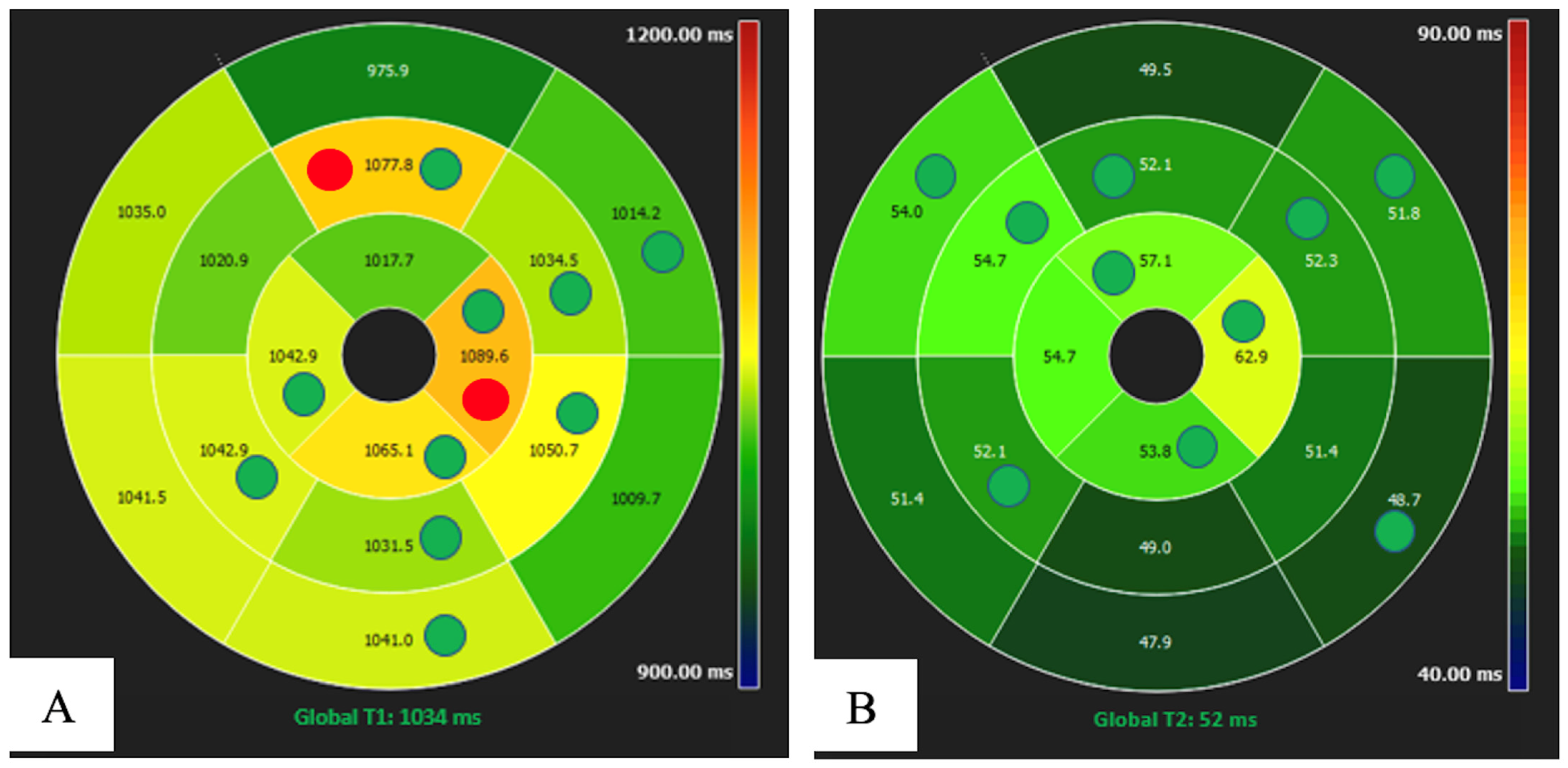
1.2. Magnetic Resonance
1.3. Future Perspective: National Networking for Big Data and Artificial Intelligence, Cardio-Radiologist in the Heart Team Using Specific Training
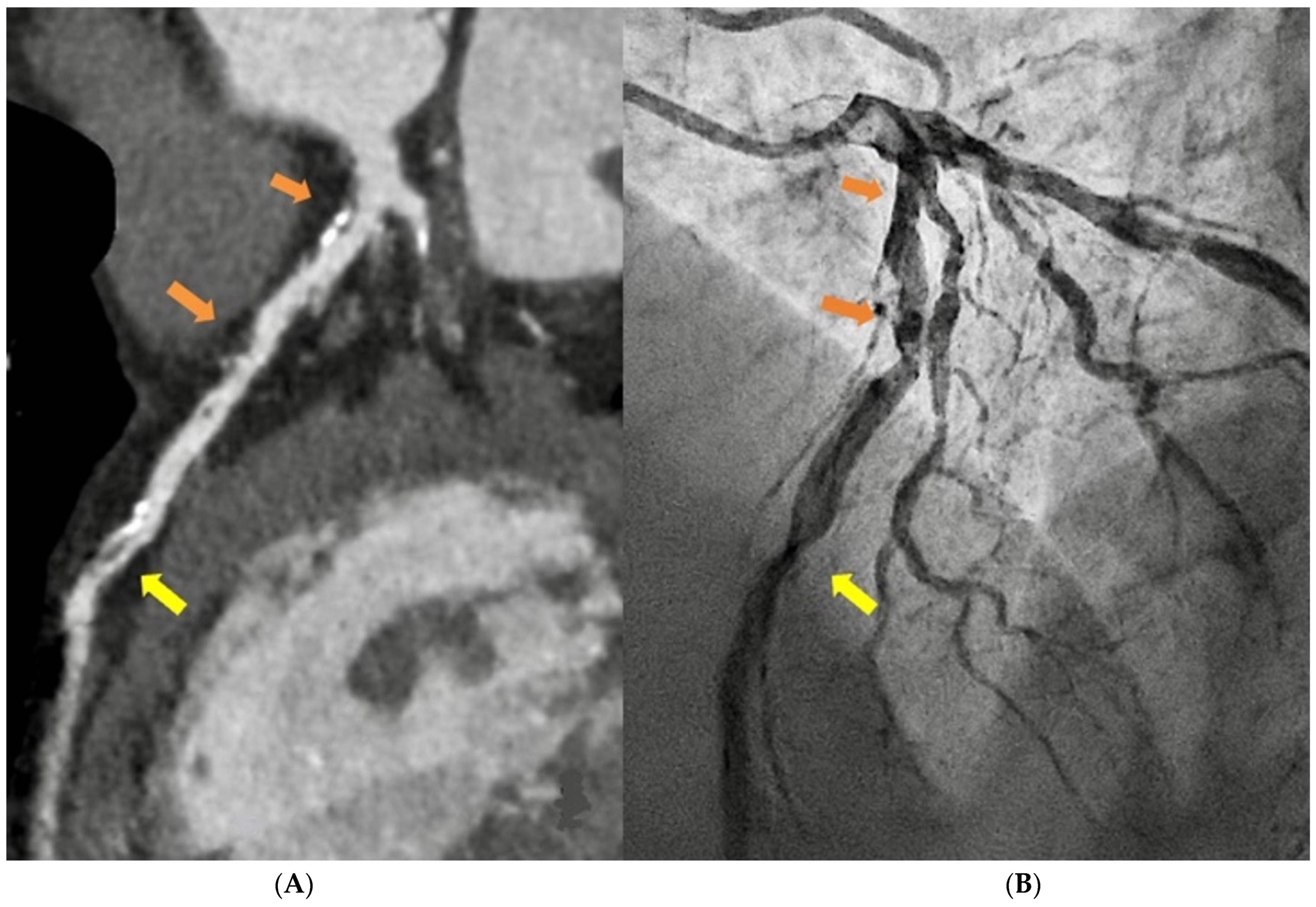
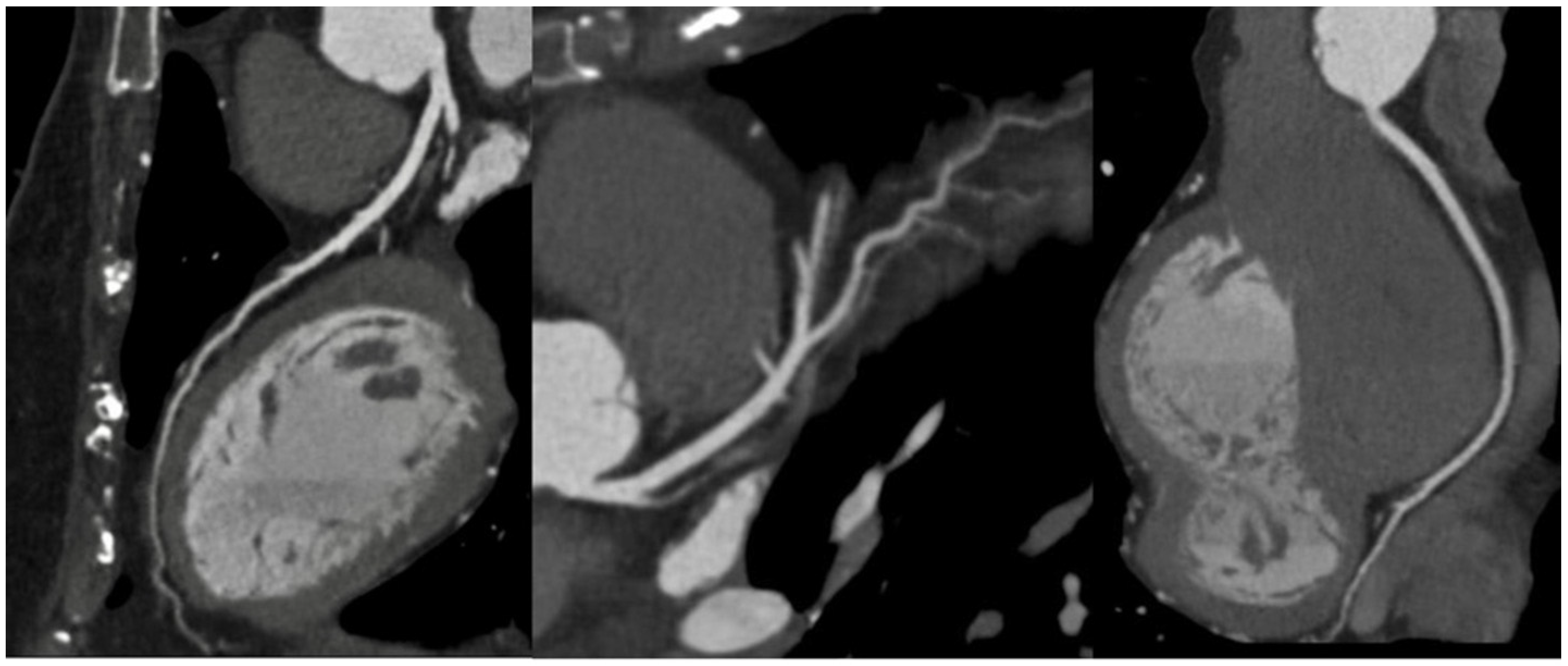
2. Vascular Imaging
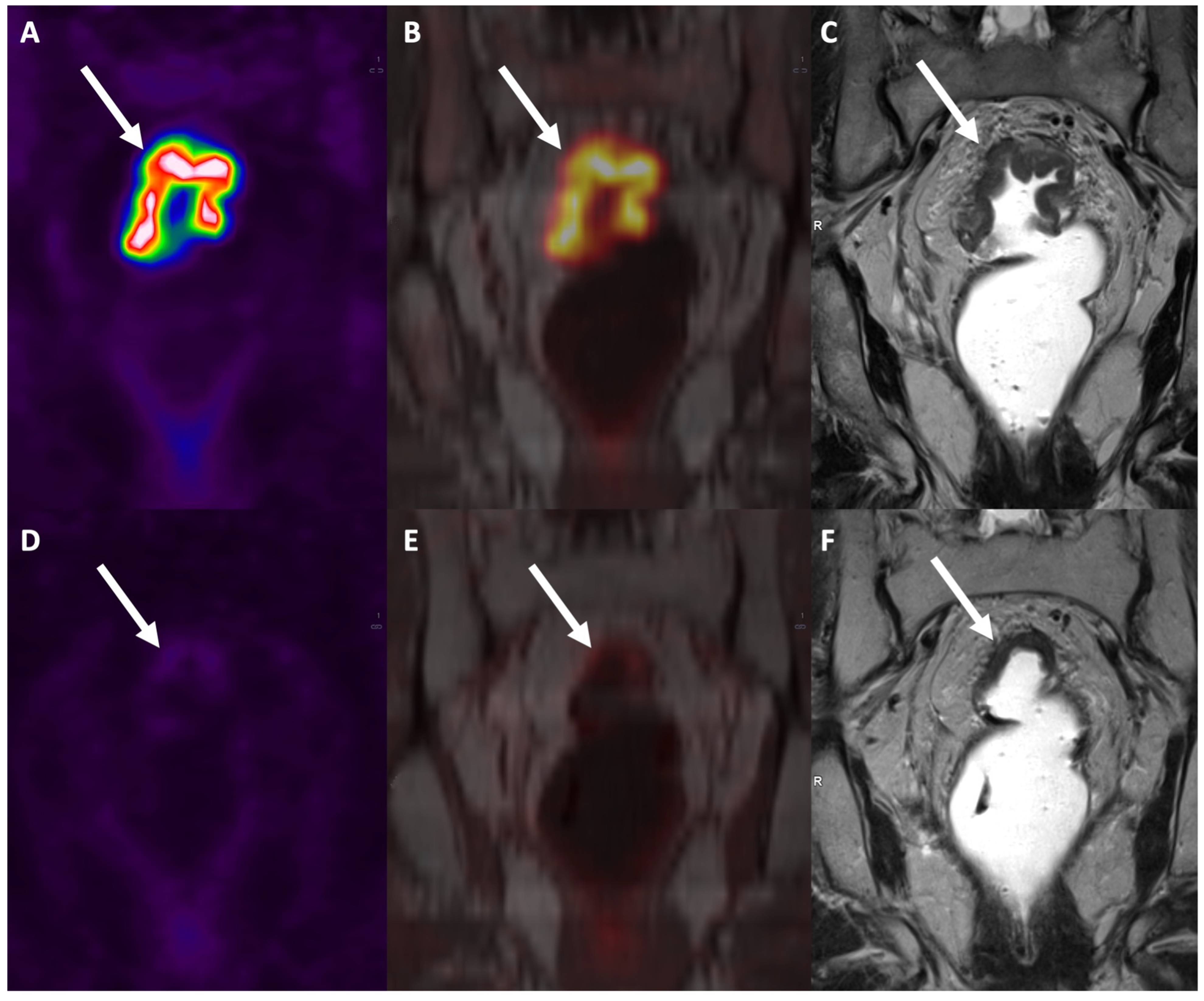
3. Rectal Imaging
3.1. T Staging
3.2. N Staging
3.3. New Techniques and Applications
4. Liver Imaging
4.1. Diffuse Liver Diseases
4.1.1. Clinical Setting
4.1.2. Imaging Approach
4.1.3. Ultrasound
4.1.4. Computed Tomography
4.1.5. Magnetic Resonance Imaging
4.1.6. Future Imaging Trends
4.2. Focal Liver Diseases
4.2.1. Clinical Setting
4.2.2. Imaging Approach
4.2.3. Ultrasound
4.2.4. Computed Tomography
4.2.5. Magnetic Resonance Imaging
4.2.6. Future Imaging Trends
Author Contributions
Funding
Institutional Review Board Statement
Informed Consent Statement
Data Availability Statement
Acknowledgments
Conflicts of Interest
References
- Reeves, R.A.; Halpern, E.J.; Rao, V.M. Cardiac Imaging Trends from 2010 to 2019 in the Medicare Population. Radiol. Cardiothorac. Imaging 2021, 3, e210156. [Google Scholar] [CrossRef]
- Cademartiri, F.; Casolo, G.; Clemente, A.; Seitun, S.; Mantini, C.; Bossone, E.; Saba, L.; Sverzellati, N.; Nistri, S.; Punzo, B.; et al. Coronary CT angiography: A guide to examination, interpretation, and clinical indications. Expert Rev. Cardiovasc. Ther. 2021, 19, 413–425. [Google Scholar] [CrossRef]
- Si-Mohamed, S.A.; Boccalini, S.; Lacombe, H.; Diaw, A.; Varasteh, M.; Rodesch, P.-A.; Dessouky, R.; Villien, M.; Tatard-Leitman, V.; Bochaton, T.; et al. Coronary CT Angiography with Photon-counting CT: First-In-Human Results. Radiology 2022, 303, 303–313. [Google Scholar] [CrossRef]
- Cademartiri, F.; Di Cesare, E.; Francone, M.; Ballerini, G.; Ligabue, G.; Maffei, E.; Romagnoli, A.; Argiolas, G.M.; Russo, V.; Buffa, V.; et al. Italian Registry of Cardiac Computed Tomography. La Radiol. Medica 2015, 120, 919–929. [Google Scholar] [CrossRef]
- Knuuti, J.; Wijns, W.; Saraste, A.; Capodanno, D.; Barbato, E.; Funck-Brentano, C.; Prescott, E.; Storey, R.F.; Deaton, C.; Cuisset, T.; et al. 2019 ESC Guidelines for the diagnosis and management of chronic coronary syndromes. Eur. Heart J. 2020, 41, 407–477. [Google Scholar] [CrossRef]
- Natale, L.; Vliegenthart, R.; Salgado, R.; Bremerich, J.; Budde, R.P.J.; Dacher, J.-N.; Francone, M.; Kreitner, K.-F.; Loewe, C.; Nikolaou, K.; et al. Cardiac radiology in Europe: Status and vision by the European Society of Cardiovascular Radiology (ESCR) and the European Society of Radiology (ESR). Eur. Radiol. 2023, 33, 5489–5497. [Google Scholar] [CrossRef]
- Cury, R.C.; Leipsic, J.; Abbara, S.; Achenbach, S.; Berman, D.; Bittencourt, M.; Budoff, M.; Chinnaiyan, K.; Choi, A.D.; Ghoshhajra, B.; et al. CAD-RADSTM 2.0—2022 Coronary Artery Disease-Reporting and Data System. J. Cardiovasc. Comput. Tomogr. 2022, 16, 536–557. [Google Scholar] [CrossRef]
- Ferencik, M.; Mayrhofer, T.; Bittner, D.O.; Emami, H.; Puchner, S.B.; Lu, M.T.; Meyersohn, N.M.; Ivanov, A.V.; Adami, E.C.; Patel, M.R.; et al. Use of High-Risk Coronary Atherosclerotic Plaque Detection for Risk Stratification of Patients With Stable Chest Pain. JAMA Cardiol. 2018, 3, 144. [Google Scholar] [CrossRef]
- Oikonomou, E.K.; Marwan, M.; Desai, M.Y.; Mancio, J.; Alashi, A.; Hutt Centeno, E.; Thomas, S.; Herdman, L.; Kotanidis, C.P.; Thomas, K.E.; et al. Non-invasive detection of coronary inflammation using computed tomography and prediction of residual cardiovascular risk (the CRISP CT study): A post-hoc analysis of prospective outcome data. Lancet 2018, 392, 929–939. [Google Scholar] [CrossRef]
- Kwiecinski, J.; Wolny, R.; Chwala, A.; Slomka, P. Advances in the Assessment of Coronary Artery Disease Activity with PET/CT and CTA. Tomography 2023, 9, 328–341. [Google Scholar] [CrossRef]
- Ordovas, K.G.; Baldassarre, L.A.; Bucciarelli-Ducci, C.; Carr, J.; Fernandes, J.L.; Ferreira, V.M.; Frank, L.; Mavrogeni, S.; Ntusi, N.; Ostenfeld, E.; et al. Cardiovascular magnetic resonance in women with cardiovascular disease: Position statement from the Society for Cardiovascular Magnetic Resonance (SCMR). J. Cardiovasc. Magn. Reson. 2021, 23, 52. [Google Scholar] [CrossRef]
- Habib, G.; Bucciarelli-Ducci, C.; Caforio, A.L.P.; Cardim, N.; Charron, P.; Cosyns, B.; Dehaene, A.; Derumeaux, G.; Donal, E.; Dweck, M.R.; et al. Multimodality Imaging in Restrictive Cardiomyopathies: An EACVI expert consensus document In collaboration with the “Working Group on myocardial and pericardial diseases” of the European Society of Cardiology Endorsed by The Indian Academy of Echocardiography. Eur. Heart J.-Cardiovasc. Imaging 2017, 18, 1090–1121. [Google Scholar] [CrossRef]
- Mavrogeni, S.; Pepe, A.; Nijveldt, R.; Ntusi, N.; Sierra-Galan, L.M.; Bratis, K.; Wei, J.; Mukherjee, M.; Markousis-Mavrogenis, G.; Gargani, L.; et al. Cardiovascular magnetic resonance in autoimmune rheumatic diseases: A clinical consensus document by the European Association of Cardiovascular Imaging. Eur. Heart J.-Cardiovasc. Imaging 2022, 23, e308–e322. [Google Scholar] [CrossRef]
- Messroghli, D.R.; Moon, J.C.; Ferreira, V.M.; Grosse-Wortmann, L.; He, T.; Kellman, P.; Mascherbauer, J.; Nezafat, R.; Salerno, M.; Schelbert, E.B.; et al. Clinical recommendations for cardiovascular magnetic resonance mapping of T1, T2, T2* and extracellular volume: A consensus statement by the Society for Cardiovascular Magnetic Resonance (SCMR) endorsed by the European Association for Cardiovascular Imaging (EACVI). J. Cardiovasc. Magn. Reson. 2017, 19, 75. [Google Scholar] [CrossRef]
- Meloni, A.; Gargani, L.; Bruni, C.; Cavallaro, C.; Gobbo, M.; D’Agostino, A.; D’Angelo, G.; Martini, N.; Grigioni, F.; Sinagra, G.; et al. Additional value of T1 and T2 mapping techniques for early detection of myocardial involvement in scleroderma. Int. J. Cardiol. 2023, 376, 139–146. [Google Scholar] [CrossRef]
- Meloni, A.; Pistoia, L.; Positano, V.; Martini, N.; Borrello, R.L.; Sbragi, S.; Spasiano, A.; Casini, T.; Bitti, P.P.; Putti, M.C.; et al. Myocardial tissue characterization by segmental T2 mapping in thalassaemia major: Detecting inflammation beyond iron. Eur. Heart J.-Cardiovasc. Imaging 2023. [Google Scholar] [CrossRef]
- Meloni, A.; Positano, V.; Ruffo, G.B.; Spasiano, A.; D’Ascola, D.G.; Peluso, A.; Keilberg, P.; Restaino, G.; Valeri, G.; Renne, S.; et al. Improvement of heart iron with preserved patterns of iron store by CMR-guided chelation therapy. Eur. Heart J.-Cardiovasc. Imaging 2015, 16, 325–334. [Google Scholar] [CrossRef]
- Pepe, A.; Pistoia, L.; Gamberini, M.R.; Cuccia, L.; Lisi, R.; Cecinati, V.; Maggio, A.; Sorrentino, F.; Filosa, A.; Rosso, R.; et al. National networking in rare diseases and reduction of cardiac burden in thalassemia major. Eur. Heart J. 2022, 43, 2482–2492. [Google Scholar] [CrossRef]
- Meloni, A.; Martini, N.; Positano, V.; D’Angelo, G.; Barison, A.; Todiere, G.; Grigoratos, C.; Barra, V.; Pistoia, L.; Gargani, L.; et al. Myocardial T1 Values at 1.5 T: Normal Values for General Electric Scanners and Sex-Related Differences. J. Magn. Reson. Imaging 2021, 54, 1486–1500. [Google Scholar] [CrossRef]
- Meloni, A.; Nicola, M.; Positano, V.; D’Angelo, G.; Barison, A.; Todiere, G.; Grigoratos, C.; Keilberg, P.; Pistoia, L.; Gargani, L.; et al. Myocardial T2 values at 1.5 T by a segmental approach with healthy aging and gender. Eur. Radiol. 2022, 32, 2962–2975. [Google Scholar] [CrossRef]
- Xu, J.; Yang, W.; Zhao, S.; Lu, M. State-of-the-art myocardial strain by CMR feature tracking: Clinical applications and future perspectives. Eur. Radiol. 2022, 32, 5424–5435. [Google Scholar] [CrossRef] [PubMed]
- Quinaglia, T.; Jerosch-Herold, M.; Coelho-Filho, O.R. State-of-the-Art Quantitative Assessment of Myocardial Ischemia by Stress Perfusion Cardiac Magnetic Resonance. Magn. Reson. Imaging Clin. N. Am. 2019, 27, 491–505. [Google Scholar] [CrossRef] [PubMed]
- Dyverfeldt, P.; Bissell, M.; Barker, A.J.; Bolger, A.F.; Carlhäll, C.-J.; Ebbers, T.; Francios, C.J.; Frydrychowicz, A.; Geiger, J.; Giese, D.; et al. 4D flow cardiovascular magnetic resonance consensus statement. J. Cardiovasc. Magn. Reson. 2015, 17, 72. [Google Scholar] [CrossRef] [PubMed]
- Hansen, K.L.; Carlsen, J.F. New Trends in Vascular Imaging. Diagnostics 2021, 11, 112. [Google Scholar] [CrossRef]
- Wielandner, A.; Beitzke, D.; Schernthaner, R.; Wolf, F.; Langenberger, C.; Stadler, A.; Loewe, C. Is ECG triggering for motion artefact reduction in dual-source CT angiography of the ascending aorta still required with high-pitch scanning? The role of ECG-gating in high-pitch dual-source CT of the ascending aorta. Br. J. Radiol. 2016, 89, 20160174. [Google Scholar] [CrossRef]
- Geffroy, Y.; Rodallec, M.H.; Boulay-Coletta, I.; Jullès, M.-C.; Ridereau-Zins, C.; Zins, M. Multidetector CT Angiography in Acute Gastrointestinal Bleeding: Why, When, and How. RadioGraphics 2011, 31, E35–E46. [Google Scholar] [CrossRef]
- Hyde, D.E.; Habets, D.F.; Fox, A.J.; Gulka, I.; Kalapos, P.; Lee, D.H.; Pelz, D.M.; Holdsworth, D.W. Comparison of maximum intensity projection and digitally reconstructed radiographic projection for carotid artery stenosis measurement. Med. Phys. 2007, 34, 2968–2974. [Google Scholar] [CrossRef]
- Francone, M.; Budde, R.P.J.; Bremerich, J.; Dacher, J.N.; Loewe, C.; Wolf, F.; Natale, L.; Pontone, G.; Redheuil, A.; Vliegenthart, R.; et al. CT and MR imaging prior to transcatheter aortic valve implantation: Standardisation of scanning protocols, measurements and reporting—A consensus document by the European Society of Cardiovascular Radiology (ESCR). Eur. Radiol. 2020, 30, 2627–2650. [Google Scholar] [CrossRef]
- Zhao, L.; Zhou, S.; Fan, T.; Li, B.; Liang, W.; Dong, H. Three-dimensional printing enhances preparation for repair of double outlet right ventricular surgery. J. Card. Surg. 2018, 33, 24–27. [Google Scholar] [CrossRef]
- Santos, M.K.; Ferreira Júnior, J.R.; Wada, D.T.; Tenório, A.P.M.; Nogueira-Barbosa, M.H.; Marques, P.M.d.A. Artificial intelligence, machine learning, computer-aided diagnosis, and radiomics: Advances in imaging towards to precision medicine. Radiol. Bras. 2019, 52, 387–396. [Google Scholar] [CrossRef]
- So, A.; Hsieh, J.; Narayanan, S.; Thibault, J.-B.; Imai, Y.; Dutta, S.; Leipsic, J.; Min, J.; LaBounty, T.; Lee, T.-Y. Dual-energy CT and its potential use for quantitative myocardial CT perfusion. J. Cardiovasc. Comput. Tomogr. 2012, 6, 308–317. [Google Scholar] [CrossRef]
- Jacobsen, M.C.; Thrower, S.L.; Ger, R.B.; Leng, S.; Court, L.E.; Brock, K.K.; Tamm, E.P.; Cressman, E.N.K.; Cody, D.D.; Layman, R.R. Multi-energy computed tomography and material quantification: Current barriers and opportunities for advancement. Med. Phys. 2020, 47, 3752–3771. [Google Scholar] [CrossRef]
- Counseller, Q.; Aboelkassem, Y. Recent technologies in cardiac imaging. Front. Med. Technol. 2023, 4, 984492. [Google Scholar] [CrossRef]
- Meloni, A.; Frijia, F.; Panetta, D.; Degiorgi, G.; De Gori, C.; Maffei, E.; Clemente, A.; Positano, V.; Cademartiri, F. Photon-Counting Computed Tomography (PCCT): Technical Background and Cardio-Vascular Applications. Diagnostics 2023, 13, 645. [Google Scholar] [CrossRef] [PubMed]
- Leng, S.; Bruesewitz, M.; Tao, S.; Rajendran, K.; Halaweish, A.F.; Campeau, N.G.; Fletcher, J.G.; McCollough, C.H. Photon-counting Detector CT: System Design and Clinical Applications of an Emerging Technology. RadioGraphics 2019, 39, 729–743. [Google Scholar] [CrossRef] [PubMed]
- Pushparajah, K.; Duong, P.; Mathur, S.; Babu-Narayan, S.V. Cardiovascular MRI and CT in congenital heart disease. Echo Res. Pract. 2019, 6, R121–R138. [Google Scholar] [CrossRef] [PubMed]
- McNally, J.S.; Sakata, A.; Alexander, M.D.; Dewitt, L.D.; Sonnen, J.A.; Menacho, S.T.; Stoddard, G.J.; Kim, S.-E.; de Havenon, A.H. Vessel Wall Enhancement on Black-Blood MRI Predicts Acute and Future Stroke in Cerebral Amyloid Angiopathy. Am. J. Neuroradiol. 2021, 42, 1038–1045. [Google Scholar] [CrossRef] [PubMed]
- Takehara, Y. Clinical Application of 4D Flow MR Imaging for the Abdominal Aorta. Magn. Reson. Med. Sci. 2022, 21, 354–364. [Google Scholar] [CrossRef]
- Qin, J.J.; Obeidy, P.; Gok, M.; Gholipour, A.; Grieve, S.M. 4D-flow MRI derived wall shear stress for the risk stratification of bicuspid aortic valve aortopathy: A systematic review. Front. Cardiovasc. Med. 2023, 9, 1075833. [Google Scholar] [CrossRef]
- Hansen, K.; Hansen, P.; Ewertsen, C.; Lönn, L.; Jensen, J.; Nielsen, M. Vector Flow Imaging Compared with Digital Subtraction Angiography for Stenosis Assessment in the Superficial Femoral Artery—A Study of Vector Concentration, Velocity Ratio and Stenosis Degree Percentage. Ultrasound Int. Open 2019, 5, E53–E59. [Google Scholar] [CrossRef]
- Lubner, M.G.; Smith, A.D.; Sandrasegaran, K.; Sahani, D.V.; Pickhardt, P.J. CT Texture Analysis: Definitions, Applications, Biologic Correlates, and Challenges. Radiographics 2017, 37, 1483–1503. [Google Scholar] [CrossRef]
- Guo, Y.; Chen, X.; Lin, X.; Chen, L.; Shu, J.; Pang, P.; Cheng, J.; Xu, M.; Sun, Z. Non-contrast CT-based radiomic signature for screening thoracic aortic dissections: A multicenter study. Eur. Radiol. 2021, 31, 7067–7076. [Google Scholar] [CrossRef]
- Wang, Y.; Xiong, F.; Leach, J.; Kao, E.; Tian, B.; Zhu, C.; Zhang, Y.; Hope, M.; Saloner, D.; Mitsouras, D. Contrast-enhanced CT radiomics improves the prediction of abdominal aortic aneurysm progression. Eur. Radiol. 2023, 33, 3444–3454. [Google Scholar] [CrossRef] [PubMed]
- Charalambous, S.; Klontzas, M.E.; Kontopodis, N.; Ioannou, C.V.; Perisinakis, K.; Maris, T.G.; Damilakis, J.; Karantanas, A.; Tsetis, D. Radiomics and machine learning to predict aggressive type 2 endoleaks after endovascular aneurysm repair: A proof of concept. Acta Radiol. 2022, 63, 1293–1299. [Google Scholar] [CrossRef]
- Tharmaseelan, H.; Froelich, M.F.; Nörenberg, D.; Overhoff, D.; Rotkopf, L.T.; Riffel, P.; Schoenberg, S.O.; Ayx, I. Influence of local aortic calcification on periaortic adipose tissue radiomics texture features—A primary analysis on PCCT. Int. J. Cardiovasc. Imaging 2022, 38, 2459–2467. [Google Scholar] [CrossRef] [PubMed]
- Dossa, F.; Chesney, T.R.; Acuna, S.A.; Baxter, N.N. A watch-and-wait approach for locally advanced rectal cancer after a clinical complete response following neoadjuvant chemoradiation: A systematic review and meta-analysis. Lancet Gastroenterol. Hepatol. 2017, 2, 501–513. [Google Scholar] [CrossRef] [PubMed]
- Pucciarelli, S.; Giandomenico, F.; De Paoli, A.; Gavaruzzi, T.; Lotto, L.; Mantello, G.; Barba, C.; Zotti, P.; Flora, S.; Del Bianco, P. Bowel function and quality of life after local excision or total mesorectal excision following chemoradiotherapy for rectal cancer. Br. J. Surg. 2016, 104, 138–147. [Google Scholar] [CrossRef] [PubMed]
- Beets-Tan, R.G.H.; Lambregts, D.M.J.; Maas, M.; Bipat, S.; Barbaro, B.; Curvo-Semedo, L.; Fenlon, H.M.; Gollub, M.J.; Gourtsoyianni, S.; Halligan, S.; et al. Magnetic resonance imaging for clinical management of rectal cancer: Updated recommendations from the 2016 European Society of Gastrointestinal and Abdominal Radiology (ESGAR) consensus meeting. Eur. Radiol. 2018, 28, 1465–1475. [Google Scholar] [CrossRef]
- Dresen, R.C.; Beets, G.L.; Rutten, H.J.T.; Engelen, S.M.E.; Lahaye, M.J.; Vliegen, R.F.A.; de Bruïne, A.P.; Kessels, A.G.H.; Lammering, G.; Beets-Tan, R.G.H. Locally Advanced Rectal Cancer: MR Imaging for Restaging after Neoadjuvant Radiation Therapy with Concomitant Chemotherapy Part I. Are We Able to Predict Tumor Confined to the Rectal Wall? Radiology 2009, 252, 71–80. [Google Scholar] [CrossRef]
- Horvat, N.; El Homsi, M.; Miranda, J.; Mazaheri, Y.; Gollub, M.J.; Paroder, V. Rectal MRI Interpretation After Neoadjuvant Therapy. J. Magn. Reson. Imaging 2023, 57, 353–369. [Google Scholar] [CrossRef]
- Xu, Q.; Xu, Y.; Sun, H.; Jiang, T.; Xie, S.; Ooi, B.Y.; Ding, Y. MRI Evaluation of Complete Response of Locally Advanced Rectal Cancer After Neoadjuvant Therapy: Current Status and Future Trends. Cancer Manag. Res. 2021, 13, 4317–4328. [Google Scholar] [CrossRef]
- Horvat, N.; Carlos Tavares Rocha, C.; Clemente Oliveira, B.; Petkovska, I.; Gollub, M.J. MRI of Rectal Cancer: Tumor Staging, Imaging Techniques, and Management. RadioGraphics 2019, 39, 367–387. [Google Scholar] [CrossRef] [PubMed]
- Bates, D.D.B.; Golia Pernicka, J.S.; Fuqua, J.L.; Paroder, V.; Petkovska, I.; Zheng, J.; Capanu, M.; Schilsky, J.; Gollub, M.J. Diagnostic accuracy of b800 and b1500 DWI-MRI of the pelvis to detect residual rectal adenocarcinoma: A multi-reader study. Abdom. Radiol. 2020, 45, 293–300. [Google Scholar] [CrossRef] [PubMed]
- Wei, M.-Z.; Zhao, Z.-H.; Wang, J.-Y. The Diagnostic Accuracy of Magnetic Resonance Imaging in Restaging of Rectal Cancer After Preoperative Chemoradiotherapy. J. Comput. Assist. Tomogr. 2020, 44, 102–110. [Google Scholar] [CrossRef] [PubMed]
- Pomerri, F.; Crimì, F.; Veronese, N.; Perin, A.; Lacognata, C.; Bergamo, F.; Boso, C.; Maretto, I. Prediction of N0 Irradiated Rectal Cancer Comparing MRI Before and After Preoperative Chemoradiotherapy. Dis. Colon Rectum 2017, 60, 1184–1191. [Google Scholar] [CrossRef]
- Heijnen, L.A.; Maas, M.; Beets-Tan, R.G.; Berkhof, M.; Lambregts, D.M.; Nelemans, P.J.; Riedl, R.; Beets, G.L. Nodal staging in rectal cancer: Why is restaging after chemoradiation more accurate than primary nodal staging? Int. J. Color. Dis. 2016, 31, 1157–1162. [Google Scholar] [CrossRef] [PubMed]
- van Heeswijk, M.M.; Lambregts, D.M.J.; Palm, W.M.; Hendriks, B.M.F.; Maas, M.; Beets, G.L.; Beets-Tan, R.G.H. DWI for Assessment of Rectal Cancer Nodes After Chemoradiotherapy: Is the Absence of Nodes at DWI Proof of a Negative Nodal Status? Am. J. Roentgenol. 2017, 208, W79–W84. [Google Scholar] [CrossRef]
- Glynne-Jones, R.; Wyrwicz, L.; Tiret, E.; Brown, G.; Rödel, C.; Cervantes, A.; Arnold, D. Rectal cancer: ESMO Clinical Practice Guidelines for diagnosis, treatment and follow-up. Ann. Oncol. 2017, 28, iv22–iv40. [Google Scholar] [CrossRef]
- Kroon, H.M.; Hoogervorst, L.A.; Hanna-Rivero, N.; Traeger, L.; Dudi-Venkata, N.N.; Bedrikovetski, S.; Kusters, M.; Chang, G.J.; Thomas, M.L.; Sammour, T. Systematic review and meta-analysis of long-term oncological outcomes of lateral lymph node dissection for metastatic nodes after neoadjuvant chemoradiotherapy in rectal cancer. Eur. J. Surg. Oncol. 2022, 48, 1475–1482. [Google Scholar] [CrossRef]
- Ogura, A.; Konishi, T.; Beets, G.L.; Cunningham, C.; Garcia-Aguilar, J.; Iversen, H.; Toda, S.; Lee, I.K.; Lee, H.X.; Uehara, K.; et al. Lateral Nodal Features on Restaging Magnetic Resonance Imaging Associated with Lateral Local Recurrence in Low Rectal Cancer After Neoadjuvant Chemoradiotherapy or Radiotherapy. JAMA Surg. 2019, 154, e192172. [Google Scholar] [CrossRef]
- Jayaprakasam, V.S.; Ince, S.; Suman, G.; Nepal, P.; Hope, T.A.; Paspulati, R.M.; Fraum, T.J. PET/MRI in colorectal and anal cancers: An update. Abdom. Radiol. 2023. [Google Scholar] [CrossRef]
- Mirshahvalad, S.A.; Hinzpeter, R.; Kohan, A.; Anconina, R.; Kulanthaivelu, R.; Ortega, C.; Metser, U.; Veit-Haibach, P. Diagnostic performance of [18F]-FDG PET/MR in evaluating colorectal cancer: A systematic review and meta-analysis. Eur. J. Nucl. Med. Mol. Imaging 2022, 49, 4205–4217. [Google Scholar] [CrossRef] [PubMed]
- Crimì, F.; Valeggia, S.; Baffoni, L.; Stramare, R.; Lacognata, C.; Spolverato, G.; Albertoni, L.; Spimpolo, A.; Evangelista, L.; Zucchetta, P.; et al. [18F]FDG PET/MRI in rectal cancer. Ann. Nucl. Med. 2021, 35, 281–290. [Google Scholar] [CrossRef] [PubMed]
- Stanzione, A.; Verde, F.; Romeo, V.; Boccadifuoco, F.; Mainenti, P.P.; Maurea, S. Radiomics and machine learning applications in rectal cancer: Current update and future perspectives. World J. Gastroenterol. 2021, 27, 5306–5321. [Google Scholar] [CrossRef] [PubMed]
- Cui, Y.; Yang, X.; Shi, Z.; Yang, Z.; Du, X.; Zhao, Z.; Cheng, X. Radiomics analysis of multiparametric MRI for prediction of pathological complete response to neoadjuvant chemoradiotherapy in locally advanced rectal cancer. Eur. Radiol. 2019, 29, 1211–1220. [Google Scholar] [CrossRef] [PubMed]
- Liu, Z.; Zhang, X.-Y.; Shi, Y.-J.; Wang, L.; Zhu, H.-T.; Tang, Z.; Wang, S.; Li, X.-T.; Tian, J.; Sun, Y.-S. Radiomics Analysis for Evaluation of Pathological Complete Response to Neoadjuvant Chemoradiotherapy in Locally Advanced Rectal Cancer. Clin. Cancer Res. 2017, 23, 7253–7262. [Google Scholar] [CrossRef] [PubMed]
- Crimì, F.; Capelli, G.; Spolverato, G.; Bao, Q.R.; Florio, A.; Milite Rossi, S.; Cecchin, D.; Albertoni, L.; Campi, C.; Pucciarelli, S.; et al. MRI T2-weighted sequences-based texture analysis (TA) as a predictor of response to neoadjuvant chemo-radiotherapy (nCRT) in patients with locally advanced rectal cancer (LARC). La Radiol. Medica 2020, 125, 1216–1224. [Google Scholar] [CrossRef]
- Capelli, G.; Campi, C.; Bao, Q.R.; Morra, F.; Lacognata, C.; Zucchetta, P.; Cecchin, D.; Pucciarelli, S.; Spolverato, G.; Crimì, F. 18F-FDG-PET/MRI texture analysis in rectal cancer after neoadjuvant chemoradiotherapy. Nucl. Med. Commun. 2022, 43, 815–822. [Google Scholar] [CrossRef]
- Lazarus, J.V.; Mark, H.E.; Anstee, Q.M.; Arab, J.P.; Batterham, R.L.; Castera, L.; Cortez-Pinto, H.; Crespo, J.; Cusi, K.; Dirac, M.A.; et al. Advancing the global public health agenda for NAFLD: A consensus statement. Nat. Rev. Gastroenterol. Hepatol. 2022, 19, 60–78. [Google Scholar] [CrossRef] [PubMed]
- Berzigotti, A.; Tsochatzis, E.; Boursier, J.; Castera, L.; Cazzagon, N.; Friedrich-Rust, M.; Petta, S.; Thiele, M. EASL Clinical Practice Guidelines on non-invasive tests for evaluation of liver disease severity and prognosis—2021 update. J. Hepatol. 2021, 75, 659–689. [Google Scholar] [CrossRef] [PubMed]
- Vernuccio, F.; Cannella, R.; Bartolotta, T.V.; Galia, M.; Tang, A.; Brancatelli, G. Advances in liver US, CT, and MRI: Moving toward the future. Eur. Radiol. Exp. 2021, 5, 52. [Google Scholar] [CrossRef] [PubMed]
- Schwartz, F.R.; Ashton, J.; Wildman-Tobriner, B.; Molvin, L.; Ramirez-Giraldo, J.C.; Samei, E.; Bashir, M.R.; Marin, D. Liver fat quantification in photon counting CT in head to head comparison with clinical MRI–First experience. Eur. J. Radiol. 2023, 161, 110734. [Google Scholar] [CrossRef] [PubMed]
- Dioguardi Burgio, M.; Bruno, O.; Agnello, F.; Torrisi, C.; Vernuccio, F.; Cabibbo, G.; Soresi, M.; Petta, S.; Calamia, M.; Papia, G.; et al. The cheating liver: Imaging of focal steatosis and fatty sparing. Expert Rev. Gastroenterol. Hepatol. 2016, 10, 671–678. [Google Scholar] [CrossRef] [PubMed]
- Positano, V.; Salani, B.; Pepe, A.; Santarelli, M.F.; De Marchi, D.; Ramazzotti, A.; Favilli, B.; Cracolici, E.; Midiri, M.; Cianciulli, P.; et al. Improved T2* assessment in liver iron overload by magnetic resonance imaging. Magn. Reson. Imaging 2009, 27, 188–197. [Google Scholar] [CrossRef]
- Meloni, A.; Positano, V.; Keilberg, P.; De Marchi, D.; Pepe, P.; Zuccarelli, A.; Campisi, S.; Romeo, M.A.; Casini, T.; Bitti, P.P.; et al. Feasibility, reproducibility, and reliability for the T*2 iron evaluation at 3 T in comparison with 1.5 T. Magn. Reson. Med. 2012, 68, 543–551. [Google Scholar] [CrossRef]
- Reeder, S.B.; Yokoo, T.; França, M.; Hernando, D.; Alberich-Bayarri, Á.; Alústiza, J.M.; Gandon, Y.; Henninger, B.; Hillenbrand, C.; Jhaveri, K.; et al. Quantification of Liver Iron Overload with MRI: Review and Guidelines from the ESGAR and SAR. Radiology 2023, 307, e221856. [Google Scholar] [CrossRef]
- Positano, V.; Meloni, A.; Santarelli, M.F.; Pistoia, L.; Spasiano, A.; Cuccia, L.; Casini, T.; Gamberini, M.R.; Allò, M.; Bitti, P.P.; et al. Deep Learning Staging of Liver Iron Content From Multiecho MR Images. J. Magn. Reson. Imaging 2023, 57, 472–484. [Google Scholar] [CrossRef]
- EASL Clinical Practice Guidelines on the management of benign liver tumours. J. Hepatol. 2016, 65, 386–398. [CrossRef]
- Vernuccio, F.; Ronot, M.; Dioguardi Burgio, M.; Cauchy, F.; Choudhury, K.R.; Dokmak, S.; Soubrane, O.; Valla, D.; Zucman-Rossi, J.; Paradis, V.; et al. Long-term Evolution of Hepatocellular Adenomas at MRI Follow-up. Radiology 2020, 295, 361–372. [Google Scholar] [CrossRef]
- Nault, J.-C.; Couchy, G.; Balabaud, C.; Morcrette, G.; Caruso, S.; Blanc, J.-F.; Bacq, Y.; Calderaro, J.; Paradis, V.; Ramos, J.; et al. Molecular Classification of Hepatocellular Adenoma Associates With Risk Factors, Bleeding, and Malignant Transformation. Gastroenterology 2017, 152, 880–894.e6. [Google Scholar] [CrossRef]
- Galle, P.R.; Forner, A.; Llovet, J.M.; Mazzaferro, V.; Piscaglia, F.; Raoul, J.-L.; Schirmacher, P.; Vilgrain, V. EASL Clinical Practice Guidelines: Management of hepatocellular carcinoma. J. Hepatol. 2018, 69, 182–236. [Google Scholar] [CrossRef] [PubMed]
- Vernuccio, F.; Ronot, M.; Dioguardi Burgio, M.; Lebigot, J.; Allaham, W.; Aubé, C.; Brancatelli, G.; Vilgrain, V. Uncommon evolutions and complications of common benign liver lesions. Abdom. Radiol. 2018, 43, 2075–2096. [Google Scholar] [CrossRef] [PubMed]
- Dietrich, C.F.; Nolsøe, C.P.; Barr, R.G.; Berzigotti, A.; Burns, P.N.; Cantisani, V.; Chammas, M.C.; Chaubal, N.; Choi, B.I.; Clevert, D.-A.; et al. Guidelines and Good Clinical Practice Recommendations for Contrast-Enhanced Ultrasound (CEUS) in the Liver–Update 2020 WFUMB in Cooperation with EFSUMB, AFSUMB, AIUM, and FLAUS. Ultrasound Med. Biol. 2020, 46, 2579–2604. [Google Scholar] [CrossRef] [PubMed]
- Vernuccio, F.; Porrello, G.; Cannella, R.; Vernuccio, L.; Midiri, M.; Giannitrapani, L.; Soresi, M.; Brancatelli, G. Benign and malignant mimickers of infiltrative hepatocellular carcinoma: Tips and tricks for differential diagnosis on CT and MRI. Clin. Imaging 2021, 70, 33–45. [Google Scholar] [CrossRef]
- Wilson, A.; Lim, A.K.P. Microvascular imaging: New Doppler technology for assessing focal liver lesions. Is it useful? Clin. Radiol. 2022, 77, e807–e820. [Google Scholar] [CrossRef]
- Patel, B.N.; Rosenberg, M.; Vernuccio, F.; Ramirez-Giraldo, J.C.; Nelson, R.; Farjat, A.; Marin, D. Characterization of Small Incidental Indeterminate Hypoattenuating Hepatic Lesions: Added Value of Single-Phase Contrast-Enhanced Dual-Energy CT Material Attenuation Analysis. Am. J. Roentgenol. 2018, 211, 571–579. [Google Scholar] [CrossRef]
- Reginelli, A.; Del Canto, M.; Clemente, A.; Gragnano, E.; Cioce, F.; Urraro, F.; Martinelli, E.; Cappabianca, S. The Role of Dual-Energy CT for the Assessment of Liver Metastasis Response to Treatment: Above the RECIST 1.1 Criteria. J. Clin. Med. 2023, 12, 879. [Google Scholar] [CrossRef]
- Seo, J.Y.; Joo, I.; Yoon, J.H.; Kang, H.J.; Kim, S.; Kim, J.H.; Ahn, C.; Lee, J.M. Deep learning-based reconstruction of virtual monoenergetic images of kVp-switching dual energy CT for evaluation of hypervascular liver lesions: Comparison with standard reconstruction technique. Eur. J. Radiol. 2022, 154, 110390. [Google Scholar] [CrossRef]
- Sartoretti, T.; Mergen, V.; Jungblut, L.; Alkadhi, H.; Euler, A. Liver Iodine Quantification With Photon-Counting Detector CT: Accuracy in an Abdominal Phantom and Feasibility in Patients. Acad. Radiol. 2023, 30, 461–469. [Google Scholar] [CrossRef]
- Pahwa, S.; Liu, H.; Chen, Y.; Dastmalchian, S.; O’Connor, G.; Lu, Z.; Badve, C.; Yu, A.; Wright, K.; Chalian, H.; et al. Quantitative perfusion imaging of neoplastic liver lesions: A multi-institution study. Sci. Rep. 2018, 8, 4990. [Google Scholar] [CrossRef]
- Ren, L.; Huber, N.; Rajendran, K.; Fletcher, J.G.; McCollough, C.H.; Yu, L. Dual-Contrast Biphasic Liver Imaging With Iodine and Gadolinium Using Photon-Counting Detector Computed Tomography. Investig. Radiol. 2022, 57, 122–129. [Google Scholar] [CrossRef] [PubMed]


Disclaimer/Publisher’s Note: The statements, opinions and data contained in all publications are solely those of the individual author(s) and contributor(s) and not of MDPI and/or the editor(s). MDPI and/or the editor(s) disclaim responsibility for any injury to people or property resulting from any ideas, methods, instructions or products referred to in the content. |
© 2023 by the authors. Licensee MDPI, Basel, Switzerland. This article is an open access article distributed under the terms and conditions of the Creative Commons Attribution (CC BY) license (https://creativecommons.org/licenses/by/4.0/).
Share and Cite
Pepe, A.; Crimì, F.; Vernuccio, F.; Cabrelle, G.; Lupi, A.; Zanon, C.; Gambato, S.; Perazzolo, A.; Quaia, E. Medical Radiology: Current Progress. Diagnostics 2023, 13, 2439. https://doi.org/10.3390/diagnostics13142439
Pepe A, Crimì F, Vernuccio F, Cabrelle G, Lupi A, Zanon C, Gambato S, Perazzolo A, Quaia E. Medical Radiology: Current Progress. Diagnostics. 2023; 13(14):2439. https://doi.org/10.3390/diagnostics13142439
Chicago/Turabian StylePepe, Alessia, Filippo Crimì, Federica Vernuccio, Giulio Cabrelle, Amalia Lupi, Chiara Zanon, Sebastiano Gambato, Anna Perazzolo, and Emilio Quaia. 2023. "Medical Radiology: Current Progress" Diagnostics 13, no. 14: 2439. https://doi.org/10.3390/diagnostics13142439
APA StylePepe, A., Crimì, F., Vernuccio, F., Cabrelle, G., Lupi, A., Zanon, C., Gambato, S., Perazzolo, A., & Quaia, E. (2023). Medical Radiology: Current Progress. Diagnostics, 13(14), 2439. https://doi.org/10.3390/diagnostics13142439