Skip to Content

Osteology

Osteology is an international, peer-reviewed, open access journal on the basic and clinical research of bone science published quarterly online by MDPI.

Get Alerted

Add your email address to receive forthcoming issues of this journal.

All Articles (127)

Distal Interlocking Long Stem in Revision Hip Arthroplasty: A Narrative Review

  • Lorenzo Milani,
  • Giulio Pieratelli and
  • Domenico Tigani
  • + 3 authors

Background/Objectives: Long interlocking cementless stems have proven useful in revision total hip arthroplasty since their introduction in the late 1980s. Their ability to provide firm fixation has been confirmed by biomechanical tests and clinical observations. The cumulative survival of older, non-coated straight stems has not always been satisfactory, especially in younger patients, and a high number of screw and implant failures has also been reported. This narrative review provides a comprehensive overview of interlocking stems in complex revision procedures, focusing on timing of weight bearing, clinical outcomes, and complications. Methods: PubMed, Google Scholar, and the Cochrane Library were searched using database-specific Boolean search strategies combining terms related to distal locking femoral stems and revision hip arthroplasty. Studies reporting outcomes of cementless distally locked stems in revision total hip arthroplasty were selected according to predefined inclusion and exclusion criteria. The reference lists of the selected articles were additionally screened to identify relevant studies not retrieved by the electronic searches. Results: Thirty-one studies were included in the review. Considerable heterogeneity was observed in study design, sample size, follow-up duration, implant characteristics, and outcome reporting. Distal interlocking stems generally provided satisfactory implant survival and stable fixation, with early or immediate weight bearing permitted in several reported series. Mechanical complications involving the stem and distal locking screws were reported predominantly with earlier implant designs, particularly straight, thin, uncoated stems. More recent curved and hydroxyapatite-coated designs showed favourable mid- and long-term outcomes. Aseptic loosening, infection, dislocation, and mechanical failures were variably reported across the included studies. Because of heterogeneity in follow-up periods, denominators, and completeness of complication reporting, pooled complication rates were not calculated. Conclusions: Satisfactory survival rates were observed, with an acceptable complication rate. Implant mechanical failure, excluding stems that were eventually withdrawn from the market, remains a marginal event that has been largely overcome by curved and coated stems. Therefore, the available literature suggests favorable mid- and long-term outcomes when interlocking stems are used for complex femoral revision procedures or periprosthetic fractures with isthmus deficiency.

Osteology

10 September 2026
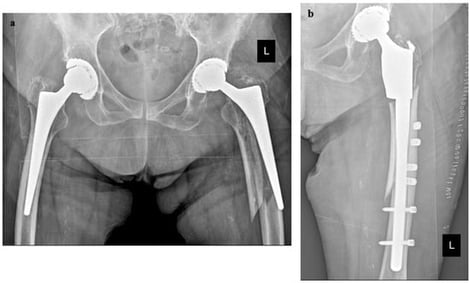
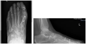
(a) The AP X-ray shows a periprosthetic fracture Vancouver B2 (b) Post-operative X-ray revision with a distal interlocking stem; (c) Four-year follow-up with consolidation of the periprosthetic fracture and absence of complications. L referred to Left.
  • Systematic Review
  • Open Access

Background/Objectives: ACL injuries in skeletally immature patients present a unique surgical challenge, with ongoing debate regarding the optimal reconstruction technique relative to the open physis. Methods: A systematic review and meta-analysis were conducted following PRISMA guidelines, searching PubMed/MEDLINE, Cochrane Library, and Google Scholar. Twenty-two studies (n = 1036 patients; 598 physeal-sparing, 438 transphyseal) were included. The evidence base consisted predominantly of non-randomized retrospective studies. Patient-reported outcomes, return-to-sport rates, growth disturbances, graft failure, and reoperation rates were pooled using a random-effects model. Results: Pooled IKDC scores were 95.14 (95% CI: 93.81–96.50) for physeal-sparing and 96.17 (95% CI: 93.98–98.42) for transphyseal reconstruction, with no significant between-group difference (p = 0.436). Lysholm scores were 95.56 versus 93.04 (p = 0.075), and Tegner scores were 6.39 versus 7.08 (p = 0.366), respectively, with no statistically significant differences. KOOSs also showed no significant difference (96.19 vs. 96.00; p = 0.948). The pooled return-to-sport rate was higher following transphyseal reconstruction than physeal-sparing reconstruction (97.8% vs. 93.9%; p = 0.034). Growth disturbances were more frequently reported in the physeal-sparing group (5.1% vs. 1.4%; p = 0.008). Graft failure (5.8% vs. 8.4%; p = 0.144) and reoperation rates (13.5% vs. 13.8%; p = 0.906) were comparable between techniques. Conclusions: Both techniques yielded comparable functional outcomes across major PROMs. Transphyseal reconstruction was associated with a higher pooled return-to-sport rate in the included studies, whereas physeal-sparing reconstruction was associated with a higher incidence of reported growth disturbances. However, these findings should be interpreted cautiously given that the evidence base consisted predominantly of non-randomized retrospective studies and was subject to potential confounding and other sources of bias.

Osteology

2 September 2026

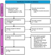
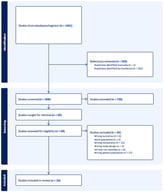
PRISMA chart showing selection and inclusion of studies.

Scapholunate Interosseous Ligament Injury: A Review of the Diagnosis, Treatment, and Outcomes

  • Abdelrahman A. Boghdady,
  • Ayotomi O. Owoeye and
  • Benjamin C. Taylor
  • + 4 authors

Background/Objectives: The scapholunate interosseous ligament (SLIL) is a C-shaped structure composed of dorsal, proximal, and volar segments and is the primary stabilizer of the scapholunate joint. Injury leads to loss of wrist motion and altered carpal mechanics, and chronic laxity may produce radiocarpal and midcarpal degenerative change. Because isolated SLIL injuries frequently lack overt radiographic findings, delayed diagnosis allows progression to fixed carpal malalignment and arthritis, substantially narrowing later treatment options. Methods: A narrative review was conducted. PubMed, Embase, and Google Scholar were searched for English-language articles published between January 1974 and 2025 using combinations of the terms “scapholunate interosseous ligament,” “scapholunate instability,” “scapholunate dissociation,” “carpal instability,” “wrist arthroscopy,” and “scapholunate reconstruction.” Articles were selected for relevance to SLIL anatomy, biomechanics, injury, and treatment, and were appraised with attention to level of evidence. Results: The SLIL is compositionally heterogeneous, ranging from cartilage-like proximally to ligamentous dorsally, and is supported by dorsal and volar secondary stabilizers. Radiographs are frequently normal in partial and predynamic tears; MRI has limited sensitivity for partial tears, arthrography improves accuracy, and arthroscopy remains the diagnostic gold standard. Management is stage-dependent: non-operative care for occult and predynamic instability, direct repair with dorsal capsulodesis for acute reducible injuries, ligament reconstruction such as three-ligament tenodesis for chronic reducible injuries, and salvage procedures for scapholunate advanced collapse. Reported outcomes are favorable but variable, and no reconstructive technique demonstrates consistent superiority. Nearly all outcome data derive from retrospective series with heterogeneous outcome measures. Conclusions: Early recognition and stage-appropriate treatment selection are central to optimizing outcomes in SLIL injury. Current recommendations rest largely on low-level, heterogeneous evidence, and prospective multicenter comparative studies using validated patient-reported and radiographic outcomes represent the field’s most important unmet need.

Osteology

18 August 2026

Anatomical illustration of the SLIL. Created in BioRender. Boghdady, A. (2026). https://www.biorender.com/1mr6wsu [21].

Background/Objectives: The primary aim of this study is to evaluate the impact of two different drilling methods on the primary stability of implants inserted into polyurethane blocks simulating different bone densities. The secondary aim is to analyse the indication provided by three different methods used to assess primary stability. Methods: The study was performed on two artificial bone blocks of solid rigid polyurethane without cortical layer and one laminated one. A total of 48 implants were placed following either a Regular Primary Stability (RPS) or an Enhanced Primary Stability (EPS) protocol based on site underpreparation. Immediately after implant placement, artificial intelligence tool (AIT) indication, peak insertion torque (pIT) and Resonance Frequency Analysis (RFA) values were recorded. The values were then categorised as suitable or not suitable for single-crown immediate loading by bone density and drilling protocol. Results: EPS drilling protocol produced significantly higher primary stability values than those in all bone densities, regardless of the primary stability measurement method used, except for the implants placed in D4 and evaluated by AIT. The grouping of values shows that implants placed in D3 and D4 never reached the immediate loading threshold for a single crown. In D2, EPS and RPS protocols both reach the threshold for all implants when evaluated with pIT or RFA; when AIT is used, RPS protocol was not able to reach the single-crown threshold in 50% of cases. Conclusions: Within the limitations of this in vitro study, underpreparation appears a viable technique to improve primary stability, nevertheless it might be insufficient to reach the threshold of immediate loading suitability in low density bone. All systems used are able to report the primary stability increase obtained by underpreparation, but AIT showed the potential to detect a loss of primary stability related to torque drop during insertion. These findings require further confirmation.

Osteology

16 August 2026

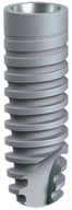
Myplant two geometry.

Featured Articles of Last Quarter

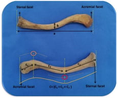
Anterior view of the clavicle (a) clavicle projection length (b) thickness at the narrowest part of the clavicle (c) true length of the clavicle, orange circle: midpoint of the acromioclavicular curvature, red circle: midpoint of the sternoclavicular curvature. c1: distance between the sternal articular surface and the midpoint of the sternoclavicular curvature, c2: distance between the midpoints of the sternoclavicular and the acromioclavicular curvatures, c3: distance between the midpoint of the acromioclavicular curvature and the acromial articular surface. The yellow line indicates the length of c and is shown in the figure.
PRISMA flow diagram.

Highly Accessed Articles

News & Conferences

Latest Issues

Open for Submission

Journal Sections

XFacebookLinkedIn
Osteology - ISSN 2673-4036