


Abstract
Background/Objectives: The scapholunate interosseous ligament (SLIL) is a C-shaped structure composed of dorsal, proximal, and volar segments and is the primary stabilizer of the scapholunate joint. Injury leads to loss of wrist motion and altered carpal mechanics, and chronic laxity may produce radiocarpal and midcarpal degenerative change. Because isolated SLIL injuries frequently lack overt radiographic findings, delayed diagnosis allows progression to fixed carpal malalignment and arthritis, substantially narrowing later treatment options. Methods: A narrative review was conducted. PubMed, Embase, and Google Scholar were searched for English-language articles published between January 1974 and 2025 using combinations of the terms “scapholunate interosseous ligament,” “scapholunate instability,” “scapholunate dissociation,” “carpal instability,” “wrist arthroscopy,” and “scapholunate reconstruction.” Articles were selected for relevance to SLIL anatomy, biomechanics, injury, and treatment, and were appraised with attention to level of evidence. Results: The SLIL is compositionally heterogeneous, ranging from cartilage-like proximally to ligamentous dorsally, and is supported by dorsal and volar secondary stabilizers. Radiographs are frequently normal in partial and predynamic tears; MRI has limited sensitivity for partial tears, arthrography improves accuracy, and arthroscopy remains the diagnostic gold standard. Management is stage-dependent: non-operative care for occult and predynamic instability, direct repair with dorsal capsulodesis for acute reducible injuries, ligament reconstruction such as three-ligament tenodesis for chronic reducible injuries, and salvage procedures for scapholunate advanced collapse. Reported outcomes are favorable but variable, and no reconstructive technique demonstrates consistent superiority. Nearly all outcome data derive from retrospective series with heterogeneous outcome measures. Conclusions: Early recognition and stage-appropriate treatment selection are central to optimizing outcomes in SLIL injury. Current recommendations rest largely on low-level, heterogeneous evidence, and prospective multicenter comparative studies using validated patient-reported and radiographic outcomes represent the field’s most important unmet need.
1. Introduction
Carpal instability can lead to devastating consequences when it comes to the wrist, severely affecting its mobility and range of motion. In 1974, Sebald et al. investigated the mechanism of injury that leads to carpal instability [1]. The authors described the functional importance of the scaphoid in maintaining carpal stability. A scaphoid fracture and loss of its proximal attachment lead to carpal destabilization, ultimately causing joint arthritis. The most common cause of carpal instability is injury to the scapholunate joint [2]. In a study by Short et al. [3], the researchers aimed to assess the biomechanics of the scapholunate joint and its associated ligaments. The study used a wrist joint movement simulator to test flexion–extension and radial–ulnar movements in eight cadaveric upper extremities. Three main ligaments (scapholunate interosseous ligament (SLIL), radioscaphocapitate ligament (RSC), and scaphotrapezial ligament (ST)) were identified, and their elimination was compared with the intact state. The results identified the SLIL as the primary stabilizer of the scapholunate joint, with the RSC and ST as secondary stabilizers.
The most common mechanism of injury was described in 1980 by Mayfield [4]. The authors described the injury as a fall onto an outstretched hand, with the hand in extension, ulnar deviation, and intercarpal supination. In this position, as Dr. White described, the carpal bones lock in a “screw-vise” mechanism, in which the hamate pushes the lunate onto the scaphoid and capitate, disrupting the scapholunate joint [5]. The injury may be isolated or associated with a distal radius fracture, scaphoid fracture, or perilunate dislocation [6].
SLIL injury can lead to degenerative changes in the scapholunate joint [2,6]. It can be difficult to diagnose because isolated SLIL injuries often do not present in a clear, visible manner, making early diagnosis challenging. Several current concept reviews have summarized SLIL anatomy or treatment individually; this review is distinguished by integrating anatomy, diagnosis, and treatment outcomes into a single critical framework, with explicit attention to the level of evidence supporting each management recommendation. This article reviews the current literature on SLIL anatomy and its mechanical significance, with the specific objectives of (1) critically appraising diagnostic modalities and their comparative accuracy, (2) synthesizing outcomes across surgical and non-surgical treatment strategies while accounting for study quality and heterogeneity, and (3) identifying gaps that should guide future research, to provide a better understanding of patient treatment and outcomes.
2. Methods
A narrative review was conducted. The PubMed, Embase, and Google Scholar databases were searched for English-language articles published between January 1974 (the earliest foundational description of carpal collapse mechanics) and 2025, using combinations of the terms “scapholunate interosseous ligament,” “scapholunate instability,” “scapholunate dissociation,” “carpal instability,” “wrist arthroscopy,” and “scapholunate reconstruction.” Articles were selected based on relevance to the anatomy and biomechanics, as well as injury and treatment of the SLIL. The BioRender tool was used to create the schematic diagrams.
3. Anatomy
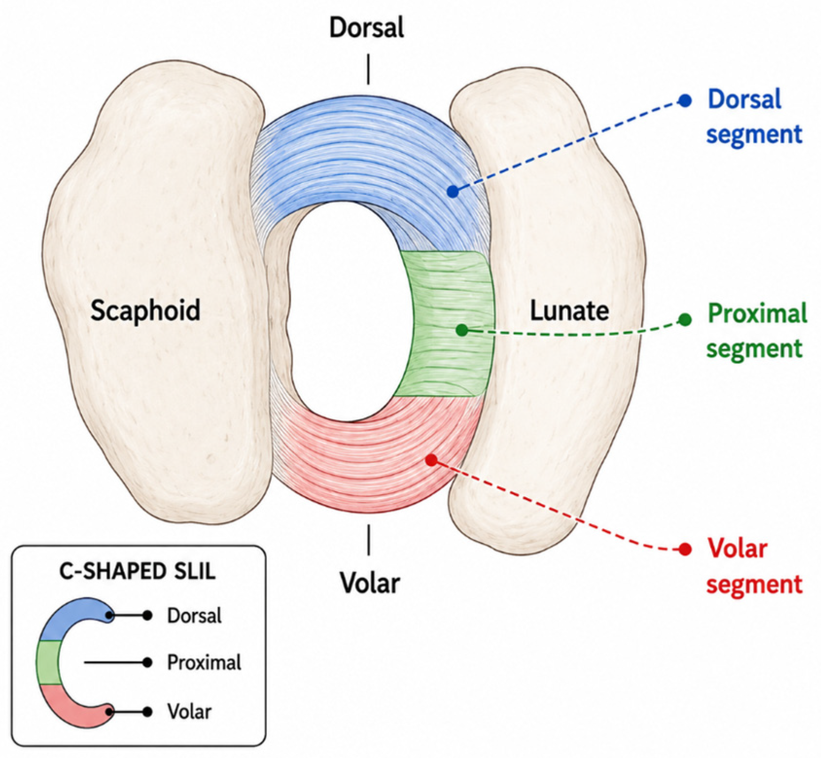
The SLIL is a C-shaped ligament connecting the scaphoid and lunate carpal bones [7]. SLIL is composed of three distinct segments: dorsal, proximal, and volar, with a lack of ligamentous attachment distally over the scapholunate articular surface (Figure 1) [8].
Berger investigated the gross anatomy and histology of the SLIL in a series of human cadaveric wrists [9]. They confirmed three distinct regions of the ligament and examined each region anatomically and histologically. The dorsal region courses between the radial margin of the dorsal horn of the lunate and the dorsal-ulnar margin of the proximal pole of the scaphoid. It is approximately 3 mm thick and 5 mm in proximal–distal length, with collagen the predominant tissue type oriented transversely [9]. Loose connective tissue surrounding the collagen fascicles provides blood supply and innervation. The proximal region is primarily fibrocartilage, which blends with the articular cartilage of the scaphoid and lunate proximally and integrates with the radioscapholunate ligament palmarly. Superficially, it is oriented longitudinally, with relatively variable tissue thickness. The volar region has an oblique fiber orientation, extending from the ulna proximally to the radius distally, with an average thickness of 1 mm. It is primarily composed of collagen fibers and is joined proximally to the combined scapholunate and radioscapholunate ligaments [9].
In a recent study examining the histological and molecular composition of the SLIL, Chato-Astrain et al. identified six distinct zones within the ligament, highlighting its extensive compositional heterogeneity. The membranous (proximal) region most closely resembled cartilage, the dorsal region partially resembled ligamentous tissue, and the palmar region was rich in fibers and scattered spindle-shaped cells [10]. These regions were further subdivided into distinct zones based on composition [10]. In summary, the researchers found that the variation in composition, from cartilage-like to ligament-like, can provide deeper insight into the ligament’s regenerative ability and its susceptibility to injury.
The SLIL is considered the primary stabilizer of the scapholunate joint [3,7]. Several other key ligaments in the dorsal complex have been identified as complementary stabilizers, including the intrinsic dorsal intercarpal (DIC), extrinsic dorsal radiocarpal (DRC), and dorsal scaphotriquetral (DST) ligaments. The distinction between DIC and DST had long been uncertain, but cadaveric dissection by Wessel et al. confirmed that the DST is an anatomically distinct structure that inserts on the dorsal capitate over a 2.9 ± 0.8 mm extension [11].
Viegas et al. reported that DRC most frequently originates from the dorsal margin of the distal radius, just distal and ulnar to Lister’s tubercle. From there, it extends medially and obliquely to the triquetrum’s dorsal tubercle. The DIC ligament has been found to originate on the triquetrum’s dorsal tubercle, attaching to the distal and dorsal aspect of the lunate, with the most common insertion on the scaphoid’s dorsal groove. Additionally, the DIC ligament was found to be attached to the SLIL and the lunotriquetral interosseous ligament (LTIL) [12]. DIC and DRC enhance wrist stability during motion, primarily due to the lateral V design, which allows significant changes in length between the DRC and DIC ligaments and their attachment points. Damage to these ligaments may lead to deficits in normal wrist motion [13]. Finally, their stabilization of the scapholunate joint was emphasized by Elsaidi et al. in a cadaveric study. When the ligaments were removed, rotatory subluxation of the scaphoid and segmental instability and collapse were reproduced [14].
Sennwald et al. examined the palmar scaphotriquetral ligament in 15 human cadaveric wrists to better understand its role in carpal stability [15]. The authors described the ligament as an intracapsular structure lying on the head of the capitate, just dorsal to the RSC ligament. It attaches to the triquetrum and lies between the attachments of the lunotriquetral and triquetrocapitate ligaments. Its attachment is fan-shaped and thin radially over the scaphoid. The palmar scaphotriquetral ligament is stretched when the wrist is dorsiflexed and relaxed when the wrist is flexed. Its structure provides stability to the wrist, especially during flexion/extension, dart-thrower’s motion, and circumduction [2,15].
The long radiolunate (LRL) ligament is located on the volar side of the wrist [16]. It is a capsular ligament that originates from the palmar rim of the scaphoid fossa on the distal radius, medial to the RSC ligament. It runs obliquely, medially, and volar to the proximal pole of the scaphoid and volar to the scapholunate joint, attaching to the radial edge of the palmar horn of the lunate [17]. Perez et al. found that the LRL contributes to wrist stability during lunate extension, and that injury can lead to dorsal intercalated segment instability (DISI) [18]. This suggests that the LRL plays a secondary role in overall wrist stability.
The scaphotrapezium–trapezoid (STT) ligament complex is also located on the volar side of the wrist, consisting of two slips that form a V shape [19]. This ligament attaches to the proximal palmar cortex of the scaphoid tubercle and trapezium via longitudinally oriented fibers, with some fibers passing obliquely and attaching dorsally to the palmar cortex of the trapezoid [17]. Drewniany et al. described the STT ligament as a complex consisting of four elements: a scaphotrapezial ligament with radial and palmar subcomponents, a scaphocapitate capsular ligament, and palmar and dorsal capsules [20]. The authors suggest that the STT ligament complex provides rotational stability to the scapholunate joint and the proximal carpal row.
Figure 1.
Anatomical illustration of the SLIL. Created in BioRender. Boghdady, A. (2026). https://www.biorender.com/1mr6wsu [21].
4. Presentation and Diagnosis
SLIL injury typically affects young individuals and most often results from a fall onto an outstretched hand (FOOSH) [4]. Patients may report pain with axial loading of an extended wrist, as well as pain and weakness with gripping [22]. Approximately 5% of wrist sprains involve an SLIL tear and are often associated with fractures of the radial styloid, also known as the Chauffeur’s fracture [23,24,25,26]. Carpal instability can be classified as either dissociative or nondissociative. Carpal instability dissociative (CID) involves impairment of articulations between carpal bones within the same row, such as the scapholunate and lunotriquetral articulations in the proximal carpal row. Carpal instability nondissociative (CIND) affects articulations outside a single carpal row, for example, the radiocarpal and mid-carpal joints [27]. As Andersson emphasized, early arthroscopic diagnosis and treatment, ideally within four to six weeks of the injury, are crucial to prevent progression to degenerative changes such as scapholunate advanced collapse (SLAC) wrist, soft-tissue attenuation, and cartilage loss [27].
Diagnosis involves both radiographic evaluation and clinical examination. Watson’s test is a commonly used provocative test for SLIL injuries. It is performed by stabilizing the patient’s ipsilateral distal radius and applying pressure to the scaphoid’s palmar prominence, as described by Jones et al. [24]. The wrist is then moved from ulnar to radial deviation while maintaining pressure on the scaphoid tubercle. A positive test is indicated by a “clunk” sound, with pain indicating instability and dorsal scaphoid subluxation suggesting a complete SLIL injury. Despite its common use, the clinical test lacks specificity, and up to 20% of healthy individuals without a history of SLIL injury may have a positive result [27]. During more thorough physical examinations, patients may experience tenderness over the SLIL, and pain is frequently elicited at the end range of radial deviation or wrist extension.
Multiple imaging modalities are available for evaluating suspected SLIL injury, each with distinct strengths and limitations. Standard posteroanterior and lateral radiographs are the first-line study and may demonstrate a widened scapholunate gap (>3 mm, the “Terry Thomas” sign) or an increased scapholunate angle (>60°) in established injuries, but they are often normal in partial or predynamic tears [28,29]. Stress radiographs (clenched-fist and radial/ulnar deviation views) or dynamic fluoroscopy can reveal dynamic instability not seen on resting films, although their accuracy depends on patient effort and positioning [30].
MRI is the most commonly used cross-sectional modality, but visualization of the thin (2 to 4 mm dorsally), C-shaped SLIL depends heavily on field strength and imaging technique [26]. Conventional 1.5T MRI detects most complete tears but has poor sensitivity for partial tears, while 3T MRI provides modest improvement [31]. MR arthrography further improves sensitivity and specificity, particularly for partial-thickness and membranous tears [32]. CT arthrography has been shown to have the highest sensitivity and specificity in SLIL tears with nearly 100% accuracy [31]. Dynamic ultrasound can demonstrate dorsal SLIL disruption but remains highly operator-dependent. Across non-arthrographic imaging modalities, partial-thickness and membranous tears are frequently missed [31]. Finally, wrist arthroscopy remains the gold standard for diagnosing SLIL injuries when noninvasive imaging methods are inconclusive [28].
5. Treatment and Outcomes
Garcia-Elias et al. introduced a seven-stage classification system for dorsal SLIL injuries based on five key factors: the integrity of the dorsal SLIL, its reparability, scaphoid posture, reducibility of carpal malalignment, and the quality of radiocarpal and midcarpal cartilage [33]; Kitay and Wolfe later expanded this framework to account for the biplanar nature of scapholunate instability [34]. This system, summarized in Table 1, remains the most widely used framework for guiding treatment selection. Management generally progresses from non-surgical care for early-stage, occult injuries toward direct repair, ligament reconstruction, and ultimately salvage procedures as ligament disruption, malalignment, and secondary degenerative change advance.
Table 1.
Stages of Scapholunate Injury, Relevant Pathology, and Management Options.
5.1. Non-Surgical Treatment
To preserve sensorimotor control and joint stability, targeted regimens combine proprioceptive exercises, neuromuscular training, and structured physiotherapy [35]. Patients with predynamic or dynamic instability may benefit the most from this approach, which may help mitigate degenerative changes that could otherwise require surgical intervention [27]. Some studies have reported success with adjusting the scaphoid followed by immobilization of the wrist in a palmar cast; however, many surgeons have found this treatment unreliable, with many patients experiencing continued scapholunate instability post-treatment [36].
Non-surgical management is best indicated for Garcia-Elias Stage 1 (occult, partial-thickness) injuries and for predynamic instability in low-demand or older patients without radiographic malalignment, when the SLIL remains grossly intact and secondary stabilizers are functioning [5]. When immobilization is used, a short-arm thumb spica or volar wrist splint with the wrist in slight flexion and ulnar deviation is typically worn for 4 to 6 weeks to unload the dorsal SLIL, followed by a structured rehabilitation program emphasizing proprioceptive and neuromuscular re-education (e.g., dart-thrower’s motion training, dynamic scaphoid-stabilization exercises) for an additional 6 to 12 weeks before return to unrestricted activity [37]. Reported criteria for converting to surgical management include persistent pain or a positive Watson’s test beyond 3 months of adherent conservative treatment, progression of the scapholunate gap or angle on serial radiographs, or new dynamic instability demonstrated on stress imaging or arthroscopy. However, these thresholds are largely based on expert opinion and small case series rather than comparative trials, and no validated protocol currently defines the optimal duration of a conservative trial before surgical referral [5].
5.2. Surgical Techniques
Acute injuries to the SLIL require rapid surgical intervention to restore carpal stability and prevent long-term sequelae, such as degenerative changes. When selecting a surgical repair plan, key considerations include tissue quality, injury severity, and timing after injury. Open surgical repair is usually preferred because it provides direct visualization of the SLIL, cartilage, and surrounding anatomical structures. However, when partial repairs are viable, minimally invasive techniques are more often used [27]. Traditionally, open SLIL repair uses a dorsal approach; common methods include direct suture fixation, posterior transosseous suturing, and bone anchors to reattach the ligament. According to a review by Carratalá et al., these techniques, while linked to favorable outcomes, are not without drawbacks. Open surgery can lead to increased soft-tissue trauma, fibrosis, joint stiffness, and may result in injury to the posterior interosseous nerve (PIN), all factors that may compromise proper healing [38].
Arthroscopic repair offers several advantages, including less disruption of surrounding tissues, improved assessment of intra-articular pathology, and potentially faster recovery. Suture anchor repair is commonly used in both open and arthroscopic approaches and provides strong initial fixation by reattaching the SLIL to its bony insertion. This approach is particularly effective for avulsion-type injuries. For added stability, suture anchor repair is often combined with dorsal capsulodesis and temporary Kirschner-wire (K-wire) pinning. Baxamusa et al. described this combined approach in detail: Once it is determined that the ligament is repairable, it is reattached to the scaphoid using suture anchors, taking care to align the scaphoid and lunate correctly [22]. Two K-wires are then inserted percutaneously across the scapholunate interval to hold the bones in place during healing. After the ligament is repaired, a dorsal capsulodesis is performed using the DIC ligament. The DIC is lifted off the triquetrum while keeping its connection to the scaphoid intact. The free end is then anchored to the dorsal lunate using a suture anchor, reinforcing the repair and helping to maintain carpal alignment.
When performed in the acute setting, this technique has demonstrated good short- and mid-term outcomes [27]. The absence of arthritis and the integrity of secondary wrist stabilizers are good prognostic markers. Overall, suture anchor repair, dorsal capsulodesis, and temporary K-wire pinning are most effective for Garcia-Elias Stage 2 SLIL injuries, where proper ligament alignment may still be achieved. Outside the 3–6-week initial injury period, however, SLIL injuries may no longer be amenable to repair with these techniques due to compromised tissue quality and carpal alignment.
In contrast, chronic SLIL injuries, often diagnosed more than six weeks after the initial injury, pose unique challenges. By this stage, carpal instability tends to be static, with additional ligament degeneration and early arthritic changes. These cases usually require more complex procedures; if carpal alignment is amenable to restoration and degenerative changes are minimal, ligament reconstruction may be employed. One well-known reconstructive method is the three-ligament tenodesis (3LT) described by Garcia-Elias et al. [33]. In this technique, a strip of the flexor carpi radialis (FCR) tendon is harvested and passed through a tunnel drilled in the scaphoid to the original insertion point of the SLIL. The graft replicates the function of the damaged ligament. A groove is then prepared on the dorsal lunate, and a suture anchor is used to secure the tendon graft. To enhance stability, the graft is also routed through the radiotriquetral ligament and sewn back onto itself under tension. Temporary K-wire fixation is used across the scapholunate and scaphoid-capitate joints to protect the reconstruction during healing [27,33].
Scapholunate Advanced Collapse (SLAC) and Scaphoid Nonunion Advanced Collapse (SNAC), which present with irreducible misalignment or severe degenerative arthritis, are examples of cases in which direct reconstruction is no longer viable. Instead, salvage procedures are indicated for pain management and preservation of partial wrist function. Proximal Row Carpectomy (PRC) and Four-Corner Fusion (4CF) are two of the most commonly performed procedures in this class [39]. PRC allows the capitate to articulate directly with the radius by removing the scaphoid, lunate, and triquetrum. This procedure can preserve wrist motion and manage pain in patients with intact cartilage at the capitate-lunate fossa. 4CF involves removal of the scaphoid and subsequent fusion of the four remaining carpal bones (capitate, lunate, triquetrum, and hamate), often using a circular plate for optimal stability. Both PRC and 4CF aim to balance pain management with preservation of overall wrist function [39].
5.3. Outcomes
Short-term and long-term outcomes in patients with scapholunate wrist injury are highly variable. This variability stems from multiple factors, including the extent of injury (partial vs. complete tears), the presence of additional injuries to other ligaments, the duration of injury, and the chosen surgical approach. Before reviewing these results, it is important to note that the vast majority of the outcome studies summarized below are retrospective case series or cohort studies (Oxford level of evidence III–IV), and that the cited systematic reviews and meta-analyses pool this same heterogeneous, largely uncontrolled evidence base; very few prospective or comparative studies with standardized, validated outcome measures exist for SLIL treatment, which should temper the strength of any conclusions about the relative superiority of one technique over another.
As noted earlier in this review, chronic injuries are typically managed with ligament reconstruction using 3LT. Garcia-Elias and colleagues reported outcomes for patients who underwent the 3LT procedure. At four-year follow-up, they reported a 75% success rate, a composite outcome the authors defined as return to prior daily activities and work combined with significant pain relief at rest; this outcome measure was not a validated patient-reported instrument, limiting comparison with later cohorts using standardized scores such as the PRWE or DASH. Patients regained 75% of wrist flexion and extension compared with their uninjured arm, and grip strength returned to 65% of normal. The 3LT technique provided long-term stability, as only 5% of patients developed carpal collapse or dorsal intercalated segment instability (DISI) [33]. Blackburn et al. conducted a cohort study to evaluate short-term outcomes in 203 patients who underwent the 3LT technique for chronic scapholunate ligament injuries [40]. Patients completed the Patient-Rated Wrist Evaluation (PRWE) questionnaire and a visual analog scale (VAS) for pain assessment before the intervention, at 3 months, and one year postoperatively. The PRWE and VAS scores showed significant improvement after surgery. The preoperative PRWE score averaged 57 points with a standard deviation of 20, and the postoperative score at 12 months averaged 26 points with a standard deviation of 21. The PRWE pain scores were 30 (10) and 15 (12) pre- and postoperatively, respectively. The PRWE function score was 26 (12) preoperatively and 11 (11) postoperatively. Finally, the VAS scores for pain were 53 (23) before surgery and 25 (24) after surgery. There were no clinically significant differences in wrist motion before and after surgery.
Rohman et al. conducted a retrospective review of 82 wrists. The authors compared outcomes of treatment for SLIL injuries, including complete and partial injuries, between acute and chronic cases and across different techniques. The authors also compared failure rates for ligament reconstruction versus repair, with or without capsulodesis, in chronic cases [41]. The acute intervention group produced better results than the late intervention group, with a 4% failure rate compared with 18%. For late management, ligament reconstruction yielded smaller radiographic scapholunate joint space gaps than the repair-only or repair with capsulodesis group at 6 weeks (2.5 mm vs. 3.7 mm) and 6 months (2.3 mm vs. 3.8 mm).
Regarding scapholunate angles, ligament reconstruction had smaller angles than the repair with or without capsulodesis group at 6 months (57° vs. 66°). The wrist extension angle was larger in the ligament reconstruction group than in the repair with or without the capsulodesis group (43° vs. 32°). The repair with or without capsulodesis group showed a trend toward higher failure rates. However, the difference between the two groups was nonsignificant. Finally, among isolated SLIL injuries specifically, 21% of chronic procedures failed, whereas none failed in the acute setting.
In a retrospective study of 29 patients with chronic SLIL instability, Moran et al. compared the outcomes of dorsal capsulodesis and tenodesis [42]. In the tenodesis group, postoperative wrist range of motion was 43° of extension (range, 20–65°), 26° of ulnar deviation (range, 10–40°), 40° of flexion (range, 10–60°), and 16° of radial deviation (range, 8–25°). Motion decreased from 89% of the unaffected side before surgery to 63% after surgery. In the capsulodesis group, postoperative wrist range of motion was 49° of extension (range, 15–70°), 26° of ulnar deviation (range, 15–35°), 44° of flexion (range, 10–55°), and 19° of radial deviation (range, 10–30°). Before surgery, wrist motion was 91% of the unaffected side, and after surgery it decreased to 64%. The mean postoperative grip strength was 37 kg (range, 14–65 kg) in the tenodesis group and 37 kg (range, 15–64 kg) in the capsulodesis group. The difference in grip strength compared with the contralateral side was not statistically significant in either group when comparing pre- and post-surgery. The Mayo wrist score was 74 and 77 in the tenodesis and capsulodesis groups, respectively. The study found no statistically significant differences between the two groups in wrist range of motion, grip strength, or Mayo wrist scores. The authors concluded that neither technique offered a clear advantage over the other.
The medical community continues to debate whether PRC or 4CF yields superior outcomes. Ahmadi et al. compared the two methods and found no meaningful differences in pain intensity, range of motion, grip strength, or reoperation rates [39]. Hones et al. conducted a systematic review and meta-analysis in accordance with PRISMA guidelines [43]. The analysis showed that PRC provided a greater range of motion (ROM) than 4CF in extension (42° vs. 34°) and ulnar deviation (29° vs. 20°). However, the two groups had comparable postoperative values for flexion, grip strength, and radial deviation. The postoperative DASH score was better with PRC (15 vs. 26), and there was no difference in QuickDASH scores, Patient-Rated Wrist and Hand Evaluation scores, or VAS scores. Based on pre- to postoperative change, PRC achieved greater improvement in extension (11° vs. −6.8°), ulnar deviation (8.0° vs. −4.7°), and flexion (21° vs. −6.0°). There was no improvement in the pre- to postoperative differences in grip strength and radial deviation between PRC and 4CF. According to Hones et al. [43], VAS scores showed greater improvement from preoperative to postoperative for PRC patients than for 4CF patients (−6.2 vs. −4.6). Chammas et al. reported that mid- to long-term results of PRC showed superior outcomes, including better flexion (weighted mean difference (WMD) = 10°) and ulnar deviation (8.7°) ROM, lower complication rates (OR = 0.3), and lower reoperation rates (OR = 0.1) [44]. These three pooled analyses do not agree: Ahmadi et al. found no difference in range of motion or reoperation rates, whereas Hones et al. and Chammas et al. favored PRC on both. These comparative results should therefore be interpreted cautiously: nearly all of the underlying studies are retrospective cohorts rather than randomized trials, and patients are not randomly allocated to PRC versus 4CF. Surgeons often select PRC for patients with preserved capitolunate cartilage and lower functional demands, while 4CF is favored in younger, higher-demand patients or those with more advanced midcarpal arthritis, so the two groups differ systematically at baseline in ways that can bias outcome comparisons independent of the procedure itself. The included systematic reviews are also heterogeneous, pooling studies with variable follow-up duration, outcome instruments, and surgical technique (e.g., plate versus staple/pin fixation for 4CF), which limits the precision of pooled effect estimates. Long-term radiographic progression to degenerative arthritis and conversion to total wrist arthrodesis are inconsistently reported across these studies, which is a notable gap given that both procedures are intended to be durable, motion-preserving alternatives to fusion.
More recent comparative evidence reinforces the difficulty of identifying a single superior reconstructive technique for chronic SLIL injury. In a 2022 systematic review and meta-analysis of 42 studies (1172 patients), Wu and Ilyas compared capsulodesis, tenodesis, and bone-ligament-bone reconstruction and found that bone-ligament-bone grafts achieved the lowest postoperative pain scores and the highest proportion of “excellent” functional outcomes, while tenodesis achieved the best PRWE scores. No technique showed a consistent advantage in grip strength or range of motion, and the authors explicitly cautioned that most included studies were retrospective case series (level III-IV evidence) with substantial heterogeneity in outcome reporting [45]. This finding is consistent with the general pattern throughout this review: individual techniques may outperform one another on specific metrics without a clear overall winner, largely because of inconsistent study design rather than because the techniques are truly equivalent. For acute and subacute injuries, arthroscopic-assisted repair techniques have continued to accumulate supportive, if still low-level, evidence. Lee et al. reported satisfactory outcomes at a minimum two-year follow-up in a case series of 19 wrists treated with arthroscopic SLIL repair and dorsal capsulodesis using suture anchors, with 94.7% of patients achieving good or excellent Modified Mayo Wrist Scores, again as a single-surgeon retrospective case series [46].
5.4. Treatment Algorithm
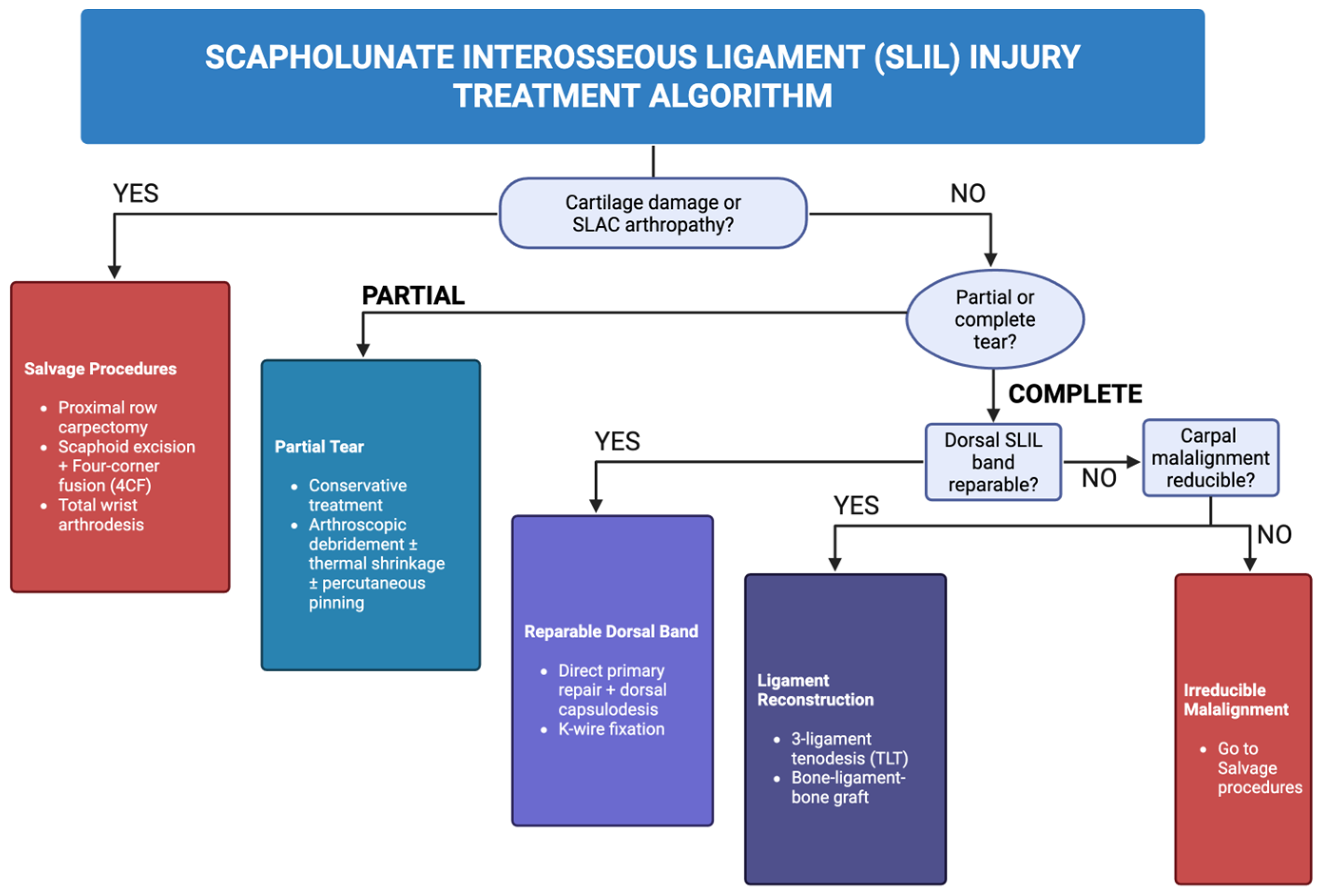
Because the treatment decisions summarized above depend on the interaction of several variables, we propose a simplified decision framework (Figure 2). For acute or subacute injuries (typically within 3–6 weeks of trauma) with a reducible scapholunate interval and preserved cartilage, direct arthroscopic or open repair with dorsal capsulodesis and temporary K-wire fixation is generally preferred, corresponding to Garcia-Elias Stage 2. For chronic injuries (more than 6 weeks) with a reducible interval and minimal degenerative change (Stages 3–4), ligament reconstruction techniques such as three-ligament tenodesis or bone-ligament-bone grafting are favored. When chronicity has produced a fixed, irreducible malalignment or DISI deformity without significant arthritis (Stages 5–6), more extensive open reconstruction with concomitant bony realignment procedures is typically required. Once radiocarpal or midcarpal arthritis has developed (Stage 7, SLAC wrist), reconstruction is no longer feasible, and salvage procedures (PRC or 4CF) are indicated. The choice between these two is informed by cartilage status at the capitolunate joint, patient age, and functional demand, as discussed above. This framework is intended as a starting point for clinical decision-making rather than a rigid protocol; it should be adapted to individual patient factors, and prospective validation of any such algorithm is an important direction for future research.
Figure 2.
Flowchart of SLIL injury treatment options. Created in BioRender. Boghdady, A. (2026) https://BioRender.com/31gefgl [47].
5.5. Future Directions
Several areas of active investigation are likely to shape the future management of SLIL injury. Arthroscopic-assisted reconstruction techniques, which aim to combine the reduced morbidity of arthroscopic surgery with more anatomic reconstruction of the dorsal SLIL, are an area of growing interest, though published series remain small, retrospective, and largely single-surgeon [46]. Suture tape augmentation (internal bracing) of primary ligament repairs has been adapted from other joints (e.g., the knee and ankle) to the wrist [48]. This technique suggests earlier mobilization and improved construct stability, but comparative data against traditional suture anchor repair or capsulodesis are still lacking. Biological augmentation strategies, including allograft or synthetic scaffolds, growth factor application, and orthobiologic adjuncts to enhance ligament healing, are being explored in early translational and small clinical studies but have not yet been validated in the SLIL literature specifically. Across these emerging techniques, the central unmet need identified throughout this review is the absence of prospective, multicenter, comparative studies using standardized, validated outcome measures (e.g., PRWE, DASH/QuickDASH, and radiographic scapholunate gap/angle) and consistent minimum follow-up. Until such studies are performed, treatment selection among the many available reconstructive options will continue to rely more on surgeon experience and individual patient factors than on high-level comparative evidence.
6. Conclusions
Carpal instability can be a devastating injury, limiting hand motion and function. The most common cause of carpal instability is injury to the SLIL, given its crucial stabilizing role at the scapholunate joint. Our objective for this narrative review was to integrate the anatomy and histology of the SLIL with a critical appraisal of diagnostic methods and a synthesis of surgical and non-surgical treatment outcomes. Early diagnosis and management can prevent the progressive degenerative changes associated with this injury. For acute injuries, direct repair yields the best outcomes. For chronic injuries, patients treated with ligament reconstruction, including techniques such as three-ligament tenodesis, show good outcomes. In cases with pre-existing osteoarthritis, salvage procedures, such as proximal row carpectomy or four-corner fusion, are effective. As detailed throughout this review, the current evidence base for SLIL treatment is dominated by retrospective case series and heterogeneous systematic reviews, with no high-quality randomized or prospective comparative trial directly comparing modern reconstructive techniques (e.g., three-ligament tenodesis, bone-ligament-bone grafting, and arthroscopic-assisted repair) using standardized outcome measures. Closing this gap with prospective, multicenter studies incorporating validated patient-reported outcomes and long-term radiographic follow-up represents the most important unmet need in the field and should be prioritized in future research.
Author Contributions
Writing—original draft, A.A.B., A.O.O. and P.G.; Writing—review & editing, A.A.B., A.O.O., P.G., T.J.M., N.S., S.E.B. and B.C.T.; Supervision—N.S., T.J.M., S.E.B. and B.C.T. All authors have read and agreed to the published version of the manuscript.
Funding
This research received no external funding.
Institutional Review Board Statement
Not applicable.
Informed Consent Statement
Not applicable.
Data Availability Statement
No new data were created or analyzed in this study. Data sharing is not applicable to this article.
Acknowledgments
The BioRender tool was used to create the schematic diagrams.
Conflicts of Interest
The authors declare no conflicts of interest.
Abbreviations
The following abbreviations are used in this manuscript:
| SLIL | Scapholunate interosseous ligament |
| RSC | Radioscaphocapitate ligament |
| ST | Scaphotrapezial ligament |
| DIC | Dorsal intercarpal |
| DRC | Dorsal radiocarpal |
| DST | Dorsal scaphotriquetral |
| LTIL | Lunotriquetral interosseous ligament |
| LRL | Long radiolunate |
| DISI | Dorsal intercalated segment instability |
| STT | Scaphotrapezium-trapezoid |
| FOOSH | fall onto an outstretched hand |
| CID | Carpal instability dissociative |
| CIND | Carpal instability non-dissociative |
| SLAC | Scapholunate advanced collapse |
| PIN | Posterior interosseous nerve |
| 3LT | Three-ligament tenodesis |
| FCR | Flexor carpi radialis |
| SNAC | Scaphoid nonunion advanced collapse |
| PRC | Proximal row carpectomy |
| 4CF | Four-corner fusion |
| PRWE | Patient-rated wrist evaluation |
| VAS | Visual analog scale |
| ROM | Range of motion |
| DASH | Disabilities of the Arm, Shoulder and Hand |
References
- Sebald, J.R.; Dobyns, J.H.; Linscheid, R.L. The natural history of collapse deformities of the wrist. Clin. Orthop. Relat. Res. 1974, 104, 140–148. [Google Scholar] [CrossRef] [Scilit] [PubMed]
- Wessel, L.E.; Wolfe, S.W. Scapholunate Instability: Diagnosis and Management—Anatomy, Kinematics, and Clinical Assessment—Part I. J. Hand Surg. Am. 2023, 48, 1139–1149. [Google Scholar] [CrossRef] [Scilit] [PubMed]
- Short, W.H.; Werner, F.W.; Green, J.K.; Masaoka, S. Biomechanical evaluation of ligamentous stabilizers of the scaphoid and lunate. J. Hand Surg. 2002, 27, 991–1002. [Google Scholar] [CrossRef] [Scilit] [PubMed]
- Mayfield, J.K. Mechanism of carpal injuries. Clin. Orthop. Relat. Res. 1980, 149, 45–54. [Google Scholar] [CrossRef] [Scilit]
- White, N.J.; Rollick, N.C. Injuries of the Scapholunate Interosseous Ligament: An Update. J. Am. Acad. Orthop. Surg. 2015, 23, 691–703. [Google Scholar] [CrossRef] [Scilit] [PubMed]
- Chim, H.; Moran, S.L. Wrist essentials: The diagnosis and management of scapholunate ligament injuries. Plast. Reconstr. Surg. 2014, 134, 312e–322e. [Google Scholar] [CrossRef] [Scilit] [PubMed]
- Rajan, P.V.; Day, C.S. Scapholunate Interosseous Ligament Anatomy and Biomechanics. J. Hand Surg. Am. 2015, 40, 1692–1702. [Google Scholar] [CrossRef] [Scilit] [PubMed]
- Logan, S.E.; Nowak, M.D. Intrinsic and extrinsic wrist ligaments: Biomechanical and functional differences. ISA Trans. 1988, 27, 37–41. [Google Scholar] [PubMed]
- Berger, R.A. The gross and histologic anatomy of the scapholunate interosseous ligament. J. Hand Surg. Am. 1996, 21, 170–178. [Google Scholar] [CrossRef] [Scilit] [PubMed]
- Chato-Astrain, J.; Roda, O.; Carriel, V.; Hita-Contreras, F.; Sanchez-Montesinos, I.; Alaminos, M.; Hernández-Cortés, P. Histological characterization of the human scapholunate ligament. Microsc. Res. Tech. 2024, 87, 257–271. [Google Scholar] [CrossRef] [Scilit] [PubMed]
- Wessel, L.E.; Kim, J.; Morse, K.W.; Loisel, F.; Koff, M.F.; Breighner, R.E.; Doty, S.B.; Wolfe, S.W. The Dorsal Ligament Complex: A Cadaveric, Histology, and Imaging Study. J. Hand Surg. Am. 2022, 47, 480e1–480e9. [Google Scholar] [CrossRef] [Scilit] [PubMed]
- Viegas, S.F.; Yamaguchi, S.; Boyd, N.L.; Patterson, R.M. The dorsal ligaments of the wrist: Anatomy, mechanical properties, and function. J. Hand Surg. Am. 1999, 24, 456–468. [Google Scholar] [CrossRef] [Scilit] [PubMed]
- Rainbow, M.J.; Crisco, J.J.; Moore, D.C.; Kamal, R.N.; Laidlaw, D.H.; Akelman, E.; Wolfe, S.W. Elongation of the dorsal carpal ligaments: A computational study of in vivo carpal kinematics. J. Hand Surg. Am. 2012, 37, 1393–1399. [Google Scholar] [CrossRef] [Scilit] [PubMed]
- Elsaidi, G.A.; Ruch, D.S.; Kuzma, G.R.; Smith, B.P. Dorsal wrist ligament insertions stabilize the scapholunate interval: Cadaver study. Clin. Orthop. Relat. Res. 2004, 425, 152–157. [Google Scholar] [CrossRef] [Scilit] [PubMed]
- Sennwald, G.R.; Zdravkovic, V.; Oberlin, C. The anatomy of the palmar scaphotriquetral ligament. J. Bone Jt. Surg. Br. 1994, 76, 147–149. [Google Scholar] [CrossRef] [Scilit]
- Taleisnik, J. The ligaments of the wrist. J. Hand Surg. Am. 1976, 1, 110–118. [Google Scholar] [CrossRef] [Scilit] [PubMed]
- Berger, R.A. The ligaments of the wrist. A current overview of anatomy with considerations of their potential functions. Hand Clin. 1997, 13, 63–82. [Google Scholar] [PubMed]
- Perez, A.J.; Jethanandani, R.G.; Vutescu, E.S.; Meyers, K.N.; Lee, S.K.; Wolfe, S.W. Role of Ligament Stabilizers of the Proximal Carpal Row in Preventing Dorsal Intercalated Segment Instability: A Cadaveric Study. J. Bone Jt. Surg. Am. 2019, 101, 1388–1396. [Google Scholar] [CrossRef] [Scilit] [PubMed]
- Holveck, A.; Wolfram-Gabel, R.; Dosch, J.C.; Sanda, R.; Antunes, A.B.; Decock, S.; Zorn, P.; Foessel, L.; Bierry, G.; Clavert, P.; et al. Scaphotrapezial ligament: Normal arthro-CT and arthro-MRI appearance with anatomical and clinical correlation. Surg. Radiol. Anat. 2011, 33, 473–480. [Google Scholar] [CrossRef] [Scilit] [PubMed]
- Drewniany, J.J.; Palmer, A.K.; Flatt, A.E. The scaphotrapezial ligament complex: An anatomic and biomechanical study. J. Hand Surg. Am. 1985, 10, 492–498. [Google Scholar] [CrossRef] [Scilit] [PubMed]
- Boghdady, A. Anatomical Illustration of the SLIL. BioRender 2026. Available online: https://BioRender.com/1mr6wsu (accessed on 19 May 2026).
- Baxamusa, T.H.; Williams, C.S. Capsulodesis of the wrist for scapholunate dissociation. Tech. Hand Up. Extrem. Surg. 2005, 9, 35–41. [Google Scholar] [CrossRef] [Scilit] [PubMed]
- Chennagiri, R.J.; Lindau, T.R. Assessment of scapholunate instability and review of evidence for management in the absence of arthritis. J. Hand Surg. (Eur. Vol.) 2013, 38, 727–738. [Google Scholar] [CrossRef] [Scilit] [PubMed]
- Jones, W.A. Beware the sprained wrist. The incidence and diagnosis of scapholunate instability. J. Bone Jt. Surg. Br. 1988, 70, 293–297. [Google Scholar] [CrossRef] [Scilit] [PubMed]
- Reichel, L.M.; Bell, B.R.; Michnick, S.M.; Reitman, C.A. Radial styloid fractures. J. Hand Surg. Am. 2012, 37, 1726–1741. [Google Scholar] [CrossRef] [Scilit] [PubMed]
- Mudgal, C.; Hastings, H. Scapho-lunate diastasis in fractures of the distal radius. Pathomechanics and treatment options. J. Hand Surg. Br. 1993, 18, 725–729. [Google Scholar] [CrossRef] [Scilit] [PubMed]
- Andersson, J.K. Treatment of scapholunate ligament injury: Current concepts. EFORT Open Rev. 2017, 2, 382–393. [Google Scholar] [CrossRef] [Scilit] [PubMed]
- Dietrich, T.J.; Toms, A.P.; Cerezal, L.; Omoumi, P.; Boutin, R.D.; Fritz, J.; Schmitt, R.; Shahabpour, M.; Becce, F.; Cotten, A.; et al. Interdisciplinary consensus statements on imaging of scapholunate joint instability. Eur. Radiol. 2021, 31, 9446–9458. [Google Scholar] [CrossRef] [Scilit] [PubMed]
- Hafezi-Nejad, N.; Carrino, J.A.; Eng, J.; Blackmore, C.; Shores, J.; Lifchez, S.D.; Farahani, S.J.; Demehri, S. Scapholunate Interosseous Ligament Tears: Diagnostic Performance of 1.5 T, 3 T MRI, and MR Arthrography—A Systematic Review and Meta-analysis. Acad. Radiol. 2016, 23, 1091–1103. [Google Scholar] [CrossRef] [Scilit] [PubMed]
- Cheriex, K.; Sulkers, G.S.I.; Terra, M.P.; Schep, N.W.L.; van Aard, B.; Strackee, S.D. Scapholunate dissociation; diagnostics made easy. Eur. J. Radiol. 2017, 92, 45–50. [Google Scholar] [CrossRef] [Scilit] [PubMed]
- Expert Panel On Musculoskeletal I; Torabi, M.; Lenchik, L.; Beaman, F.D.; Wessell, D.E.; Bussell, J.K.; Cassidy, R.C.; Czuczman, G.J.; Demertzis, J.L.; Khurana, B.; et al. ACR Appropriateness Criteria® Acute Hand and Wrist Trauma. J. Am. Coll. Radiol. 2019, 16, S7–S17. [Google Scholar] [CrossRef] [Scilit] [PubMed]
- Stensby, J.D.; Fox, M.G.; Nacey, N.; Blankenbaker, D.G.; Frick, M.A.; Jawetz, S.T.; Raizman, N.M.; Said, N.; Stephens, L.A.; Subhas, N.; et al. ACR Appropriateness Criteria® Chronic Hand and Wrist Pain: 2023 Update. J. Am. Coll. Radiol. 2024, 21, S65–S78. [Google Scholar] [CrossRef] [Scilit] [PubMed]
- Garcia-Elias, M.; Lluch, A.L.; Stanley, J.K. Three-ligament tenodesis for the treatment of scapholunate dissociation: Indications and surgical technique. J. Hand Surg. Am. 2006, 31, 125–134. [Google Scholar] [CrossRef] [Scilit] [PubMed]
- Kitay, A.; Wolfe, S.W. Scapholunate instability: Current concepts in diagnosis and management. J. Hand Surg. Am. 2012, 37, 2175–2196. [Google Scholar] [CrossRef] [Scilit] [PubMed]
- Hagert, E.; Rein, S. Wrist proprioception-An update on scientific insights and clinical implications in rehabilitation of the wrist. J. Hand Ther. 2024, 37, 257–268. [Google Scholar] [CrossRef] [Scilit] [PubMed]
- Manuel, J.; Moran, S.L. The diagnosis and treatment of scapholunate instability. Orthop. Clin. N. Am. 2007, 38, 261–277. [Google Scholar] [CrossRef] [Scilit] [PubMed]
- Anderson, H.; Hoy, G. Orthotic intervention incorporating the dart-thrower’s motion as part of conservative management guidelines for treatment of scapholunate injury. J. Hand Ther. 2016, 29, 199–204. [Google Scholar] [CrossRef] [Scilit] [PubMed]
- Carratala, V.; Lucas, F.J.; Miranda, I.; Sanchez Alepuz, E.; Gonzalez Jofre, C. Arthroscopic Scapholunate Capsuloligamentous Repair: Suture with Dorsal Capsular Reinforcement for Scapholunate Ligament Lesion. Arthrosc. Tech. 2017, 6, e113–e120. [Google Scholar] [CrossRef] [Scilit] [PubMed]
- Ahmadi, A.R.; Duraku, L.S.; van der Oest, M.J.W.; Hundepool, C.A.; Selles, R.W.; Zuidam, J.M. The never-ending battle between proximal row carpectomy and four corner arthrodesis: A systematic review and meta-analysis for the final verdict. J. Plast. Reconstr. Aesthet. Surg. 2022, 75, 711–721. [Google Scholar] [CrossRef] [Scilit] [PubMed]
- Blackburn, J.; van der Oest, M.J.W.; Poelstra, R.; Selles, R.W.; Chen, N.C.; Feitz, R.; Hand-Wrist Study Group. Three-ligament tenodesis for chronic scapholunate injuries: Short-term outcomes in 203 patients. J. Hand Surg. (Eur. Vol.) 2020, 45, 383–388. [Google Scholar] [CrossRef] [Scilit] [PubMed]
- Rohman, E.M.; Agel, J.; Putnam, M.D.; Adams, J.E. Scapholunate interosseous ligament injuries: A retrospective review of treatment and outcomes in 82 wrists. J. Hand Surg. Am. 2014, 39, 2020–2026. [Google Scholar] [CrossRef] [Scilit] [PubMed]
- Moran, S.L.; Ford, K.S.; Wulf, C.A.; Cooney, W.P. Outcomes of dorsal capsulodesis and tenodesis for treatment of scapholunate instability. J. Hand Surg. Am. 2006, 31, 1438–1446. [Google Scholar] [CrossRef] [Scilit] [PubMed]
- Hones, K.M.; Hao, K.A.; Rakauskas, T.R.; Densley, S.; Hampton, H.; Kim, J.; Wright, T.W.; Chim, H. Four-Corner Fusion Versus Proximal Row Carpectomy for Scapholunate Advanced Collapse and Scaphoid Nonunion Advanced Collapse Wrist: A Systematic Review and Meta-Analysis. J. Hand Surg. Am. 2024, 49, 633–638. [Google Scholar] [CrossRef] [Scilit] [PubMed]
- Chammas, P.E.; Hadouiri, N.; Chammas, M.; Ramos-Pascual, S.; Stirling, P.; Nover, L.; Klouche, S. Proximal row carpectomy generates better mid- to long-term outcomes than four-corner arthrodesis for post-traumatic wrist arthritis: A meta-analysis. Orthop. Traumatol. Surg. Res. 2022, 108, 103373. [Google Scholar] [CrossRef] [Scilit] [PubMed]
- Wu, M.; Ilyas, A. Comparison of Outcomes of Scapholunate Ligament Reconstruction Techniques. J. Wrist Surg. 2023, 12, 558–568. [Google Scholar] [CrossRef] [Scilit] [PubMed]
- Lee, Y.C.; Shih, Y.C.; Lo, I.N.; Shih, J.T. Arthroscopic scapholunate ligament repair and dorsal capsulodesis with suture anchor in acute and subacute scapholunate dissociation. J. Orthop. Surg. Res. 2023, 18, 661. [Google Scholar] [CrossRef] [Scilit] [PubMed]
- Boghdady, A. Flowchart of SLIL Injury Treatment Options. BioRender 2026. Available online: https://BioRender.com/31gefgl (accessed on 19 May 2026).
- Jackson, G.R.; Opara, O.; Tuthill, T.; Khan, Z.A.; Hevesi, M.; Mameri, E.; Jawanda, H.; Batra, A.K.; Schundler, S.; McCormick, J.; et al. Suture Augmentation in Orthopaedic Surgery Offers Improved Time-Zero Biomechanics and Promising Short-Term Clinical Outcomes. Arthrosc.-J. Arthrosc. Relat. Surg. 2023, 39, 1357–1365. [Google Scholar] [CrossRef] [Scilit] [PubMed]

Disclaimer/Publisher’s Note: The statements, opinions and data contained in all publications are solely those of the individual author(s) and contributor(s) and not of MDPI and/or the editor(s). MDPI and/or the editor(s) disclaim responsibility for any injury to people or property resulting from any ideas, methods, instructions or products referred to in the content. |
© 2026 by the authors. Licensee MDPI, Basel, Switzerland. This article is an open access article distributed under the terms and conditions of the Creative Commons Attribution (CC BY) license.