


Abstract
Background/Objectives: A short scarf osteotomy involves the conventional “Z”-shaped osteotomy which is shorter in length with promising results. Fixation can be performed with a single screw or two screws. This observational study evaluated the clinical effectiveness of single-screw versus traditional two-screw scarf osteotomies for the correction of hallux valgus deformities. Methods: Forty-eight hallux valgus cases treated between 2019 and 2024 were reviewed. Data collected included patient demographics, operative details, radiological outcomes, and patient-reported outcome measures (PROMs). PROMs were assessed using the Manchester Oxford Foot Questionnaire (MOxFQ) and the visual analogue scale (VAS). A cost analysis was also performed. Results: Fourteen patients (29.2%) underwent traditional two-screw scarf osteotomies. In this group, the hallux valgus angle (HVA) improved from 37.4° to 19.6° (p < 0.001), and the intermetatarsal angle (IMA) improved from 16.5° to 8.9° (p = 0.004). MOxFQ scores decreased from 37.8 to 27.4 (p = 0.139), and VAS scores improved from 7.4 to 3.3 (p = 0.012). Complications were limited to two superficial infections (14.3%). The remaining 34 patients (70.8%) underwent modified single-screw scarf osteotomies. This group demonstrated an HVA improvement from 34.7° to 12.6° (p < 0.001) and an IMA improvement from 15.5° to 7.8° (p < 0.001). MOxFQ scores improved from 39.4 to 10.4 (p < 0.001), and VAS scores decreased from 6.6 to 2.5 (p = 0.002). Complications included three superficial infections (8.8%), two intraoperative fractures (5.9%), and two cases of complex regional pain syndrome (5.9%). Conclusions: The modified single-screw scarf osteotomy appears clinically non-inferior to the traditional two-screw technique. Although wider validation is required, these findings suggest that the single-screw technique may offer an alternative that maintains clinical outcomes while supporting more sustainable healthcare resource use.
1. Introduction
A hallux valgus deformity involves the lateral deviation of the proximal phalanx with a corresponding medial deviation of the metatarsal head, resulting in a prominence that instigates inflammation [1,2]. Over time, more than 100 variant procedures for hallux valgus correction have been described leading to variable techniques adapted by foot and ankle surgeons [2,3,4].
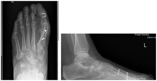
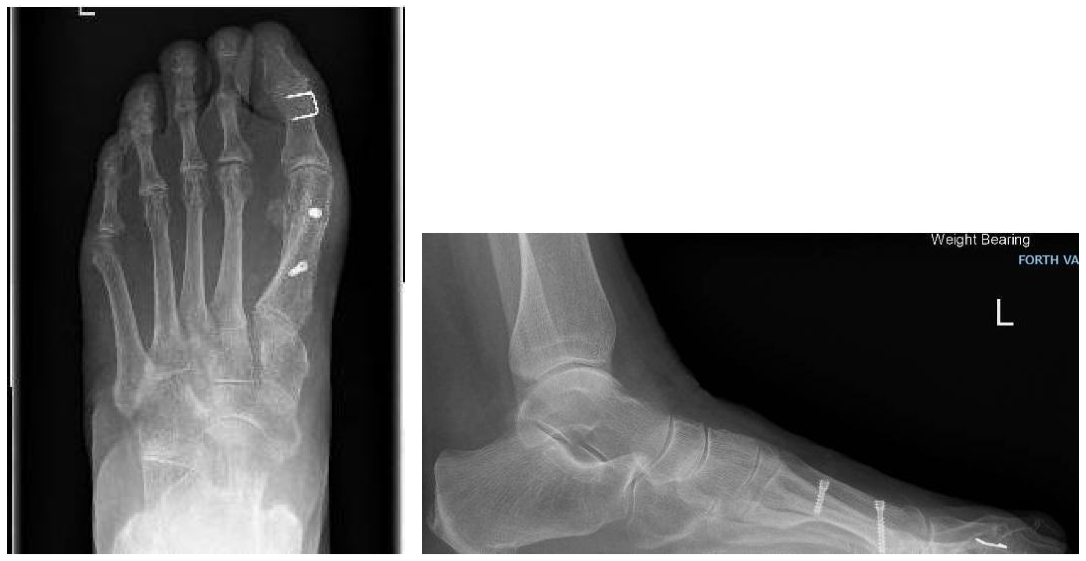
Scarf osteotomies are frequently used to correct moderate to severe hallux valgus deformities, as they are extremely powerful and versatile [5]. Traditionally, this involves a “Z”-shaped bone cut of the metatarsal body with two-screw fixation after translation at the desired position [2] (Figure 1). This surgical procedure requires extensive exposure of the entire first metatarsal ray with significant stripping of the surrounding soft tissues associated with risks such as avascular necrosis and potentially prolonged recovery times [6]. A systematic review demonstrated 10.6% dissatisfaction among patients that underwent first ray corrective osteotomy [7].
Figure 1.
Postoperative X-rays following traditional scarf osteotomy.
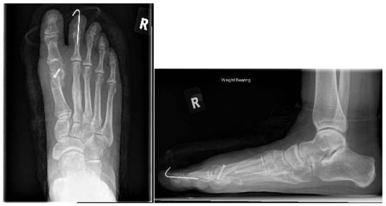
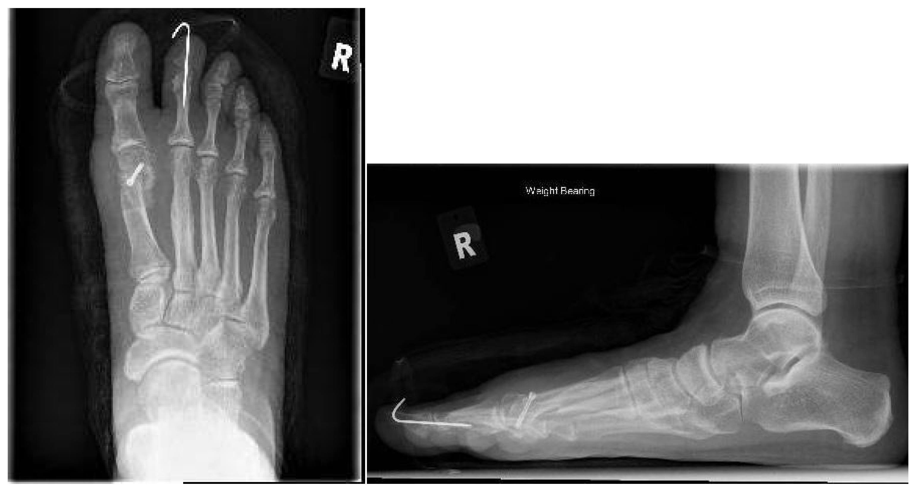
A short scarf osteotomy has been described as an alternative approach with promising results [8,9]. This involves the conventional “Z”-shaped osteotomy which is shorter in length performed via a smaller skin incision. This modification aims to minimize soft tissue dissection and potentially reduce complications associated with it. Fixation can be achieved with a single or double screws depending on surgeon’s preference (Figure 2).
Figure 2.
Postoperative X-rays following short scarf osteotomy with single screw fixation.
The existing literature shows favourable outcomes on this emerging technique; however, it is limited with no superiority of one technique [9,10]. This is a single-centred observational study aiming to compare the clinical effectiveness of the short scarf osteotomies fixed with a single screw in comparison to the traditional long scarf fixed with two screws.
2. Materials and Methods
This is an observational study involving patients with hallux valgus deformities undergoing a scarf osteotomy from June 2019 to September 2024 in Forth Valley Royal Hospital, Scotland. Patients’ demographics, such as age, sex, body mass index (BMI) and smoking status, were documented. Those with neuromuscular disease, uncontrolled diabetes and peripheral vascular disease were excluded from the study. The severity of the hallux deformity was not part of the selection criteria.
For all the included patients the operative details, including complications, additional osteotomies, operative time and additional procedures in the early postoperative time, were reviewed via the local electronic database. Radiological measurements such as the hallux valgus angle (HVA) and the intermetatarsal angle (IMA) were calculated on weight-bearing radiographs both preoperatively and postoperatively, three, six or twelve months postoperatively, on the Picture Archiving Communication System (PACS). These were classified according to their severity as per the American Orthopaedic Foot & Ankle Society [11]. Patient Reported Outcome Measures (PROMs) were obtained through the Manchester-Oxford Foot Questionnaire (MOxFQ 2011; Isis Innovation Lt, Oxford University, Oxford, UK) and the visual analogue scale (VAS) pain score both preoperatively and postoperatively at either 3, 6 or 12 months.
Both long and short osteotomies were conducted under general anaesthetic with regional ankle block. Prophylactic antibiotics were administered. All patients were positioned supine with either a thigh or a calf tourniquet. Patients with reduced abduction in the first metatarsophalangeal joint had a lateral capsular release. A medial incision was made along the first metatarsal head, and its extension depended on whether a short or a long scarf osteotomy was carried out. The joint capsule was released with attention to preserve the metatarsal head blood supply. Additional bone spurs and the medial eminence were excised. Either a short or a long scarf osteotomy was then performed, ensuring the proximal and distal bone cuts were at a 60° angle to the shaft of the metatarsal. The bone fragments were displaced and a Kirschner wire was applied to provisionally stabilise the fragments at the desired correction. Cases with a long scarf osteotomy were fixed with two headless compression screws (Arthex INCTM, Naples, FL, USA) whilst the ones with a short osteotomy required only one. Based on the intraoperative radiologic imaging, an Akin osteotomy of the proximal phalanx was performed when required. This was fixed either with an additional headless compression screw or a varisation staple, based on the surgeon’s preference. Further correction of the deformity was achieved with corrective capsulorrhaphy using Vicryl 1 suture (Ethicon, Somerville, NJ, USA). Following skin closure, all patients received corrective bandage for two weeks.
Postoperatively, all patients were advised to mobilise fully weight-bearing on a forefoot offloading shoe for a total of six to eight weeks. The surgical wound site was reviewed by a specialist nurse two weeks postoperatively and the patient received further consultation by the operating surgeon six to eight weeks after the procedure.
All patient data was collected on Microsoft Excel (Microsoft, Redmond, WA, USA) as well as their variables such as sex, severity of deformity, side, osteotomy type, and additional osteotomy. The data was exported to IBM Statistical Package of Social Sciences (version 29) for statistical analysis. Cross-tabulation and Pearson’s chi-square test (X2) were used to establish the association between the variables. Ordered categories such as smoking status and postoperative complications required cross-tabulations and X2 for linear trends. Fisher’s exact test was performed for categorical variables with reduced number of cases and the Mann–Whitney U test for continuous variables such as age, BMI and surgical procedure time. Radiological outcomes and PROMs preoperatively and postoperatively were analysed with the Wilcoxon signed-rank test due to their non-parametric nature whilst the Mann–Whitney U was further used to compare their differences in long and short scarf osteotomies. Statistical significance was considered as a value of p < 0.05.
3. Results
This study involved 48 scarf operations, short or long, performed on 44 patients, of whom 40 were females. The average age was 52.7 years old (standard deviation (SD) = 13.6) whilst the average BMI was 28 (SD = 5.4). In our cohort, eight patients were active smokers and 10 had ceased smoking in the past. The procedures were performed on average within 89.9 min (SD 17.9) and 29 of them were performed on the left foot. With regard to the radiological findings, none of the cases were classified as mild severity, 38 were considered moderate and 10 severe. A short scarf osteotomy was performed in 34 cases whilst 14 received a traditional long scarf. Akin osteotomies were performed in 25 cases and 14 cases received an additional procedure. Complications were assessed, and five cases had a superficial skin infection treated with antibiotics, two cases developed complex regional pain syndrome (CRPS) and two cases had an intraoperative fracture resulting in the utilisation of a forefoot offloading shoe in the initial postoperative period. The above results can be found in Table 1. Further analysis between the long and short scarf groups indicated no statistical significance as seen in Table 2.
Table 1.
Patients’ demographics and perioperative details within this study cohort.
Table 2.
Comparison of demographic characteristics and operative details of patients who underwent long versus short scarf osteotomy.
The radiological outcomes were analysed preoperatively and postoperatively at 12 months after the operation. Preoperatively the average HVA was calculated at 35.5° (SD = 7.8) and postoperatively it was calculated to be 14.7° (7.9) (p < 0.001). The IMA average was 15.8° (SD = 3.4) preoperatively and 8.2° (SD = 5.2) postoperatively (p < 0.001) (Table 3). These outcomes were analysed further with regard to the long and short scarf groups (Table 4). Within the long scarf group, the preoperative HVA was 37.4° (SD = 6.7) on average, whilst postoperatively it was 19.6° (SD = 9.9) (p < 0.001). The average IMA was 16.5° (SD = 17.7) preoperatively and 8.9° (SD = 3.6) postoperatively (p = 0.004). Within the short scarf group the preoperative HVA was 34.7° (SD = 8.2) on average whilst postoperatively it was 12.6° (SD = 5.9) (p < 0.001). The average IMA was 15.5° (SD = 2.6) preoperatively and 7.8° (SD = 5.7) postoperatively (p < 0.001). The HVA and IMA differences between the long and short scarf osteotomy groups were not statistically significant (Table 5).
Table 3.
Patients’ radiological and reported outcomes preoperatively and postoperatively.
Table 4.
(A). Patients’ radiological and reported outcomes preoperatively and postoperatively between the long scarf osteotomy group. (B). Patients’ radiological and reported outcomes preoperatively and postoperatively between the short scarf osteotomy group.
Table 5.
Statistical significance of the radiological outcomes and the patient reported outcomes between the long and short scarf osteotomy patients.
The PROMs were reported using MOxFQ and VAS scores which were completed preoperatively and at three, six and twelve months postoperatively for all the patients. The statistical analysis was performed based on the longest follow up appointment, which was at 12 months postoperatively. As seen in Table 3 the mean MOxFQ score was 39.0 (SD = 14.8) preoperatively and 16.0 (SD = 2.4) postoperatively (p < 0.001). The mean VAS score preoperatively was 6.8 (SD = 2.4) and 2.7 (SD = 2.5) postoperatively (p < 0.001). This was further analysed within the long and short scarf osteotomy groups (Table 4). Bilateral cases were excluded from the subanalysis to avoid bias between the two operated sides. The difference in MOxFQ scores pre- and postoperatively in the long scarf group were not statistically significant whilst the mean VAS score was 7.4 (SD = 2.2) preoperatively and 3.3 (SD = 3.0) postoperatively (p = 0.012). The short scarf group had a mean MOxFQ of 39.4 (SD = 13.9) preoperatively and 10.4 (SD = 14.1) postoperatively (p < 0.001). The VAS score was also calculated, this was 6.6 (SD = 2.5) preoperatively and 2.5 (SD = 2.3) postoperatively (p = 0.002). The difference in the MOxFQ and VAS scores between the long and short scarf osteotomies were not statistically significant (Table 5).
4. Discussion
This study presents the results between patients undergoing long and short scarf osteotomies for correction of a hallux valgus deformity. These were assessed in terms of complications and radiological results as well as PROMs.
Out of 48 patients, 29.2% received the traditional long scarf osteotomy fixed with two screws and 70.8% received a short scarf osteotomy fixed with a single screw. The patients’ demographics such as age, sex, BMI, affected side and smoking status were comparable between the groups. The surgical details such as the procedure time, deformity severity, complications, need for Akin osteotomy and additional procedures did not show significant differences between the two groups. The preoperative HVA was compared between the long and short scarf groups, and this was not statistically significant (p > 0.2). Similarly, the comparison of IMA showed that p > 0.4. The above results highlight that short scarf osteotomies have comparable results to the traditional technique, even in correction of severe deformities. This is in accordance with the existing literature suggesting this is an effective technique [8,10,12,13].
Izzo et al. conducted a systematic review looking into the number of screws used for fixing a scarf osteotomy and how these influence the radiological results and overall complications [12]. They concluded that screw number did not impact the final correction and were not associated with complications; however, it was noticed that single screw fixation had an increased risk of recurrence in comparison to screwless and two-screw fixation techniques [12]. This result, however, was not statistically significant and was likely impacted by studies with longer follow-up periods recorded. Recurrence of hallux valgus was not noticed in our cohort; however, long-term follow-up was not arranged beyond one year.
Additional procedures, such as Akin osteotomies, have been utilised to improve deformities and reduce recurrence. Butler et al. performed a systematic review comparing the effectiveness of scarf osteotomies versus the ones in conjunction with an Akin osteotomy [14]. It was concluded that a combination of the two procedures had better long-term benefits [15]. In our study, 64.3% of the long scarf cases and 47.1% of the short scarf cases received an Akin osteotomy, too. This requirement was not statistically significant between the two groups. Additional procedures were also investigated. In around 29% of cases from each group an additional procedure was indicated; this was not statistically significant.
The radiological improvements on postoperative X-rays for those with moderate to severe deformities were comparable to those of previous studies [10,15]. Adam et al. demonstrated a mean difference of 19.7° and 8.6° in HVA and IMA respectively following the traditional scarf osteotomy; this was 17.8° and 7.6° respectively in our cohort [15]. In short scarf osteotomies, Poutoglidou et al. indicated a mean difference of 18.0° in HVA and 7.0° in IMA between preoperative and postoperative X-rays. In our cohort these were 22.1° and 7.7° respectively. Comparison of the mean difference in HVA and IMA improvement between the long and short osteotomies demonstrated no statistically significant results, indicating the lack of superiority between the two techniques.
In our study, complication rates were overall low in both groups with no statistically significant results. Superficial skin infection developed in two patients from the long scarf group compared to three in the short scarf group. CRPS and intraoperative fracture were only noted in the short scarf cases with two cases for each. Additional complications, such as stress fractures, troughing and metatarsal splitting, have been previously reported following traditional scarf osteotomy [10,12]. Fixation with a second screw can deliver effective compression; however, it may potentially increase the risk of complications.
MOxFQ and VAS were selected to assess the patient reported outcomes as efficient and representative of the patients’ perspectives. In the long scarf osteotomy group the improvement in the MOxFQ was not statistically significant, whilst the VAS improved by 4.1. Respectively, in the short scarf group the MOxFQ improved by 29.0 and the VAS by 4.1. Comparison of these results between the two groups showed no statistically significant difference, indicating that both techniques were equally effective.
The limitations of this study are mainly associated with the lack of multiple centre participation, limiting the patient selection and potentially its application to a wider population. The long and short scarf cohorts were not equally sized which may have impacted the statistical power whilst the procedure selection was based on the surgeon’s preference and the equipment availability. Lastly, a few of the MOxFQ and VAS questionnaires were unavailable, potentially leading to a slight alteration in the PROMs.
5. Conclusions
To our knowledge, this is the first study comparing the outcomes between the traditional scarf osteotomy fixed with two screws compared with the proposed short scarf fixed with a single screw. Both groups were compared in aspects such as the demographics, surgical details, the pre- and postoperative radiological findings and PROMs. It was noted that there were no statistically significant results indicating the superiority of one technique. It appears that a short scarf osteotomy can produce similar radiological results and patient reported outcomes as the traditional scarf osteotomy.
Author Contributions
Conceptualisation, E.D.; Methodology, P.Z., C.M. and E.D.; Formal analysis, C.M. and E.D.; Investigation, C.M.; Resources, C.M.; Data curation, C.M.; Writing—original draft, P.Z.; Writing—review and editing, P.Z.; Supervision, T.S. and E.D.; Project administration, E.D. All authors have read and agreed to the published version of the manuscript.
Funding
This research received no external funding.
Institutional Review Board Statement
The project was approved as a service evaluation/audit by NHS Forth Valley under the title “Audit of the clinical, financial, and environmental implications of modified short scarf osteotomies in comparison to traditional long scarf osteotomies”. Caldicott and Data Protection approval was granted via the NHS Forth Valley Caldicott/Data Protection Release Form (Reference Number: CAL/24032). The Research and Development Committee was informed and exemption from needing approval was confirmed as the surgical technique described is a recognised and widely accepted method for the correction of hallux valgus and does not constitute an experimental intervention but the project constitutes a service evaluation/audit of standard clinical practice.
Informed Consent Statement
Informed consent was obtained from all patients, including consent for the collection and use of their data in an anonymised manner and the Caldicott Guardian was informed.
Data Availability Statement
The original contributions presented in this study are included in the article. Further inquiries can be directed to the corresponding author.
Conflicts of Interest
The authors declare no conflicts of interest.
Abbreviations
The following abbreviations are used in this manuscript:
| HVA | Hallux valgus angle |
| IMA | Intermetatarsal angle |
| PACS | Picture Archiving Communication System |
| PROMs | Patient Reported Outcome Measures |
| MOxFQ | Manchester-Oxford Foot Questionnaire |
| VAS | Visual analogue scale |
References
- Bunions. National Institute for Health and Care Excellence. Available online: https://cks.nice.org.uk/topics/bunions/ (accessed on 2 December 2024).
- Ray, J.J.; Friedmann, A.J.; Hanselman, A.E.; Vaida, J.; Dayton, P.D.; Hatch, D.J.; Smith, B.; Santrock, R.D. Hallux Valgus. Foot Ankle Orthop. 2019, 4, 2473011419838500. [Google Scholar] [CrossRef] [Scilit]
- Li, Y.; Wang, Y.; Wang, F.; Tang, K.; Tao, X. Biochemical Comparison between Rotational Scarf Osteotomy and Traditional Scarf Osteotomy: A Finite Element Analysis. Orthop. Surg. 2023, 15, 3243–3253. [Google Scholar] [CrossRef] [Scilit]
- Nair, A.; Bence, M.; Saleem, J.; Yousaf, A.; Al-Hilfi, L.; Kunasingam, K. A Systematic Review of Open and Minimally Invasive Surgery for Treating Recurrent Hallux Valgus. Surg. J. 2022, 8, e350–e356. [Google Scholar] [CrossRef] [Scilit]
- Molloy, A.; Widnall, J. Scarf osteotomy. Foot Ankle Clin. 2014, 19, 165–180. [Google Scholar] [CrossRef] [Scilit]
- Dereymaeker, G. Scarf osteotomy for correction of hallux valgus. Surgical technique and results as compared to distal chevron osteotomy. Foot Ankle Clin. 2000, 5, 513–524. [Google Scholar]
- Barg, A.; Harmer, J.R.; Presson, A.P.; Zhang, C.; Lackey, M.; Saltzman, C.L. Unfavorable Outcomes Following Surgical Treatment of Hallux Valgus Deformity: A Systematic Literature Review. J. Bone Jt. Surg. Am. 2018, 100, 1563–1573. [Google Scholar] [CrossRef] [Scilit]
- Rajeev, A.; Tumia, N. Three-year follow-up results of combined short Scarf Osteotomy with Akin procedure for Hallux Valgus. J. Foot Ankle Surg. 2019, 58, 837–841. [Google Scholar] [CrossRef] [Scilit]
- Dalal, R.; Rachha, R.; Leonard, D.; Chourasia, A.; Javed, D. Short Scarf Osteotomy for Hallux Valgus: Short-term and Medium-term Results. J. Foot Ankle Surg. 2017, 4, 14–18. [Google Scholar]
- Poutoglidou, F.; Khan, I.; Scott, A.; Hamilton, P.; Jeong, S.; Yousaf, S. Patient-reported and radiological outcomes of short scarf osteotomy fixed with a single screw: A prospective study of 271 osteotomies. Foot 2024, 61, 102130. [Google Scholar] [CrossRef] [Scilit] [PubMed]
- Coughlin, M.J.; Saltzman, C.L.; Nunley, J.A., 2nd. Angular measurements in the evaluation of hallux valgus deformities: A report of the ad hoc committee of the American Orthopaedic Foot & Ankle Society on angular measurements. Foot Ankle Int. 2002, 23, 68–74. [Google Scholar] [CrossRef] [Scilit] [PubMed]
- Izzo, A.; Manzi, G.; Di Vico, G.; Mariconda, M.; Bernasconi, A. Does the number of screws to fix scarf osteotomy influence the radiographic outcome and the complication rate in hallux valgus? A proportional meta-analysis and systematic review. Joints 2023, 1, e812. [Google Scholar]
- Qasim, S.N.; Mangwani, J. Short versus ‘Standard’ Scarf Osteotomy for Hallux Valgus Correction. Foot Ankle Orthop. 2016, 1, 2473011416S00275. [Google Scholar] [CrossRef] [Scilit]
- Butler, J.J.; Hartman, H.; Rettig, S.; Konar, K.; Randall, G.; Samsonov, A.P.; Kennedy, J.G. Similar outcomes following scarf-Akin osteotomy compared to scarf-alone osteotomy for the treatment of hallux valgus: A systematic review and meta-analysis. Foot Ankle Surg. 2024, 30, 299–308. [Google Scholar] [CrossRef] [Scilit] [PubMed]
- Adam, S.P.; Choung, S.C.; Gu, Y.; O’Malley, M.J. Outcomes after scarf osteotomy for treatment of adult hallux valgus deformity. Clin. Orthop. Relat. Res. 2011, 469, 854–859. [Google Scholar] [CrossRef] [Scilit] [PubMed]

Disclaimer/Publisher’s Note: The statements, opinions and data contained in all publications are solely those of the individual author(s) and contributor(s) and not of MDPI and/or the editor(s). MDPI and/or the editor(s) disclaim responsibility for any injury to people or property resulting from any ideas, methods, instructions or products referred to in the content. |
© 2026 by the authors. Licensee MDPI, Basel, Switzerland. This article is an open access article distributed under the terms and conditions of the Creative Commons Attribution (CC BY) license.