1. Introduction
The clavicle is the first bone to ossify in the human body. Bone formation, which begins in the fifth week of pregnancy, continues until around the age of 26. This S-shaped bone articulates medially with the sternum and laterally with the acromion on the scapula [
1,
2]. It is the most commonly fractured long bone in childhood and the second most commonly fractured bone in adulthood after the radius. Clavicular fractures occur more frequently in men than in women [
3,
4,
5,
6,
7]. Because the clavicle is innervated by the supraclavicular nerves, sensory disturbances such as numbness over the chest wall may be observed following clavicular fractures [
8].
Treatment of these fractures is mostly conservative. Although it was previously believed that nearly all clavicular fractures heal without surgical treatment, recent studies suggest that this rate is lower than previously assumed [
9].
Surgical options for clavicular fractures include intramedullary nails, plates and Kirschner wires, depending on the fracture characteristics [
10,
11]. A detailed understanding of clavicular morphometry is essential during surgical intervention. Preservation and restoration of the pre-fracture morphometric structure of the clavicle are important in preventing postoperative functional loss. Furthermore, because the degree of clavicular shortening plays a key role in surgical decision-making, it is necessary to determine morphometric differences between the right and left clavicles separately [
10,
12]. Accurate restoration of clavicular length is particularly important for optimal functional outcomes.
A thorough understanding of clavicular morphometry is essential for both determining the indication for surgical treatment and for accurate intraoperative planning. Although our study provides anatomical morphometric data, it has not been clinically validated. In cases of unilateral clavicle fracture, the contralateral intact clavicle is commonly assumed to be symmetrical and is therefore used as a reference for assessment and implant selection. However, bilateral clavicular symmetry is not consistently present, which may lead to inaccurate estimations of length, alignment, and implant dimensions. For this reason, independent morphometric evaluation of the right and left clavicle may provide more reliable anatomical guidance. However, no distinction was made between genders in our study, and this may limit the study’s reliability. The aim of the present study is to characterize side-specific clavicular morphometric parameters and key bony landmarks, providing clinically relevant data primarily for orthopaedic surgeons and secondarily for radiologists involved in preoperative assessment and surgical planning.
2. Materials and Methods
This study examined 179 dried adult human clavicles (84 right, 95 left), obtained from the osteological collection of the Department of Anatomy, Faculty of Medicine, Ankara University. The age and sex of the specimens were unknown. Damaged or deformed bones were excluded. The skeletal material is part of an established academic collection used for educational and research purposes. The study was conducted on fully anonymized dry bone specimens without any identifiable personal information. The research was performed in accordance with the ethical principles of the Declaration of Helsinki. The protocol for this study was approved by the local Ethics Committee of the Ankara University Faculty of Medicine (Approval Number: İ02-201-26).
All measurements were performed by a single experienced investigator in order to ensure methodological consistency and minimize inter-observer variability. The measurements were repeated 2 times by the same person using a 0.01 mm precision digital caliper, and the data obtained were averaged. The intraclass correlation coefficient (ICC) was calculated for intra-observer reliability analysis. A two-way mixed-methods model and absolute agreement approach were used. The same measurement protocol and instruments were used throughout the study. Morphometric measurements were obtained using a Mitutoyo digital caliper. Angular measurements were performed using the Image J programme (LOCI, University of Wisconsin, Madison, WI, USA). The measurement of clavicle length was performed in two ways: as both projected and actual length. The reason for this is that, in clavicle reconstructions, if an incorrect angle is created in the bone, the change in projected length can be detected even though the actual length remains the same. However, the accuracy of this method has not yet been established.
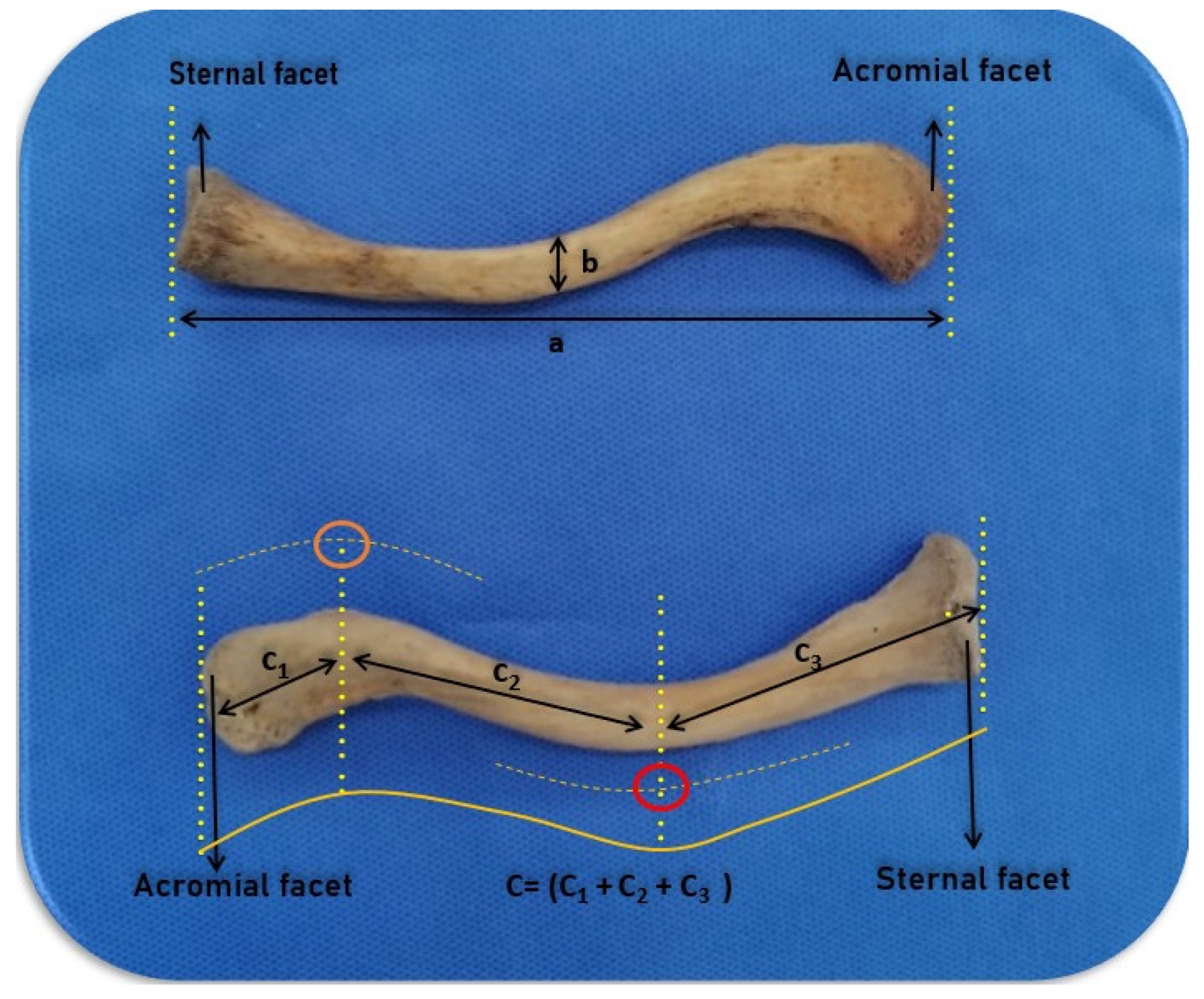
The projection length of the clavicle was defined as the linear distance between the most medial point of the sternal articular surface and the most lateral point of the acromial articular surface.
The actual clavicular length was assessed by dividing the bone into three sections based on the midpoints of its curvatures. These sections are:
- −
First part: The distance between the sternal articular surface and the midpoint of the sternoclavicular curvature.
- −
Second part: The distance between the midpoints of the sternoclavicular and the acromioclavicular curvatures.
- −
Third part: The distance between the midpoint of the acromioclavicular curvature and the acromial articular surface.
Parameters that were measured:
A1: Distance between the sternal articular surface and the midpoint of the sternoclavicular curvature.
A2: Distance between the midpoints of the sternoclavicular and the acromioclavicular curvatures.
A3: Distance between the midpoint of the acromioclavicular curvature and the acromial articular surface.
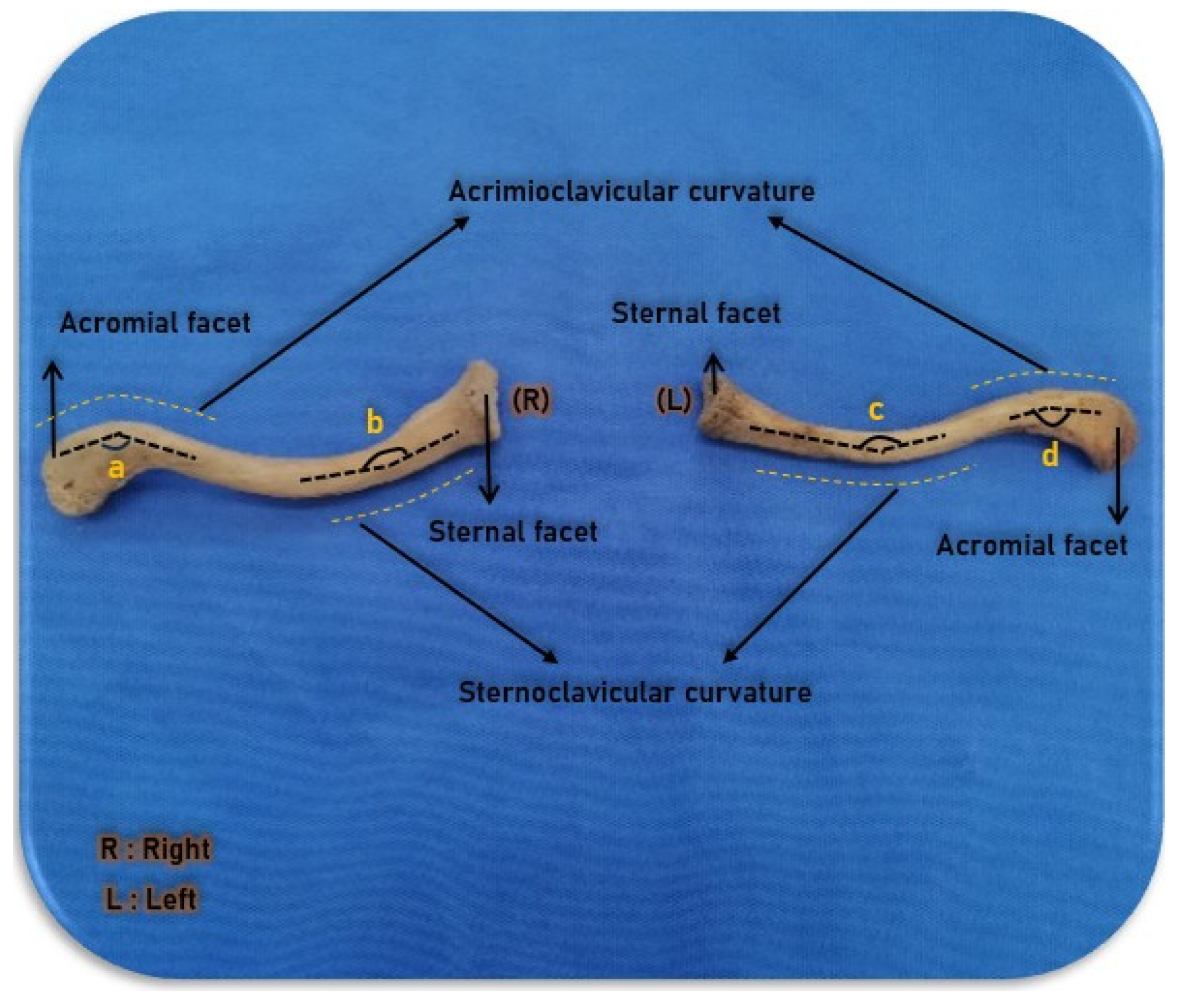
B: Sternoclavicular angle.
C: Acromioclavicular angle.
D: Thickness of the narrowest middle part of the clavicle.
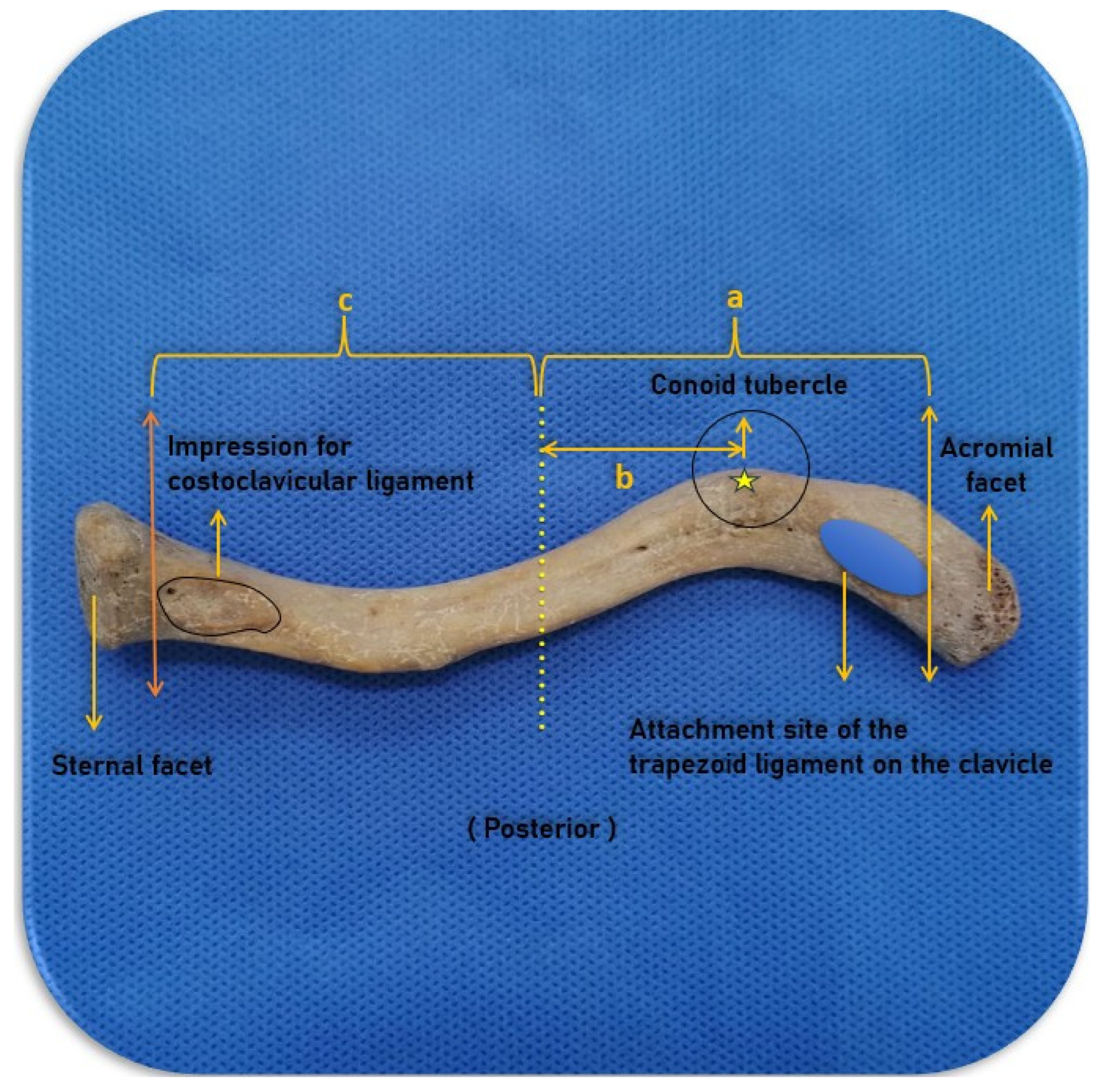
E: Projection distance between the middle part of the clavicle and the most prominent point of the conoid tubercle.
F: Distance between the middle part of the clavicle and the medial aspect of the trapezoid line.
G: Distance between the midline of the clavicle and the lateral impression for the costoclavicular ligament.
H: Projection length of the clavicle.
Statistical Analysis
Statistical analyses were performed using SPSS v.30.0. A
p value < 0.05 was considered statistically significant. Depending on the normality of distribution between groups, comparisons were conducted using an independent samples
t-test or a Mann–Whitney U test. The measurements were repeated 2 times by the same person using a 0.01 mm precision digital caliper, and the data obtained were averaged. (ICC = 0.991; %95 GA: 0.985–0.994;
p < 0.001). Minimum and maximum values, as well as the mean and standard deviation values, were calculated for each measured parameter. Measurement landmarks are illustrated in
Figure 1,
Figure 2 and
Figure 3.
3. Results
The mean distance between the sternal articular surface and the midpoint of the sternoclavicular curvature was found to be 49.54 ± 8.29 mm on the right side and 48.95 ± 9.37 mm on the left side. The mean distance between the midpoints of the sternoclavicular and the acromioclavicular curvatures was 66.29 ± 12.49 mm on the right side and 64.16 ± 11.86 mm on the left side. The mean distance between the midpoint of the acromioclavicular curvature and the acromial articular surface was 41.81 ± 5.05 mm on the right side and 41.38 ± 4.65 mm on the left side. The longest section on both sides was the second (midshaft) segment. The mean actual length of the right clavicle was found to be 157.66 ± 15.55 mm, and on the left clavicle, 154.50 ± 16.36 mm. No significant difference was observed between the mean actual length measurements of the right-sided and left-sided clavicles (
p > 0.05,
Table 1).
The mean sternoclavicular angle was found to be 150.67 ± 9.91° on the right side and 155.65 ± 8.57° on the left. The mean acromioclavicular angle was 120.71 ± 11.53° on the right side and 119.28 + 10.37° on the left. On both sides, the sternoclavicular angle was found to be greater than the acromioclavicular angle. A significant difference was observed between the right and left sides for the sternoclavicular angle (
p < 0.05,
Table 1), while no significant differences were observed for the acromioclavicular angle between the two sides (
p > 0.05,
Table 1).
The mean thickness of the narrowest midshaft part of the clavicle was found to be 11.47 ± 1.71 mm on the right side and 11.74 ± 1.81 mm on the left. No significant difference was observed between the thickness of the narrowest midshaft part of the right and left clavicles (
p > 0.05,
Table 1).
The mean distance between the midshaft part of the clavicle and the most prominent point of the conoid tubercle was 41.72 ± 6.14 mm on the right side and 45.65 ± 5.94 mm on the left, while the distance from the midshaft of the clavicle to the medial trapezoid ligament was found to be 48.31 ± 6.26 mm on average on the right side and 51.69 ± 5.95 mm on average on the left. This difference was found to be statistically significant (
p < 0.05,
Table 1). The mean distance between the midshaft of the clavicle and the lateral impression for the costoclavicular ligament was 42.18 ± 5.55 mm on the right side and 42.38 ± 6.19 mm on the left, with no significant difference between the two sides (
p > 0.05,
Table 1).
The clavicle projection length was found to be an average of 140.06 ± 11.31 mm on the right side and 145.15 ± 12.68 mm on the left. This difference between the two sides was found to be statistically significant (
p < 0.05,
Table 1). Descriptive statistics of the measured parameters are presented in
Table 1.
No correlation was observed between the sternoclavicular angle and the acromioclavicular angle on the right side (p = 0.188) or on the left side (p = 0.108).
On the right side, a significant correlation was observed only between the distance from the sternal articular surface and the midpoint of the sternoclavicular curvature and the distance from the midpoints of the sternoclavicular and the acromioclavicular curvatures (p = 0.00). On the left side, however, a significant correlation was found between the distance from the sternal articular surface and the midpoint of the sternoclavicular curvature and the distance from the midpoint of the acromioclavicular curvature and the acromial articular surface (p = 0.021).
A significant correlation was observed between the projection distance between the middle part of the clavicle and the most prominent point of the conoid tubercle and the distance between the middle part of the clavicle and the medial aspect of the trapezoid line (right and left: p = 0.00).
There is a significant correlation between the projection length and the true length of the clavicle on the right and left sides (right and left: p = 0.00).
4. Discussion
Accurate determination of the clavicular length is critical for assessing fracture-related shortening, which is a key criterion in surgical decision-making [
10]. In addition, shortening of the bone may also occur from malunion due to overlapping at fracture ends, which can be challenging to detect radiologically. Therefore, during assessment, the fractured clavicle is often compared to the contralateral healthy side under the assumption of bilateral symmetry [
12,
13]. However, as demonstrated in this study, right−left asymmetry can be more common than previously assumed, potentially leading to inaccurate evaluations and suboptimal decision-making. Thus, knowledge of separate morphometric data for each clavicle is essential. We believe that the findings of our study, which demonstrate that plates to be applied to the right side should be chosen to be longer than those for the left side, will serve as a guide for orthopaedic surgeons. We note that functional deficits may arise in treatments administered without taking right−left asymmetry into account. However, these results have been obtained from a single population, and their generalisability is limited.
There are numerous studies in the literature reporting clavicle length, and in these studies [
13,
14,
15,
16,
17,
18,
19,
20,
21,
22,
23] length measurement was performed as a projection or an attempt was made to calculate the actual length radiologically. King et al. [
24], however, measured the actual clavicle length radiologically but did not assess projection length. Kotekar et al. discussed the importance of clavicle length for maintaining shoulder biomechanics, but they did not measure the true length of the bone [
20]. In contrast, the current study evaluated both actual and projected lengths of the clavicle, which may provide a more accurate assessment of clavicular length and side-to-side differences relevant to postoperative evaluation. Although true length is preserved in reconstructions, if projection length is not preserved, functional loss may occur as a result of incorrect alignment of the bone. We therefore believe that measuring both lengths together may be clinically valuable.
Furthermore, the present study revealed that the actual length of each of the three parts of the clavicle was greater on the right side. However, when measured as a projection, the opposite was found, with the left side being longer. Mathieu et al. [
18] found that the length of the right clavicle was greater than that of the left. Lu et al. [
14] measured only left-sided clavicles and reported greater values than those observed in the left clavicles of the present study. Zhang et al. [
13] and King et al. [
24], in accordance with the current study, found that the length of the left clavicle was greater than that of the right clavicle. Akbulut et al. [
15] also found that the length of the left clavicle was greater. Our study was conducted on an Anatolian population. Whilst our findings are consistent with those of Akbulut et al. [
15], who worked with the same population, they do not appear to be consistent with the results of the study by Lu et al. This suggests that the morphometric characteristics of the clavicle may vary between different populations. In their study, Siheng-Qiu et al. [
19], as well as Kotekar et al. [
20], also highlighted the differences between populations.
Intramedullary fixation and plates are commonly used in the treatment of clavicle fractures, and successful application requires detailed morphometric knowledge [
25]. In addition to the external morphometry of the clavicle, knowledge of the medullary canal thickness is also important for intramedullary fixation via nailing [
18]. Mathieu et al. [
18] examined the medullary diameter of the bone, along with the thickness measurement at the midpoint of the clavicle, and found that this thickness was greater in the right-sided bones. Patted et al. [
25] examined the thickness measurement at the narrowest part of the clavicle in their study, and their findings are consistent with the results of the present study. Bachaura et al. [
17] performed radiological measurements of the cortex diameter in the middle part of the clavicle and did not measure the diameter externally. In this study, the diameter measurement was performed at the narrowest point of the middle part of the clavicle and the left-sided clavicles were found to be thicker at that point compared to the right ones. Most of the studies in the literature appear to be consistent with the findings of our study. Overall, these findings suggest that right-sided clavicles, characterized by smaller thickness, may be more prone to fracture.
A thorough understanding of the morphometry of the ligaments on the underside of the clavicle is essential, as some reconstructions may require ligament replacement and precise knowledge of their attachment sites [
15,
16]. The coracoclavicular ligament consists of two separate ligaments: the conoid ligament and the trapezoid ligament. The conoid ligament provides the most effective stabilisation against impacts to the superior and anterior parts of the bone [
7]. Akbulut et al. [
15] measured the distances from the conoid ligament’s widest attachment region to the acromial facet of the clavicle. In the current study, we instead measured the projection distance from the most prominent point of the conoid tubercle to the midpoint of the clavicle. The same approach was applied to the trapezoid ligament: whereas Akbulut et al. measured distances from the medial attachment to the anterior and posterior borders of the clavicle, we measured the distance to the clavicle’s midshaft. Measuring relative to the midpoint provides a consistent reference that can help define the lateral and medial attachment boundaries for ligament reconstruction. However, this has not been clinically confirmed.
Another ligament contributing to the stabilisation of the sternoclavicular joint is the costoclavicular ligament. This ligament forms the impression of the costoclavicular ligament on the clavicle [
7]. Güçlü et al. measured the length and width of the impression for the costoclavicular ligament [
26]. In the present study, we measured the distance between the middle part of the bone and the most distal part of the impression for the costoclavicular ligament, again using the midshaft of the clavicle as a reference point. We believe that identifying an anatomical reference point in ligament reconstructions will enhance surgical safety. However, we also note that the findings of this study need to be supported by clinical trials.
There are two curvatures in the clavicle. The first is the sternoclavicular curvature, which is convex on the medial side, and the second is the acromioclavicular curvature, which is concave on the lateral side [
24]. There is a distinct angle in the areas where these curvatures are located. If these angles are incorrectly reconstructed, bone lengthening or shortening may occur [
9,
12]. Furthermore, in intramedullary fixation, the sternoclavicular angle and acromioclavicular angle may cause difficulties in advancing the nail [
25]. In our study, the sternoclavicular angle was approximately 30–36° larger than the acromioclavicular angle on both sides. This difference suggests that undercorrection of the sternoclavicular angle during intramedullary fixation may lead to relative clavicular lengthening and potential functional impairment, whereas undercorrection of the smaller acromioclavicular angle is less likely to produce significant length discrepancies. These findings highlight the need for careful attention to the sternoclavicular angle in surgical planning. We also believe that this will serve as a guide for surgeons regarding the need for greater sternoclavicular angulation compared to acromioclavicular angulation during reconstructions.
A few limitations of this study should be considered. The specimens represent a single population, and the sex and age of the individuals were unknown. This limits the generalisability of the study. Moreover, all measurements were performed by a single observer; therefore, intra- and inter-observer reliability were not formally assessed. Furthermore, these morphometric data have not been clinically validated.