


Bioengineering, Volume 12, Issue 8 (August 2025) – 111 articles
Cover Story (view full-size image):
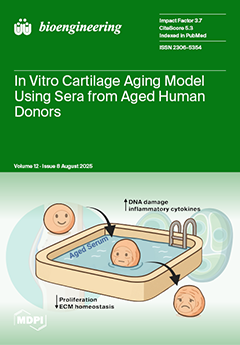
Osteoarthritis (OA) affects millions of people worldwide, with its prevalence increasing significantly as individuals age. However, the specific mechanisms by which aging leads to cartilage degeneration remain unclear. Despite decades of research, no in vitro model has successfully captured cartilage aging in a physiologically relevant manner. In this study, we address this gap by developing a reproducible system that utilizes human serum from elderly donors to simulate cartilage aging. After treatment with the aged serum, the cartilage constructs display key phenotypes associated with aging, including loss of the extracellular matrix, increased inflammation, and decreased regenerative capacity. This innovative platform provides a valuable, human-relevant tool for identifying the underlying factors driving cartilage aging and for accelerating the development of targeted therapies for OA. View this paper
- Issues are regarded as officially published after their release is announced to the table of contents alert mailing list.
- You may sign up for e-mail alerts to receive table of contents of newly released issues.
- PDF is the official format for papers published in both, html and pdf forms. To view the papers in pdf format, click on the "PDF Full-text" link, and use the free Adobe Reader to open them.
Previous Issue
Next Issue