Next Issue
Volume 14, January-1
Previous Issue
Volume 13, December-1


Diagnostics, Volume 13, Issue 24 (December-2 2023) – 92 articles

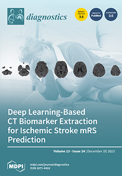
Cover Story (view full-size image): Accurately predicting stroke recovery outcomes, measured by the modified Rankin Scale (mRS), from brain CT scans remains challenging yet clinically valuable. We tested deep learning models to predict a patient's mRS at 3 months post-stroke. We experimented with image-only models that make predictions directly from CT scans, and hybrid models that incorporate clinical and demographic information along with imaging data. In the hybrid models, we first extracted quantitative imaging biomarkers reflective of stroke damage from CT scans using deep learning. These imaging features were then incorporated into prognostic machine learning models for making outcome predictions. This approach could help address the challenges faced by image-only approach and also make the resulting model more interpretable. View this paper
  • Issues are regarded as officially published after their release is announced to the table of contents alert mailing list.
  • You may sign up for e-mail alerts to receive table of contents of newly released issues.
  • PDF is the official format for papers published in both, html and pdf forms. To view the papers in pdf format, click on the "PDF Full-text" link, and use the free Adobe Reader to open them.
Order results
Result details
Section
Select all
Export citation of selected articles as:
14 pages, 3260 KiB  
Article
Impact of Voxel Normalization on a Machine Learning-Based Method: A Study on Pulmonary Nodule Malignancy Diagnosis Using Low-Dose Computed Tomography (LDCT)
by Chia-Chi Hsiao, Chen-Hao Peng, Fu-Zong Wu and Da-Chuan Cheng
Diagnostics 2023, 13(24), 3690; https://doi.org/10.3390/diagnostics13243690 - 18 Dec 2023
Cited by 1 | Viewed by 1331
Abstract
Lung cancer (LC) stands as the foremost cause of cancer-related fatality rates worldwide. Early diagnosis significantly enhances patient survival rate. Nowadays, low-dose computed tomography (LDCT) is widely employed on the chest as a tool for large-scale lung cancer screening. Nonetheless, a large amount [...] Read more.
Lung cancer (LC) stands as the foremost cause of cancer-related fatality rates worldwide. Early diagnosis significantly enhances patient survival rate. Nowadays, low-dose computed tomography (LDCT) is widely employed on the chest as a tool for large-scale lung cancer screening. Nonetheless, a large amount of chest radiographs creates an onerous burden for radiologists. Some computer-aided diagnostic (CAD) tools can provide insight to the use of medical images for diagnosis and can augment diagnostic speed. However, due to the variation in the parameter settings across different patients, substantial discrepancies in image voxels persist. We found that different voxel sizes can create a compromise between model generalization and diagnostic efficacy. This study investigates the performance disparities of diagnostic models trained on original images and LDCT images reconstructed to different voxel sizes while making isotropic. We examined the ability of our method to differentiate between benign and malignant nodules. Using 11 features, a support vector machine (SVM) was trained on LDCT images using an isotropic voxel with a side length of 1.5 mm for 225 patients in-house. The result yields a favorable model performance with an accuracy of 0.9596 and an area under the receiver operating characteristic curve (ROC/AUC) of 0.9855. In addition, to furnish CAD tools for clinical application, future research including LDCT images from multi-centers is encouraged. Full article
(This article belongs to the Section Medical Imaging and Theranostics)
Show Figures

Figure 1

12 pages, 285 KiB  
Review
Clinical Diagnosis and Treatment of Chronic Pain
by Sadiq Rahman, Ali Kidwai, Emiliya Rakhamimova, Murad Elias, William Caldwell and Sergio D. Bergese
Diagnostics 2023, 13(24), 3689; https://doi.org/10.3390/diagnostics13243689 - 18 Dec 2023
Viewed by 1595
Abstract
More than 600 million people globally are estimated to be living with chronic pain. It is one of the most common complaints seen in an outpatient setting, with over half of patients complaining of pain during a visit. Failure to properly diagnose and [...] Read more.
More than 600 million people globally are estimated to be living with chronic pain. It is one of the most common complaints seen in an outpatient setting, with over half of patients complaining of pain during a visit. Failure to properly diagnose and manage chronic pain is associated with substantial morbidity and mortality, especially when opioids are involved. Furthermore, it is a tremendous financial strain on the healthcare system, as over USD 100 billion is spent yearly in the United States on healthcare costs related to pain management and opioids. This exceeds the costs of diabetes, heart disease, and cancer-related care combined. Being able to properly diagnose, manage, and treat chronic pain conditions can substantially lower morbidity, mortality, and healthcare costs in the United States. This review will outline the current definitions, biopsychosocial model, subclassifications, somatosensory assessments, imaging, clinical prediction models, and treatment modalities associated with chronic pain. Full article
(This article belongs to the Special Issue Clinical Diagnosis and Treatment of Chronic Pain)
12 pages, 985 KiB  
Article
Performance Evaluation of Three Antibody Binding Assays, a Neutralizing Antibody Assay, and an Interferon-Gamma Release Assay for SARS-CoV-2 According to Vaccine Type in Vaccinated Group
by Minjeong Nam, Jae Hyun Cha, Sang-Wook Kim, Sun Bean Kim, Ki-Byung Lee, You-Seung Chung, Seung Gyu Yun, Myung-Hyun Nam, Chang Kyu Lee and Yunjung Cho
Diagnostics 2023, 13(24), 3688; https://doi.org/10.3390/diagnostics13243688 - 18 Dec 2023
Viewed by 869
Abstract
We evaluated the performance of SARS-CoV-2 assays in the vaccinated group using receptor-binding domain antibody assays (RBD Ab assay), neutralizing antibody assay (nAb assay), and interferon-gamma release assay (IGR assay). We also compared the performance of the SARS-CoV-2 assays based on vaccine type [...] Read more.
We evaluated the performance of SARS-CoV-2 assays in the vaccinated group using receptor-binding domain antibody assays (RBD Ab assay), neutralizing antibody assay (nAb assay), and interferon-gamma release assay (IGR assay). We also compared the performance of the SARS-CoV-2 assays based on vaccine type in a large population. We collected 1851 samples from vaccinated individuals with vector, mix-and-match (MM), and mRNA vaccines. The performance of the RBD Ab assays was assessed by SARS-CoV-2 IgG II Quant (Abbott Laboratories, Sligo, Ireland), SARS-CoV-2 IgG (Beckman Coulter, CA, USA), and anti-SARS-CoV-2 S (Roche Diagnostics GmbH, Mannheim, Germany). The nAb assay was assessed by cPass SARS-CoV-2 neutralization antibody detection kits (GenScript, NJ, USA). The IGR assay was assessed by QuantiFERON (Qiagen, Venlo, The Netherlands). Median values of the RBD Ab assays and nAb assay sequentially increased after the first and second vaccinations. RBD Ab assays and nAb assay showed very strong correlations. The median values of the RBD Ab, nAb, and IGR were higher in the mRNA vaccine group than in the vector and MM vaccine groups. The agreement and correlation among the RBD Ab assays, nAb assay, and IGR assay were higher in the mRNA vaccine group than in the vector and MM vaccine groups. We compared the performance of the RBD Ab assay, nAb assay, and IGR assay based on the vaccine types using the RBD Ab, nAb, and IGR assays. This study provides a better understanding of the assessment of humoral and cellular immune responses after vaccination. Full article
(This article belongs to the Section Diagnostic Microbiology and Infectious Disease)
Show Figures

Figure 1

14 pages, 1653 KiB  
Article
Comparison of Automated Keratometer and Scheimpflug Tomography for Predicting Refractive Astigmatism in Pseudophakic Eyes
by Kyung-Sun Na, Giacomo Savini, Woong-Joo Whang and Kristian Næser
Diagnostics 2023, 13(24), 3687; https://doi.org/10.3390/diagnostics13243687 - 18 Dec 2023
Viewed by 707
Abstract
Purpose: To analyse the correspondence between refractive astigmatism and corneal astigmatism in pseudophakic eyes with non-toric intraocular lenses. Setting: Yeouido St. Mary hospital, Seoul, Republic of Korea. Design: Evaluation of a diagnostic test instrument. Methods: This retrospective study included 95 eyes of 95 [...] Read more.
Purpose: To analyse the correspondence between refractive astigmatism and corneal astigmatism in pseudophakic eyes with non-toric intraocular lenses. Setting: Yeouido St. Mary hospital, Seoul, Republic of Korea. Design: Evaluation of a diagnostic test instrument. Methods: This retrospective study included 95 eyes of 95 patients. Corneal astigmatism was measured with an automated keratometer (RK-5, Canon) and Scheimpflug tomography (Pentacam HR, Oculus). Refractive astigmatism was compared to keratometric astigmatism (based on anterior corneal measurements only), equivalent K-reading, and total corneal astigmatism (both based on anterior and posterior corneal measurements). Vector analysis was carried out by Næser’s polar value method. The accuracy was defined as the average magnitude of the vectorial difference in astigmatism (DA). Each corneal measurement was optimized in retrospect by a multiple linear regression equation between refractive and corneal astigmatism. Results: Keratometric astigmatism overestimated with-the-rule (WTR) refractive astigmatism and underestimated against-the-rule (ATR) refractive astigmatism. Several measurements based on both corneal surfaces’ values did not show any statistically significant difference with respect to refractive astigmatism. The mean corneal astigmatism by total corneal refractive power (TCRP) at 4.0 mm (zone/pupil) produced the lowest mean arithmetic DA and the highest percentage of eyes with a DA ≤ 0.50 dioptre. After optimization, the accuracies of automated KA and TCRP 4.0 mm (zone/pupil) were similar. Conclusions: Total corneal astigmatism measured by Scheimpflug tomography at a 4.0 mm zone centered on the pupil accurately reflects the refractive astigmatism in pseudophakic eyes. However, the accuracy of total corneal astigmatism is not different from automated KA after optimization. Full article
Show Figures

Figure 1

10 pages, 900 KiB  
Brief Report
Performance of a Point-of-Care Fluorescence Immunoassay Test to Measure the Anti-Severe Acute Respiratory Syndrome Corona Virus 2 Spike, Receptor Binding Domain Antibody Level
by Marita Restie Tiara, Hofiya Djauhari, Febi Ramdhani Rachman, Antonius Christianus Rettob, Darmastuti Utami, Fahda Cintia Suci Pulungan, Heru Purwanta, Rudi Wisaksana, Bachti Alisjahbana and Agnes Rengga Indrati
Diagnostics 2023, 13(24), 3686; https://doi.org/10.3390/diagnostics13243686 - 18 Dec 2023
Cited by 1 | Viewed by 707
Abstract
Quantitative determination of anti-SARS-CoV2-S-RBD is necessary for the evaluation of vaccination effectiveness. The surrogate viral neutralization test (SVNT) is approved for measuring anti-SARS-CoV2-S-RBD, but a point-of-care platform is needed to simplify anti-SARS-CoV-2-S-RBD measurement. We aimed to evaluate the performance of a rapid fluorescent [...] Read more.
Quantitative determination of anti-SARS-CoV2-S-RBD is necessary for the evaluation of vaccination effectiveness. The surrogate viral neutralization test (SVNT) is approved for measuring anti-SARS-CoV2-S-RBD, but a point-of-care platform is needed to simplify anti-SARS-CoV-2-S-RBD measurement. We aimed to evaluate the performance of a rapid fluorescent immunoassay-based kit, FastBio-RBDTM, compared to the SVNT. During April–September 2021, we enrolled two groups of subjects, convalescent subjects and subjects without a COVID-19 history. The subjects were tested for the anti-SARS-CoV2-S-RBD antibody using FastBio-RBDTM and the GenScript-cPASSTM SVNT. We measured the correlation coefficient and conducted an ROC analysis to determine the best cut-off value of anti-SARS-CoV2-S-RBD against the SVNT percent inhibition levels of 30% and 60%. We included 109 subjects. Anti-SARS-CoV-2-S-RBD strongly correlated to SVNT % inhibition with an R value of 0.866 (p < 0.0001). The ROC analysis showed that the anti-SARS-CoV-2-S-RBD of 6.71 AU/mL had 95.7% sensitivity and 87.5% specificity to detect a percentage inhibition of 30%. The anti-SARS-CoV-2-S-RBD of 59.76 AU/mL had a sensitivity of 88.1% and specificity of 97.0% to detect a percentage inhibition of 60%. FastBio-RBDTM could determine the presence and level of anti-SARS-CoV-2-S-RBD with good sensitivity and specificity. It has the potential to be deployed in health facilities with limited resources. Full article
(This article belongs to the Section Diagnostic Microbiology and Infectious Disease)
Show Figures

Figure 1

13 pages, 2898 KiB  
Article
Immunohistochemical Stain-Aided Annotation Accelerates Machine Learning and Deep Learning Model Development in the Pathologic Diagnosis of Nasopharyngeal Carcinoma
by Tai-Pei Lin, Chiou-Ying Yang, Ko-Jiunn Liu, Meng-Yuan Huang and Yen-Lin Chen
Diagnostics 2023, 13(24), 3685; https://doi.org/10.3390/diagnostics13243685 - 18 Dec 2023
Viewed by 1070
Abstract
Nasopharyngeal carcinoma (NPC) is an epithelial cancer originating in the nasopharynx epithelium. Nevertheless, annotating pathology slides remains a bottleneck in the development of AI-driven pathology models and applications. In the present study, we aim to demonstrate the feasibility of using immunohistochemistry (IHC) for [...] Read more.
Nasopharyngeal carcinoma (NPC) is an epithelial cancer originating in the nasopharynx epithelium. Nevertheless, annotating pathology slides remains a bottleneck in the development of AI-driven pathology models and applications. In the present study, we aim to demonstrate the feasibility of using immunohistochemistry (IHC) for annotation by non-pathologists and to develop an efficient model for distinguishing NPC without the time-consuming involvement of pathologists. For this study, we gathered NPC slides from 251 different patients, comprising hematoxylin and eosin (H&E) slides, pan-cytokeratin (Pan-CK) IHC slides, and Epstein–Barr virus-encoded small RNA (EBER) slides. The annotation of NPC regions in the H&E slides was carried out by a non-pathologist trainee who had access to corresponding Pan-CK IHC slides, both with and without EBER slides. The training process utilized ResNeXt, a deep neural network featuring a residual and inception architecture. In the validation set, NPC exhibited an AUC of 0.896, with a sensitivity of 0.919 and a specificity of 0.878. This study represents a significant breakthrough: the successful application of deep convolutional neural networks to identify NPC without the need for expert pathologist annotations. Our results underscore the potential of laboratory techniques to substantially reduce the workload of pathologists. Full article
(This article belongs to the Special Issue Applications of Artificial Intelligence in Healthcare Monitoring)
Show Figures

Figure 1

9 pages, 3054 KiB  
Brief Report
The Histology-Driven Differential Diagnosis in Bowel Inflammatory Conditions Is Not All That Obvious: Evidence from a Survey Based on Digital Slides
by Gabriella Canavese, Enrico Costantino Falco, Nuria Perez-Diaz-del-Campo, Gian Paolo Caviglia, Fabrizia Di Giovanni and Davide Giuseppe Ribaldone
Diagnostics 2023, 13(24), 3684; https://doi.org/10.3390/diagnostics13243684 - 18 Dec 2023
Viewed by 930
Abstract
(1) Background: when the pathologist faces histologic slides from colonoscopies in daily practice, given the large number of entities and etiologies under inflammatory bowel conditions, in-depth definition of the histological spectrum and the recommendations of current guidelines are often not enough to conclusively [...] Read more.
(1) Background: when the pathologist faces histologic slides from colonoscopies in daily practice, given the large number of entities and etiologies under inflammatory bowel conditions, in-depth definition of the histological spectrum and the recommendations of current guidelines are often not enough to conclusively define a diagnostic framework. Histological patterns should be organized hierarchically in flowcharts that consider the correlation with clinical data. We conducted an online survey asking a group of gastroenteropathologists to apply a pattern classification based on the most significant lesions in colitis differential diagnosis: crypt distortion and activity. (2) Methods: digital slides from 20 endoscopy samples were analyzed by twenty pathologists and classified according to the occurrence of crypt distortion (nondestructive–destructive colitis) and subsequently to the evidence of activity (ND1-2-3, D1-2). (3) Results: in 8 out of 20 (40%) cases, the participants reached a full agreement regarding the evaluation of crypt distortion (5 cases: nondestructive colitis; 3 cases: destructive colitis). The calculated agreement was k = 0.432. In the second-level quiz (ND1-2-3 and D1-2), full agreement between participants was achieved for 7 of the 28 (25%) possible classifications, with k = 0.229. (4) Conclusions: The findings from this survey are indicative of an unexpectedly low consensus, even among dedicated pathologists, about the recognition of histological changes that are commonly considered critical lesions in the histologic identification of bowel non-neoplastic diseases. In our opinion, these divergences imply a significant risk of misdiagnosis of bowel inflammatory conditions, hampering the usefulness of histological assessment. Full article
(This article belongs to the Section Pathology and Molecular Diagnostics)
Show Figures

Figure 1

12 pages, 2780 KiB  
Article
ECMO in Myocardial Infarction-Associated Cardiogenic Shock: Blood Biomarkers as Predictors of Mortality
by Thomas Senoner, Benedikt Treml, Robert Breitkopf, Ulvi Cenk Oezpeker, Nicole Innerhofer, Christine Eckhardt, Aleksandra Radovanovic Spurnic and Sasa Rajsic
Diagnostics 2023, 13(24), 3683; https://doi.org/10.3390/diagnostics13243683 - 17 Dec 2023
Viewed by 744
Abstract
Background: Veno-arterial extracorporeal membrane oxygenation (va-ECMO) can provide circulatory and respiratory support in patients with cardiogenic shock. The main aim of this work was to investigate the association of blood biomarkers with mortality in patients with myocardial infarction needing va-ECMO support. Methods: We [...] Read more.
Background: Veno-arterial extracorporeal membrane oxygenation (va-ECMO) can provide circulatory and respiratory support in patients with cardiogenic shock. The main aim of this work was to investigate the association of blood biomarkers with mortality in patients with myocardial infarction needing va-ECMO support. Methods: We retrospectively analyzed electronic medical charts from patients receiving va-ECMO support in the period from 2008 to 2021 at the Medical University Innsbruck, Department of Anesthesiology and Intensive Care Medicine. Results: Of 188 patients, 57% (108/188) survived to discharge, with hemorrhage (46%) and thrombosis (27%) as the most frequent adverse events. Procalcitonin levels were markedly higher in non-survivors compared with survivors during the observation period. The multivariable model identified higher blood levels of procalcitonin (HR 1.01, p = 0.002) as a laboratory parameter associated with a higher risk of mortality. Conclusions: In our study population of patients with myocardial infarction-associated cardiogenic shock, deceased patients had increased levels of inflammatory blood biomarkers throughout the whole study period. Increased procalcitonin levels have been associated with a higher risk of mortality. Future studies are needed to show the role of procalcitonin in patients receiving ECMO support. Full article
(This article belongs to the Special Issue Diagnostics in Critical Care)
Show Figures

Figure 1

13 pages, 3340 KiB  
Systematic Review
Clinical Validity of Anti-Proteinase 3 Antibodies in Patients with Inflammatory Bowel Disease: A Short Meta-Analysis
by Carmen Andalucía, Laura Martínez-Prat, Chelsea Bentow, Mary Ann Aure, Michael P. Horn and Michael Mahler
Diagnostics 2023, 13(24), 3682; https://doi.org/10.3390/diagnostics13243682 - 16 Dec 2023
Viewed by 1310
Abstract
Anti-neutrophil cytoplasmic antibodies (ANCA) directed to proteinase 3 (PR3) represent highly established markers for patients with ANCA-associated vasculitis (AAV). PR3-ANCA have also demonstrated utility in the management of inflammatory bowel disease (IBD). More specifically, PR3-ANCA discriminate individuals with ulcerative colitis (UC) from Crohn’s [...] Read more.
Anti-neutrophil cytoplasmic antibodies (ANCA) directed to proteinase 3 (PR3) represent highly established markers for patients with ANCA-associated vasculitis (AAV). PR3-ANCA have also demonstrated utility in the management of inflammatory bowel disease (IBD). More specifically, PR3-ANCA discriminate individuals with ulcerative colitis (UC) from Crohn’s disease (CD) patients and are associated with disease severity, activity, and treatment non-response. Here, we aim to summarize the current data on the diagnostic utility of PR3-ANCA in IBD. A structured, systematic literature review, including three electronic databases, was conducted on June 6th, 2023, to identify studies assessing the diagnostic accuracy of the QUANTA Flash® PR3 assay in UC vs. CD patients. Electronic searches were supplemented by hand searching. A hierarchical, bivariate, mixed-effect meta-analysis was conducted using the metandi function, as per the Cochrane collaboration recommendations. Study quality was assessed using the QUADAS-2 tool, which considers the risk of bias and applicability. Six out of a hundred and eleven citations met the inclusion criteria and reported QUANTA Flash® PR3 diagnostic accuracy in UC vs. CD (UC, n = 667, CD, n = 682 patients). The sensitivity/specificity point estimate for UC was 34.9%/95.9%. This resulted in a Diagnostic Odds Ratio (DOR) of 12.6. The risk of bias was low in the index test and reference standard domains. Four of the six studies (67%) showed an unclear risk of bias in patient selection and in flow and timing domains. All studies had low concerns about applicability in all the domains. PR3-ANCA measured with the QUANTA Flash® PR3 assay represent novel diagnostic markers in IBD and enables discrimination between UC and CD. Full article
Show Figures

Figure 1

13 pages, 2157 KiB  
Article
Prehospital Targeting of 1-Year Mortality in Acute Chest Pain by Cardiac Biomarkers
by Daniel Zalama-Sánchez, Francisco Martín-Rodríguez, Raúl López-Izquierdo, Juan F. Delgado Benito, Irene Sánchez Soberón, Carlos del Pozo Vegas and Ancor Sanz-García
Diagnostics 2023, 13(24), 3681; https://doi.org/10.3390/diagnostics13243681 - 16 Dec 2023
Viewed by 736
Abstract
The identification and appropriate management of patients at risk of suffering from acute chest pain (ACP) in prehospital care are not straightforward. This task could benefit, as occurs in emergency departments (EDs), from cardiac enzyme assessment. The aim of the present work was [...] Read more.
The identification and appropriate management of patients at risk of suffering from acute chest pain (ACP) in prehospital care are not straightforward. This task could benefit, as occurs in emergency departments (EDs), from cardiac enzyme assessment. The aim of the present work was to derive and validate a scoring system based on troponin T (cTnT), N-terminal pro B-type natriuretic peptide (NT-proBNP), and D-dimer to predict 1-year mortality in patients with ACP. This was a prospective, multicenter, ambulance-based cohort study of adult patients with a prehospital ACP diagnosis who were evacuated by ambulance to the ED between October 2019 and July 2021. The primary outcome was 365-day cumulative mortality. A total of 496 patients fulfilled the inclusion criteria. The mortality rate was 12.1% (60 patients). The scores derived from cTnT, NT-proBNP, and D-dimer presented an AUC of 0.802 (95% CI: 0718-0.886) for 365-day mortality. This AUC was superior to that of each individual cardiac enzyme. Our study provides promising evidence for the predictive value of a risk score based on cTnT, NT-proBNP, and D-dimer for the prediction of 1-year mortality in patients with ACP. The implementation of this score has the potential to benefit emergency medical service care and facilitate the on-scene decision-making process. Full article
(This article belongs to the Special Issue Novel Methods and Devices for Monitoring in Critical Care)
Show Figures

Figure 1

18 pages, 317 KiB  
Review
Evolution of Systemic Therapy in Medulloblastoma Including Irradiation-Sparing Approaches
by Naureen Mushtaq, Rahat Ul Ain, Syed Ahmer Hamid and Eric Bouffet
Diagnostics 2023, 13(24), 3680; https://doi.org/10.3390/diagnostics13243680 - 16 Dec 2023
Cited by 1 | Viewed by 1249
Abstract
The management of medulloblastoma in children has dramatically changed over the past four decades, with the development of chemotherapy protocols aiming at improving survival and reducing long-term toxicities of high-dose craniospinal radiotherapy. While the staging and treatment of medulloblastoma were until recently based [...] Read more.
The management of medulloblastoma in children has dramatically changed over the past four decades, with the development of chemotherapy protocols aiming at improving survival and reducing long-term toxicities of high-dose craniospinal radiotherapy. While the staging and treatment of medulloblastoma were until recently based on the modified Chang’s system, recent advances in the molecular biology of medulloblastoma have revolutionized approaches in the management of this increasingly complex disease. The evolution of systemic therapies is described in this review. Full article
(This article belongs to the Special Issue Medulloblastoma—Existing and Evolving Landscape)
13 pages, 1917 KiB  
Article
Whole Brain and Corpus Callosum Fractional Anisotropy Differences in Patients with Cognitive Impairment
by Kalvis Kaļva, Nauris Zdanovskis, Kristīne Šneidere, Andrejs Kostiks, Guntis Karelis, Ardis Platkājis and Ainārs Stepens
Diagnostics 2023, 13(24), 3679; https://doi.org/10.3390/diagnostics13243679 - 16 Dec 2023
Viewed by 1460
Abstract
Diffusion tensor imaging (DTI) is an MRI analysis method that could help assess cognitive impairment (CI) in the ageing population more accurately. In this research, we evaluated fractional anisotropy (FA) of whole brain (WB) and corpus callosum (CC) in patients with normal cognition [...] Read more.
Diffusion tensor imaging (DTI) is an MRI analysis method that could help assess cognitive impairment (CI) in the ageing population more accurately. In this research, we evaluated fractional anisotropy (FA) of whole brain (WB) and corpus callosum (CC) in patients with normal cognition (NC), mild cognitive impairment (MCI), and moderate/severe cognitive impairment (SCI). In total, 41 participants were included in a cross-sectional study and divided into groups based on Montreal Cognitive Assessment (MoCA) scores (NC group, nine participants, MCI group, sixteen participants, and SCI group, sixteen participants). All participants underwent an MRI examination that included a DTI sequence. FA values between the groups were assessed by analysing FA value and age normative percentile. We did not find statistically significant differences between the groups when analysing CC FA values. Both approaches showed statistically significant differences in WB FA values between the MCI-SCI and MCI-NC groups, where the MCI group participants showed the highest mean FA and highest mean FA normative percentile results in WB. Full article
(This article belongs to the Special Issue Neuropathology, Neuroimaging and Biomarkers in Neurological Disease)
Show Figures

Figure 1

12 pages, 562 KiB  
Article
Development of Quality Indicators for the Ultrasound Department through a Modified Delphi Method
by Aiping Zeng, Yang Gu, Li Ma, Xixi Tao, Luying Gao, Jianchu Li, Hongyan Wang and Yuxin Jiang
Diagnostics 2023, 13(24), 3678; https://doi.org/10.3390/diagnostics13243678 - 15 Dec 2023
Viewed by 804
Abstract
This study aims to establish precise quality indicators for evaluating and enhancing ultrasound performance, employing a methodology based on a comprehensive review of the literature, expert insights, and practical application experiences. We conducted a thorough review of both the domestic and international literature [...] Read more.
This study aims to establish precise quality indicators for evaluating and enhancing ultrasound performance, employing a methodology based on a comprehensive review of the literature, expert insights, and practical application experiences. We conducted a thorough review of both the domestic and international literature on ultrasound quality control to identify potential indicators. A dedicated team was formed to oversee the complete indicator development process. Utilizing a three-round modified Delphi method, we sought expert opinions through personalized email correspondence. Subsequently, data from diverse hospital indicators were collected to validate and assess feasibility. A novel set of seven indicators was compiled initially, followed by the convening of a 36-member nationally representative expert panel. After three rounds of meticulous revisions, consensus was reached on 13 indicators across three domains. These finalized indicators underwent application in various hospital settings, demonstrating their initial validity and feasibility. The development of thirteen ultrasound quality indicators represents a significant milestone in evaluating ultrasound performance. These indicators empower hospitals to monitor changes in quality effectively, fostering efficient quality management practices. Full article
(This article belongs to the Section Medical Imaging and Theranostics)
Show Figures

Figure 1

22 pages, 3367 KiB  
Systematic Review
Artificial Intelligence and Its Clinical Applications in Orthodontics: A Systematic Review
by Gianna Dipalma, Alessio Danilo Inchingolo, Angelo Michele Inchingolo, Fabio Piras, Vincenzo Carpentiere, Grazia Garofoli, Daniela Azzollini, Merigrazia Campanelli, Gregorio Paduanelli, Andrea Palermo and Francesco Inchingolo
Diagnostics 2023, 13(24), 3677; https://doi.org/10.3390/diagnostics13243677 - 15 Dec 2023
Cited by 1 | Viewed by 1347
Abstract
This review aims to analyze different strategies that make use of artificial intelligence to enhance diagnosis, treatment planning, and monitoring in orthodontics. Orthodontics has seen significant technological advancements with the introduction of digital equipment, including cone beam computed tomography, intraoral scanners, and software [...] Read more.
This review aims to analyze different strategies that make use of artificial intelligence to enhance diagnosis, treatment planning, and monitoring in orthodontics. Orthodontics has seen significant technological advancements with the introduction of digital equipment, including cone beam computed tomography, intraoral scanners, and software coupled to these devices. The use of deep learning in software has sped up image processing processes. Deep learning is an artificial intelligence technology that trains computers to analyze data like the human brain does. Deep learning models are capable of recognizing complex patterns in photos, text, audio, and other data to generate accurate information and predictions. Materials and Methods: Pubmed, Scopus, and Web of Science were used to discover publications from 1 January 2013 to 18 October 2023 that matched our topic. A comparison of various artificial intelligence applications in orthodontics was generated. Results: A final number of 33 studies were included in the review for qualitative analysis. Conclusions: These studies demonstrate the effectiveness of AI in enhancing orthodontic diagnosis, treatment planning, and assessment. A lot of articles emphasize the integration of artificial intelligence into orthodontics and its potential to revolutionize treatment monitoring, evaluation, and patient outcomes. Full article
(This article belongs to the Special Issue Artificial Intelligence in Dental Medicine)
Show Figures

Figure 1

4 pages, 1818 KiB  
Interesting Images
Immunohistochemistry in an Adult Case of Bitot’s Spots Caused by Vitamin A Deficiency
by Hideki Fukuoka, Norihiko Yokoi and Chie Sotozono
Diagnostics 2023, 13(24), 3676; https://doi.org/10.3390/diagnostics13243676 - 15 Dec 2023
Viewed by 906
Abstract
Bitot’s spots (BS) are the buildup of superficially located keratin in the conjunctiva and are early indicators of vitamin A deficiency (VAD), primarily due to malnutrition and malabsorption, thus leading to xerophthalmia. BS are particularly prevalent in developing countries, and their presence necessitates [...] Read more.
Bitot’s spots (BS) are the buildup of superficially located keratin in the conjunctiva and are early indicators of vitamin A deficiency (VAD), primarily due to malnutrition and malabsorption, thus leading to xerophthalmia. BS are particularly prevalent in developing countries, and their presence necessitates prompt vitamin A supplementation to avert blindness, with the immunohistochemical characteristics of BS aiding in understanding the extent of epithelial abnormalities and the efficacy of vitamin A supplementation. We describe the case of a 34-year-old male with persistent BS despite extensive vitamin A supplementation and topical treatments who underwent surgical excision of the BS followed by amniotic membrane transplantation, thus resulting in symptom relief and epithelialization, with no recurrence observed during follow-up. Histopathologic and immunohistochemical evaluations revealed expression of keratinization-related proteins, along with an absence of mucin-5AC-positive cells, suggesting impaired differentiation into goblet cells due to VAD. This case highlights the potential age-related disparity in the efficacy of vitamin A supplementation, emphasizing the need for early detection and a multidisciplinary approach in the management of VAD, especially in young adults. The favorable outcome of surgical intervention highlights its viability in the management of persistent BS and encourages further investigation to optimize therapeutic strategies for VAD-related ocular manifestations. Full article
(This article belongs to the Special Issue Diagnosis, Treatment and Management of Eye Diseases)
Show Figures

Figure 1

13 pages, 1008 KiB  
Article
Variety of Serotonin Levels in Pediatric Gastrointestinal Disorders
by Loredana Matiș, Lucia Georgeta Daina, Lavinia Maris, Timea Claudia Ghitea, Daniela Florina Trifan, Ioana Moga and Radu Fodor
Diagnostics 2023, 13(24), 3675; https://doi.org/10.3390/diagnostics13243675 - 15 Dec 2023
Viewed by 959
Abstract
(1) Serotonin primarily regulates our emotions. A complex process, which includes dysfunctions in gastrointestinal motility and deregulation of the gene responsible for serotonin reuptake (SERT), is implicated in the pathophysiology of irritable bowel syndrome (IBS). This also encompasses changes in intestinal microbiota, the [...] Read more.
(1) Serotonin primarily regulates our emotions. A complex process, which includes dysfunctions in gastrointestinal motility and deregulation of the gene responsible for serotonin reuptake (SERT), is implicated in the pathophysiology of irritable bowel syndrome (IBS). This also encompasses changes in intestinal microbiota, the response to stress, the intricate interplay between the brain and the digestive tract, heightened sensitivity to visceral stimuli, and low-grade inflammation. This paper aims to investigate the effectiveness of probiotic therapy in managing gastrointestinal and neuropsychiatric symptoms related to serotonin levels, with a focus on individuals with serotonin deficiency and those with normal serotonin levels experiencing gastrointestinal disorders. (2) The study involved 135 pediatric patients aged 5–18 years with gastrointestinal disturbances, including constipation, diarrhea, and other symptoms, such as nausea, flatulence, feeling full, or gastrointestinal pain. (3) Serotonin testing was performed, and administering probiotics appeared to be effective in addressing serotonin deficiency and other gastrointestinal disorders. (4) Serotonin’s pivotal role in regulating neurotransmitter secretion and its impact on neuropsychiatric health, coupled with gender differences and age-related declines, underscore the complexity of their influence on gastrointestinal and neuropsychiatric conditions. Full article
(This article belongs to the Section Pathology and Molecular Diagnostics)
Show Figures

Graphical abstract

11 pages, 1199 KiB  
Article
Cardiac Involvement in Classical Organic Acidurias: Clinical Profile and Outcome in a Pediatric Cohort
by Silvia Passantino, Serena Chiellino, Francesca Girolami, Mattia Zampieri, Giovanni Battista Calabri, Gaia Spaziani, Elena Bennati, Giulio Porcedda, Elena Procopio, Iacopo Olivotto and Silvia Favilli
Diagnostics 2023, 13(24), 3674; https://doi.org/10.3390/diagnostics13243674 - 15 Dec 2023
Viewed by 681
Abstract
Background: Cardiac involvement is reported in a significant proportion of patients with classical organic acidurias (OAs), contributing to disability and premature death. Different cardiac phenotypes have been described, among which dilated cardiomyopathy (DCM) is predominant. Despite recent progress in diagnosis and treatment, the [...] Read more.
Background: Cardiac involvement is reported in a significant proportion of patients with classical organic acidurias (OAs), contributing to disability and premature death. Different cardiac phenotypes have been described, among which dilated cardiomyopathy (DCM) is predominant. Despite recent progress in diagnosis and treatment, the natural history of patients with OAs remains unresolved, specifically with regard to the impact of cardiac complications. We therefore performed a retrospective study to address this issue at our Referral Center for Pediatric Inherited Errors of Metabolism. Methods: Sixty patients with OAs (propionic (PA), methylmalonic (MMA) and isovaleric acidemias and maple syrup urine disease) diagnosed from 2000 to 2022 were systematically assessed at baseline and at follow-up. Results: Cardiac anomalies were found in 23/60 OA patients, all with PA or MMA, represented by DCM (17/23 patients) and/or acquired long QT syndrome (3/23 patients). The presence of DCM was associated with the worst prognosis. The rate of occurrence of major adverse cardiac events (MACEs) at 5 years was 55% in PA with cardiomyopathy; 35% in MMA with cardiomyopathy; and 23% in MMA without cardiomyopathy. Liver transplantation was performed in seven patients (12%), all with PA or MMA, due to worsening cardiac impairment, and led to the stabilization of metabolic status and cardiac function. Conclusions: Cardiac involvement was documented in about one third of children diagnosed with classical OAs, confined to PA and MMA, and was often associated with poor outcome in over 50%. Etiological diagnosis of OAs is essential in guiding management and risk stratification. Full article
Show Figures

Figure 1

21 pages, 721 KiB  
Review
Advancing Colorectal Cancer Diagnosis with AI-Powered Breathomics: Navigating Challenges and Future Directions
by Ioannis K. Gallos, Dimitrios Tryfonopoulos, Gidi Shani, Angelos Amditis, Hossam Haick and Dimitra D. Dionysiou
Diagnostics 2023, 13(24), 3673; https://doi.org/10.3390/diagnostics13243673 - 15 Dec 2023
Cited by 1 | Viewed by 1280
Abstract
Early detection of colorectal cancer is crucial for improving outcomes and reducing mortality. While there is strong evidence of effectiveness, currently adopted screening methods present several shortcomings which negatively impact the detection of early stage carcinogenesis, including low uptake due to patient discomfort. [...] Read more.
Early detection of colorectal cancer is crucial for improving outcomes and reducing mortality. While there is strong evidence of effectiveness, currently adopted screening methods present several shortcomings which negatively impact the detection of early stage carcinogenesis, including low uptake due to patient discomfort. As a result, developing novel, non-invasive alternatives is an important research priority. Recent advancements in the field of breathomics, the study of breath composition and analysis, have paved the way for new avenues for non-invasive cancer detection and effective monitoring. Harnessing the utility of Volatile Organic Compounds in exhaled breath, breathomics has the potential to disrupt colorectal cancer screening practices. Our goal is to outline key research efforts in this area focusing on machine learning methods used for the analysis of breathomics data, highlight challenges involved in artificial intelligence application in this context, and suggest possible future directions which are currently considered within the framework of the European project ONCOSCREEN. Full article
Show Figures

Figure 1

20 pages, 9173 KiB  
Article
Design and Simulate Intracranial Support to Guide Maxillo Surgery: A Study Based on Bioengineering
by Maria Giulia Cristofaro, Elvis Kallaverja, Francesco Ferragina and Ida Barca
Diagnostics 2023, 13(24), 3672; https://doi.org/10.3390/diagnostics13243672 - 14 Dec 2023
Viewed by 790
Abstract
Background: Intraoperative navigation allows for the creation of a real-time relationship between the anatomy imagined during diagnosis/planning and the site of surgical interest. This procedure takes place by identifying and registering trustworthy anatomical markers on planning images and using a point locator during [...] Read more.
Background: Intraoperative navigation allows for the creation of a real-time relationship between the anatomy imagined during diagnosis/planning and the site of surgical interest. This procedure takes place by identifying and registering trustworthy anatomical markers on planning images and using a point locator during the operation. The locator is calibrated in the workspace by placing a Dynamic Reference Frame (DRF) sensor. Objective: This study aims to calculate the localization accuracy of an electromagnetic locator of neuro-maxillofacial surgery, moving the standard sensor position to a different position more suitable for maxillofacial surgery. Materials and Methods: The upper dental arch was chosen as an alternative fixed point for the positioning of the sensor. The prototype of a bite support device was designed and generated via 3D printing. CT images of a skull phantom with 10 anatomical landmarks were acquired. The testing procedure consisted of 10 measurements for each position of the sensor: precisely 10 measurements with the sensor placed on the forehead and 10 measurements with the sensor placed on the bite support device. It also evaluated the localization error by comparing the two procedures. Results: The localization error, when the sensor was placed on the bite support device, was lower in the sphere located on the temporal bone. It was the same in the spheres located on the maxillary bone. The test analysis of the data of the new device showed that it is reliable; the tests are reproducible and can be considered as accurate as the traditional ones. In addition, the sensor mounted on this device has proven to be slightly superior in terms of accuracy and accuracy in areas such as the middle third of the face and jaw. Discussion and Conclusion: The realization of the bite support device allowed the sensor to change position concerning its natural site. This procedure allows us to explore structures, such as the frontal site, which were initially difficult to approach with neuronavigation and improves the approach to midface structures, already studied with neuronavigation. The new calibration, with the position of the sensor on the support device in the same reference points sphere, highlighted the reduction in the location error. We can say that the support proposed in this study lays the foundations for a new navigation approach for patients in maxillofacial surgery, by changing the position of the sensor. It has strong points in improving the localization error for some reference points without determining disadvantages both in the calibration and in the surgical impediment. Full article
(This article belongs to the Section Point-of-Care Diagnostics and Devices)
Show Figures

Figure 1

4 pages, 205 KiB  
Editorial
AI/ML-Based Medical Image Processing and Analysis
by Jaafar Alghazo and Ghazanfar Latif
Diagnostics 2023, 13(24), 3671; https://doi.org/10.3390/diagnostics13243671 - 14 Dec 2023
Viewed by 1115
Abstract
The medical field is experiencing remarkable advancements, notably with the latest technologies—artificial intelligence (AI), big data, high-performance computing (HPC), and high-throughput computing (HTC)—that are in place to offer groundbreaking solutions to support medical professionals in the diagnostic process [...] Full article
(This article belongs to the Special Issue AI/ML-Based Medical Image Processing and Analysis)
18 pages, 1681 KiB  
Article
The Challenge to Stabilize, Extract and Analyze Urinary Cell-Free DNA (ucfDNA) during Clinical Routine
by Ivonne Nel, Carolin Münch, Saikal Shamkeeva, Mitja L. Heinemann, Berend Isermann and Bahriye Aktas
Diagnostics 2023, 13(24), 3670; https://doi.org/10.3390/diagnostics13243670 - 14 Dec 2023
Viewed by 971
Abstract
Background: The “Liquid Biopsy” has become a powerful tool for cancer research during the last decade. Circulating cell-free DNA (cfDNA) that originates from tumors has emerged as one of the most promising analytes. In contrast to plasma-derived cfDNA, only a few studies have [...] Read more.
Background: The “Liquid Biopsy” has become a powerful tool for cancer research during the last decade. Circulating cell-free DNA (cfDNA) that originates from tumors has emerged as one of the most promising analytes. In contrast to plasma-derived cfDNA, only a few studies have investigated urinary cfDNA. One reason might be rapid degradation and hence inadequate concentrations for downstream analysis. In this study, we examined the stability of cfDNA in urine using different methods of preservation under various storage conditions. Methodology: To mimic patient samples, a pool of healthy male and female urine donors was spiked with a synthetic cfDNA reference standard (fragment size 170 bp) containing the T790M mutation in the EGFR gene. Spiked samples were preserved with three different buffers and with no buffer over four different storage periods (0 h; 4 h; 12 h; 24 h) at room temperature vs. 4 °C. The preservatives used were Urinary Analyte Stabilizer (UAS, Novosanis, Wijnegem, Belgium), Urine Conditioning Buffer (UCB, Zymo, Freiburg, Germany) and a self-prepared buffer called “AlloU”. CfDNA was extracted using the QIAamp MinElute ccfDNA Mini Kit (Qiagen, Hilden, Germany). CfDNA concentration was measured using the Qubit™ 4 fluorometer (Thermo Fisher Scientific, Waltham, MA, USA). Droplet digital PCR (ddPCR) was used for detection and quantification of the T790M mutation. Results: Almost no spiked cfDNA was recoverable from samples with no preservation buffer and the T790M variant was not detectable in these samples. These findings indicate that cfDNA was degraded below the detection limit by urinary nucleases. Stabilizing buffers showed varying efficiency in preventing this degradation. The most effective stabilizing buffer under all storage conditions was the UAS, enabling adequate recovery of the T790M variant using ddPCR. Conclusion: From a technical point of view, stabilizing buffers and adequate storage conditions are a prerequisite for translation of urinary cfDNA diagnostics into clinical routine. Full article
(This article belongs to the Topic Biomarker Development and Application)
Show Figures

Figure 1

15 pages, 4918 KiB  
Article
Computer-Aided Diagnosis of Laryngeal Cancer Based on Deep Learning with Laryngoscopic Images
by Zhi-Hui Xu, Da-Ge Fan, Jian-Qiang Huang, Jia-Wei Wang, Yi Wang and Yuan-Zhe Li
Diagnostics 2023, 13(24), 3669; https://doi.org/10.3390/diagnostics13243669 - 14 Dec 2023
Viewed by 1003
Abstract
Laryngeal cancer poses a significant global health burden, with late-stage diagnoses contributing to reduced survival rates. This study explores the application of deep convolutional neural networks (DCNNs), specifically the Densenet201 architecture, in the computer-aided diagnosis of laryngeal cancer using laryngoscopic images. Our dataset [...] Read more.
Laryngeal cancer poses a significant global health burden, with late-stage diagnoses contributing to reduced survival rates. This study explores the application of deep convolutional neural networks (DCNNs), specifically the Densenet201 architecture, in the computer-aided diagnosis of laryngeal cancer using laryngoscopic images. Our dataset comprised images from two medical centers, including benign and malignant cases, and was divided into training, internal validation, and external validation groups. We compared the performance of Densenet201 with other commonly used DCNN models and clinical assessments by experienced clinicians. Densenet201 exhibited outstanding performance, with an accuracy of 98.5% in the training cohort, 92.0% in the internal validation cohort, and 86.3% in the external validation cohort. The area under the curve (AUC) values consistently exceeded 92%, signifying robust discriminatory ability. Remarkably, Densenet201 achieved high sensitivity (98.9%) and specificity (98.2%) in the training cohort, ensuring accurate detection of both positive and negative cases. In contrast, other DCNN models displayed varying degrees of performance degradation in the external validation cohort, indicating the superiority of Densenet201. Moreover, Densenet201’s performance was comparable to that of an experienced clinician (Clinician A) and outperformed another clinician (Clinician B), particularly in the external validation cohort. Statistical analysis, including the DeLong test, confirmed the significance of these performance differences. Our study demonstrates that Densenet201 is a highly accurate and reliable tool for the computer-aided diagnosis of laryngeal cancer based on laryngoscopic images. The findings underscore the potential of deep learning as a complementary tool for clinicians and the importance of incorporating advanced technology in improving diagnostic accuracy and patient care in laryngeal cancer diagnosis. Future work will involve expanding the dataset and further optimizing the deep learning model. Full article
Show Figures

Figure 1

11 pages, 1235 KiB  
Article
Clinical Evaluation of Direct Reverse Transcription PCR for Detection of SARS-CoV-2 Compared to Conventional RT-PCR in Patients with Positive Rapid Antigen Test Results during Circulation of Emerging Viral Variants
by Ming-Jr Jian, Chi-Sheng Chen, Hsing-Yi Chung, Chih-Kai Chang, Cherng-Lih Perng and Hung-Sheng Shang
Diagnostics 2023, 13(24), 3668; https://doi.org/10.3390/diagnostics13243668 - 14 Dec 2023
Viewed by 776
Abstract
The emergence of the Omicron (B.1.1.529) variant of SARS-CoV-2 has precipitated a new global wave of the COVID-19 pandemic. The rapid identification of SARS-CoV-2 infection is imperative for the effective mitigation of transmission. Diagnostic modalities such as rapid antigen testing and real-time reverse [...] Read more.
The emergence of the Omicron (B.1.1.529) variant of SARS-CoV-2 has precipitated a new global wave of the COVID-19 pandemic. The rapid identification of SARS-CoV-2 infection is imperative for the effective mitigation of transmission. Diagnostic modalities such as rapid antigen testing and real-time reverse transcription polymerase chain reaction (RT-PCR) offer expedient turnaround times of 10–15 min and straightforward implementation. This preliminary study assessed the correlation between outcomes of commercially available rapid antigen tests for home use and conventional reverse transcription polymerase chain reaction (RT-PCR) assays using a limited set of clinical specimens. Patients aged 5–99 years presenting to the emergency department for SARS-CoV-2 testing were eligible for enrollment (n = 5652). Direct PCR and conventional RT-PCR were utilized for the detection of SARS-CoV-2. The entire cohort of 5652 clinical specimens was assessed by both modalities to determine the clinical utility of the direct RT-PCR assay. Timely confirmation of SARS-CoV-2 infection may attenuate viral propagation and guide therapeutic interventions. Additionally, direct RT-PCR as a secondary confirmatory test for at-home rapid antigen test results demonstrated sensitivity comparable to conventional RT-PCR, indicating utility for implementation in laboratories globally, especially in resource-limited settings with constraints on reagents, equipment, and skilled personnel. In summary, direct RT-PCR enables the detection of SARS-CoV-2 with a sensitivity approaching that of conventional RT-PCR while offering expedient throughput and shorter turnaround times. Moreover, direct RT-PCR provides an open-source option for diagnostic laboratories worldwide, particularly in low- and middle-income countries. Full article
(This article belongs to the Special Issue Detection and Assessment of SARS-CoV-2 Variants)
Show Figures

Figure 1

17 pages, 5885 KiB  
Article
Artificial Intelligence Image Recognition System for Preventing Wrong-Site Upper Limb Surgery
by Yi-Chao Wu, Chao-Yun Chang, Yu-Tse Huang, Sung-Yuan Chen, Cheng-Hsuan Chen and Hsuan-Kai Kao
Diagnostics 2023, 13(24), 3667; https://doi.org/10.3390/diagnostics13243667 - 14 Dec 2023
Viewed by 820
Abstract
Our image recognition system employs a deep learning model to differentiate between the left and right upper limbs in images, allowing doctors to determine the correct surgical position. From the experimental results, it was found that the precision rate and the recall rate [...] Read more.
Our image recognition system employs a deep learning model to differentiate between the left and right upper limbs in images, allowing doctors to determine the correct surgical position. From the experimental results, it was found that the precision rate and the recall rate of the intelligent image recognition system for preventing wrong-site upper limb surgery proposed in this paper could reach 98% and 93%, respectively. The results proved that our Artificial Intelligence Image Recognition System (AIIRS) could indeed assist orthopedic surgeons in preventing the occurrence of wrong-site left and right upper limb surgery. At the same time, in future, we will apply for an IRB based on our prototype experimental results and we will conduct the second phase of human trials. The results of this research paper are of great benefit and research value to upper limb orthopedic surgery. Full article
(This article belongs to the Special Issue Artificial Intelligence in Orthopedic Surgery and Sport Medicine)
Show Figures

Figure 1

17 pages, 4181 KiB  
Review
Focus on Paediatric Restrictive Cardiomyopathy: Frequently Asked Questions
by Mattia Zampieri, Chiara Di Filippo, Chiara Zocchi, Vera Fico, Cristina Golinelli, Gaia Spaziani, Giovanni Calabri, Elena Bennati, Francesca Girolami, Alberto Marchi, Silvia Passantino, Giulio Porcedda, Guglielmo Capponi, Alessia Gozzini, Iacopo Olivotto, Luca Ragni and Silvia Favilli
Diagnostics 2023, 13(24), 3666; https://doi.org/10.3390/diagnostics13243666 - 14 Dec 2023
Viewed by 994
Abstract
Restrictive cardiomyopathy (RCM) is characterized by restrictive ventricular pathophysiology determined by increased myocardial stiffness. While suspicion of RCM is initially raised by clinical evaluation and supported by electrocardiographic and echocardiographic findings, invasive hemodynamic evaluation is often required for diagnosis and management of patients [...] Read more.
Restrictive cardiomyopathy (RCM) is characterized by restrictive ventricular pathophysiology determined by increased myocardial stiffness. While suspicion of RCM is initially raised by clinical evaluation and supported by electrocardiographic and echocardiographic findings, invasive hemodynamic evaluation is often required for diagnosis and management of patients during follow-up. RCM is commonly associated with a poor prognosis and a high incidence of heart failure, and PH is reported in paediatric patients with RCM. Currently, only a few therapies are available for specific RCM aetiologies. Early referral to centres for advanced heart failure treatment is often necessary. The aim of this review is to address questions frequently asked when facing paediatric patients with RCM, including issues related to aetiologies, clinical presentation, diagnostic process and prognosis. Full article
Show Figures

Figure 1

14 pages, 654 KiB  
Systematic Review
Efficacy of Handheld Ultrasound in Medical Education: A Comprehensive Systematic Review and Narrative Analysis
by Mariam Haji-Hassan, Roxana-Denisa Capraș and Sorana D. Bolboacă
Diagnostics 2023, 13(24), 3665; https://doi.org/10.3390/diagnostics13243665 (registering DOI) - 14 Dec 2023
Viewed by 906
Abstract
Miniaturization has made ultrasound (US) technology ultraportable and, in association with their relatively low cost, made handheld devices attractive for medical education training programs. However, performing an ultrasound requires complex skills, and it is unclear whether handheld devices are suitable for the training [...] Read more.
Miniaturization has made ultrasound (US) technology ultraportable and, in association with their relatively low cost, made handheld devices attractive for medical education training programs. However, performing an ultrasound requires complex skills, and it is unclear whether handheld devices are suitable for the training of novices. Our study aimed to identify to what extent handheld US devices can be employed in medical undergraduates’ and residents’ education. We selected studies that evaluate the results obtained by students and/or residents who have participated in ultrasound training programs using handheld devices. The studies were included if they reported post-test (pre-test optional) achievements or a comparison with a control group (a group of experts or novices who underwent a different intervention). Twenty-six studies were selected, and their characteristics were summarized. Handheld ultrasound devices were used in training programs to learn echocardiography, abdominal, and/or musculoskeletal ultrasound. Statistically significant progress was noted in the ability of naïve participants to capture and interpret ultrasound images, but training duration influenced the outcomes. While ultrasound training using handheld devices has proven to be feasible for various body regions and purposes (e.g., better understanding of anatomy, clinical applications, etc.), the long-term impacts of handheld education interventions must be considered in addition to the short-term results to outline guidelines for targeted educational needs. Full article
(This article belongs to the Special Issue The Use of Portable Devices in Emergency Medicine)
Show Figures

Figure 1

12 pages, 702 KiB  
Article
Plasma 25-Hydroxyvitamin D and 1,25-Dihydroxyvitamin D Levels in Breast Cancer Risk in Mali: A Case–Control Study
by Aboubacar D. T. Bissan, Madani Ly, Awo Emmanuela H. Amegonou, Fatoumata M. Sidibe, Bocary S. Koné, Nènè Oumou K. Barry, Madiné Tall, Lassana G. Timbiné, Bourèma Kouriba, Pascal Reynier and Zahra Ouzzif
Diagnostics 2023, 13(24), 3664; https://doi.org/10.3390/diagnostics13243664 - 14 Dec 2023
Viewed by 772
Abstract
(1) Background: Breast cancer is the most prevalent cancer found in women in Mali. The aim of the current study was to determine the association between metabolites circulating in the blood, 25(OH)D and 1,25(OH)2D, and vitamin D levels with the risk [...] Read more.
(1) Background: Breast cancer is the most prevalent cancer found in women in Mali. The aim of the current study was to determine the association between metabolites circulating in the blood, 25(OH)D and 1,25(OH)2D, and vitamin D levels with the risk of breast cancer in Malian women. (2) Methods: We conducted a prospective case–control study from August 2021 to March 2022. Control subjects were matched to cases according to age (within 5 years). The patients’ clinical stage was determined by the oncologist according to the tumour–nodes–metastasis (TNM) classification system. (3) Results: We observed no differences in the mean 25(OH)D (p = 0.221) and 1,25(OH)2D (p = 0.285) between cases and controls. However, our findings indicate a more pronounced inverse association in the first level of plasma 25(OH)D, while the risk function decreases at higher levels. This observation takes strength with 1,25(OH)2D by a significant association between the first quartile and breast cancer as a risk factor (p = 0.03; OR = 71.84; CI: 1.36–3785.34). (4) Conclusions: These outcomes showed a possible association between 25(OH)D and 1,25(OH)2D in decreasing the risk of breast cancer. Full article
(This article belongs to the Section Clinical Laboratory Medicine)
Show Figures

Figure 1

19 pages, 6429 KiB  
Article
Rare Pancreatic/Peripancreatic Cystic Lesions Can Be Accurately Characterized by EUS with Through-the-Needle Biopsy—A Unique Pictorial Essay with Clinical and Histopathological Correlations
by Maria Cristina Conti Bellocchi, Erminia Manfrin, Alessandro Brillo, Laura Bernardoni, Andrea Lisotti, Pietro Fusaroli, Alice Parisi, Sokol Sina, Antonio Facciorusso, Armando Gabbrielli and Stefano Francesco Crinò
Diagnostics 2023, 13(24), 3663; https://doi.org/10.3390/diagnostics13243663 - 14 Dec 2023
Cited by 1 | Viewed by 827
Abstract
Due to their aspecific macroscopic appearance, uncommon pancreatic cystic lesions (PCLs) are often misdiagnosed as mucinous lesions and improperly resected. We aimed to evaluate the endoscopic ultrasound (EUS)-guided through-the-needle biopsy (TTNB) capacity of the preoperative diagnosis of uncommon PCLs. Overall, 136 patients with [...] Read more.
Due to their aspecific macroscopic appearance, uncommon pancreatic cystic lesions (PCLs) are often misdiagnosed as mucinous lesions and improperly resected. We aimed to evaluate the endoscopic ultrasound (EUS)-guided through-the-needle biopsy (TTNB) capacity of the preoperative diagnosis of uncommon PCLs. Overall, 136 patients with PCLs who underwent EUS-TTNB between 2016 and 2022 were retrospectively identified. Common histotypes (e.g., IPMN, serous cystadenoma, and mucinous cystadenoma) were excluded and 26 (19.1%) patients (15 female, mean age 52.9 ± 10.4) were analyzed. The EUS findings, adverse events (AEs), and TTNB outcomes in uncommon PCLs were evaluated. The cysts histotype was accurately diagnosed by TTNB in 24/26 (92.3%) cases (seven cystic neuroendocrine tumors, four squamoid cysts, three acinar cells cystadenomas, two lymphoepithelial cysts, two mucinous non-neoplastic cysts, two bronchogenic cysts, two cystic lymphangiomas, one solid-pseudopapillary neoplasm, and one schwannoma). In the remaining two cases, lymphangioma was eventually diagnosed after resection. Surgery was performed in 15/26 (57.7%) patients. The mean follow-up of non-surgical patients was 32.5 months. One severe acute case of pancreatitis (3.8%) that required surgery occurred after EUS-TTNB. Uncommon pancreatic/peripancreatic lesions represent the 19.1% of PCLs in our series, with mainly benign histotypes. TTNB demonstrated a high diagnostic performance with a low rate of AEs in this setting, representing a reliable tool with which to avoid useless surgery. Full article
(This article belongs to the Special Issue Endoscopic Ultrasound Guided Tissue Sampling of Tumors)
Show Figures

Figure 1

12 pages, 1271 KiB  
Review
Diagnostic Significance of Tryptase for Suspected Mast Cell Disorders
by Michiel Beyens, Alessandro Toscano, Didier Ebo, Theo Gülen and Vito Sabato
Diagnostics 2023, 13(24), 3662; https://doi.org/10.3390/diagnostics13243662 - 14 Dec 2023
Viewed by 2130
Abstract
Tryptase has proven to be a very useful and specific marker to demonstrate mast cell activation and degranulation when an acute (i.e., within 4 h after the event) and baseline value (i.e., at least 24 h after the event) are compared and meet [...] Read more.
Tryptase has proven to be a very useful and specific marker to demonstrate mast cell activation and degranulation when an acute (i.e., within 4 h after the event) and baseline value (i.e., at least 24 h after the event) are compared and meet the consensus formula (i.e., an increase of 20% + 2). The upper limit of normal determined by the manufacturer is 11.4 ng/mL; however, this boundary has been the subject of debate. According to ECNM and AIM experts, the normal range of baseline tryptase should be 1 to 15 ng/mL. A genetic trait, hereditary alpha tryptasemia, characterized by an increased alpha coding TPSAB1 copy number is associated with a baseline value above 8 ng/mL. Elevated tryptase can also be found in chronic kidney disease, obesity, and hematological neoplasms. A tryptase > 20 ng/mL serves as a minor criterion to diagnose systemic mastocytosis and an increase in tryptase > 20% + 2 during an acute event is a required criterion in the diagnosis of mast cell activation syndrome. The goal of this review is to demonstrate the (in)significance of tryptase using some clinical vignettes and to provide a practical guide on how to manage and interpret an elevated tryptase level. Full article
(This article belongs to the Section Clinical Laboratory Medicine)
Show Figures

Figure 1

13 pages, 2474 KiB  
Article
Attenuation Correction of Long Axial Field-of-View Positron Emission Tomography Using Synthetic Computed Tomography Derived from the Emission Data: Application to Low-Count Studies and Multiple Tracers
by Maria Elkjær Montgomery, Flemming Littrup Andersen, Sabrina Honoré d’Este, Nanna Overbeck, Per Karkov Cramon, Ian Law, Barbara Malene Fischer and Claes Nøhr Ladefoged
Diagnostics 2023, 13(24), 3661; https://doi.org/10.3390/diagnostics13243661 - 14 Dec 2023
Viewed by 920
Abstract
Recent advancements in PET/CT, including the emergence of long axial field-of-view (LAFOV) PET/CT scanners, have increased PET sensitivity substantially. Consequently, there has been a significant reduction in the required tracer activity, shifting the primary source of patient radiation dose exposure to the attenuation [...] Read more.
Recent advancements in PET/CT, including the emergence of long axial field-of-view (LAFOV) PET/CT scanners, have increased PET sensitivity substantially. Consequently, there has been a significant reduction in the required tracer activity, shifting the primary source of patient radiation dose exposure to the attenuation correction (AC) CT scan during PET imaging. This study proposes a parameter-transferred conditional generative adversarial network (PT-cGAN) architecture to generate synthetic CT (sCT) images from non-attenuation corrected (NAC) PET images, with separate networks for [18F]FDG and [15O]H2O tracers. The study includes a total of 1018 subjects (n = 972 [18F]FDG, n = 46 [15O]H2O). Testing was performed on the LAFOV scanner for both datasets. Qualitative analysis found no differences in image quality in 30 out of 36 cases in FDG patients, with minor insignificant differences in the remaining 6 cases. Reduced artifacts due to motion between NAC PET and CT were found. For the selected organs, a mean average error of 0.45% was found for the FDG cohort, and that of 3.12% was found for the H2O cohort. Simulated low-count images were included in testing, which demonstrated good performance down to 45 s scans. These findings show that the AC of total-body PET is feasible across tracers and in low-count studies and might reduce the artifacts due to motion and metal implants. Full article
Show Figures

Figure 1

Previous Issue
Back to TopTop