Next Issue
Volume 13, December-2
Previous Issue
Volume 13, November-2


Diagnostics, Volume 13, Issue 23 (December-1 2023) – 110 articles

Cover Story (view full-size image): 
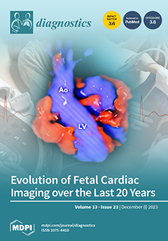
Ultrasound imaging technologies have advanced rapidly over the past few decades, enabling the reconstruction of realistic images of the fetal heart and blood vessels. Currently, these developments have evolved into a useful tool that enables the prenatal detection of a wide variety of cardiac defects by improving the resolution of cardiovascular anatomical details. Advances in fetal cardiac imaging are an exciting and promising field. View this paper
  • Issues are regarded as officially published after their release is announced to the table of contents alert mailing list.
  • You may sign up for e-mail alerts to receive table of contents of newly released issues.
  • PDF is the official format for papers published in both, html and pdf forms. To view the papers in pdf format, click on the "PDF Full-text" link, and use the free Adobe Reader to open them.
Order results
Result details
Section
Select all
Export citation of selected articles as:
9 pages, 1543 KiB  
Article
Diagnosis of Eosinophilic Otitis Media Using Blood Eosinophil Levels
by Yeonsu Jeong, Gina Na, Jong-Gyun Ha, Dachan Kim, Junyup Kim and Seonghoon Bae
Diagnostics 2023, 13(23), 3598; https://doi.org/10.3390/diagnostics13233598 - 04 Dec 2023
Viewed by 965
Abstract
Eosinophilic otitis media (EOM) is a rare middle ear disease with unfavorable outcomes. Under the current diagnostic criteria of EOM, it is challenging to suspect EOM before tympanostomy. Therefore, this study attempted to use blood eosinophil levels for the differential diagnosis of EOM [...] Read more.
Eosinophilic otitis media (EOM) is a rare middle ear disease with unfavorable outcomes. Under the current diagnostic criteria of EOM, it is challenging to suspect EOM before tympanostomy. Therefore, this study attempted to use blood eosinophil levels for the differential diagnosis of EOM from other conditions. Three disease groups with features of recurrent otorrhea were categorized, which included the following: EOM (n = 9), granulomatosis with polyangiitis (GPA, n = 12), and primary ciliary dyskinesia (PCD, n = 6). Clinical and radiological characteristics were analyzed in the three groups. Patients who underwent ventilation tube insertion due to serous otitis media were enrolled as the control group (n = 225) to evaluate the diagnostic validity of blood eosinophilia. The EOM group showed a significantly higher blood eosinophil concentration (p < 0.001) and blood eosinophil count (p < 0.001) compared to the GPA and PCD groups. The estimated sensitivity and specificity for diagnosing EOM from OME patients who underwent ventilation tube insertion were 100% and 95.6%, respectively. In addition, EOM tended to have protympanic space soft tissue density and a relatively clear retrotympanic space in temporal bone computerized tomography. Blood eosinophil evaluation is a significant clinical indicator of EOM. Furthermore, the assessment of exclusive protympanic soft tissue density can provide an additional diagnostic clue. Full article
Show Figures

Figure 1

5 pages, 1349 KiB  
Interesting Images
Myocardial Edema: A Rare but Specific Manifestation of Neonatal Capillary Leak Syndrome
by Jing Liu and Yue-Qiao Gao
Diagnostics 2023, 13(23), 3597; https://doi.org/10.3390/diagnostics13233597 - 04 Dec 2023
Cited by 1 | Viewed by 832
Abstract
Capillary leak syndrome (CLS) is a rare, potentially life-threatening systemic disease with a mortality rate of more than 30%. Its major clinical manifestation and diagnostic basis are systemic hyperedema. However, we lack knowledge about the presence of severe myocardial edema in patients with [...] Read more.
Capillary leak syndrome (CLS) is a rare, potentially life-threatening systemic disease with a mortality rate of more than 30%. Its major clinical manifestation and diagnostic basis are systemic hyperedema. However, we lack knowledge about the presence of severe myocardial edema in patients with CLS. If myocardial edema cannot be detected, it will become a dangerous hidden condition that threatens the safety of patient lives. With the routine application of point-of-care critical ultrasound (POC-CUS) in clinical practice, we found that 2 of 37 (5.41%) CLS patients had severe myocardial edema as the main manifestation. It is also necessary to distinguish it from myocardial noncompaction in newborn infants with severe myocardial edema. This paper will help us to have a deeper understanding and correct management of CLS and, thus, help us to improve the prognosis of patients. This article also suggests the necessity of routine implementation of POC-CUS in the neonatal intensive care unit. Full article
(This article belongs to the Special Issue Advances in Diagnostic Medical Imaging in 2023)
Show Figures

Figure 1

17 pages, 299 KiB  
Article
Barriers and Facilitators in Pricing and Funding Policies of European Countries That Impact the Use of Point-of-Care Diagnostics for Acute Respiratory Tract Infections in Outpatient Practices
by Caroline Steigenberger, Friederike Windisch and Sabine Vogler
Diagnostics 2023, 13(23), 3596; https://doi.org/10.3390/diagnostics13233596 - 04 Dec 2023
Cited by 1 | Viewed by 797
Abstract
Antimicrobial resistance is a major global health threat, which is increased by the irrational use of antibiotics, for example, in the treatment of respiratory tract infections in community care. By using rapid point-of-care diagnostics, overuse can be avoided. However, the diagnostic tests are [...] Read more.
Antimicrobial resistance is a major global health threat, which is increased by the irrational use of antibiotics, for example, in the treatment of respiratory tract infections in community care. By using rapid point-of-care diagnostics, overuse can be avoided. However, the diagnostic tests are rarely used in most European countries. We mapped potential barriers and facilitators in health technology assessment (HTA), pricing, and funding policies related to the use of rapid diagnostics in patients with community-acquired acute respiratory tract infections. Expert interviews were conducted with representatives of public authorities from five European case study countries: Austria, Estonia, France, Poland, and Sweden. Barriers to the HTA process include the lack of evidence and limited transferability of methods established for medicines to diagnostics. There was no price regulation for the studied diagnostics in the case study countries, but prices were usually indirectly determined via procurement. The lack of price regulation and weak purchasing power due to regional procurement processes were mentioned as pricing-related barriers. Regarding funding, coverage (reimbursement) of the diagnostic tests and the optimized remuneration of physicians in their use were mentioned as facilitators. There is potential to strengthen peri-launch policies, as optimized policies may promote the uptake of POCT. Full article
(This article belongs to the Section Point-of-Care Diagnostics and Devices)
17 pages, 2539 KiB  
Article
Tumor Segmentation in Colorectal Ultrasound Images Using an Ensemble Transfer Learning Model: Towards Intra-Operative Margin Assessment
by Freija Geldof, Constantijn W. A. Pruijssers, Lynn-Jade S. Jong, Dinusha Veluponnar, Theo J. M. Ruers and Behdad Dashtbozorg
Diagnostics 2023, 13(23), 3595; https://doi.org/10.3390/diagnostics13233595 - 04 Dec 2023
Cited by 1 | Viewed by 973
Abstract
Tumor boundary identification during colorectal cancer surgery can be challenging, and incomplete tumor removal occurs in approximately 10% of the patients operated for advanced rectal cancer. In this paper, a deep learning framework for automatic tumor segmentation in colorectal ultrasound images was developed, [...] Read more.
Tumor boundary identification during colorectal cancer surgery can be challenging, and incomplete tumor removal occurs in approximately 10% of the patients operated for advanced rectal cancer. In this paper, a deep learning framework for automatic tumor segmentation in colorectal ultrasound images was developed, to provide real-time guidance on resection margins using intra-operative ultrasound. A colorectal ultrasound dataset was acquired consisting of 179 images from 74 patients, with ground truth tumor annotations based on histopathology results. To address data scarcity, transfer learning techniques were used to optimize models pre-trained on breast ultrasound data for colorectal ultrasound data. A new custom gradient-based loss function (GWDice) was developed, which emphasizes the clinically relevant top margin of the tumor while training the networks. Lastly, ensemble learning methods were applied to combine tumor segmentation predictions of multiple individual models and further improve the overall tumor segmentation performance. Transfer learning outperformed training from scratch, with an average Dice coefficient over all individual networks of 0.78 compared to 0.68. The new GWDice loss function clearly decreased the average tumor margin prediction error from 1.08 mm to 0.92 mm, without compromising the segmentation of the overall tumor contour. Ensemble learning further improved the Dice coefficient to 0.84 and the tumor margin prediction error to 0.67 mm. Using transfer and ensemble learning strategies, good tumor segmentation performance was achieved despite the relatively small dataset. The developed US segmentation model may contribute to more accurate colorectal tumor resections by providing real-time intra-operative feedback on tumor margins. Full article
(This article belongs to the Special Issue Application of Deep Learning in Medical Ultrasound)
Show Figures

Figure 1

13 pages, 831 KiB  
Article
Predicting Teeth Extraction after Concurrent Chemoradiotherapy in Locally Advanced Nasopharyngeal Cancer Patients Using the Novel GLUCAR Index
by Efsun Somay, Erkan Topkan, Busra Yilmaz, Ali Ayberk Besen, Hüseyin Mertsoylu and Ugur Selek
Diagnostics 2023, 13(23), 3594; https://doi.org/10.3390/diagnostics13233594 - 04 Dec 2023
Cited by 1 | Viewed by 763
Abstract
To evaluate the value of the newly created GLUCAR index in predicting tooth extraction rates after concurrent chemoradiotherapy (C-CRT) in locally advanced nasopharyngeal carcinomas (LA-NPCs). Methods: A total of 187 LA-NPC patients who received C-CRT were retrospectively analyzed. The GLUCAR index was defined [...] Read more.
To evaluate the value of the newly created GLUCAR index in predicting tooth extraction rates after concurrent chemoradiotherapy (C-CRT) in locally advanced nasopharyngeal carcinomas (LA-NPCs). Methods: A total of 187 LA-NPC patients who received C-CRT were retrospectively analyzed. The GLUCAR index was defined as ′GLUCAR = (Fasting Glucose × CRP/Albumin Ratio) by utilizing measures of glucose, C-reactive protein (CRP), and albumin obtained on the first day of C-CRT. Results: The optimal GLUCAR cutoff was 31.8 (area under the curve: 78.1%; sensitivity: 70.5%; specificity: 70.7%, Youden: 0.412), dividing the study cohort into two groups: GLUCAR ˂ 1.8 (N = 78) and GLUCAR ≥ 31.8 (N = 109) groups. A comparison between the two groups found that the tooth extraction rate was significantly higher in the group with a GLUCAR ≥ 31.8 (84.4% vs. 47.4% for GLUCAR ˂ 31.8; odds ratio (OR):1.82; p < 0.001). In the univariate analysis, the mean mandibular dose ≥ 38.5 Gy group (76.5% vs. 54.9% for <38.5 Gy; OR: 1.45; p = 0.008), mandibular V55.2 Gy group ≥ 40.5% (80.3 vs. 63.5 for <40.5%, p = 0.004, OR; 1.30), and being diabetic (71.8% vs. 57.9% for nondiabetics; OR: 1.23; p = 0.007) appeared as the additional factors significantly associated with higher tooth extraction rates. All four characteristics remained independent predictors of higher tooth extraction rates after C-CRT in the multivariate analysis (p < 0.05 for each). Conclusions: The GLUCAR index, first introduced here, may serve as a robust new biomarker for predicting post-C-CRT tooth extraction rates and stratifying patients according to their tooth loss risk after treatment. Full article
(This article belongs to the Special Issue Evaluating Novel Biomarkers for Personalized Medicine)
Show Figures

Figure 1

13 pages, 1842 KiB  
Article
The Associations between the Maxillary Sinus Volume, Infraorbital Ethmoid Cells, and the Infraorbital Canal: A CT-Based Study
by Einat Kedar, Ilan Koren, Bahaa Medlej and Israel Hershkovitz
Diagnostics 2023, 13(23), 3593; https://doi.org/10.3390/diagnostics13233593 - 04 Dec 2023
Viewed by 1889
Abstract
This CT-based study aimed to characterize and explain the existence of two anatomical structures positioned near the maxillary sinuses, which are of clinical relevance in rhinology and maxillofacial surgery. A total of 182 head scans (92 males and 90 females) were inspected for [...] Read more.
This CT-based study aimed to characterize and explain the existence of two anatomical structures positioned near the maxillary sinuses, which are of clinical relevance in rhinology and maxillofacial surgery. A total of 182 head scans (92 males and 90 females) were inspected for infraorbital ethmoid cells (IECs) and for the type (route) of infraorbital canal (IOC). The maxillary sinuses were segmented, and their volumes were measured. Statistical analysis was conducted to reveal the associations between the two anatomical variations, namely, sex and the maxillary sinus volume. Infraorbital ethmoid cells were noted in 43.9% of the individuals studied; they were more frequent in males (53.3%) than in females (34.4%). The descending infraorbital nerve (type 3 IOC) was found in 13.2% of individuals and was independent of sex. Infraorbital ethmoid cells were associated with the IOC types. The maxillary sinus volume was found to be sex-dependent. A large sinus volume is significantly associated with IOC Type 3 (the descending canal) and the presence of IEC. Dentists, radiologists, and surgeons should be aware that individuals with extensive pneumatization of the maxillary sinuses are more likely to display a descending IOC and IEC. These findings should be studied, along with CT scans, before treatment and surgery. Full article
(This article belongs to the Special Issue Advances in Diagnostic Imaging of Head and Neck Tumors)
Show Figures

Figure 1

39 pages, 1385 KiB  
Review
Exploring Huntington’s Disease Diagnosis via Artificial Intelligence Models: A Comprehensive Review
by Sowmiyalakshmi Ganesh, Thillai Chithambaram, Nadesh Ramu Krishnan, Durai Raj Vincent, Jayakumar Kaliappan and Kathiravan Srinivasan
Diagnostics 2023, 13(23), 3592; https://doi.org/10.3390/diagnostics13233592 - 03 Dec 2023
Cited by 1 | Viewed by 1918
Abstract
Huntington’s Disease (HD) is a devastating neurodegenerative disorder characterized by progressive motor dysfunction, cognitive impairment, and psychiatric symptoms. The early and accurate diagnosis of HD is crucial for effective intervention and patient care. This comprehensive review provides a comprehensive overview of the utilization [...] Read more.
Huntington’s Disease (HD) is a devastating neurodegenerative disorder characterized by progressive motor dysfunction, cognitive impairment, and psychiatric symptoms. The early and accurate diagnosis of HD is crucial for effective intervention and patient care. This comprehensive review provides a comprehensive overview of the utilization of Artificial Intelligence (AI) powered algorithms in the diagnosis of HD. This review systematically analyses the existing literature to identify key trends, methodologies, and challenges in this emerging field. It also highlights the potential of ML and DL approaches in automating HD diagnosis through the analysis of clinical, genetic, and neuroimaging data. This review also discusses the limitations and ethical considerations associated with these models and suggests future research directions aimed at improving the early detection and management of Huntington’s disease. It also serves as a valuable resource for researchers, clinicians, and healthcare professionals interested in the intersection of machine learning and neurodegenerative disease diagnosis. Full article
Show Figures

Figure 1

11 pages, 2271 KiB  
Article
Inferior Vena Caval Measures Do Not Correlate with Carotid Artery Corrected Flow Time Change Measured Using a Wireless Doppler Patch in Healthy Volunteers
by Jon-Emile S. Kenny, Ross Prager, Philippe Rola, Garett McCulloch, Sarah Atwi, Chelsea E. Munding, Joseph K. Eibl and Korbin Haycock
Diagnostics 2023, 13(23), 3591; https://doi.org/10.3390/diagnostics13233591 - 03 Dec 2023
Viewed by 1111
Abstract
(1) Background: The inspiratory collapse of the inferior vena cava (IVC), a non-invasive surrogate for right atrial pressure, is often used to predict whether a patient will augment stroke volume (SV) in response to a preload challenge. There is a correlation between changing [...] Read more.
(1) Background: The inspiratory collapse of the inferior vena cava (IVC), a non-invasive surrogate for right atrial pressure, is often used to predict whether a patient will augment stroke volume (SV) in response to a preload challenge. There is a correlation between changing stroke volume (SV) and corrected flow time of the common carotid artery (ccFT). (2) Objective: We studied the relationship between IVC collapsibility and ccFT in healthy volunteers during preload challenges. (3) Methods: A prospective, observational, pilot study in euvolemic, healthy volunteers with no cardiovascular history was undertaken in a local physiology lab. Using a tilt-table, we studied two degrees of preload augmentation from (a) supine to 30-degrees head-down and (b) fully-upright to 30-degrees head down. In the supine position, % of IVC collapse with respiration, sphericity index and portal vein pulsatility was calculated. The common carotid artery Doppler pulse was continuously captured using a wireless, wearable ultrasound system. (4) Results: Fourteen subjects were included. IVC % collapse with respiration ranged between 10% and 84% across all subjects. Preload responsiveness was defined as an increase in ccFT of at least 7 milliseconds. A total of 79% (supine baseline) and 100% (head-up baseline) of subjects were preload-responsive. No supine venous measures (including IVC % collapse) were significantly related to ccFT. (5) Conclusions: From head-up baseline, 100% of healthy subjects were ‘preload-responsive’ as per the ccFT. Based on the 42% and 25% IVC collapse thresholds in the supine position, only 50% and 71% would have been labeled ‘preload-responsive’. Full article
(This article belongs to the Section Point-of-Care Diagnostics and Devices)
Show Figures

Figure 1

11 pages, 3344 KiB  
Article
Line-Field Confocal Optical Coherence Tomography Evaluation of Eyelid Skin Lesions
by Alessandro Di Stefani, Simone Cappilli, Giovanni Cuffaro, Bruno Fionda, Monica Maria Pagliara, Andrea Paradisi, Costantino Ricci, Ernesto Rossi, Maria Grazia Sammarco, Giovanni Schinzari, Luca Tagliaferri, Maria Antonietta Blasi, Elisa Cinotti, Alessandro Moro, Gustavo Savino, Mariano Suppa and Ketty Peris
Diagnostics 2023, 13(23), 3590; https://doi.org/10.3390/diagnostics13233590 - 03 Dec 2023
Viewed by 1033
Abstract
Background: Periocular malignancies may be clinically different from the examples arising at other sites, with possible delayed diagnosis and greater challenges for treatment and repair. Line-field confocal optical coherence tomography (LC-OCT) is a recently developed technique characterized by an unprecedented capacity to acquire [...] Read more.
Background: Periocular malignancies may be clinically different from the examples arising at other sites, with possible delayed diagnosis and greater challenges for treatment and repair. Line-field confocal optical coherence tomography (LC-OCT) is a recently developed technique characterized by an unprecedented capacity to acquire high-definition images in vertical and horizontal modes. In this study, we aimed to investigate the LC-OCT morphological features of a series of eyelid skin lesions, correlating them to histopathological findings. Methods: Patients with biopsy-proven equivocal skin lesion in the eyelid area, previously investigated by means of LC-OCT, were included in the study. Percentage overall agreement was estimated for LC-OCT and histopathological diagnosis for study cases. Results: A total of 51 patients (28 women, 23 men; mean age 66.4 years old), for a total of 51 skin lesions, were assessed. The histopathological diagnosis consisted of 30 malignant and 21 benign tumors. Different entities were characterized by peculiar findings in LC-OCT, alike to histopathological features, allowing for an accurate “in vivo” classification in almost all cases, with a diagnostic concordance with histopathology of 92.1% (47/51). Conclusions: By integrating this new imaging technique into the assessment of suspicious tumors in this area, diagnostic accuracy may increase, improving strategies adopted in multidisciplinary meetings and patient-centered care. Full article
Show Figures

Figure 1

11 pages, 2309 KiB  
Article
Detection of Factors Related to the Development of Osteochondritis Dissecans in Youth Baseball Players Screening
by Shotaro Teruya, Takeshi Ogawa, Hiroki Yamada, Hiromitsu Tsuge, Ryuhei Michinobu, Kazuhiro Ikeda, Yuki Hara, Hiroshi Kamada, Masashi Yamazaki and Yuichi Yoshii
Diagnostics 2023, 13(23), 3589; https://doi.org/10.3390/diagnostics13233589 - 03 Dec 2023
Viewed by 803
Abstract
On-field screening for ‘elbow injury in baseball’, a condition commonly seen in youth baseball players, was conducted over two years on 160 elementary school students in Ibaraki Prefecture, Japan. This on-field screening was conducted in collaboration with the Ibaraki Prefecture High School Baseball [...] Read more.
On-field screening for ‘elbow injury in baseball’, a condition commonly seen in youth baseball players, was conducted over two years on 160 elementary school students in Ibaraki Prefecture, Japan. This on-field screening was conducted in collaboration with the Ibaraki Prefecture High School Baseball Federation. Pitchers, catchers, symptomatic players, and players who had previously experienced elbow pain were given a comprehensive evaluation that included a physical exam and ultrasound. Out of the 135 students who were successfully screened, 10 were diagnosed with osteochondritis dissecans of the humeral capitellum (OCD). Notably, seven among these were asymptomatic. This assessment identified limited range of motion and pain when extending their elbow as significant risk factors for OCD. An attempt at on-field screening for baseball elbow injuries in collaboration with the local baseball federation was introduced. The risk factors for OCD were identified. Considering these factors, more efficient screening will be possible in the next attempt. Full article
(This article belongs to the Special Issue Recent Advances in Bone and Joint Imaging—2nd Edition)
Show Figures

Figure 1

11 pages, 3324 KiB  
Article
Comparative Analysis of CT Texture in Lumbar and Femur and Its Correlation with Bone Mineral Density and Content over Time: An Exploratory Study
by Min Woo Kim, Young Min Noh, Jung Wook Huh, Han Eol Seo and Dong Ha Lee
Diagnostics 2023, 13(23), 3588; https://doi.org/10.3390/diagnostics13233588 - 03 Dec 2023
Viewed by 748
Abstract
Background: This research explores the application of morphometric texture analysis in chest Computed Tomography (CT) scans for determining Bone Mineral Content (BMC) and its temporal changes, both crucial in diagnosing osteoporosis. The study establishes an innovative approach to osteoporosis screening by leveraging Hounsfield [...] Read more.
Background: This research explores the application of morphometric texture analysis in chest Computed Tomography (CT) scans for determining Bone Mineral Content (BMC) and its temporal changes, both crucial in diagnosing osteoporosis. The study establishes an innovative approach to osteoporosis screening by leveraging Hounsfield Units (HUs) in CT scans to evaluate BMC, offering a comparison with dual-energy X-ray absorptiometry (DXA)-based BMC. Methods: A total of 806 instances (encompassing 379 individuals) were meticulously compiled from a sole institution, during the period stretching from 6 May 2012 to 30 June 2020. In this detailed analysis, each participant was subjected to a pair of chest CT scans, sequentially pursued by a DXA scan, spread over two years. Focused records of BMC values at the inaugural lumbar vertebra (L1) were secured from both the DXA and CT axial slices across all instances. A meticulous selection process pinpointed the largest trabecular section from the L1 vertebral body, whereupon 45 distinctive texture attributes were harvested utilizing gray-level co-occurrence matrix methodologies. Utilizing these amassed 45 attributes, a regression architecture was devised, aiming to forecast the precise BMC values individually. Moreover, an alternative regression framework was engaged, leveraging 90 distinct features, to gauge the BMC fluctuations observed between the duo of scans administered to each participant. Results: The precision of the cultivated regression frameworks was scrupulously assessed, benchmarking against the correlation coefficient (CC) and the mean absolute deviation (MAE) in comparison to the DXA-established references. The regression apparatus employed for estimating BMC unveiled a CC of 0.754 and an MAE of 1.641 (g), respectively. Conversely, the regression mechanism devoted to discerning the variations in BMC manifested a CC of 0.680, coupled with an MAE of 0.528 (g), respectively. Conclusion: The innovative methodology utilizing morphometric texture analysis in CT HUs offers an indirect, yet promising, approach for osteoporosis screening by providing estimations of BMC and its temporal changes. The estimations demonstrate moderate positive correlations with DXA measures, suggesting a potential alternative in circumstances where DXA scanning is limited. Full article
(This article belongs to the Section Medical Imaging and Theranostics)
Show Figures

Figure 1

14 pages, 1212 KiB  
Article
A Logistic Regression Model for Predicting the Risk of Subsequent Surgery among Patients with Newly Diagnosed Crohn’s Disease Using a Brute Force Method
by Kohei Ogasawara, Hiroto Hiraga, Yoshihiro Sasaki, Noriko Hiraga, Naoki Higuchi, Keisuke Hasui, Shinji Ota, Takato Maeda, Yasuhisa Murai, Tetsuya Tatsuta, Hidezumi Kikuchi, Daisuke Chinda, Tatsuya Mikami, Masashi Matsuzaka, Hirotake Sakuraba and Shinsaku Fukuda
Diagnostics 2023, 13(23), 3587; https://doi.org/10.3390/diagnostics13233587 - 03 Dec 2023
Viewed by 790
Abstract
Surgery avoidance is an important goal in Crohn’s disease (CD) treatment and predicting the risk of subsequent surgery is important to determine adequate therapeutic strength for patients with newly diagnosed CD. Herein, we aimed to construct a prediction model for the risk of [...] Read more.
Surgery avoidance is an important goal in Crohn’s disease (CD) treatment and predicting the risk of subsequent surgery is important to determine adequate therapeutic strength for patients with newly diagnosed CD. Herein, we aimed to construct a prediction model for the risk of subsequent surgery based on disease characteristics at the patients’ initial visit. We retrospectively collected disease characteristic data from 93 patients with newly diagnosed CD. A logistic regression model with a brute force method was used to maximize the area under the receiver operating characteristic curve (auROC) by employing a combination of potential predictors from 14 covariates (16,383). The auROC remained almost constant when one to 12 covariates were considered, reaching a peak of 0.89 at four covariates (small-bowel patency, extensive small-bowel lesions, main lesions, and the number of poor prognostic factors), and it decreased with increasing covariate size. The most significant predictors were small-bowel patency, extensive small-bowel lesions, and age or major lesions. Therefore, this prediction model using covariates may be helpful in determining the likelihood that a patient with newly diagnosed CD will require surgery, which can aid in appropriate treatment selection for high-risk patients. Full article
(This article belongs to the Special Issue Diagnosis and Prognosis of Gastrointestinal Diseases)
Show Figures

Figure 1

19 pages, 8320 KiB  
Article
Auto-Segmentation and Quantification of Non-Cavitated Enamel Caries Imaged with Swept-Source Optical Coherence Tomography
by Tamer Abdelrehim, Maha Salah, Heather J. Conrad and Hooi Pin Chew
Diagnostics 2023, 13(23), 3586; https://doi.org/10.3390/diagnostics13233586 - 03 Dec 2023
Viewed by 789
Abstract
(1) Background: OCT imaging has been used to assess enamel demineralization in dental research, but it is not yet developed enough to qualify as a diagnostic technique in clinics. The current capabilities of most commercial acquisition software allow for visual and qualitative assessments. [...] Read more.
(1) Background: OCT imaging has been used to assess enamel demineralization in dental research, but it is not yet developed enough to qualify as a diagnostic technique in clinics. The current capabilities of most commercial acquisition software allow for visual and qualitative assessments. There is a need for a fast and verified batch-processing algorithm to segment and analyze demineralized enamel. This study suggests a GUI MATLAB algorithm for the processing and quantitative analysis of demineralized enamel. (2) Methods: A group of artificially demineralized human enamels was in vitro scanned under the OCT, and ROI frames were extracted. By using a selected intensity threshold colormap, Inter- (Ie) and Intra- (Ia) prismatic demineralization can be segmented. A set of quantitative measurements for the average demineralized depth, average line profile, and integrated reflectivity can be obtained for an accurate assessment. Real and simulated OCT frames were used for algorithm verification. (3) Results: A strong correlation between the automated and known Excel measurements for the average demineralization depth was found (R2 > 0.97). (4) Conclusions: OCT image segmentation and quantification of the enamel demineralization zones are possible. The algorithm can assess the future development of a real-time assessment of dental diagnostics using an oral probe OCT. Full article
(This article belongs to the Special Issue Advances in Dental Imaging)
Show Figures

Figure 1

10 pages, 1481 KiB  
Article
Changes in Vertebrobasilar Artery Dissection Visible with High-Resolution Vessel Wall Imaging: A Serial Follow-Up Study
by Eunjeong Cho, Youjin Won, Ui Yun Lee, Seung Bae Hwang and Hyo Sung Kwak
Diagnostics 2023, 13(23), 3585; https://doi.org/10.3390/diagnostics13233585 - 01 Dec 2023
Viewed by 763
Abstract
Background: High-resolution vessel wall imaging (HR-VWI) can identify vertebrobasilar artery dissections (VBADs) due to its good intramural hematoma and intimal flap visualization. Although the clinical course of VBADs is known to be benign, changes in VBADs visible using HR-VWI at follow-up are unknown. [...] Read more.
Background: High-resolution vessel wall imaging (HR-VWI) can identify vertebrobasilar artery dissections (VBADs) due to its good intramural hematoma and intimal flap visualization. Although the clinical course of VBADs is known to be benign, changes in VBADs visible using HR-VWI at follow-up are unknown. Thus, this study aimed to assess serial changes in VBADs using HR-VWI at follow-up. Materials and methods: Patients with neurological symptoms from VBADs who had undergone both initial and follow-up HR-VWI examinations were retrospectively enrolled. Enrolled patients with VBADs at the initial HR-VWI after acute symptom onset underwent serial follow-up with HR-VWI at 3, 6, 12, and 24 months. Patients were classified into three groups based on the results of follow-up HR-VWI examinations: type 1 = wall thickness of the dissected artery; type 2 = no interval change; and type 3 = occlusion. Results: Fifteen patients (median age: 50 years, nine males) were enrolled in this study. All patients initially showed an intimal flap and a double lumen. Twelve (80%) patients showed strong wall enhancement. Nine (60%) patients had an intramural hematoma. During serial follow-up, nine (60.0%) patients showed type 1 lesions due to attachment of the intimal flap to the vessel wall, five (33.3%) showed type 2, and one showed type 3. Four patients with BA dissection showed type 2 lesions without change in the intimal flap or the double lumen. Conclusions: Changes in VBADs in HR-VWI were observed during the follow-up period. Most patients with VBADs showed the healing process, such as the disappearance of the intimal flap and the double lumen. Full article
(This article belongs to the Section Medical Imaging and Theranostics)
Show Figures

Figure 1

12 pages, 855 KiB  
Article
Emergency Endoscopic Interventions in Acute Upper Gastrointestinal Bleeding: A Cohort Study
by Anna Mackiewicz-Pracka, Piotr Nehring and Adam Przybyłkowski
Diagnostics 2023, 13(23), 3584; https://doi.org/10.3390/diagnostics13233584 - 01 Dec 2023
Viewed by 867
Abstract
Introduction: Acute upper gastrointestinal bleeding is a common cause of emergency department admissions. The standard approach for the diagnosis and treatment of acute upper gastrointestinal bleeding (AUGIB) involves an endoscopy of the upper gastrointestinal tract. While daytime emergency endoscopy has been well studied, [...] Read more.
Introduction: Acute upper gastrointestinal bleeding is a common cause of emergency department admissions. The standard approach for the diagnosis and treatment of acute upper gastrointestinal bleeding (AUGIB) involves an endoscopy of the upper gastrointestinal tract. While daytime emergency endoscopy has been well studied, there is limited evidence regarding its effectiveness during the nighttime. Patients and Methods: We conducted a retrospective cohort study at a single center, analyzing adult patients with AUGIB referred for emergency endoscopy outside of regular hospital hours. Patients treated with endoscopic hemostatic methods were categorized into day-hours and night-hours groups based on the timing of the gastroscopy. The primary clinical endpoint was 120-day all-cause mortality, with secondary endpoints including hemostasis and recurrence. Results: In the population of 752 enrolled patients with acute upper gastrointestinal bleeding symptoms, 592 had a gastroscopy during the day hours between 8.00 a.m. and 10.00 p.m., while 160 had procedures performed at night between 10:00 p.m. and 8:00 a.m. In the day-hours group, the median time from symptom onset to endoscopy was 10 h (IQR 6–15), compared to 6 h (IQR 4–16) in the night-hours group. The gastroscopy duration (time to reach hemostasis during endoscopy) was significantly shorter during the night hours (p < 0.001). In both groups, endoscopic intervention after the sixth hour from symptom onset yielded improved outcomes, while treatment before the fifth hour resulted in poorer outcomes. Although the night-hours group had higher 120-day all-cause mortality, the difference was not statistically significant. Conclusions: Our findings indicate that emergency therapeutic gastroscopy for acute upper gastrointestinal bleeding is similarly effective during both day and night hours, particularly when performed after the sixth hour from symptom onset. Full article
(This article belongs to the Special Issue Advances in Endoscopic Diagnosis and Tissue Resection Techniques)
Show Figures

Figure 1

21 pages, 8382 KiB  
Review
Current Challenges in the Diagnosis of Pediatric Cutaneous Mastocytosis
by Hanna Ługowska-Umer, Justyna Czarny, Agnieszka Rydz, Roman J. Nowicki and Magdalena Lange
Diagnostics 2023, 13(23), 3583; https://doi.org/10.3390/diagnostics13233583 - 01 Dec 2023
Viewed by 1006
Abstract
Pediatric mastocytosis is mostly a cutaneous disease classified as cutaneous mastocytosis (CM), which is characterized by mast cell (MCs) accumulation in the skin and the absence of extracutaneous involvement. Based on the morphology of skin lesions, CM can be divided into three major [...] Read more.
Pediatric mastocytosis is mostly a cutaneous disease classified as cutaneous mastocytosis (CM), which is characterized by mast cell (MCs) accumulation in the skin and the absence of extracutaneous involvement. Based on the morphology of skin lesions, CM can be divided into three major forms: maculopapular CM (MPCM), diffuse CM (DCM) and mastocytoma of the skin. A positive Darier’s sign is pathognomonic for all forms of CM. MPCM is the most common form, presenting with red-brown macules or slightly raised papules. Mastocytoma is characterized by solitary or a maximum of three nodular or plaque lesions. DCM is a rare, severe form which presents as erythroderma, pachydermia and blistering in the infantile period of the disease. CM is associated with MC mediator-related symptoms, most commonly including pruritus, flushing, blistering, diarrhea and cramping. Anaphylactic shock occurs rarely, mainly in patients with extensive skin lesions and a significantly elevated serum tryptase level. Childhood-onset MPCM and mastocytoma are usually benign diseases, associated with a tendency for spontaneous regression, while DCM is associated with severe mediator-related symptoms, an increased risk of anaphylaxis and, in some cases, underlying systemic mastocytosis (SM). In contrast to adults, SM is a rare finding in children, most commonly presenting as indolent SM. However, advanced SM sporadically occurs. Full article
(This article belongs to the Special Issue Current Challenges in Diagnosis and Management of Mast Cell Disorders)
Show Figures

Figure 1

12 pages, 1008 KiB  
Review
Breast Imaging Physics in Mammography (Part II)
by Noemi Fico, Graziella Di Grezia, Vincenzo Cuccurullo, Antonio Alessandro Helliot Salvia, Aniello Iacomino, Antonella Sciarra, Daniele La Forgia and Gianluca Gatta
Diagnostics 2023, 13(23), 3582; https://doi.org/10.3390/diagnostics13233582 - 01 Dec 2023
Viewed by 1580
Abstract
One of the most frequently detected neoplasms in women in Italy is breast cancer, for which high-sensitivity diagnostic techniques are essential for early diagnosis in order to minimize mortality rates. As addressed in Part I of this work, we have seen how conditions [...] Read more.
One of the most frequently detected neoplasms in women in Italy is breast cancer, for which high-sensitivity diagnostic techniques are essential for early diagnosis in order to minimize mortality rates. As addressed in Part I of this work, we have seen how conditions such as high glandular density or limitations related to mammographic sensitivity have driven the optimization of technology and the use of increasingly advanced and specific diagnostic methodologies. While the first part focused on analyzing the use of a mammography machine from a physical and dosimetric perspective, in this paper, we will examine other techniques commonly used in breast imaging: contrast-enhanced mammography, digital breast tomosynthesis, radio imaging, and include some notes on image processing. We will also explore the differences between these various techniques to provide a comprehensive overview of breast lesion detection techniques. We will examine the strengths and weaknesses of different diagnostic modalities and observe how, with the implementation of improvements over time, increasingly effective diagnoses can be achieved. Full article
(This article belongs to the Special Issue Exploring Gynecological Pathology and Imaging)
Show Figures

Figure 1

11 pages, 1212 KiB  
Case Report
Isolated Intramural Hematoma of Superior Mesenteric Artery: Case Reports and a Review of Literature
by Marta Ascione, Rocco Cangiano, Alireza Mohseni, Andrea Molinari, Antonio Marzano, Alessia Di Girolamo, Luca Di Marzo and Wassim Mansour
Diagnostics 2023, 13(23), 3581; https://doi.org/10.3390/diagnostics13233581 - 01 Dec 2023
Viewed by 844
Abstract
(1) Background: Spontaneous isolated intramural hematoma of the superior mesenteric artery (SIHSMA) is a rare entity often considered as a subset of spontaneous isolated dissection of the superior mesenteric artery (SIDSMA). It is characterized by a completely thrombosed false lumen with or without [...] Read more.
(1) Background: Spontaneous isolated intramural hematoma of the superior mesenteric artery (SIHSMA) is a rare entity often considered as a subset of spontaneous isolated dissection of the superior mesenteric artery (SIDSMA). It is characterized by a completely thrombosed false lumen with or without an ulcer-like projection with computed tomography (CT) imaging. The recent literature describes few reports with a relatively short-term follow-up. The natural course, prognosis, and treatment options for SIHSMA still lack consensus. We present two cases of acute abdominal pain in a young man due to IMH of the superior mesenteric artery with an extensive literature review. (2) Case report: A 46-year-old male patient was submitted to an urgent CTA for acute abdominal pain, showing the presence of an isolated dissection of the superior mesenteric artery, determining significant stenosis of the vessel with collateral vessel patency. The patient referred to a recent COVID-19 infection, whose course was paucisymptomatic. He was conservatively treated with antiplatelet therapy and corticosteroid treatments, and, after a few days, the symptomatology completely regressed; also, the 2-month-control CTA showed complete IMH regression and the absence of any signs of residual stenosis. The second patient was a 61-year-old male patient who was submitted to an urgent CTA for acute abdominal pain, showing the presence of an isolated dissection of the superior mesenteric artery, not determining significant vessel stenosis. He was conservatively treated with antiplatelet therapy and corticosteroid treatment, and after a few days, the symptomatology completely regressed and the radiological control showed complete dissection regression. (3) Conclusion: SISHSMA is a rare entity of vascular pathology, and conservative management represents the best medical strategy. We propose corticosteroid treatment as one of the most appropriate tools in the conservative treatment of SISHSMA. Full article
(This article belongs to the Special Issue Recent Advances in the Diagnosis and Treatment of Vascular Diseases)
Show Figures

Figure 1

13 pages, 3466 KiB  
Article
Evaluation of the Reliability and the Performance of Magnetic Resonance Imaging Radiomics in the Presence of Randomly Generated Irrelevant Features for Prostate Cancer
by Cindy Xue, Jing Yuan, Gladys G. Lo, Darren M. C. Poon and Winnie C. W. Chu
Diagnostics 2023, 13(23), 3580; https://doi.org/10.3390/diagnostics13233580 - 01 Dec 2023
Viewed by 796
Abstract
Radiomics has the potential to aid prostate cancer (PC) diagnoses and prediction by analyzing and modeling quantitative features extracted from clinical imaging. However, its reliability has been a concern, possibly due to its high-dimensional nature. This study aims to quantitatively investigate the impact [...] Read more.
Radiomics has the potential to aid prostate cancer (PC) diagnoses and prediction by analyzing and modeling quantitative features extracted from clinical imaging. However, its reliability has been a concern, possibly due to its high-dimensional nature. This study aims to quantitatively investigate the impact of randomly generated irrelevant features on MRI radiomics feature selection, modeling, and performance by progressively adding randomly generated features. Two multiparametric-MRI radiomics PC datasets were used (dataset 1 (n = 260), dataset 2 (n = 100)). The endpoint was to differentiate pathology-confirmed clinically significant (Gleason score (GS) ≥ 7) from insignificant (GS < 7) PC. Random features were generated at 12 levels with a 10% increment from 0% to 100% and an additional 5%. Three feature selection algorithms and two classifiers were used to build the models. The area under the curve and accuracy were used to evaluate the model’s performance. Feature importance was calculated to assess features’ contributions to the models. The metrics of each model were compared using an ANOVA test with a Bonferroni correction. A slight tendency to select more random features with the increasing number of random features introduced to the datasets was observed. However, the performance of the radiomics-built models was not significantly affected, which was partially due to the higher contribution of radiomics features toward the models compared to the random features. These reliability effects also vary among datasets. In conclusion, while the inclusion of additional random features may still slightly impact the performance of the feature selection, it may not have a substantial impact on the MRI radiomics model performance. Full article
Show Figures

Figure 1

7 pages, 4659 KiB  
Interesting Images
A Pineal Germinoma with Rapid Enlargement following Tumor Resection
by Chia-Jung Hsu, Hsiang-Chih Liao, Dueng-Yuan Hueng and Kuan-Yin Tseng
Diagnostics 2023, 13(23), 3579; https://doi.org/10.3390/diagnostics13233579 - 01 Dec 2023
Viewed by 824
Abstract
The natural course of pineal germ cell tumors (GCTs), particularly their post-operative progression, is not well understood. We report a rare case of pineal region GCT showing rapid enlargement within 2 weeks following surgical resection. A young adult male presented with progressive headache [...] Read more.
The natural course of pineal germ cell tumors (GCTs), particularly their post-operative progression, is not well understood. We report a rare case of pineal region GCT showing rapid enlargement within 2 weeks following surgical resection. A young adult male presented with progressive headache and diplopia for several weeks. Although elevation of β-human chorionic gonadotropin (β-HCG) and α-fetoprotein (AFP) levels suggested that a large pineal mass lesion observed on magnetic resonance imaging (MRI) might be a β-HCG/AFP-producing tumor, whether the mass was truly a GCT remained unclear. We performed an endoscopy-assisted suboccipital infratentorial approach with removal of the tumor that was diagnosed as germinoma via histopathological investigation. During the week preceding chemotherapy, the patient’s consciousness rapidly worsened. MRI showed that the residual pineal germinoma had enlarged and even compressed the tectum and thalamus. Emergency chemotherapy and radiotherapy were prescribed, and the patient received invasive ventilation for respiratory failure. Unexpectedly, the patient recovered within a short period. Importantly, total regression of the pineal germinoma, accompanied by β-HCG and AFP levels returning to normal range, was observed 4 months after chemotherapy. These phenomena suggest that the rapid enlargement of the pineal germinoma, which might be induced by aggressive surgical cytoreduction, responds well to chemoradiotherapy. Full article
(This article belongs to the Section Medical Imaging and Theranostics)
Show Figures

Figure 1

12 pages, 1563 KiB  
Article
Efficacy of Computed Tomography and Magnetic Resonance Imaging in the Assessment of Depth of Invasion in Oral Squamous Cell Carcinoma: Analysis of 125 Clinical Cases
by Adam Michcik, Adam Polcyn, Łukasz Garbacewicz, Tomasz Wach, Maciej Sikora, Marta Bień and Barbara Drogoszewska
Diagnostics 2023, 13(23), 3578; https://doi.org/10.3390/diagnostics13233578 - 01 Dec 2023
Viewed by 810
Abstract
Radiological diagnosis of oral squamous cell carcinoma (OSCC) is one of the main steps in treatment planning. T (tumor size), DOI (depth of invasion) (AJCC 8th edition), and nodal metastases (N+) were evaluated using CT and MRI to assess the most effective imaging [...] Read more.
Radiological diagnosis of oral squamous cell carcinoma (OSCC) is one of the main steps in treatment planning. T (tumor size), DOI (depth of invasion) (AJCC 8th edition), and nodal metastases (N+) were evaluated using CT and MRI to assess the most effective imaging method. The effectiveness of the radiological imaging methods was compared with histopathological results. Imaging diagnostic studies were performed and retrospectively analyzed in 125 patients with OSCC (CT n = 54 and MRI n = 71). Histopathology evaluated T, DOI, and N+. The radiological T results of CT in comparison with histopathological examination showed agreement in 62.5% of cases for T1, 56.25% for T2, 25% for T3, and 42.8% for T4 (p-value = 0.07), and regarding MRI, 52.2% for T1, 36.4% for T2, 33.3% in T3, and 33.3% for T4. The DOI results of CT and MRI juxtaposed against the histopathological findings were as follows: for CT, n = 18 for DOI ≤ 10 mm and n = 36 for >10 mm; for MRI, n = 29 for DOI ≤ 10 mm and n = 42 for >10 mm (DOI CT vs. DOI hist. pat. p-value = 0.23; DOI MRI vs. DOI hist. pat. p-value = 0.006). Regarding nodal metastasis, n = 21 for N0 and n = 32 for N+ for CT (p-value = 0.02), and n = 49 for N0 and n = 22 for N+ for MRI (p-value = 0.1). In the radiological N+ group, the histopathological findings coincided with the results of MRI and CT in 27% and 62.5% of cases, respectively (N0: 83.6% for MRI; 85.7% for CT). Upon evaluating T, a decreasing percentage of overlapping results with an increasing tumor size was observed. The accuracy of both imaging studies was at a similar level, with a slight advantage for MRI. Among the patients on whom CT was performed, DOI analysis did not show statistically significant differences. This led to the conclusion that, in most cases, the DOI results based on CT overlapped with those described via histopathological examination. However, among the group of patients with MRI as the imaging method of choice, the differences proved to be statistically significant (p-value = 0.006). The results of this study indicate that CT is a more accurate method for DOI assessment. The results of the radiologic metastasis evaluation (N+ group) overlapped more in the CT group, while the percentage of corresponding results in the radiologic N0 vs. hist. pat. N0 group was high and similar in both groups. These results indicate that it is easier to confirm the absence of a metastasis than its presence. Full article
(This article belongs to the Special Issue Advances in Diagnostic Imaging of Head and Neck Tumors)
Show Figures

Figure 1

12 pages, 1012 KiB  
Article
The Swedish Standardized Course of Care—Diagnostic Efficacy in Esophageal and Gastric Cancer
by Philip Kanold, Nils Nyhlin, Eva Szabo and Michiel van Nieuwenhoven
Diagnostics 2023, 13(23), 3577; https://doi.org/10.3390/diagnostics13233577 - 01 Dec 2023
Viewed by 739
Abstract
Fast-track pathways for diagnosing esophageal or gastric cancer (EGC) have been implemented in several European countries. In Sweden, symptoms such as dysphagia, early satiety, and other alarm symptoms call for a referral for gastroscopy, according to the Swedish Standardized Course of Care (SCC). [...] Read more.
Fast-track pathways for diagnosing esophageal or gastric cancer (EGC) have been implemented in several European countries. In Sweden, symptoms such as dysphagia, early satiety, and other alarm symptoms call for a referral for gastroscopy, according to the Swedish Standardized Course of Care (SCC). The aim of this study was to evaluate the diagnostic yield of the SCC criteria for EGC, to review all known EGC cases in Region Örebro County between March 2017 and February 2021, and to compare referral indication(s), waiting times, and tumor stage. In our material, EGC was found in 6.2% of the SCC referrals. Esophageal dysphagia had a positive predictive value (PPV) of 5.6%. The criterion with the highest PPV for EGC was suspicious radiological findings, with a PPV of 24.5%. A total of 139 EGCs were diagnosed, 99 (71%) through other pathways than via the SCC. Waiting times were approximately 14 days longer for patients evaluated via non-SCC pathways. There was no statistically significant association between referral pathway and primary tumor characteristics. The results show that a majority of the current SCC criteria are poor predictors of EGC, and some alarm symptoms lack a sufficiently specific definition, e.g., dysphagia. Referral through this fast track does not seem to have a positive impact on disease outcomes. Full article
(This article belongs to the Special Issue Endoscopy in Diagnosis of Gastrointestinal Disorders)
Show Figures

Figure 1

11 pages, 1015 KiB  
Article
Feasibility of Optical Genome Mapping from Placental and Umbilical Cord Sampled after Spontaneous or Therapeutic Pregnancy Termination
by Carole Goumy, Zangbéwendé Guy Ouedraogo, Elodie Bellemonte, Eleonore Eymard-Pierre, Gwendoline Soler, Isabelle Perthus, Céline Pebrel-Richard, Laetitia Gouas, Gaëlle Salaun, Lauren Véronèse, Hélène Laurichesse, Claude Darcha and Andrei Tchirkov
Diagnostics 2023, 13(23), 3576; https://doi.org/10.3390/diagnostics13233576 - 30 Nov 2023
Cited by 1 | Viewed by 824
Abstract
Optical genome mapping (OGM) is an alternative to classical cytogenetic techniques to improve the detection rate of clinically significant genomic abnormalities. The isolation of high-molecular-weight (HMW) DNA is critical for a successful OGM analysis. HMW DNA quality depends on tissue type, sample size, [...] Read more.
Optical genome mapping (OGM) is an alternative to classical cytogenetic techniques to improve the detection rate of clinically significant genomic abnormalities. The isolation of high-molecular-weight (HMW) DNA is critical for a successful OGM analysis. HMW DNA quality depends on tissue type, sample size, and storage conditions. We assessed the feasibility of OGM analysis of DNA from nine umbilical cord (UC) and six chorionic villus (CV) samples collected after the spontaneous or therapeutic termination of pregnancy. We analyzed quality control metrics provided by the Saphyr system (Bionano Genomics) and assessed the length of extracted DNA molecules using pulsed-field capillary electrophoresis. OMG data were successfully analyzed for all six CV samples. Five of the UC samples did not meet the Saphyr quality criteria, mainly due to poor DNA quality. In this regard, we found that DNA quality assessment with pulsed-field capillary electrophoresis can predict a successful OGM analysis. OGM data were fully concordant with the results of standard cytogenetic methods. Moreover, OGM detected an average of 14 additional structural variants involving OMIM genes per sample. On the basis of our results, we established the optimal conditions for sample storage and preparation required for a successful OGM analysis. We recommend checking DNA quality before analysis with pulsed-field capillary electrophoresis if the storage conditions were not ideal or if the quality of the sample is poor. OGM can therefore be performed on fetal tissue harvested after the termination of pregnancy, which opens up the perspective for improved diagnostic yield. Full article
(This article belongs to the Special Issue Fetal Medicine: From Basic Science to Prenatal Diagnosis and Therapy)
Show Figures

Figure 1

14 pages, 4616 KiB  
Article
Myocardial and Vascular Involvement in Patients with Takayasu Arteritis: A Cardiovascular MRI Study
by Simin Almasi, Sanaz Asadian, Leila Hosseini, Nahid Rezaeian, Shakiba Ghasemi asl, Abdolmohammad Ranjbar, Seyyed-Reza Sadat-Ebrahimi, Behnaz Mahmoodieh and Alireza Salmanipour
Diagnostics 2023, 13(23), 3575; https://doi.org/10.3390/diagnostics13233575 - 30 Nov 2023
Cited by 1 | Viewed by 872
Abstract
We aimed to explore the cardiovascular magnetic resonance (CMR) of Takayasu arteritis (TA) and its cardiovascular complications. CMR was conducted on 37 TA patients and 28 healthy individuals. We evaluated the CMR findings and adverse cardiovascular complications at the time of the CMR [...] Read more.
We aimed to explore the cardiovascular magnetic resonance (CMR) of Takayasu arteritis (TA) and its cardiovascular complications. CMR was conducted on 37 TA patients and 28 healthy individuals. We evaluated the CMR findings and adverse cardiovascular complications at the time of the CMR (ACCCMR). After 8 to 26 months, the major adverse cardiac and cerebrovascular events (MACCEs) were evaluated. The TA included 25 women (67.6%), aged 36 ± 16 years old, and 28 age- and sex-matched healthy controls. Left ventricular (LV) ejection fraction was significantly lower in the TA group than in the control group (51 ± 9% vs. 58 ± 1.7%; p < 0.001). Aortic mural edema was present in 34 patients (92%) and aortic mural hyperenhancement in 36 (97%). Left ventricular global longitudinal strain (LVGLS) was significantly lower in the TA group (median [interquartile range] = 13.70 [3.27] vs. 18.08 [1.35]; p < 0.001). ACCCMR was seen in 13 TA patients (35.1%), with the most common cardiac complication being myocarditis (16.2%). During a median follow-up of 18 months (8–26 months), nine patients developed MACCEs, of which the most common was cerebrovascular accident in five (13.5%). The LVGLS of the CMR had the strongest association with complications. Myocardial strain values, especially LVGLS, can reveal concurrent and future cardiovascular complications in TA patients. Full article
Show Figures

Figure 1

10 pages, 2518 KiB  
Article
Human AI Teaming for Coronary CT Angiography Assessment: Impact on Imaging Workflow and Diagnostic Accuracy
by Florian Andre, Philipp Fortner, Matthias Aurich, Sebastian Seitz, Ann-Kathrin Jatsch, Max Schöbinger, Michael Wels, Martin Kraus, Mehmet Akif Gülsün, Norbert Frey, Andre Sommer, Johannes Görich and Sebastian J. Buss
Diagnostics 2023, 13(23), 3574; https://doi.org/10.3390/diagnostics13233574 - 30 Nov 2023
Viewed by 964
Abstract
As the number of coronary computed tomography angiography (CTA) examinations is expected to increase, technologies to optimize the imaging workflow are of great interest. The aim of this study was to investigate the potential of artificial intelligence (AI) to improve clinical workflow and [...] Read more.
As the number of coronary computed tomography angiography (CTA) examinations is expected to increase, technologies to optimize the imaging workflow are of great interest. The aim of this study was to investigate the potential of artificial intelligence (AI) to improve clinical workflow and diagnostic accuracy in high-volume cardiac imaging centers. A total of 120 patients (79 men; 62.4 (55.0–72.7) years; 26.7 (24.9–30.3) kg/m2) undergoing coronary CTA were randomly assigned to a standard or an AI-based (human AI) coronary analysis group. Severity of coronary artery disease was graded according to CAD-RADS. Initial reports were reviewed and changes were classified. Both groups were similar with regard to age, sex, body mass index, heart rate, Agatston score, and CAD-RADS. The time for coronary CTA assessment (142.5 (106.5–215.0) s vs. 195.0 (146.0–265.5) s; p < 0.002) and the total reporting time (274.0 (208.0–377.0) s vs. 350 (264.0–445.5) s; p < 0.02) were lower in the human AI than in the standard group. The number of cases with no, minor, or CAD-RADS relevant changes did not differ significantly between groups (52, 7, 1 vs. 50, 8, 2; p = 0.80). AI-based analysis significantly improves clinical workflow, even in a specialized high-volume setting, by reducing CTA analysis and overall reporting time without compromising diagnostic accuracy. Full article
Show Figures

Graphical abstract

13 pages, 3784 KiB  
Article
Calculation of Kidney Volumes with Magnetic Resonance in Patients with Autosomal Dominant Polycystic Kidney Disease: Comparison between Methods
by Stefano Di Pietro, Alfredo Gaetano Torcitto, Carmelita Marcantoni, Gabriele Giordano, Christian Campisi, Giovanni Failla, Licia Saporito, Rosa Giunta, Massimiliano Veroux, Pietro Valerio Foti, Stefano Palmucci and Antonio Basile
Diagnostics 2023, 13(23), 3573; https://doi.org/10.3390/diagnostics13233573 - 30 Nov 2023
Viewed by 887
Abstract
Autosomal dominant polycystic renal disease (ADPKD) is the most frequent kidney inheritable disease, characterized by the presence of numerous bilateral renal cysts, causing a progressive increase in total kidney volume (TKV) and a progressive loss of renal function. Several methods can be used [...] Read more.
Autosomal dominant polycystic renal disease (ADPKD) is the most frequent kidney inheritable disease, characterized by the presence of numerous bilateral renal cysts, causing a progressive increase in total kidney volume (TKV) and a progressive loss of renal function. Several methods can be used to measure TKV by using MRI, and they differ in complexity, accuracy and time consumption. This study was performed to assess the performance of the ellipsoid method and the semi-automatic segmentation method, both for TKV and SKV (single kidney volume) computation. In total, 40 patients were enrolled, and 78 polycystic kidneys analyzed. Two independent operators with different levels of experience evaluated renal volumetry using both methods. Mean error for ellipsoid method for SKV computation was −2.74 ± 11.79% and 3.25 ± 10.02% for the expert and the beginner operator, respectively (p = 0.0008). A Wilcoxon test showed a statistically significant difference between the two operators for both methods (SKV p = 0.0371 and 0.0034; TKV p = 0.0416 and 0.0171 for the expert and the beginner operator, respectively). No inter-operator significant difference was found for the semi-automatic method, in contrast to the ellipsoid method. Both with a Wilcoxon test and Bland–Altman plot, statistically significant differences were found when comparing SKV and TKV measurements obtained with the two methods for both operators, even if the differences are stronger for the beginner operator than for the expert one. The semi-automatic segmentation method showed more inter-observer reproducibility. The ellipsoid method, in contrast, appears to be affected by greater inter-observer variability, especially when performed by operators with limited experience. Full article
(This article belongs to the Section Medical Imaging and Theranostics)
Show Figures

Figure 1

10 pages, 1787 KiB  
Article
Association of Late Radiographic Assessment of Lung Edema Score with Clinical Outcome in Patients with Influenza-Associated Acute Respiratory Distress Syndrome
by Hsiao-Chin Shen, Chun-Chia Chen, Wei-Chih Chen, Wen-Kuang Yu, Kuang-Yao Yang and Yuh-Min Chen
Diagnostics 2023, 13(23), 3572; https://doi.org/10.3390/diagnostics13233572 - 30 Nov 2023
Viewed by 770
Abstract
Background: Influenza virus infection leads to acute pulmonary injury and acute respiratory distress syndrome (ARDS). The Radiographic Assessment of Lung Edema (RALE) score has been proposed as a reliable tool for the evaluation of the opacity of chest X-rays (CXRs). This study aimed [...] Read more.
Background: Influenza virus infection leads to acute pulmonary injury and acute respiratory distress syndrome (ARDS). The Radiographic Assessment of Lung Edema (RALE) score has been proposed as a reliable tool for the evaluation of the opacity of chest X-rays (CXRs). This study aimed to examine the RALE scores and outcomes in patients with influenza-associated ARDS. Methods: Patients who were newly diagnosed with influenza-associated ARDS from December 2015 to March 2016 were enrolled. Two independent reviewers scored the CXRs obtained on the day of ICU admission and on days 2 and 7 after intensive care unit (ICU) admission. Results: During the study, 47 patients had influenza-associated ARDS. Five died within 7 days of ICU admission. Of the remaining 42, non-survivors (N = 12) had higher Sequential Organ Failure Assessment scores (SOFA) at ICU admission and higher day 7 RALE scores than survivors (N = 30). The day 7 RALE score independently related to late in-hospital mortality (aOR = 1.121, 95% CI: 1.014–1.240, p = 0.025). Conclusions: The RALE score for the evaluation of opacity on CXRs is a highly reproducible tool. Moreover, RALE score on day 7 was an independent predictor of late in-hospital mortality in patients with influenza-associated ARDS. Full article
(This article belongs to the Special Issue Imaging and Chest Diseases)
Show Figures

Figure 1

5 pages, 4127 KiB  
Interesting Images
Primary Chronic Sclerosing Osteomyelitis: A New Diagnostic Tool
by Anne-Sophie Lacagne, Laurence May, Marie Nicod Lalonde, John O. Prior and Martin Broome
Diagnostics 2023, 13(23), 3571; https://doi.org/10.3390/diagnostics13233571 - 29 Nov 2023
Viewed by 721
Abstract
Aims: Primary chronic sclerosing osteomyelitis is a rare and complex pathology and remains a diagnostic and therapeutic challenge. Our aim is to show our experience with a new diagnostic tool. Material and Methods: Four patients aged from 26 to 67 were referred to [...] Read more.
Aims: Primary chronic sclerosing osteomyelitis is a rare and complex pathology and remains a diagnostic and therapeutic challenge. Our aim is to show our experience with a new diagnostic tool. Material and Methods: Four patients aged from 26 to 67 were referred to the department of oral and maxillofacial surgery of University Hospital CHUV in Lausanne between January 2010 and December 2018 for chronic mandibular pain without infectious signs nor symptoms. All patients underwent three-phase bone scintigraphy and anti-granulocyte antibody scintigraphy. Results: Three-phase bone scintigraphy demonstrated radiotracer uptake at the zone of pain, whereas anti-granulocyte antibody scintigraphy showed no uptake, thus rendering an infectious origin unlikely. Conclusion: A combination of the two different scintigraphies should be considered in order to guide the clinician in the diagnosis of primary chronic sclerosing osteomyelitis, thus preventing patients from undergoing unnecessary imagery and useless treatment, and also allowing an early diagnosis. Full article
(This article belongs to the Special Issue Advances in Oral and Maxillofacial Diagnostic Imaging)
Show Figures

Figure 1

9 pages, 580 KiB  
Article
Predictive Significance of the ABC Score for Early Re-Hemorrhage and In-Hospital Mortality in High-Risk Variceal Bleeding among Cirrhotic Patients
by Thai Doan Ky, Nguyen Thi Huyen Trang and Mai Thanh Binh
Diagnostics 2023, 13(23), 3570; https://doi.org/10.3390/diagnostics13233570 - 29 Nov 2023
Viewed by 681
Abstract
(1) Background: Upper gastrointestinal bleeding due to ruptured varices is a severe complication in patients with cirrhosis, with high rates of recurrent hemorrhage and in-hospital mortality. This study aimed to evaluate the value of the ABC score in predicting two events among 201 [...] Read more.
(1) Background: Upper gastrointestinal bleeding due to ruptured varices is a severe complication in patients with cirrhosis, with high rates of recurrent hemorrhage and in-hospital mortality. This study aimed to evaluate the value of the ABC score in predicting two events among 201 cirrhotic patients with high-risk variceal hemorrhage. (2) Methods: The ABC score was calculated and categorized into risk groups of patients, and the association between the ABC score and the rates of early hemorrhagic recurrence and clinic mortality were analyzed. (3) Results: Among 201 patients, 8.0% experienced early rebleeding within five days of admission, and 10.4% died in the hospital. Patients who experienced events had higher average ABC scores compared to those who did not experience these events (p < 0.001), especially in the high-risk group (with ABC score ≥ 8). The ABC score showed an excellent predictive value for in-hospital mortality with an AUROC of 0.804, with the optimal cutoff point being 8 points. Additionally, the ABC score demonstrated an acceptable predictive value for early rebleeding with an AUROC of 0.744, and the best cutoff point was 9 points. (4) Conclusions: The ABC score is closely associated with the rates of early re-hemorrhage and in-hospital mortality in cirrhotic patients with variceal bleeding. This scoring system has the potential for clinical application, aiding in early risk stratification for recurrent bleeding and mortality and allowing for more aggressive interventions in high-risk cases. Full article
(This article belongs to the Section Clinical Laboratory Medicine)
Show Figures

Figure 1

15 pages, 3837 KiB  
Article
Tumor Heterogeneity of Breast Cancer Assessed with Computed Tomography Texture Analysis: Association with Disease-Free Survival and Clinicopathological Prognostic Factor
by Hyeongyu Yoo, Kyu Ran Cho, Sung Eun Song, Yongwon Cho, Seung Pil Jung and Kihoon Sung
Diagnostics 2023, 13(23), 3569; https://doi.org/10.3390/diagnostics13233569 - 29 Nov 2023
Viewed by 696
Abstract
Breast cancer is a heterogeneous disease, and computed tomography texture analysis (CTTA), which reflects the tumor heterogeneity, may predict the prognosis. We investigated the usefulness of CTTA for the prediction of disease-free survival (DFS) and prognostic factors in patients with invasive breast cancer. [...] Read more.
Breast cancer is a heterogeneous disease, and computed tomography texture analysis (CTTA), which reflects the tumor heterogeneity, may predict the prognosis. We investigated the usefulness of CTTA for the prediction of disease-free survival (DFS) and prognostic factors in patients with invasive breast cancer. A total of 256 consecutive women who underwent preoperative chest CT and surgery in our institution were included. The Cox proportional hazards model was used to determine the relationship between textural features and DFS. Logistic regression analysis was used to reveal the relationship between textural features and prognostic factors. Of 256 patients, 21 (8.2%) had disease recurrence over a median follow-up of 60 months. For the prediction of shorter DFS, higher histological grade (hazard ratio [HR], 6.12; p < 0.001) and lymphovascular invasion (HR, 2.93; p = 0.029) showed significance, as well as textural features such as lower mean attenuation (HR, 4.71; p = 0.003) and higher entropy (HR, 2.77; p = 0.036). Lower mean attenuation showed a correlation with higher tumor size, and higher entropy showed correlations with higher tumor size and Ki-67. In conclusion, CTTA-derived textural features can be used as a noninvasive imaging biomarker to predict shorter DFS and prognostic factors in patients with invasive breast cancer. Full article
Show Figures

Figure 1

Previous Issue
Back to TopTop