Next Issue
Volume 12, August
Previous Issue
Volume 12, June


Toxins, Volume 12, Issue 7 (July 2020) – 43 articles

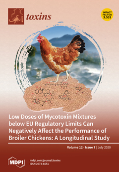
Cover Story (view full-size image): Mycotoxins are naturally occurring toxins produced by certain fungi. They appear in the feed supply chain when crops experience fungal infections both before and after harvest. Although several studies have found that mycotoxins can cause a wide range of adverse health effects in farm animals, little is known about how mycotoxin can effect health at low concentrations. This study shows that mycotoxin mixtures at levels below the European Union guidance values—which is representative of the field conditions in many countries—can adversely affect broiler chicken performance. Therefore, it is necessary to have continuous monitoring and mitigation against the economic, welfare, and health impacts of these compounds on farm animals. View this paper
  • Issues are regarded as officially published after their release is announced to the table of contents alert mailing list.
  • You may sign up for e-mail alerts to receive table of contents of newly released issues.
  • PDF is the official format for papers published in both, html and pdf forms. To view the papers in pdf format, click on the "PDF Full-text" link, and use the free Adobe Reader to open them.
Order results
Result details
Section
Select all
Export citation of selected articles as:
21 pages, 5949 KiB  
Article
Assessing the Aflatoxins Mitigation Efficacy of Blueberry Pomace Biosorbent in Buffer, Gastrointestinal Fluids and Model Wine
by Usman Rasheed, Qurat Ul Ain, Muhammad Yaseen, Sayantan Santra, Xiaohua Yao and Bin Liu
Toxins 2020, 12(7), 466; https://doi.org/10.3390/toxins12070466 - 21 Jul 2020
Cited by 19 | Viewed by 2771
Abstract
Blueberry (BB) and cherry pomace were investigated as new biosorbents for aflatoxins (AFs) sequestration from buffered solutions, gastrointestinal fluids and model wine. Among the tested biosorbents, BB exhibited the maximum adsorption performance for AFs and hence was further selected for the optimization of [...] Read more.
Blueberry (BB) and cherry pomace were investigated as new biosorbents for aflatoxins (AFs) sequestration from buffered solutions, gastrointestinal fluids and model wine. Among the tested biosorbents, BB exhibited the maximum adsorption performance for AFs and hence was further selected for the optimization of experimental parameters like pH, dosage, time and initial concentration of AFs. Material characterizations via scanning electron microscopy (SEM), Fourier transform infrared (FTIR) spectroscopy, N2 adsorption-desorption isothermal studies, thermogravimetric analysis (TGA) and X-ray photon spectroscopy (XPS) techniques revealed useful information about the texture and chemical composition of the biosorbents. The fitting of isothermal data with different models showed the model suitability trend as: Sips model > Langmuir model > Freundlich model, where the theoretical maximum adsorption capacity calculated from the Sips model was 4.6, 2.9, 2.7 and 2.4 mg/g for AFB1, AFB2, AFG1 and AFG2, respectively. Kinetics study revealed the fast AFs uptake by BB (50–90 min) while thermodynamics studies suggested the exothermic nature of the AFs adsorption from both, single as well as multi-toxin buffer systems, gastrointestinal fluids and model wine. Accrediting to the fast and efficient adsorption performance, green and facile fabrication approach and cost-effectiveness, the newly designed BB pomace can be counted as a promising contender for the sequestration of AFs and other organic pollutants. Full article
(This article belongs to the Section Mycotoxins)
Show Figures

Graphical abstract

16 pages, 1096 KiB  
Article
Copepod Prey Selection and Grazing Efficiency Mediated by Chemical and Morphological Defensive Traits of Cyanobacteria
by Luciana M. Rangel, Lúcia H. S. Silva, Elisabeth J. Faassen, Miquel Lürling and Kemal Ali Ger
Toxins 2020, 12(7), 465; https://doi.org/10.3390/toxins12070465 - 21 Jul 2020
Cited by 17 | Viewed by 3360
Abstract
Phytoplankton anti-grazer traits control zooplankton grazing and are associated with harmful blooms. Yet, how morphological versus chemical phytoplankton defenses regulate zooplankton grazing is poorly understood. We compared zooplankton grazing and prey selection by contrasting morphological (filament length: short vs. long) and chemical (saxitoxin: [...] Read more.
Phytoplankton anti-grazer traits control zooplankton grazing and are associated with harmful blooms. Yet, how morphological versus chemical phytoplankton defenses regulate zooplankton grazing is poorly understood. We compared zooplankton grazing and prey selection by contrasting morphological (filament length: short vs. long) and chemical (saxitoxin: STX- vs. STX+) traits of a bloom-forming cyanobacterium (Raphidiopsis) offered at different concentrations in mixed diets with an edible phytoplankton to a copepod grazer. The copepod selectively grazed on the edible prey (avoidance of cyanobacteria) even when the cyanobacterium was dominant. Avoidance of the cyanobacterium was weakest for the “short STX-” filaments and strongest for the other three strains. Hence, filament size had an effect on cyanobacterial avoidance only in the STX- treatments, while toxin production significantly increased cyanobacterial avoidance regardless of filament size. Moreover, cyanobacterial dominance reduced grazing on the edible prey by almost 50%. Results emphasize that the dominance of filamentous cyanobacteria such as Raphidiopsis can interfere with copepod grazing in a trait specific manner. For cyanobacteria, toxin production may be more effective than filament size as an anti-grazer defense against selectively grazing zooplankton such as copepods. Our results highlight how multiple phytoplankton defensive traits interact to regulate the producer-consumer link in plankton ecosystems. Full article
(This article belongs to the Section Marine and Freshwater Toxins)
Show Figures

Figure 1

31 pages, 5779 KiB  
Article
Anthrax Edema and Lethal Toxins Differentially Target Human Lung and Blood Phagocytes
by Vineet I. Patel, J. Leland Booth, Mikhail Dozmorov, Brent R. Brown and Jordan P. Metcalf
Toxins 2020, 12(7), 464; https://doi.org/10.3390/toxins12070464 - 20 Jul 2020
Cited by 4 | Viewed by 3823
Abstract
Bacillus anthracis, the causative agent of inhalation anthrax, is a serious concern as a bioterrorism weapon. The vegetative form produces two exotoxins: Lethal toxin (LT) and edema toxin (ET). We recently characterized and compared six human airway and alveolar-resident phagocyte (AARP) subsets [...] Read more.
Bacillus anthracis, the causative agent of inhalation anthrax, is a serious concern as a bioterrorism weapon. The vegetative form produces two exotoxins: Lethal toxin (LT) and edema toxin (ET). We recently characterized and compared six human airway and alveolar-resident phagocyte (AARP) subsets at the transcriptional and functional levels. In this study, we examined the effects of LT and ET on these subsets and human leukocytes. AARPs and leukocytes do not express high levels of the toxin receptors, tumor endothelium marker-8 (TEM8) and capillary morphogenesis protein-2 (CMG2). Less than 20% expressed surface TEM8, while less than 15% expressed CMG2. All cell types bound or internalized protective antigen, the common component of the two toxins, in a dose-dependent manner. Most protective antigen was likely internalized via macropinocytosis. Cells were not sensitive to LT-induced apoptosis or necrosis at concentrations up to 1000 ng/mL. However, toxin exposure inhibited B. anthracis spore internalization. This inhibition was driven primarily by ET in AARPs and LT in leukocytes. These results support a model of inhalation anthrax in which spores germinate and produce toxins. ET inhibits pathogen phagocytosis by AARPs, allowing alveolar escape. In late-stage disease, LT inhibits phagocytosis by leukocytes, allowing bacterial replication in the bloodstream. Full article
Show Figures

Figure 1

19 pages, 8846 KiB  
Article
Effects of Deoxynivalenol and Zearalenone on the Histology and Ultrastructure of Pig Liver
by Natalia Skiepko, Barbara Przybylska-Gornowicz, Magdalena Gajęcka, Maciej Gajęcki and Bogdan Lewczuk
Toxins 2020, 12(7), 463; https://doi.org/10.3390/toxins12070463 - 20 Jul 2020
Cited by 36 | Viewed by 4347
Abstract
The purpose of this study was to determine the effects of single and combined administrations of deoxynivalenol (DON) and zearalenone (ZEN) on the histology and ultrastructure of pig liver. The study was performed on immature gilts, which were divided into four equal groups. [...] Read more.
The purpose of this study was to determine the effects of single and combined administrations of deoxynivalenol (DON) and zearalenone (ZEN) on the histology and ultrastructure of pig liver. The study was performed on immature gilts, which were divided into four equal groups. Animals in the experimental groups received DON at a dose of 12 μg/kg body weight (BW) per day, ZEN at 40 μg/kg BW per day, or a mixture of DON (12 μg/kg BW per day) and ZEN (40 μg/kg BW). The control group received vehicle. The animals were killed after 1, 3, and 6 weeks of experiment. Treatment with mycotoxins resulted in several changes in liver histology and ultrastructure, including: (1) an increase in the thickness of the perilobular connective tissue and its penetration to the lobules in gilts receiving DON and DON + ZEN; (2) an increase in the total microscopic liver score (histology activity index (HAI)) in pigs receiving DON and DON + ZEN; (3) dilatation of hepatic sinusoids in pigs receiving ZEN, DON and DON + ZEN; (4) temporary changes in glycogen content in all experimental groups; (5) an increase in iron accumulation in the hepatocytes of gilts treated with ZEN and DON + ZEN; (6) changes in endoplasmic reticulum organization in the hepatocytes of pigs receiving toxins; (7) changes in morphology of Browicz–Kupffer cells after treatment with ZEN, DON, and DON + ZEN. The results show that low doses of mycotoxins used in the present study, even when applied for a short period, affected liver morphology. Full article
(This article belongs to the Special Issue Mycotoxins Occurence in Feed and Their Influence on Animal Health)
Show Figures

Figure 1

14 pages, 740 KiB  
Article
Development of an Improved Method of Sample Extraction and Quantitation of Multi-Mycotoxin in Feed by LC-MS/MS
by Bahar Nakhjavan, Nighat Sami Ahmed and Maryam Khosravifard
Toxins 2020, 12(7), 462; https://doi.org/10.3390/toxins12070462 - 19 Jul 2020
Cited by 27 | Viewed by 5145
Abstract
A multi-mycotoxin chromatographic method was developed and validated for the simultaneous quantitation of aflatoxins (AFB1, AFB2, AFG1 and AFG2), ochratoxin A (OTA), zearalenone (ZON), deoxynivalenol (DON), nivalenol (NIV), diacetoxyscirpenol (DAS), fumonisins (FB1, FB2 and FB3), T-2 toxin (T-2) and HT-2 toxin (HT-2) in [...] Read more.
A multi-mycotoxin chromatographic method was developed and validated for the simultaneous quantitation of aflatoxins (AFB1, AFB2, AFG1 and AFG2), ochratoxin A (OTA), zearalenone (ZON), deoxynivalenol (DON), nivalenol (NIV), diacetoxyscirpenol (DAS), fumonisins (FB1, FB2 and FB3), T-2 toxin (T-2) and HT-2 toxin (HT-2) in feed. The three most popular sample preparation techniques for determination of mycotoxins have been evaluated, and the method with highest recoveries was selected and optimized. This modified QuEChERS (quick, easy, cheap, effective, rugged and safe) approach was based on the extraction with acetonitrile, salting-out and cleanup with lipid removal. A reconstitution process in methanol/water was used to improve the MS responses and then the extracts were analyzed by LC-MS/MS. In this method, the recovery range is 70–100% for DON, DAS, FB1, FB2, FB3, HT-2, T-2, OTA, ZON, AFG1, AFG2, AFB1 and AFB2 and 55% for NIV in the spike range of 2–80 µg/kg. Method robustness was determined with acceptable z-scores in proficiency tests and validation experiments. Full article
(This article belongs to the Special Issue Rapid Detection of Mycotoxin Contamination)
Show Figures

Figure 1

28 pages, 757 KiB  
Review
Mycotoxins and the Enteric Nervous System
by Sławomir Gonkowski, Magdalena Gajęcka and Krystyna Makowska
Toxins 2020, 12(7), 461; https://doi.org/10.3390/toxins12070461 - 19 Jul 2020
Cited by 20 | Viewed by 4549
Abstract
Mycotoxins are secondary metabolites produced by various fungal species. They are commonly found in a wide range of agricultural products. Mycotoxins contained in food enter living organisms and may have harmful effects on many internal organs and systems. The gastrointestinal tract, which first [...] Read more.
Mycotoxins are secondary metabolites produced by various fungal species. They are commonly found in a wide range of agricultural products. Mycotoxins contained in food enter living organisms and may have harmful effects on many internal organs and systems. The gastrointestinal tract, which first comes into contact with mycotoxins present in food, is particularly vulnerable to the harmful effects of these toxins. One of the lesser-known aspects of the impact of mycotoxins on the gastrointestinal tract is the influence of these substances on gastrointestinal innervation. Therefore, the present study is the first review of current knowledge concerning the influence of mycotoxins on the enteric nervous system, which plays an important role, not only in almost all regulatory processes within the gastrointestinal tract, but also in adaptive and protective reactions in response to pathological and toxic factors in food. Full article
(This article belongs to the Special Issue Mycotoxins Occurence in Feed and Their Influence on Animal Health)
Show Figures

Figure 1

20 pages, 4344 KiB  
Article
Qualifying the T-2 Toxin-Degrading Properties of Seven Microbes with Zebrafish Embryo Microinjection Method
by Edina Garai, Anita Risa, Emese Varga, Mátyás Cserháti, Balázs Kriszt, Béla Urbányi and Zsolt Csenki
Toxins 2020, 12(7), 460; https://doi.org/10.3390/toxins12070460 - 18 Jul 2020
Cited by 4 | Viewed by 2770
Abstract
T-2 mycotoxin degradation and detoxification efficiency of seven bacterial strains were investigated with zebrafish microinjection method in three steps ((1) determination of mycotoxin toxicity baseline, (2) examination of bacterial metabolites toxicity, (3) identification of degradation products toxicity). Toxicity of T-2 was used as [...] Read more.
T-2 mycotoxin degradation and detoxification efficiency of seven bacterial strains were investigated with zebrafish microinjection method in three steps ((1) determination of mycotoxin toxicity baseline, (2) examination of bacterial metabolites toxicity, (3) identification of degradation products toxicity). Toxicity of T-2 was used as a baseline of toxic effects, bacterial metabolites of strains as control of bacterial toxicity and degradation products of toxin as control of biodegradation were injected into one-cell stage embryos in the same experiment. The results of in vivo tests were checked and supplemented with UHPLC-MS/MS measurement of T-2 concentration of samples. Results showed that the Rhodococcus erythropolis NI1 strain was the only one of the seven tested (R. gordoniae AK38, R. ruber N361, R. coprophilus N774, R. rhodochrous NI2, R. globerulus N58, Gordonia paraffinivorans NZS14), which was appropriated to criteria all aspects (bacterial and degradation metabolites of strains caused lower toxicity effects than T-2, and strains were able to degrade T-2 mycotoxin). Bacterial and degradation metabolites of the NI1 strain caused slight lethal and sublethal effects on zebrafish embryos at 72- and 120-h postinjection. Results demonstrated that the three-step zebrafish microinjection method is well-suited to the determination and classification of different bacterial strains by their mycotoxin degradation and detoxification efficiency. Full article
(This article belongs to the Section Mycotoxins)
Show Figures

Figure 1

11 pages, 4373 KiB  
Article
Cytotoxicity of the Sesquiterpene Lactones, Ivalin and Parthenolide in Murine Muscle Cell Lines and Their Effect on Desmin, a Cytoskeletal Intermediate Filament
by Christo J. Botha, Y. Zethu Mathe, Gezina C. H. Ferreira and E. Annette Venter
Toxins 2020, 12(7), 459; https://doi.org/10.3390/toxins12070459 - 18 Jul 2020
Cited by 1 | Viewed by 2180
Abstract
Vermeersiekte or “vomiting disease” is an economically important disease of ruminants following ingestion of Geigeria (G.) species in South Africa. Sheep are more susceptible, and poisoning is characterized by stiffness, regurgitation, bloat, paresis, and paralysis. Various sesquiterpene lactones have been implicated [...] Read more.
Vermeersiekte or “vomiting disease” is an economically important disease of ruminants following ingestion of Geigeria (G.) species in South Africa. Sheep are more susceptible, and poisoning is characterized by stiffness, regurgitation, bloat, paresis, and paralysis. Various sesquiterpene lactones have been implicated as the cause of poisoning. The in vitro cytotoxicity of two sesquiterpene lactones, namely, ivalin (purified from Geigeria aspera) and parthenolide (a commercially available sesquiterpene lactone), were compared using mouse skeletal myoblast (C2C12) and rat embryonic cardiac myocyte (H9c2) cell lines, representing the oesophageal, skeletal and cardiac muscles, which are affected in sheep. For 24, 48, and 72 h, both cell lines were exposed. A colorimetric viability assay, 3-(4,5-dimethylthiazol-2-yl)-2,5-diphenyltetrazolium bromide (MTT), was used to assess cytotoxicity. A concentration-dependent cytotoxic response was observed in both cell lines, however, the C2C12 cells were more sensitive, with the half-maximal effective concentrations (EC50s) ranging between 2.7 and 3.3 µM. In addition, the effect that ivalin and parthenolide has on desmin, an important cytoskeletal intermediate filament in myocytes, was evaluated using the C2C12 myoblasts. Disorganization and aggregation of desmin were caused by both sesquiterpene lactones, which could clarify some of the ultrastructural lesions described in vermeersiekte. Full article
(This article belongs to the Special Issue Plant Toxins Affecting Animal Health and Production)
Show Figures

Figure 1

15 pages, 347 KiB  
Article
Comparative Toxigenicity and Associated Mutagenicity of Aspergillus fumigatus and Aspergillus flavus Group Isolates Collected from the Agricultural Environment
by Caroline Lanier, David Garon, Natacha Heutte, Valérie Kientz and Véronique André
Toxins 2020, 12(7), 458; https://doi.org/10.3390/toxins12070458 - 17 Jul 2020
Cited by 4 | Viewed by 2266
Abstract
The mutagenic patterns of A. flavus, A. parasiticus and A. fumigatus extracts were evaluated. These strains of toxigenic Aspergillus were collected from the agricultural environment. The Ames test was performed on Salmonella typhimurium strains TA98, TA100 and TA102, without and with S9mix (exogenous [...] Read more.
The mutagenic patterns of A. flavus, A. parasiticus and A. fumigatus extracts were evaluated. These strains of toxigenic Aspergillus were collected from the agricultural environment. The Ames test was performed on Salmonella typhimurium strains TA98, TA100 and TA102, without and with S9mix (exogenous metabolic activation system). These data were compared with the mutagenicity of the corresponding pure mycotoxins tested alone or in reconstituted mixtures with equivalent concentrations, in order to investigate the potential interactions between these molecules and/or other natural metabolites. At least 3 mechanisms are involved in the mutagenic response of these aflatoxins: firstly, the formation of AFB1-8,9-epoxide upon addition of S9mix, secondly the likely formation of oxidative damage as indicated by significant responses in TA102, and thirdly, a direct mutagenicity observed for higher doses of some extracts or associated mycotoxins, which does not therefore involve exogenously activated intermediates. Besides the identified mycotoxins (AFB1, AFB2 and AFM1), additional “natural” compounds contribute to the global mutagenicity of the extracts. On the other hand, AFB2 and AFM1 modulate negatively the mutagenicity of AFB1 when mixed in binary or tertiary mixtures. Thus, the evaluation of the mutagenicity of “natural” mixtures is an integrated parameter that better reflects the potential impact of exposure to toxigenic Aspergilli. Full article
(This article belongs to the Special Issue Effects of Single and Combined Mycotoxins)
33 pages, 2759 KiB  
Review
Secondary Metabolites of Lasiodiplodia theobromae: Distribution, Chemical Diversity, Bioactivity, and Implications of Their Occurrence
by Maria Michela Salvatore, Artur Alves and Anna Andolfi
Toxins 2020, 12(7), 457; https://doi.org/10.3390/toxins12070457 - 17 Jul 2020
Cited by 57 | Viewed by 5766
Abstract
Lasiodiplodia theobromae is a plant pathogenic fungus from the family Botryosphaeriaceae that is commonly found in tropical and subtropical regions. It has been associated with many hosts, causing diverse diseases and being responsible for serious damages on economically important crops. A diverse array [...] Read more.
Lasiodiplodia theobromae is a plant pathogenic fungus from the family Botryosphaeriaceae that is commonly found in tropical and subtropical regions. It has been associated with many hosts, causing diverse diseases and being responsible for serious damages on economically important crops. A diverse array of bioactive low molecular weight compounds has been described as being produced by L. theobromae cultures. In this review, the existing literature on secondary metabolites of L. theobromae, their bioactivity, and the implications of their occurrence are compiled. Moreover, the effects of abiotic factors (e.g., temperature, nutrient availability) on secondary metabolites production are highlighted, and possible avenues for future research are presented. Currently, a total of 134 chemically defined compounds belonging to the classes of secondary metabolites and fatty acids have been reported from over 30 L. theobromae isolates. Compounds reported include cyclohexenes and cyclohexenones, indoles, jasmonates, lactones, melleins, phenols, and others. Most of the existing bioactivity studies of L. theobromae metabolites have assessed their potential phytotoxic, cytotoxic, and antimicrobial activities. In fact, its host adaptability and its ability to cause diseases in plants as well as in humans may be related to the capacity to produce bioactive compounds directly involved in host–fungus interactions. Full article
(This article belongs to the Special Issue Phytopathogenic Fungi and Toxicity)
Show Figures

Figure 1

9 pages, 1047 KiB  
Article
Effect of Botulinum Toxin Injection on Asymmetric Lower Face with Chin Deviation
by Dongwook Kim, Ju-Hyun Park, Vittorio Favero, James Mah, Young-Soo Jung and Seong Taek Kim
Toxins 2020, 12(7), 456; https://doi.org/10.3390/toxins12070456 - 17 Jul 2020
Cited by 5 | Viewed by 2786
Abstract
The purpose of this study was to compare the efficacy of botulinum toxin (BoNT) in masseter muscle reduction depending on the amount of chin deviation. Exploring distinctive effects of BoNT relative to the characteristics of facial asymmetry will aid in planning and predicting [...] Read more.
The purpose of this study was to compare the efficacy of botulinum toxin (BoNT) in masseter muscle reduction depending on the amount of chin deviation. Exploring distinctive effects of BoNT relative to the characteristics of facial asymmetry will aid in planning and predicting treatment outcomes. Sixteen adult volunteers were classified into two groups according to the degree of menton deviation observed in posteroanterior cephalograms. Eight had a menton deviation of 3 mm or more and the other eight had less than 3 mm. A total of 25 Units of BoNT was injected into the unilateral masseter muscle of the prominent side for each participant. Changes in the volume and bulkiest height of the lower face on each side were measured with a 3D laser scan at four time points: before and 4, 8, and 12 weeks after the injection. Two-way mixed ANOVA was employed for analyses. The volume and bulkiest height of the injected side decreased over time in both types of asymmetry, with significant differences at each time point. The reductions in the volume and bulkiest height were significantly greater in subjects without chin deviation. The reductions in the volume and bulkiest height of the lower face using BoNT are more effective for subjects without chin deviation. Full article
(This article belongs to the Special Issue Application of Botulinum Toxin in Clinical Medicine)
Show Figures

Figure 1

24 pages, 3106 KiB  
Article
Neotropical Rattlesnake (Crotalus simus) Venom Pharmacokinetics in Lymph and Blood Using an Ovine Model
by Edgar Neri-Castro, Melisa Bénard-Valle, Dayanira Paniagua, Leslie V. Boyer, Lourival D. Possani, Fernando López-Casillas, Alejandro Olvera, Camilo Romero, Fernando Zamudio and Alejandro Alagón
Toxins 2020, 12(7), 455; https://doi.org/10.3390/toxins12070455 - 17 Jul 2020
Cited by 11 | Viewed by 4176
Abstract
The most abundant protein families in viper venoms are Snake Venom Metalloproteases (SVMPs), Snake Venom Serine Proteases (SVSPs) and Phospholipases (PLA2s). These are primarily responsible for the pathophysiology caused by the bite of pit-vipers; however, there are few studies that analyze [...] Read more.
The most abundant protein families in viper venoms are Snake Venom Metalloproteases (SVMPs), Snake Venom Serine Proteases (SVSPs) and Phospholipases (PLA2s). These are primarily responsible for the pathophysiology caused by the bite of pit-vipers; however, there are few studies that analyze the pharmacokinetics (PK) of whole venom (WV) and its protein families. We studied the pathophysiology, PK profile and differential absorption of representative toxins from venom of Neotropical Rattlesnake (Crotalus simus) in a large animal model (ovine). Toxins studied included crotoxin (the main lethal component), which causes moderate to severe neurotoxicity; SVSPs, which deplete fibrinogen; and SVMPs, which cause local tissue damage and local and systemic hemorrhage. We found that Whole Venom (WV) was highly bioavailable (86%) 60 h following intramuscular (IM) injection, and extrapolation suggests that bioavailability may be as high as 92%. PK profiles of individual toxins were consistent with their physicochemical properties and expected clinical effects. Lymph cannulated animals absorbed 1.9% of WV through lymph during the first 12 h. Crotoxin was minimally detectable in serum after intravenous (IV) injection; however, following IM injection it was detected in lymph but not in blood. This suggests that crotoxin is quickly released from the blood toward its tissue targets. Full article
(This article belongs to the Section Animal Venoms)
Show Figures

Figure 1

18 pages, 2102 KiB  
Review
Characterization of Bacillus cereus in Dairy Products in China
by Xiao-Ye Liu, Qiao Hu, Fei Xu, Shuang-Yang Ding and Kui Zhu
Toxins 2020, 12(7), 454; https://doi.org/10.3390/toxins12070454 - 14 Jul 2020
Cited by 45 | Viewed by 9766
Abstract
Bacillus cereus is a common and ubiquitous foodborne pathogen with an increasing prevalence rate in dairy products in China. High and unmet demands for such products, particularly milk, raise the risk of B. cereus associated contamination. The presence of B. cereus and its [...] Read more.
Bacillus cereus is a common and ubiquitous foodborne pathogen with an increasing prevalence rate in dairy products in China. High and unmet demands for such products, particularly milk, raise the risk of B. cereus associated contamination. The presence of B. cereus and its virulence factors in dairy products may cause food poisoning and other illnesses. Thus, this review first summarizes the epidemiological characteristics and analytical assays of B. cereus from dairy products in China, providing insights into the implementation of intervention strategies. In addition, the recent achievements on the cytotoxicity and mechanisms of B. cereus are also presented to shed light on the therapeutic options for B. cereus associated infections. Full article
(This article belongs to the Special Issue Bacillus cereus Toxins)
Show Figures

Figure 1

2 pages, 197 KiB  
Editorial
A Long Road to Safer Food
by Filippo Rossi
Toxins 2020, 12(7), 453; https://doi.org/10.3390/toxins12070453 - 14 Jul 2020
Viewed by 1629
Abstract
As a side eect of food production, mycotoxins have always accompanied humanity, even if the danger posed by these molecules has only recently been understood and new research has begun to identify and study ways to reduce their presence in food. [...] Full article
(This article belongs to the Special Issue Mycotoxins in Feed and Food Chain: Present Status and Future Concerns)
17 pages, 1081 KiB  
Article
Multi-Toxin Quantitative Analysis of Paralytic Shellfish Toxins and Tetrodotoxins in Bivalve Mollusks with Ultra-Performance Hydrophilic Interaction LC-MS/MS—An In-House Validation Study
by Fadillah Putri Patria, Heidi Pekar and Aida Zuberovic-Muratovic
Toxins 2020, 12(7), 452; https://doi.org/10.3390/toxins12070452 - 13 Jul 2020
Cited by 16 | Viewed by 3346
Abstract
Ultra-performance hydrophilic interaction liquid chromatography tandem mass spectrometry system (UP-HILIC–MS/MS) was used in multi-toxin analysis of paralytic shellfish toxins (PSTs) and tetrodotoxins (TTXs) in sample matrices from bivalve molluscan species commercially produced for human consumption in Sweden. The method validation includes 17 toxins [...] Read more.
Ultra-performance hydrophilic interaction liquid chromatography tandem mass spectrometry system (UP-HILIC–MS/MS) was used in multi-toxin analysis of paralytic shellfish toxins (PSTs) and tetrodotoxins (TTXs) in sample matrices from bivalve molluscan species commercially produced for human consumption in Sweden. The method validation includes 17 toxins of which GTX6 and two TTX analogues, TTX and 4,9-anhydroTTX, were previously not analyzed together with hydrophilic PSTs. 11-deoxyTTX was monitored qualitatively with a non-certified reference standard. The performance of the method was evaluated for selectivity, repeatability, and linearity by analyzing spiked samples which generated linear calibration curves across the concentration ranges used (R2 > 0.99). The in-house reproducibility (RSD) was satisfactory including the LOD and LOQ for both PST and TTX toxins being far below their regulatory action limits. The major advantage of the method is that it allows direct confirmation of the toxin identity and specific toxin quantification using a derivatization-free approach. Unlike the PST-chemical methods used in routine regulatory monitoring until now for food control, the UP-HILIC-MS/MS approach enables the calibration set-up for each of the toxin analogs separately, thereby providing the essential flexibility and specificity in analysis of this challenging group of toxins. The method is suitable to implement in food monitoring for PSTs and TTXs in bivalves, and can serve as a fast and cost-efficient screening method. However, positive samples would, for regulatory reasons still need to be confirmed using the AOAC official method (2005.06). Full article
(This article belongs to the Special Issue Isolation and Characterization of Marine Toxins)
Show Figures

Figure 1

17 pages, 916 KiB  
Review
Antimicrobial Properties of Apis mellifera’s Bee Venom
by Hesham El-Seedi, Aida Abd El-Wahed, Nermeen Yosri, Syed Ghulam Musharraf, Lei Chen, Moustafa Moustafa, Xiaobo Zou, Saleh Al-Mousawi, Zhiming Guo, Alfi Khatib and Shaden Khalifa
Toxins 2020, 12(7), 451; https://doi.org/10.3390/toxins12070451 - 11 Jul 2020
Cited by 65 | Viewed by 9480
Abstract
Bee venom (BV) is a rich source of secondary metabolites from honeybees (Apis mellifera L.). It contains a variety of bioactive ingredients including peptides, proteins, enzymes, and volatile metabolites. The compounds contribute to the venom’s observed biological functions as per its anti-inflammatory [...] Read more.
Bee venom (BV) is a rich source of secondary metabolites from honeybees (Apis mellifera L.). It contains a variety of bioactive ingredients including peptides, proteins, enzymes, and volatile metabolites. The compounds contribute to the venom’s observed biological functions as per its anti-inflammatory and anticancer effects. The antimicrobial action of BV has been shown in vitro and in vivo experiments against bacteria, viruses, and fungi. The synergistic therapeutic interactions of BV with antibiotics has been reported. The synergistic effect contributes to a decrease in the loading and maintenance dosage, a decrease in the side effects of chemotherapy, and a decrease in drug resistance. To our knowledge, there have been no reviews on the impact of BV and its antimicrobial constituents thus far. The purpose of this review is to address the antimicrobial properties of BV and its compounds. Full article
Show Figures

Graphical abstract

13 pages, 1846 KiB  
Article
Vaccination with VLPs Presenting a Linear Neutralizing Domain of S. aureus Hla Elicits Protective Immunity
by Jason A. Joyner, Seth M. Daly, Julianne Peabody, Kathleen D. Triplett, Srijana Pokhrel, Bradley O. Elmore, Diane Adebanjo, David S. Peabody, Bryce Chackerian and Pamela R. Hall
Toxins 2020, 12(7), 450; https://doi.org/10.3390/toxins12070450 - 11 Jul 2020
Cited by 5 | Viewed by 3894
Abstract
The pore-forming cytotoxin α-hemolysin, or Hla, is a critical Staphylococcus aureus virulence factor that promotes infection by causing tissue damage, excessive inflammation, and lysis of both innate and adaptive immune cells, among other cellular targets. In this study, we asked whether a virus-like [...] Read more.
The pore-forming cytotoxin α-hemolysin, or Hla, is a critical Staphylococcus aureus virulence factor that promotes infection by causing tissue damage, excessive inflammation, and lysis of both innate and adaptive immune cells, among other cellular targets. In this study, we asked whether a virus-like particle (VLP)-based vaccine targeting Hla could attenuate S. aureus Hla-mediated pathogenesis. VLPs are versatile vaccine platforms that can be used to display target antigens in a multivalent array, typically resulting in the induction of high titer, long-lasting antibody responses. In the present study, we describe the first VLP-based vaccines that target Hla. Vaccination with either of two VLPs displaying a 21 amino-acid linear neutralizing domain (LND) of Hla protected both male and female mice from subcutaneous Hla challenge, evident by reduction in lesion size and neutrophil influx to the site of intoxication. Antibodies elicited by VLP-LND vaccination bound both the LND peptide and the native toxin, effectively neutralizing Hla and preventing toxin-mediated lysis of target cells. We anticipate these novel and promising vaccines being part of a multi-component S. aureus vaccine to reduce severity of S. aureus infection. Full article
(This article belongs to the Special Issue Staphylococcus aureus Toxins: Promoter or Handicap during Infection)
Show Figures

Figure 1

16 pages, 2511 KiB  
Article
Shiga Toxin Uptake and Sequestration in Extracellular Vesicles Is Mediated by Its B-Subunit
by Annie Willysson, Anne-lie Ståhl, Daniel Gillet, Julien Barbier, Jean-Christophe Cintrat, Valérie Chambon, Anne Billet, Ludger Johannes and Diana Karpman
Toxins 2020, 12(7), 449; https://doi.org/10.3390/toxins12070449 - 10 Jul 2020
Cited by 12 | Viewed by 3727
Abstract
Shiga toxin (Stx)-stimulated blood cells shed extracellular vesicles (EVs) which can transfer the toxin to the kidneys and lead to hemolytic uremic syndrome. The toxin can be taken up by renal cells within EVs wherein the toxin is released, ultimately leading to cell [...] Read more.
Shiga toxin (Stx)-stimulated blood cells shed extracellular vesicles (EVs) which can transfer the toxin to the kidneys and lead to hemolytic uremic syndrome. The toxin can be taken up by renal cells within EVs wherein the toxin is released, ultimately leading to cell death. The mechanism by which Stx is taken up, translocated, and sequestered in EVs was addressed in this study utilizing the B-subunit that binds to the globotriaosylceramide (Gb3) receptor. We found that Stx1B was released in EVs within minutes after stimulation of HeLa cells or red blood cells, detected by live cell imaging and flow cytometry. In the presence of Retro-2.1, an inhibitor of intracellular retrograde trafficking, a continuous release of Stx-positive EVs occurred. EVs from HeLa cells possess the Gb3 receptor on their membrane, and EVs from cells that were treated with a glycosylceramide synthase inhibitor, to reduce Gb3, bound significantly less Stx1B. Stx1B was detected both on the membrane and within the shed EVs. Stx1B was incubated with EVs derived from blood cells, in the absence of cells, and was shown to bind to, and be taken up by, these EVs, as demonstrated by electron microscopy. Using a membrane translocation assay we demonstrated that Stx1B was taken up by blood cell- and HeLa-derived EVs, an effect enhanced by chloropromazine or methyl-ß-cyclodextrin, suggesting toxin transfer within the membrane. This is a novel mechanism by which EVs derived from blood cells can sequester their toxic content, possibly to evade the host response. Full article
Show Figures

Figure 1

3 pages, 181 KiB  
Editorial
Fate of Free and Modified Forms of Mycotoxins during Food Processing
by Michele Suman
Toxins 2020, 12(7), 448; https://doi.org/10.3390/toxins12070448 - 10 Jul 2020
Cited by 2 | Viewed by 1874
Abstract
International trade is highly affected by mycotoxin contaminations, which result in an annual 5% to 10% loss of global crop production [...] Full article
24 pages, 3609 KiB  
Article
Toxic Potential and Metabolic Profiling of Two Australian Biotypes of the Invasive Plant Parthenium Weed (Parthenium hysterophorus L.)
by Ali Ahsan Bajwa, Paul A. Weston, Saliya Gurusinghe, Sajid Latif, Steve W. Adkins and Leslie A. Weston
Toxins 2020, 12(7), 447; https://doi.org/10.3390/toxins12070447 - 10 Jul 2020
Cited by 15 | Viewed by 3887
Abstract
Parthenium weed (Parthenium hysterophorus L.) is an invasive plant species in around 50 countries and a ‘Weed of National Significance’ in Australia. This study investigated the relative toxicity of the leaf, shoot and root extracts of two geographically separate and morphologically distinct [...] Read more.
Parthenium weed (Parthenium hysterophorus L.) is an invasive plant species in around 50 countries and a ‘Weed of National Significance’ in Australia. This study investigated the relative toxicity of the leaf, shoot and root extracts of two geographically separate and morphologically distinct biotypes of parthenium weed in Queensland, Australia. Parthenium weed exhibited higher phytotoxic, cytotoxic and photocytotoxic activity in leaf tissue extracts in contrast to shoot and root. The germination and seedling growth of a dicot species (garden cress) were inhibited more than those of a monocot species (annual ryegrass) using a phytotoxicity bioassay. The cytotoxicity of leaf extracts was assessed in a mouse fibroblast cell suspension assay and increased under high ultraviolet A(UV-A) radiation. A major secondary metabolite, parthenin, was found in abundance in leaf extracts and was positively correlated with cytotoxicity but not with photocytotoxicity or phytotoxicity. Ambrosin and chlorogenic acid were also detected and were positively correlated with germination inhibition and the inhibition of radicle elongation, respectively. In addition, other currently unidentified compounds in the leaf extracts were positively correlated with phytotoxicity, cytotoxicity and photocytotoxicity with two to three molecules strongly correlated in each case. Both parthenium weed biotypes investigated did not differ with respect to their relative toxicity, despite their reported differences in invasive potential in the field. This suggests that secondary chemistry plays a limited role in their invasion success. Full article
(This article belongs to the Special Issue Identification and Functional Characterization of Plant Toxins)
Show Figures

Figure 1

3 pages, 212 KiB  
Editorial
Introduction to the Toxins Special Issue on Toxicological Effects of Mycotoxin on Target Cells
by Ana Juan-García
Toxins 2020, 12(7), 446; https://doi.org/10.3390/toxins12070446 - 10 Jul 2020
Cited by 1 | Viewed by 1854
Abstract
Mycotoxins are toxic secondary metabolites produced by filamentous fungi from Fusarium, Alternaria and Penicillium spp [...] Full article
(This article belongs to the Special Issue Toxicological Effects of Mycotoxins on Target Cells)
12 pages, 596 KiB  
Article
AFLA-PISTACHIO: Development of a Mechanistic Model to Predict the Aflatoxin Contamination of Pistachio Nuts
by Michail D. Kaminiaris, Marco Camardo Leggieri, Dimitrios I. Tsitsigiannis and Paola Battilani
Toxins 2020, 12(7), 445; https://doi.org/10.3390/toxins12070445 - 10 Jul 2020
Cited by 20 | Viewed by 4402
Abstract
In recent years, very many incidences of contamination with aflatoxin B1 (AFB1) in pistachio nuts have been reported as a major global problem for the crop. In Europe, legislation is in force and 12 μg/kg of AFB1 is the [...] Read more.
In recent years, very many incidences of contamination with aflatoxin B1 (AFB1) in pistachio nuts have been reported as a major global problem for the crop. In Europe, legislation is in force and 12 μg/kg of AFB1 is the maximum limit set for pistachios to be subjected to physical treatment before human consumption. The goal of the current study was to develop a mechanistic, weather-driven model to predict Aspergillus flavus growth and the AFB1 contamination of pistachios on a daily basis from nut setting until harvest. The planned steps were to: (i) build a phenology model to predict the pistachio growth stages, (ii) develop a prototype model named AFLA-pistachio (model transfer from AFLA-maize), (iii) collect the meteorological and AFB1 contamination data from pistachio orchards, (iv) run the model and elaborate a probability function to estimate the likelihood of overcoming the legal limit, and (v) manage a preliminary validation. The internal validation of AFLA-pistachio indicated that 75% of the predictions were correct. In the external validation with an independent three-year dataset, 95.6% of the samples were correctly predicted. According to the results, AFLA-pistachio seems to be a reliable tool to follow the dynamic of AFB1 contamination risk throughout the pistachio growing season. Full article
(This article belongs to the Collection Understanding Mycotoxin Occurrence in Food and Feed Chains)
Show Figures

Figure 1

14 pages, 1570 KiB  
Article
Anti-Biofilm Activity of the Fungal Phytotoxin Sphaeropsidin A against Clinical Isolates of Antibiotic-Resistant Bacteria
by Emanuela Roscetto, Marco Masi, Matilde Esposito, Roberta Di Lecce, Antonella Delicato, Lucia Maddau, Viola Calabrò, Antonio Evidente and Maria Rosaria Catania
Toxins 2020, 12(7), 444; https://doi.org/10.3390/toxins12070444 - 08 Jul 2020
Cited by 25 | Viewed by 3286
Abstract
Many pathogens involved in human infection have rapidly increased their antibiotic resistance, reducing the effectiveness of therapies in recent decades. Most of them can form biofilms and effective drugs are not available to treat these formations. Natural products could represent an efficient solution [...] Read more.
Many pathogens involved in human infection have rapidly increased their antibiotic resistance, reducing the effectiveness of therapies in recent decades. Most of them can form biofilms and effective drugs are not available to treat these formations. Natural products could represent an efficient solution in discovering and developing new drugs to overcome antimicrobial resistance and treat biofilm-related infections. In this study, 20 secondary metabolites produced by pathogenic fungi of forest plants and belonging to diverse classes of naturally occurring compounds were evaluated for the first time against clinical isolates of antibiotic-resistant Gram-negative and Gram-positive bacteria. epi-Epoformin, sphaeropsidone, and sphaeropsidin A showed antimicrobial activity on all test strains. In particular, sphaeropsidin A was effective at low concentrations with Minimum Inhibitory Concentration (MIC) values ranging from 6.25 μg/mL to 12.5 μg/mL against all reference and clinical test strains. Furthermore, sphaeropsidin A at sub-inhibitory concentrations decreased methicillin-resistant S. aureus (MRSA) and P. aeruginosa biofilm formation, as quantified by crystal violet staining. Interestingly, mixtures of sphaeropsidin A and epi-epoformin have shown antimicrobial synergistic effects with a concomitant reduction of cytotoxicity against human immortalized keratinocytes. Our data show that sphaeropsidin A and epi-epoformin possess promising antimicrobial properties. Full article
(This article belongs to the Special Issue Microbial and Plant Phytotoxins)
Show Figures

Figure 1

23 pages, 2504 KiB  
Article
Detection of Chaetomium globosum, Ch. cochliodes and Ch. rectangulare during the Diversity Tracking of Mycotoxin-Producing Chaetomium-like Isolates Obtained in Buildings in Finland
by Johanna M. Salo, Orsolya Kedves, Raimo Mikkola, László Kredics, Maria A. Andersson, Jarek Kurnitski and Heidi Salonen
Toxins 2020, 12(7), 443; https://doi.org/10.3390/toxins12070443 - 08 Jul 2020
Cited by 18 | Viewed by 5316
Abstract
The diversity of Chaetomium-like isolates in buildings in Finland is poorly documented. This paper describes a set of methods for rapid diversity tracking of 42 indoor Chaetomium-like isolates. These isolates were categorized based on their fluorescence emission, ascomatal hair morphology, responses [...] Read more.
The diversity of Chaetomium-like isolates in buildings in Finland is poorly documented. This paper describes a set of methods for rapid diversity tracking of 42 indoor Chaetomium-like isolates. These isolates were categorized based on their fluorescence emission, ascomatal hair morphology, responses in three bioassays and resistance/sensitivity to the wetting agent Genapol X-080. Thirty-nine toxigenic isolates were identified [Ch. globosum (n = 35), Ch. cochliodes (n = 2) and Ch. rectangulare (n = 2)]. These isolates were identified down to the species level by tef1α gene sequencing. The major toxic substances in the ethanol extracts of the Ch. globosum and Ch. cochliodes strains were chaetoglobosin, chaetoviridin A and C, chaetomugilin D and chaetomin, identified based on HPLC-UV and mass spectrometry data (MS and MS/MS). Ethanol extracts from pure Ch. globosum cultures exhibited a toxicological profile in the boar sperm motility inhibition assay (BSMI), sperm membrane integrity damage assay (SMID) and inhibition of cell proliferation (ICP) assay, similar to that exhibited by pure chaetoglobosin A. Overall, differences in fluorescence, morphology, toxicity profile, mycotoxin production and sensitivity to chemicals were consistent with those in tef1α sequencing results for species identification. The results indicate the presence of Ch. cochliodes and Ch. rectangulare in Finnish buildings, representing a new finding. Full article
(This article belongs to the Section Mycotoxins)
Show Figures

Figure 1

14 pages, 3385 KiB  
Article
Change in Paralytic Shellfish Toxins in the Mussel Mytilus galloprovincialis Depending on Dynamics of Harmful Alexandrium catenella (Group I) in the Geoje Coast (South Korea) during Bloom Season
by Seung Ho Baek, Jung Min Choi, Minji Lee, Bum Soo Park, Yuchengmin Zhang, Osamu Arakawa, Tomohiro Takatani, Joong-Kyun Jeon and Young Ok Kim
Toxins 2020, 12(7), 442; https://doi.org/10.3390/toxins12070442 - 07 Jul 2020
Cited by 12 | Viewed by 3600
Abstract
Paralytic shellfish toxins (PSTs) produced by Alexandrium catenella (formerly A. tamarense) in Korean coastal waters caused the deaths of four people (in 1986 and 1996) who consumed contaminated mussels (Mytilus edulis). This led to more detailed consideration of the risks [...] Read more.
Paralytic shellfish toxins (PSTs) produced by Alexandrium catenella (formerly A. tamarense) in Korean coastal waters caused the deaths of four people (in 1986 and 1996) who consumed contaminated mussels (Mytilus edulis). This led to more detailed consideration of the risks of PST outbreaks and incidents in Korea, including the introduction of shellfish collection bans. In this study, we investigated the relationships between A. catenella population dynamics and PST accumulation in the mussel M. galloprovincialis. Discharges from the Nakdong River affect the environmental conditions along the Geoje coast, resulting in low salinity and high nutrient levels that trigger blooms of A. catenella. At the toxin peak on 24 April 2017, the toxins detected in A. catenella cells were C1, gonyautoxin (GTX)1 and GTX2, whereas the concentrations of PSTs in M. galloprovincialis were high and in the order of GTX4 > GTX1 > GTX3 > saxitoxin (STX) > GTX2 > neoSTX > decarbamoylgonyautoxin (dcGTX)2 > dc GTX3. The PST level in mussels was also high. At 15 °C, the PSTs are constantly found to be higher (10-fold higher in 2017 and 30-fold higher in 2018) than safe levels for human consumption (80 μg STX diHCl equivalents 100 g−1). Full article
(This article belongs to the Special Issue Isolation and Characterization of Marine Toxins)
Show Figures

Figure 1

20 pages, 1952 KiB  
Article
Spread of Jacobaea vulgaris and Occurrence of Pyrrolizidine Alkaloids in Regionally Produced Honeys from Northern Germany: Inter- and Intra-Site Variations and Risk Assessment for Special Consumer Groups
by Christoph Gottschalk, Florian Kaltner, Matthias Zimmermann, Rainer Korten, Oliver Morris, Karin Schwaiger and Manfred Gareis
Toxins 2020, 12(7), 441; https://doi.org/10.3390/toxins12070441 - 07 Jul 2020
Cited by 15 | Viewed by 3837
Abstract
Pyrrolizidine alkaloids (PA) and PA N-oxides (PANO) are secondary plant metabolites exhibiting genotoxic and carcinogenic properties. Apart from the roots and leaves, PA/PANO are particularly present in pollen and nectar. Therefore, the spread of Jacobaea vulgaris in certain regions of northern Germany [...] Read more.
Pyrrolizidine alkaloids (PA) and PA N-oxides (PANO) are secondary plant metabolites exhibiting genotoxic and carcinogenic properties. Apart from the roots and leaves, PA/PANO are particularly present in pollen and nectar. Therefore, the spread of Jacobaea vulgaris in certain regions of northern Germany has an impact on the safety of honey produced in that region. In this study, raw honey samples (n = 437) were collected from usually three individual beehives per site (n = 73) in the district of Ostholstein and analyzed for 25 PA/PANO. The results reveal mean levels of 8.4, 1.5, and 72.6 µg/kg and maximum levels of 111, 59.4, and 3313 µg/kg, depending on the season (summer 2015 and spring/summer 2016, respectively). As far as individual data are concerned, sites near areas with J. vulgaris growth did not necessarily result in high PA/PANO values. Furthermore, intra-site investigations revealed remarkable differences in PA/PANO levels of raw honey collected by different bee colonies at the same site. Consumption of these regionally produced honeys entails an increased exposure to PA/PANO, especially in children and high consumers. Margin of exposure values of <10,000 and an exceedance of the health-based guidance value highlight that regionally produced and marketed honey must be considered with care for a proper risk assessment and risk management. Full article
(This article belongs to the Section Plant Toxins)
Show Figures

Figure 1

19 pages, 729 KiB  
Review
Over 25 Years of Pediatric Botulinum Toxin Treatments: What Have We Learned from Injection Techniques, Doses, Dilutions, and Recovery of Repeated Injections?
by Heli Sätilä
Toxins 2020, 12(7), 440; https://doi.org/10.3390/toxins12070440 - 06 Jul 2020
Cited by 24 | Viewed by 6585
Abstract
Botulinum toxin type A (BTXA) has been used for over 25 years in the management of pediatric lower and upper limb hypertonia, with the first reports in 1993. The most common indication is the injection of the triceps surae muscle for the correction [...] Read more.
Botulinum toxin type A (BTXA) has been used for over 25 years in the management of pediatric lower and upper limb hypertonia, with the first reports in 1993. The most common indication is the injection of the triceps surae muscle for the correction of spastic equinus gait in children with cerebral palsy. The upper limb injection goals include improvements in function, better positioning of the arm, and facilitating the ease of care. Neurotoxin type A is the most widely used serotype in the pediatric population. After being injected into muscle, the release of acetylcholine at cholinergic nerve endings is blocked, and a temporary denervation and atrophy ensues. Targeting the correct muscle close to the neuromuscular junctions is considered essential and localization techniques have developed over time. However, each technique has its own limitations. The role of BTXA is flexible, but limited by the temporary mode of action as a focal spasticity treatment and the restrictions on the total dose deliverable per visit. As a mode of treatment, repeated BTXA injections are needed. This literature reviewed BTXA injection techniques, doses and dilutions, the recovery of muscles and the impact of repeated injections, with a focus on the pediatric population. Suggestions for future studies are also discussed. Full article
(This article belongs to the Special Issue Drug Development Using Natural Toxins)
Show Figures

Figure 1

27 pages, 901 KiB  
Review
Immune Dysfunction in Uremia 2020
by Gerald Cohen
Toxins 2020, 12(7), 439; https://doi.org/10.3390/toxins12070439 - 05 Jul 2020
Cited by 77 | Viewed by 6079
Abstract
Cardiovascular disease and infections are major causes for the high incidence of morbidity and mortality of patients with chronic kidney disease. Both complications are directly or indirectly associated with disturbed functions or altered apoptotic rates of polymorphonuclear leukocytes, monocytes, lymphocytes, and dendritic cells. [...] Read more.
Cardiovascular disease and infections are major causes for the high incidence of morbidity and mortality of patients with chronic kidney disease. Both complications are directly or indirectly associated with disturbed functions or altered apoptotic rates of polymorphonuclear leukocytes, monocytes, lymphocytes, and dendritic cells. Normal responses of immune cells can be reduced, leading to infectious diseases or pre-activated/primed, giving rise to inflammation and subsequently to cardiovascular disease. This review summarizes the impact of kidney dysfunction on the immune system. Renal failure results in disturbed renal metabolic activities with reduced renin, erythropoietin, and vitamin D production, which adversely affects the immune system. Decreased kidney function also leads to reduced glomerular filtration and the retention of uremic toxins. A large number of uremic toxins with detrimental effects on immune cells have been identified. Besides small water-soluble and protein-bound compounds originating from the intestinal microbiome, several molecules in the middle molecular range, e.g., immunoglobulin light chains, retinol-binding protein, the neuropeptides Met-enkephalin and neuropeptide Y, endothelin-1, and the adipokines leptin and resistin, adversely affect immune cells. Posttranslational modifications such as carbamoylation, advanced glycation products, and oxidative modifications contribute to uremic toxicity. Furthermore, high-density lipoprotein from uremic patients has an altered protein profile and thereby loses its anti-inflammatory properties. Full article
(This article belongs to the Special Issue Immune Dysfunction in Uremia)
Show Figures

Figure 1

15 pages, 5829 KiB  
Article
Structural and Functional Insights into the C-terminal Fragment of Insecticidal Vip3A Toxin of Bacillus thuringiensis
by Kun Jiang, Yan Zhang, Zhe Chen, Dalei Wu, Jun Cai and Xiang Gao
Toxins 2020, 12(7), 438; https://doi.org/10.3390/toxins12070438 - 05 Jul 2020
Cited by 20 | Viewed by 4104
Abstract
The vegetative insecticidal proteins (Vips) secreted by Bacillus thuringiensis are regarded as the new generation of insecticidal toxins because they have different insecticidal properties compared with commonly applied insecticidal crystal proteins (Cry toxins). Vip3A toxin, representing the vast majority of Vips, has been [...] Read more.
The vegetative insecticidal proteins (Vips) secreted by Bacillus thuringiensis are regarded as the new generation of insecticidal toxins because they have different insecticidal properties compared with commonly applied insecticidal crystal proteins (Cry toxins). Vip3A toxin, representing the vast majority of Vips, has been used commercially in transgenic crops and bio-insecticides. However, the lack of both structural information on Vip3A and a clear understanding of its insecticidal mechanism at the molecular level limits its further development and broader application. Here we present the first crystal structure of the C-terminal fragment of Vip3A toxin (Vip3Aa11200–789). Since all members of this insecticidal protein family are highly conserved, the structure of Vip3A provides unique insight into the general domain architecture and protein fold of the Vip3A family of insecticidal toxins. Our structural analysis reveals a four-domain organization, featuring a potential membrane insertion region, a receptor binding domain, and two potential glycan binding domains of Vip3A. In addition, cytotoxicity assays and insect bioassays show that the purified C-terminal fragment of Vip3Aa toxin alone have no insecticidal activity. Taken together, these findings provide insights into the mode of action of the Vip3A family of insecticidal toxins and will boost the development of Vip3A into more efficient bio-insecticides. Full article
Show Figures

Figure 1

12 pages, 1481 KiB  
Article
Quantitative Measurement of Melittin in Asian Honeybee Venom Using a New Method Including UPLC-QqTOF-MS
by Sheng Huang, Jianhua Wang, Zeqin Guo, Yan Wang and Chundong Liu
Toxins 2020, 12(7), 437; https://doi.org/10.3390/toxins12070437 - 04 Jul 2020
Cited by 9 | Viewed by 3561
Abstract
Asian honeybee venom is widely used in traditional oriental medicine. Melittin is the main component of Asian honeybee venom. In the present study, an ultra-performance liquid chromatography-quadrupole time-of-flight mass spectrometry (UPLC-QqTOF-MS) method was used for accurate qualitative and quantitative analyses of melittin in [...] Read more.
Asian honeybee venom is widely used in traditional oriental medicine. Melittin is the main component of Asian honeybee venom. In the present study, an ultra-performance liquid chromatography-quadrupole time-of-flight mass spectrometry (UPLC-QqTOF-MS) method was used for accurate qualitative and quantitative analyses of melittin in Asian honeybee venom. The results showed that the dynamic linear range of melittin was from 0.094 to 20 μg/mL, and the limit of quantification was 0.3125 μg/mL. The spiking recovery of melittin in honeybee venom ranged from 84.88% to 93.05%. Eighteen Asian honeybee venom samples in eighteen batches were collected from two different zones of China, and their melittin contents were measured. The contents of melittin in Asian honeybee venom samples was 33.9–46.23% of dry weight. This method proved a useful tool for the rapid evaluation of the authenticity and quality of Asian honeybee venom in terms of the melittin contents, and will contribute to a broader understanding of Asian honeybee venom. Full article
Show Figures

Graphical abstract

Previous Issue
Next Issue
Back to TopTop