Next Issue
Volume 9, November
Previous Issue
Volume 9, September


Viruses, Volume 9, Issue 10 (October 2017) – 47 articles

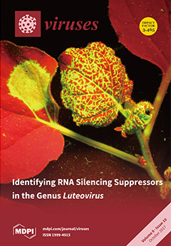
Cover Story (view full-size image): Viruses use silencing suppressor proteins to combat or avoid a plant’s intrinsic RNA silencing defense mechanism. In the family Luteoviridae, members of the genera Polerovirus and Enamovirus have a strong suppressor of local and systemic RNA silencing, named P0. Members of the genus Luteovirus, however, lack the corresponding ORF 0. We show that, in the absence of P0, luteoviruses have evolved a P4 movement protein that also displays strong systemic silencing suppression activity by inhibiting the accumulation of small RNAs. View this paper
  • Issues are regarded as officially published after their release is announced to the table of contents alert mailing list.
  • You may sign up for e-mail alerts to receive table of contents of newly released issues.
  • PDF is the official format for papers published in both, html and pdf forms. To view the papers in pdf format, click on the "PDF Full-text" link, and use the free Adobe Reader to open them.
Order results
Result details
Section
Select all
Export citation of selected articles as:
18 pages, 1744 KiB  
Review
Tomato Leaf Curl New Delhi Virus: An Emerging Virus Complex Threatening Vegetable and Fiber Crops
by Enrique Moriones 1,*, Shelly Praveen 2 and Supriya Chakraborty 3
1 Subtropical and Mediterranean Horticulture Institute “La Mayora” (IHSM-UMA-CSIC), Consejo Superior de Investigaciones Científicas, La Mayora Experimental Station, 29750 Algarrobo-Costa, Málaga, Spain
2 Advanced Center for Plant Virology, Division of Plant Pathology, Indian Agricultural Research Institute, New Delhi 110 012, India
3 Molecular Virology Laboratory, School of Life Sciences, Jawaharlal Nehru University, New Delhi 110 067, India
Viruses 2017, 9(10), 264; https://doi.org/10.3390/v9100264 - 21 Sep 2017
Cited by 131 | Viewed by 12638
Abstract
The tomato leaf curl New Delhi virus (ToLCNDV) (genus Begomovirus, family Geminiviridae) represents an important constraint to tomato production, as it causes the most predominant and economically important disease affecting tomato in the Indian sub-continent. However, in recent years, ToLCNDV has [...] Read more.
The tomato leaf curl New Delhi virus (ToLCNDV) (genus Begomovirus, family Geminiviridae) represents an important constraint to tomato production, as it causes the most predominant and economically important disease affecting tomato in the Indian sub-continent. However, in recent years, ToLCNDV has been fast extending its host range and spreading to new geographical regions, including the Middle East and the western Mediterranean Basin. Extensive research on the genome structure, protein functions, molecular biology, and plant–virus interactions of ToLCNDV has been conducted in the last decade. Special emphasis has been given to gene silencing suppression ability in order to counteract host plant defense responses. The importance of the interaction with DNA alphasatellites and betasatellites in the biology of the virus has been demonstrated. ToLCNDV genetic variability has been analyzed, providing new insights into the taxonomy, host adaptation, and evolution of this virus. Recombination and pseudorecombination have been shown as motors of diversification and adaptive evolution. Important progress has also been made in control strategies to reduce disease damage. This review highlights these various achievements in the context of the previous knowledge of begomoviruses and their interactions with plants. Full article
(This article belongs to the Special Issue Geminiviruses)
Show Figures

Figure 1

12 pages, 854 KiB  
Communication
A Recombinant Measles Vaccine with Enhanced Resistance to Passive Immunity
by Emily Julik and Jorge Reyes-del Valle *,‡
1 School of Life Sciences, Arizona State University, Tempe, AZ 85287, USA
Present Address: Institute for Molecular Virology, University of Minnesota, Minneapolis, MN 55455, USA
Present Address: Process Development Department, Virus and Gene Therapy, Merck KGaA, 64293 Darmstadt, Germany
Viruses 2017, 9(10), 265; https://doi.org/10.3390/v9100265 - 21 Sep 2017
Cited by 4 | Viewed by 4841
Abstract
Current measles vaccines suffer from poor effectiveness in young infants due primarily to the inhibitory effect of residual maternal immunity on vaccine responses. The development of a measles vaccine that resists such passive immunity would strongly contribute to the stalled effort toward measles [...] Read more.
Current measles vaccines suffer from poor effectiveness in young infants due primarily to the inhibitory effect of residual maternal immunity on vaccine responses. The development of a measles vaccine that resists such passive immunity would strongly contribute to the stalled effort toward measles eradication. In this concise communication, we show that a measles virus (MV) with enhanced hemagglutinin (H) expression and incorporation, termed MVvac2-H2, retained its enhanced immunogenicity, previously established in older mice, when administered to very young, genetically modified, MV-susceptible mice in the presence of passive anti-measles immunity. This immunity level mimics the sub-neutralizing immunity prevalent in infants too young to be vaccinated. Additionally, toward a more physiological small animal model of maternal anti-measles immunity interference, we document vertical transfer of passive anti-MV immunity in genetically-modified, MV susceptible mice and show in this physiological model a better MVvac2-H2 immunogenic profile than that of the parental vaccine strain. In sum, these data support the notion that enhancing MV hemagglutinin incorporation can circumvent in vivo neutralization. This strategy merits additional exploration as an alternative pediatric measles vaccine. Full article
(This article belongs to the Section Viral Immunology, Vaccines, and Antivirals)
Show Figures

Figure 1

15 pages, 6440 KiB  
Article
Oncolytic Reovirus Infection Is Facilitated by the Autophagic Machinery
by Vera Kemp, Iris J. C. Dautzenberg, Ronald W. Limpens, Diana J. M. Van den Wollenberg and Rob C. Hoeben *
Department of Molecular Cell Biology, Leiden University Medical Center, P.O. Box 9600, 2300 RC Leiden, The Netherlands
Viruses 2017, 9(10), 266; https://doi.org/10.3390/v9100266 - 21 Sep 2017
Cited by 13 | Viewed by 5900
Abstract
Mammalian reovirus is a double-stranded RNA virus that selectively infects and lyses transformed cells, making it an attractive oncolytic agent. Despite clinical evidence for anti-tumor activity, its efficacy as a stand-alone therapy remains to be improved. The success of future trials can be [...] Read more.
Mammalian reovirus is a double-stranded RNA virus that selectively infects and lyses transformed cells, making it an attractive oncolytic agent. Despite clinical evidence for anti-tumor activity, its efficacy as a stand-alone therapy remains to be improved. The success of future trials can be greatly influenced by the identification and the regulation of the cellular pathways that are important for reovirus replication and oncolysis. Here, we demonstrate that reovirus induces autophagy in several cell lines, evident from the formation of Atg5-Atg12 complexes, microtubule-associated protein 1 light chain 3 (LC3) lipidation, p62 degradation, the appearance of acidic vesicular organelles, and LC3 puncta. Furthermore, in electron microscopic images of reovirus-infected cells, autophagosomes were observed without evident association with viral factories. Using UV-inactivated reovirus, we demonstrate that a productive reovirus infection facilitates the induction of autophagy. Importantly, knock-out cell lines for specific autophagy-related genes revealed that the expression of Atg3 and Atg5 but not Atg13 facilitates reovirus replication. These findings highlight a central and Atg13-independent role for the autophagy machinery in facilitating reovirus infection and contribute to a better understanding of reovirus-host interactions. Full article
(This article belongs to the Section Animal Viruses)
Show Figures

Graphical abstract

10 pages, 739 KiB  
Review
Natural History of HPV Infection across the Lifespan: Role of Viral Latency
by Patti E. Gravitt 1,* and Rachel L. Winer 2
1 Department of Global Health, George Washington University Milken Institute School of Public Health, Washington, DC 20052, USA
2 Department of Epidemiology, University of Washington School of Public Health, Seattle, WA 98195, USA
Viruses 2017, 9(10), 267; https://doi.org/10.3390/v9100267 - 21 Sep 2017
Cited by 188 | Viewed by 24910
Abstract
Large-scale epidemiologic studies have been invaluable for elaboration of the causal relationship between persistent detection of genital human papillomavirus (HPV) infection and the development of invasive cervical cancer. However, these studies provide limited data to adequately inform models of the individual-level natural history [...] Read more.
Large-scale epidemiologic studies have been invaluable for elaboration of the causal relationship between persistent detection of genital human papillomavirus (HPV) infection and the development of invasive cervical cancer. However, these studies provide limited data to adequately inform models of the individual-level natural history of HPV infection over the course of a lifetime, and particularly ignore the biological distinction between HPV-negative tests and lack of infection (i.e., the possibility of latent, undetectable HPV infection). Using data from more recent epidemiological studies, this review proposes an alternative model of the natural history of genital HPV across the life span. We argue that a more complete elucidation of the age-specific probabilities of the alternative transitions is highly relevant with the expanded use of HPV testing in cervical cancer screening. With routine HPV testing in cervical cancer screening, women commonly transition in and out of HPV detectability, raising concerns for the patient and the provider regarding the source of the positive test result, its prognosis, and effective strategies to prevent future recurrence. Alternative study designs and analytic frameworks are proposed to better understand the frequency and determinants of these transition pathways. Full article
(This article belongs to the Special Issue Expert Views on HPV Infection)
Show Figures

Figure 1

16 pages, 1157 KiB  
Review
Why Human Papillomaviruses Activate the DNA Damage Response (DDR) and How Cellular and Viral Replication Persists in the Presence of DDR Signaling
by Molly L. Bristol 1, Dipon Das 1 and Iain M. Morgan 1,2,*
1 VCU Philips Institute for Oral Health Research, Virginia Commonwealth University School of Dentistry, Department of Oral and Craniofacial Molecular Biology, Richmond, VA 23298, USA
2 Massey Cancer Center, Virginia Commonwealth University, Richmond, VA 23298, USA
Viruses 2017, 9(10), 268; https://doi.org/10.3390/v9100268 - 21 Sep 2017
Cited by 43 | Viewed by 7984
Abstract
Human papillomaviruses (HPV) require the activation of the DNA damage response (DDR) in order to undergo a successful life cycle. This activation presents a challenge for the virus and the infected cell: how does viral and host replication proceed in the presence of [...] Read more.
Human papillomaviruses (HPV) require the activation of the DNA damage response (DDR) in order to undergo a successful life cycle. This activation presents a challenge for the virus and the infected cell: how does viral and host replication proceed in the presence of a DDR that ordinarily arrests replication; and how do HPV16 infected cells retain the ability to proliferate in the presence of a DDR that ordinarily arrests the cell cycle? This raises a further question: why do HPV activate the DDR? The answers to these questions are only partially understood; a full understanding could identify novel therapeutic strategies to target HPV cancers. Here, we propose that the rapid replication of an 8 kb double stranded circular genome during infection creates aberrant DNA structures that attract and activate DDR proteins. Therefore, HPV replication in the presence of an active DDR is a necessity for a successful viral life cycle in order to resolve these DNA structures on viral genomes; without an active DDR, successful replication of the viral genome would not proceed. We discuss the essential role of TopBP1 in this process and also how viral and cellular replication proceeds in HPV infected cells in the presence of DDR signals. Full article
(This article belongs to the Special Issue Viruses and the DNA Damage Response)
Show Figures

Figure 1

9 pages, 749 KiB  
Article
Live Attenuated Influenza Vaccine contains Substantial and Unexpected Amounts of Defective Viral Genomic RNA
by Philip S. Gould 1, Andrew J. Easton 2,* and Nigel J. Dimmock 2,*
1 Faculty of Health and Life Sciences, Coventry University, Science and Health Building, 20 Whitefriars Street Coventry CV1 2DS, UK
2 School of Life Sciences, University of Warwick, Coventry CV4 7AL, UK
Viruses 2017, 9(10), 269; https://doi.org/10.3390/v9100269 - 21 Sep 2017
Cited by 40 | Viewed by 8652
Abstract
The live attenuated influenza vaccine FluMist® was withdrawn in the USA by the Centers for Disease Control and Prevention after its failure to provide adequate protective immunity during 2013–2016. The vaccine uses attenuated core type A and type B viruses, reconfigured each [...] Read more.
The live attenuated influenza vaccine FluMist® was withdrawn in the USA by the Centers for Disease Control and Prevention after its failure to provide adequate protective immunity during 2013–2016. The vaccine uses attenuated core type A and type B viruses, reconfigured each year to express the two major surface antigens of the currently circulating viruses. Here Fluenz™ Tetra, the European version of this vaccine, was examined directly for defective-interfering (DI) viral RNAs. DI RNAs are deleted versions of the infectious virus genome, and have powerful biological properties including attenuation of infection, reduction of infectious virus yield, and stimulation of some immune responses. Reverse transcription polymerase chain reaction followed by cloning and sequencing showed that Fluenz™ vaccine contains unexpected and substantial amounts of DI RNA arising from both its influenza A and influenza B components, with 87 different DI RNA sequences identified. Flu A DI RNAs from segment 3 replaced the majority of the genomic full-length segment 3, thus compromising its infectivity. DI RNAs arise during vaccine production and non-infectious DI virus replaces infectious virus pro rata so that fewer doses of the vaccine can be made. Instead the vaccine carries a large amount of non-infectious but biologically active DI virus. The presence of DI RNAs could significantly reduce the multiplication in the respiratory tract of the vaccine leading to reduced immunizing efficacy and could also stimulate the host antiviral responses, further depressing vaccine multiplication. The role of DI viruses in the performance of this and other vaccines requires further investigation. Full article
(This article belongs to the Section Viral Immunology, Vaccines, and Antivirals)
Show Figures

Figure 1

25 pages, 1971 KiB  
Review
Canonical and Non-Canonical Autophagy in HIV-1 Replication Cycle
by Olivier Leymarie 1,2,3,4, Leslie Lepont 1,2,3,4 and Clarisse Berlioz-Torrent 1,2,3,4,*
1 Inserm, U1016, Institut Cochin, 75014 Paris, France
2 CNRS, UMR8104, 75014 Paris, France
3 University Paris Descartes, Sorbonne Paris Cité, 75006 Paris, France
4 Institut Cochin, Department Infection, Immunity, Inflammation, 75014 Paris, France
Viruses 2017, 9(10), 270; https://doi.org/10.3390/v9100270 - 23 Sep 2017
Cited by 31 | Viewed by 12630
Abstract
Autophagy is a lysosomal-dependent degradative process essential for maintaining cellular homeostasis, and is a key player in innate and adaptive immune responses to intracellular pathogens such as human immunodeficiency virus type 1 (HIV-1). In HIV-1 target cells, autophagy mechanisms can (i) selectively direct [...] Read more.
Autophagy is a lysosomal-dependent degradative process essential for maintaining cellular homeostasis, and is a key player in innate and adaptive immune responses to intracellular pathogens such as human immunodeficiency virus type 1 (HIV-1). In HIV-1 target cells, autophagy mechanisms can (i) selectively direct viral proteins and viruses for degradation; (ii) participate in the processing and presentation of viral-derived antigens through major histocompatibility complexes; and (iii) contribute to interferon production in response to HIV-1 infection. As a consequence, HIV-1 has evolved different strategies to finely regulate the autophagy pathway to favor its replication and dissemination. HIV-1 notably encodes accessory genes encoding Tat, Nef and Vpu proteins, which are able to perturb and hijack canonical and non-canonical autophagy mechanisms. This review outlines the current knowledge on the complex interplay between autophagy and HIV-1 replication cycle, providing an overview of the autophagy-mediated molecular processes deployed both by infected cells to combat the virus and by HIV-1 to evade antiviral response. Full article
(This article belongs to the Special Issue Viruses and Autophagy)
Show Figures

Figure 1

15 pages, 4754 KiB  
Article
Antiviral Properties of Chemical Inhibitors of Cellular Anti-Apoptotic Bcl-2 Proteins
by Daria Bulanova 1,†, Aleksandr Ianevski 1,†, Andrii Bugai 2,†, Yevhen Akimov 1, Suvi Kuivanen 3, Henrik Paavilainen 4, Laura Kakkola 4, Jatin Nandania 1, Laura Turunen 1, Tiina Ohman 5, Hanna Ala-Hongisto 6, Hanna M. Pesonen 6, Marika S. Kuisma 6, Anni Honkimaa 7, Emma L. Walton 8, Valentyn Oksenych 8, Martina B. Lorey 9, Dmitry Guschin 10, Jungmin Shim 10, Jinhee Kim 10, Thoa T. Than 10, So Young Chang 10, Veijo Hukkanen 4, Evgeny Kulesskiy 1, Varpu S. Marjomaki 11, Ilkka Julkunen 4, Tuula A. Nyman 5,12, Sampsa Matikainen 9, Jani S. Saarela 1, Famara Sane 13, Didier Hober 13, Gülsah Gabriel 14, Jef K. De Brabander 15, Miika Martikainen 16, Marc P. Windisch 10, Ji-Young Min 10, Roberto Bruzzone 10,17,18, Tero Aittokallio 1, Markus Vähä-Koskela 1, Olli Vapalahti 19,20, Arto Pulk 21, Vidya Velagapudi 1 and Denis E. Kainov 1,8,10,21,*add Show full author list remove Hide full author list
1 Institute for Molecular Medicine Finland, FIMM, University of Helsinki, Helsinki 00290, Finland
2 Department of Biochemistry and Developmental Biology, University of Helsinki, Helsinki 00290, Finland
3 Department of Virology, University of Helsinki, Helsinki 00290, Finland
4 Department of Virology, University of Turku, Turku 20520, Finland
5 Institute of Biotechnology, University of Helsinki, Helsinki 00014, Finland
6 Biomedicum Functional Genomics Unit (FuGU), Helsinki, Helsinki 00290, Finland
7 Department of Virology, University of Tampere, Tampere 33520, Finland
8 Department of Clinical and Molecular Medicine, Norwegian University of Science and Technology, Trondheim 7028, Norway
9 University of Helsinki and Helsinki University Hospital, Rheumatology, Helsinki 00290, Finland
10 Institut Pasteur Korea, Gyeonggi-do 13488, Korea
11 Department of Biological and Environmental Science/Nanoscience center, University of Jyväskylä, Jyväskylä 40500, Finland
12 Department of Immunology, University of Oslo, Oslo 0424, Norway
13 University of Lille, CHU Lille laboratoire de Virologie, EA3610, F-59037 Lille, France
14 Heinrich Pette Institute, Leibniz Institute for Experimental Virology, Hamburg 20251, Germany
15 Department of Biochemistry, University of Texas Southwestern Medical Center, Dallas, TX 75390-9038, USA
16 Department of Immunology, Genetics and Pathology, Science for Life Laboratory, Uppsala University, Uppsala 75237, Sweden
17 HKU-Pasteur Research Pole, School of Public Health, University of Hong Kong, Hong Kong, China
18 Department of Cell Biology and Infection, Institut Pasteur, Paris 75015, France
19 Department of Virology and Immunology, University of Helsinki and Helsinki University Hospital, Helsinki 00014, Finland
20 Department of Veterinary Biosciences, University of Helsinki, Helsinki 00014, Finland
21 Institute of Technology, University of Tartu, Tartu 50090, Estonia
These authors contributed equally to this work.
add Show full affiliation list remove Hide full affiliation list
Viruses 2017, 9(10), 271; https://doi.org/10.3390/v9100271 - 25 Sep 2017
Cited by 32 | Viewed by 8678
Abstract
Viral diseases remain serious threats to public health because of the shortage of effective means of control. To combat the surge of viral diseases, new treatments are urgently needed. Here we show that small-molecules, which inhibit cellular anti-apoptotic Bcl-2 proteins (Bcl-2i), induced the [...] Read more.
Viral diseases remain serious threats to public health because of the shortage of effective means of control. To combat the surge of viral diseases, new treatments are urgently needed. Here we show that small-molecules, which inhibit cellular anti-apoptotic Bcl-2 proteins (Bcl-2i), induced the premature death of cells infected with different RNA or DNA viruses, whereas, at the same concentrations, no toxicity was observed in mock-infected cells. Moreover, these compounds limited viral replication and spread. Surprisingly, Bcl-2i also induced the premature apoptosis of cells transfected with viral RNA or plasmid DNA but not of mock-transfected cells. These results suggest that Bcl-2i sensitizes cells containing foreign RNA or DNA to apoptosis. A comparison of the toxicity, antiviral activity, and side effects of six Bcl-2i allowed us to select A-1155463 as an antiviral lead candidate. Thus, our results pave the way for the further development of Bcl-2i for the prevention and treatment of viral diseases. Full article
(This article belongs to the Section Viral Immunology, Vaccines, and Antivirals)
Show Figures

Graphical abstract

9 pages, 1465 KiB  
Article
The Evolutionary History and Spatiotemporal Dynamics of the NC Lineage of Citrus Tristeza Virus
by María José Benítez-Galeano, Matías Castells and Rodney Colina *
1 Laboratorio de Virología Molecular, Centro Universitario Regional Litoral Norte (CENUR Litoral Norte), Universidad de la República (UdelaR), Rivera 1350, 50000 Salto, Uruguay
These authors contributed equally to this work.
Viruses 2017, 9(10), 272; https://doi.org/10.3390/v9100272 - 12 Oct 2017
Cited by 9 | Viewed by 5620
Abstract
Citrus tristeza virus (CTV) is a major pathogen affecting citrus trees worldwide. However, few studies have focused on CTV’s evolutionary history and geographic behavior. CTV is locally dispersed by an aphid vector and long distance dispersion due to transportation of contaminated material. With [...] Read more.
Citrus tristeza virus (CTV) is a major pathogen affecting citrus trees worldwide. However, few studies have focused on CTV’s evolutionary history and geographic behavior. CTV is locally dispersed by an aphid vector and long distance dispersion due to transportation of contaminated material. With the aim to delve deeper into the CTV-NC (New Clade) genotype evolution, we estimated an evolution rate of 1.19 × 10⁻³ subs/site/year and the most common recent ancestor in 1977. Furthermore, the place of origin of the genotype was in the United States, and a great expansion of the population was observed in Uruguay. This expansion phase could be a consequence of the increment in the number of naïve citrus trees in Uruguayan orchards encompassing citrus industry growth in the past years. Full article
(This article belongs to the Section Viruses of Plants, Fungi and Protozoa)
Show Figures

Figure 1

19 pages, 810 KiB  
Review
The Incredible Journey of Begomoviruses in Their Whitefly Vector
by Henryk Czosnek 1,*, Aliza Hariton-Shalev 1, Iris Sobol 1, Rena Gorovits 1 and Murad Ghanim 2
1 Institute of Plant Sciences and Genetics in Agriculture, Robert H. Smith Faculty of Agriculture, Food and Environment, The Hebrew University of Jerusalem, Rehovot, 7610001, Israel
2 Department of Entomology, Agricultural Research Organization, Volcani Center, HaMaccabim Road 68, Rishon LeZion, 7505101, Israel
Viruses 2017, 9(10), 273; https://doi.org/10.3390/v9100273 - 24 Sep 2017
Cited by 165 | Viewed by 13904
Abstract
Begomoviruses are vectored in a circulative persistent manner by the whitefly Bemisia tabaci. The insect ingests viral particles with its stylets. Virions pass along the food canal and reach the esophagus and the midgut. They cross the filter chamber and the midgut into [...] Read more.
Begomoviruses are vectored in a circulative persistent manner by the whitefly Bemisia tabaci. The insect ingests viral particles with its stylets. Virions pass along the food canal and reach the esophagus and the midgut. They cross the filter chamber and the midgut into the haemolymph, translocate into the primary salivary glands and are egested with the saliva into the plant phloem. Begomoviruses have to cross several barriers and checkpoints successfully, while interacting with would-be receptors and other whitefly proteins. The bulk of the virus remains associated with the midgut and the filter chamber. In these tissues, viral genomes, mainly from the tomato yellow leaf curl virus (TYLCV) family, may be transcribed and may replicate. However, at the same time, virus amounts peak, and the insect autophagic response is activated, which in turn inhibits replication and induces the destruction of the virus. Some begomoviruses invade tissues outside the circulative pathway, such as ovaries and fat cells. Autophagy limits the amounts of virus associated with these organs. In this review, we discuss the different sites begomoviruses need to cross to complete a successful circular infection, the role of the coat protein in this process and the sites that balance between virus accumulation and virus destruction. Full article
(This article belongs to the Special Issue Geminiviruses)
Show Figures

Figure 1

15 pages, 6444 KiB  
Article
Immunogenicity of eGFP-Marked Recombinant Lactobacillus casei against Transmissible Gastroenteritis Virus and Porcine Epidemic Diarrhea Virus
by Meiling Yu 1,†, Li Wang 1,†, Sunting Ma 1, Xiaona Wang 1, Yusai Wang 1, Ya Xiao 1, Yanping Jiang 1, Xinyuan Qiao 1,2, Lijie Tang 1,2, Yigang Xu 1,2,* and Yijing Li 1,2,*
1 College of Veterinary Medicine, Northeast Agricultural University, Harbin 150030, China
2 Heilongjiang Key Laboratory for Animal Disease Control and Pharmaceutical Development, Harbin 150030, China
These authors contributed equally to this work.
Viruses 2017, 9(10), 274; https://doi.org/10.3390/v9100274 - 25 Sep 2017
Cited by 38 | Viewed by 6249
Abstract
Porcine transmissible gastroenteritis virus (TGEV) and porcine epidemic diarrhea virus (PEDV) are the causative agents of highly fatal acute diarrhea in pigs, resulting in enormous losses in the pig industry worldwide. To develop an effective bivalent oral vaccine against TGEV and PEDV infection, [...] Read more.
Porcine transmissible gastroenteritis virus (TGEV) and porcine epidemic diarrhea virus (PEDV) are the causative agents of highly fatal acute diarrhea in pigs, resulting in enormous losses in the pig industry worldwide. To develop an effective bivalent oral vaccine against TGEV and PEDV infection, the D antigenic site of the TGEV spike (S) protein and the major antigen site (core neutralizing epitope—COE) of the PEDV S protein were used as immunogens, and the enhanced green fluorescent protein (eGFP) gene was used as a reporter to construct genetically engineered Lactobacillus casei rLpPGF-T7g10-eGFP-6D-COE. The expression of proteins of interest by the recombinant L. casei was confirmed by confocal laser scanning microscopy and a Western blot assay, and the immunogenicity of rLpPGF-T7g10-eGFP-6D-COE in orally immunized mice was evaluated. The results showed that levels of anti-PEDV and anti-TGEV serum immunoglobulin G (IgG) and mucosal secreted immunoglobulin A (sIgA) antibodies obtained from the mice immunized with rLpPGF-T7g10-eGFP-6D-COE, as well as the proliferation levels of lymphocytes, were significantly higher than those in mice orally administered phosphate-buffered saline (PBS) or rLpPG-T7g10. Moreover, the serum IgG antibodies showed neutralizing effects against PEDV and TGEV. Our data suggest that the antibiotic resistance-free genetically engineered L. casei bivalent oral vaccine provides a safe and promising strategy for vaccine development against PEDV and TGEV. Full article
(This article belongs to the Section Viral Immunology, Vaccines, and Antivirals)
Show Figures

Figure 1

15 pages, 6568 KiB  
Article
Inference of a Geminivirus−Host Protein−Protein Interaction Network through Affinity Purification and Mass Spectrometry Analysis
by Liping Wang 1,2,†, Xue Ding 1,2,†, Jiajing Xiao 1,2,†, Tamara Jiménez-Gόngora 1,2, Renyi Liu 1,* and Rosa Lozano-Durán 1,*
1 Shanghai Center for Plant Stress Biology, Chinese Academy of Sciences, Shanghai 201602, China
2 University of the Chinese Academy of Sciences, Beijing 100049, China
These authors contributed equally to this work.
Viruses 2017, 9(10), 275; https://doi.org/10.3390/v9100275 - 25 Sep 2017
Cited by 26 | Viewed by 8113
Abstract
Viruses reshape the intracellular environment of their hosts, largely through protein-protein interactions, to co-opt processes necessary for viral infection and interference with antiviral defences. Due to genome size constraints and the concomitant limited coding capacity of viruses, viral proteins are generally multifunctional and [...] Read more.
Viruses reshape the intracellular environment of their hosts, largely through protein-protein interactions, to co-opt processes necessary for viral infection and interference with antiviral defences. Due to genome size constraints and the concomitant limited coding capacity of viruses, viral proteins are generally multifunctional and have evolved to target diverse host proteins. Inference of the virus-host interaction network can be instrumental for understanding how viruses manipulate the host machinery and how re-wiring of specific pathways can contribute to disease. Here, we use affinity purification and mass spectrometry analysis (AP-MS) to define the global landscape of interactions between the geminivirus Tomato yellow leaf curl virus (TYLCV) and its host Nicotiana benthamiana. For this purpose, we expressed tagged versions of each of TYLCV-encoded proteins (C1/Rep, C2/TrAP, C3/REn, C4, V2, and CP) in planta in the presence of the virus. Using a quantitative scoring system, 728 high-confidence plant interactors were identified, and the interaction network of each viral protein was inferred; TYLCV-targeted proteins are more connected than average, and connect with other proteins through shorter paths, which would allow the virus to exert large effects with few interactions. Comparative analyses of divergence patterns between N. benthamiana and potato, a non-host Solanaceae, showed evolutionary constraints on TYLCV-targeted proteins. Our results provide a comprehensive overview of plant proteins targeted by TYLCV during the viral infection, which may contribute to uncovering the underlying molecular mechanisms of plant viral diseases and provide novel potential targets for anti-viral strategies and crop engineering. Interestingly, some of the TYLCV-interacting proteins appear to be convergently targeted by other pathogen effectors, which suggests a central role for these proteins in plant-pathogen interactions, and pinpoints them as potential targets to engineer broad-spectrum resistance to biotic stresses. Full article
(This article belongs to the Special Issue Geminiviruses)
Show Figures

Figure 1

4 pages, 919 KiB  
Comment
Comment: Characterization of Two Historic Smallpox Specimens from a Czech Museum
by Ashleigh F. Porter 1, Ana T. Duggan 2, Hendrik N. Poinar 2,3 and Edward C. Holmes 1,*
1 Marie Bashir Institute for Infectious Diseases and Biosecurity, Charles Perkins Centre, School of Life and Environmental Sciences and Sydney Medical School, The University of Sydney, Sydney, NSW 2006, Australia
2 McMaster Ancient DNA Centre, Department of Anthropology, McMaster University, Hamilton, ON L8S 4L9, Canada
3 Michael G. DeGroote Institute for Infectious Disease Research and the Department of Biochemistry, McMaster University, Hamilton, ON L8S 4L8, Canada
Viruses 2017, 9(10), 276; https://doi.org/10.3390/v9100276 - 28 Sep 2017
Cited by 16 | Viewed by 4828
Abstract
The complete genome sequences of two strains of variola virus (VARV) sampled from human smallpox specimens present in the Czech National Museum, Prague, were recently determined, with one of the sequences estimated to date to the mid-19th century. Using molecular clock methods, the [...] Read more.
The complete genome sequences of two strains of variola virus (VARV) sampled from human smallpox specimens present in the Czech National Museum, Prague, were recently determined, with one of the sequences estimated to date to the mid-19th century. Using molecular clock methods, the authors of this study go on to infer that the currently available strains of VARV share an older common ancestor, at around 1350 AD, than some recent estimates based on other archival human samples. Herein, we show that the two Czech strains exhibit anomalous branch lengths given their proposed age, and by assuming a constant rate of evolutionary change across the rest of the VARV phylogeny estimate that their true age in fact lies between 1918 and 1937. We therefore suggest that the age of the common ancestor of currently available VARV genomes most likely dates to late 16th and early 17th centuries and not ~1350 AD. Full article
Show Figures

Figure 1

12 pages, 2445 KiB  
Article
Effects of Amprenavir on HIV-1 Maturation, Production and Infectivity Following Drug Withdrawal in Chronically-Infected Monocytes/Macrophages
by Ana Borrajo 1,2, Alessandro Ranazzi 1, Michela Pollicita 1, Rosalinda Bruno 3, Andrea Modesti 4, Claudia Alteri 1, Carlo Federico Perno 1, Valentina Svicher 1,† and Stefano Aquaro 3,*,†
1 Department of Experimental Medicine and Surgery, University of Rome Tor Vergata, 00133 Roma, Italy
2 Clinical Virology Group, Institute of Biomedical Research of A Coruña (INIBIC)-University Hospital of A Coruña (CHUAC), Sergas, University of A Coruña (UDC), 15001 A Coruña, Spain
3 Department of Pharmacy, Health and Nutritional Sciences, University of Calabria, 87036 Rende, Italy
4 Department of Clinical Sciences and Translational Medicine University of Rome Tor Vergata, 00133 Roma, Italy
These authors contributed equally to this work.
Viruses 2017, 9(10), 277; https://doi.org/10.3390/v9100277 - 28 Sep 2017
Cited by 11 | Viewed by 4118
Abstract
A paucity of information is available on the activity of protease inhibitors (PI) in chronically-infected monocyte-derived macrophages (MDM) and on the kinetics of viral-rebound after PI removal in vitro. To fill this gap, the activity of different concentrations of amprenavir (AMP) was evaluated [...] Read more.
A paucity of information is available on the activity of protease inhibitors (PI) in chronically-infected monocyte-derived macrophages (MDM) and on the kinetics of viral-rebound after PI removal in vitro. To fill this gap, the activity of different concentrations of amprenavir (AMP) was evaluated in chronically-infected MDM by measuring p24-production every day up to 12 days after drug administration and up to seven days after drug removal. Clinically-relevant concentrations of AMP (4 and 20 μM) drastically decreased p24 amount released from chronically-infected MDM from Day 2 up to Day 12 after drug administration. The kinetics of viral-rebound after AMP-removal (4 and 20 μM) showed that, despite an initial increase, p24-production over time never reached the level observed for untreated-MDM, suggesting a persistent intracellular drug activity. In line with this, after AMP-removal, human immunodeficiency virus 1 (HIV-1) infectivity and intracellular the p24/p55 ratio (reflecting virion-maturation) were remarkably lower than observed for untreated MDM. Overall, AMP shows high efficacy in blocking HIV-1 replication in chronically-infected MDM, persisting even after drug-removal. This highlights the role of protease inhibitors in preventing the establishment of this important HIV-1 reservoir, thus reducing viral-dissemination in different anatomical compartments. Full article
(This article belongs to the Section Viral Immunology, Vaccines, and Antivirals)
Show Figures

Figure 1

12 pages, 1902 KiB  
Article
A Cross-Sectional Serosurvey of Anti-Orthopoxvirus Antibodies in Central and Western Africa
by Siv Aina J. Leendertz 1,2,†, Daniel Stern 3,†, Dennis Theophil 3, Etile Anoh 4,5, Arsène Mossoun 4,6, Grit Schubert 1, Lidewij Wiersma 1, Chantal Akoua-Koffi 5, Emmanuel Couacy-Hymann 6, Jean-Jacques Muyembe-Tamfum 7, Stomy Karhemere 7, Maude Pauly 1,8, Livia Schrick 3, Fabian H. Leendertz 1 and Andreas Nitsche 3,*
1 Epidemiology of Highly Pathogenic Microorganisms, Robert Koch Institute, 13353 Berlin, Germany
2 Department of Infectious Disease Epidemiology, Robert Koch Institute, 13353 Berlin, Germany
3 Centre for Biological Threats and Special Pathogens ZBS 1, Highly Pathogenic Viruses Centre for Biological Threats and Special Pathogens, Robert Koch Institute, 13353 Berlin, Germany
4 Université Felix Houphouët Boigny, Abidjan BP 1174, Cote D’Ivoire
5 Centre de Recherche pour le Développement, Université Alassane Ouattara, Bouaké BP 1174, Cote D’Ivoire
6 Laboratoire National D’appui au Développement Agricole/Laboratoire Central de Pathologie Animale, Bingerville BP 206, Cote D’Ivoire
7 Institut National de Recherche Biomédicale, Kinshasa BP 1197, Democratic Republic of the Congo
8 Department of Infection and Immunity, Luxembourg Institute of Health, 4354 Esch-sur-Alzette, Luxembourg
These authors contributed equally to this work.
Viruses 2017, 9(10), 278; https://doi.org/10.3390/v9100278 - 29 Sep 2017
Cited by 18 | Viewed by 5234
Abstract
Since the eradication of smallpox and the subsequent discontinuation of the worldwide smallpox vaccination program, other Orthopoxviruses beside Variola virus have been increasingly representing a risk to human health. To investigate the extent of natural contact with Orthopoxviruses and possible demographic risk factors [...] Read more.
Since the eradication of smallpox and the subsequent discontinuation of the worldwide smallpox vaccination program, other Orthopoxviruses beside Variola virus have been increasingly representing a risk to human health. To investigate the extent of natural contact with Orthopoxviruses and possible demographic risk factors for such an exposure, we performed a cross-sectional serosurvey of anti-Orthopoxvirus IgG antibodies in West and Central Africa. To this end, people living in forest regions in Côte d’Ivoire (CIV, n = 737) and the Democratic Republic of the Congo (COD, n = 267) were assigned into groups according to their likely smallpox vaccination status. The overall prevalence of anti-Orthopoxvirus antibodies was 51% in CIV and 60% in COD. High rates of seropositivity among the vaccinated part of the population (80% in CIV; 96% COD) indicated a long-lasting post vaccination immune response. In non-vaccinated participants, seroprevalences of 19% (CIV) and 26% (COD) indicated regular contact with Orthopoxviruses. Multivariate logistic regression revealed that the antibody level in the vaccinated part of the population was higher in COD than in CIV, increased with age and was slightly higher in females than males. In the unvaccinated part of the population none of these factors influenced antibody level significantly. In conclusion, our results confirm expectedly high anti-Orthopoxvirus seroprevalences in previously smallpox-vaccinated people living in CIV and the COD but more unexpectedly imply regular contact with Orthopoxviruses both in Western and Central Africa, even in the absence of recognized outbreaks. Full article
(This article belongs to the Special Issue Smallpox and Emerging Zoonotic Orthopoxviruses: What Is Coming Next?)
Show Figures

Graphical abstract

16 pages, 709 KiB  
Review
Venezuelan Equine Encephalitis Virus Capsid—The Clever Caper
by Lindsay Lundberg, Brian Carey and Kylene Kehn-Hall *
National Center for Biodefense and Infectious Diseases, School of Systems Biology, George Mason University, Manassas, VA 20110, USA
Viruses 2017, 9(10), 279; https://doi.org/10.3390/v9100279 - 29 Sep 2017
Cited by 40 | Viewed by 11192
Abstract
Venezuelan equine encephalitis virus (VEEV) is a New World alphavirus that is vectored by mosquitos and cycled in rodents. It can cause disease in equines and humans characterized by a febrile illness that may progress into encephalitis. Like the capsid protein of other [...] Read more.
Venezuelan equine encephalitis virus (VEEV) is a New World alphavirus that is vectored by mosquitos and cycled in rodents. It can cause disease in equines and humans characterized by a febrile illness that may progress into encephalitis. Like the capsid protein of other viruses, VEEV capsid is an abundant structural protein that binds to the viral RNA and interacts with the membrane-bound glycoproteins. It also has protease activity, allowing cleavage of itself from the growing structural polypeptide during translation. However, VEEV capsid protein has additional nonstructural roles within the host cell functioning as the primary virulence factor for VEEV. VEEV capsid inhibits host transcription and blocks nuclear import in mammalian cells, at least partially due to its complexing with the host CRM1 and importin α/β1 nuclear transport proteins. VEEV capsid also shuttles between the nucleus and cytoplasm and is susceptible to inhibitors of nuclear trafficking, making it a promising antiviral target. Herein, the role of VEEV capsid in viral replication and pathogenesis will be discussed including a comparison to proteins of other alphaviruses. Full article
(This article belongs to the Collection Advances in Ebolavirus, Marburgvirus, and Cuevavirus Research)
Show Figures

Figure 1

12 pages, 2666 KiB  
Review
An Insight into Cotton Leaf Curl Multan Betasatellite, the Most Important Component of Cotton Leaf Curl Disease Complex
by Muhammad Zubair 1,2,†, Syed Shan-e-Ali Zaidi 1,2,3,†, Sara Shakir 1,4, Imran Amin 1 and Shahid Mansoor 1,*
1 National Institute for Biotechnology and Genetic Engineering, 38000 Faisalabad, Pakistan
2 Pakistan Institute of Engineering and Applied Sciences, Nilore, 45650 Islamabad, Pakistan
3 AgroBioChem Department, Gembloux Agro-Bio Tech, University of Liège, 5030 Gembloux, Belgium
4 Boyce Thompson Institute, 533 Tower Rd, Ithaca, NY 14853, USA
These authors contributed equally to this work.
Viruses 2017, 9(10), 280; https://doi.org/10.3390/v9100280 - 29 Sep 2017
Cited by 45 | Viewed by 14191
Abstract
Cotton leaf curl disease (CLCuD) is one of the most economically important diseases and is a constraint to cotton production in major producers, Pakistan and India. CLCuD is caused by monopartite plant viruses belonging to the family Geminiviridae (genus Begomovirus), in association [...] Read more.
Cotton leaf curl disease (CLCuD) is one of the most economically important diseases and is a constraint to cotton production in major producers, Pakistan and India. CLCuD is caused by monopartite plant viruses belonging to the family Geminiviridae (genus Begomovirus), in association with an essential, disease-specific satellite, Cotton leaf curl Multan betasatellite (CLCuMuB) belonging to a newly-established family Tolecusatellitidae (genus Betasatellite). CLCuMuB has a small genome (ca. 1350 nt) with a satellite conserved region, an adenine-rich region and a single gene that encodes for a multifunctional βC1 protein. CLCuMuB βC1 protein has a major role in pathogenicity and symptom determination, and alters several host cellular functions like autophagy, ubiquitination, and suppression of gene silencing, to assist CLCuD infectivity. Efficient trans-replication ability of CLCuMuB with several monopartite and bipartite begomoviruses, is also associated with the rapid evolution and spread of CLCuMuB. In this article we comprehensively reviewed the role of CLCuMuB in CLCuD, focusing on the βC1 functions and its interactions with host proteins. Full article
(This article belongs to the Special Issue Geminiviruses)
Show Figures

Figure 1

14 pages, 636 KiB  
Review
Current Peptide and Protein Candidates Challenging HIV Therapy beyond the Vaccine Era
by Koollawat Chupradit 1,2, Sutpirat Moonmuang 1,2,3, Sawitree Nangola 2,4, Kuntida Kitidee 2,5, Umpa Yasamut 1,2, Marylène Mougel 3,* and Chatchai Tayapiwatana 1,2,*
1 Division of Clinical Immunology, Department of Medical Technology, Faculty of Associated Medical Sciences, Chiang Mai University, Chiang Mai 50200, Thailand
2 Center of Biomolecular Therapy and Diagnostic, Faculty of Associated Medical Sciences, Chiang Mai University, Chiang Mai 50200, Thailand
3 Institute of Research in Infectious Diseases, CNRS UMR9004, University of Montpellier, 34293 Montpellier, France
4 Division of Clinical Immunology and Transfusion Sciences, School of Allied Health Sciences, University of Phayao, Phayao 56000, Thailand
5 Center for Research and Innovation, Faculty of Medical Technology, Mahidol University, Nakhon Pathom 73170, Thailand
Viruses 2017, 9(10), 281; https://doi.org/10.3390/v9100281 - 29 Sep 2017
Cited by 22 | Viewed by 6907
Abstract
Human immunodeficiency virus (HIV) is a causative agent of acquired immune deficiency syndrome (AIDS). Highly active antiretroviral therapy (HAART) can slow down the replication of HIV-1, leading to an improvement in the survival of HIV-1-infected patients. However, drug toxicities and poor drug administration [...] Read more.
Human immunodeficiency virus (HIV) is a causative agent of acquired immune deficiency syndrome (AIDS). Highly active antiretroviral therapy (HAART) can slow down the replication of HIV-1, leading to an improvement in the survival of HIV-1-infected patients. However, drug toxicities and poor drug administration has led to the emergence of a drug-resistant strain. HIV-1 immunotherapy has been continuously developed, but antibody therapy and HIV vaccines take time to improve its efficiency and have limitations. HIV-1-specific chimeric antigen receptor (CAR)-based immunotherapy founded on neutralizing antibodies is now being developed. In HIV-1 therapy, anti-HIV chimeric antigen receptors showed promising data in the suppression of HIV-1 replication; however, autologous transfusion is still a problem. This has led to the development of effective peptides and proteins for an alternative HIV-1 treatment. In this paper, we provide a comprehensive review of potent anti-HIV-1 peptides and proteins that reveal promising therapeutic activities. The inhibitory mechanisms of each therapeutic molecule in the different stages of the HIV-1 life cycle will be discussed herein. Full article
(This article belongs to the Special Issue Homage to Mark Wainberg)
Show Figures

Graphical abstract

36 pages, 5505 KiB  
Article
3D Spatially Resolved Models of the Intracellular Dynamics of the Hepatitis C Genome Replication Cycle
by Markus M. Knodel 1,2,*, Sebastian Reiter 2, Paul Targett-Adams 4, Alfio Grillo 1, Eva Herrmann 5,† and Gabriel Wittum 2,3,†
1 Dipartimento di Scienze Matematiche (DISMA) “G.L. Lagrange”, Politecnico di Torino, Corso Duca degli Abruzzi, 24, 10129 Torino (TO), Italy
2 Goethe Center for Scientific Computing (G-CSC), Goethe Universität Frankfurt, Kettenhofweg 139, 60325 Frankfurt am Main, Germany
3 Applied Mathematics and Computational Science, Computer, Electrical and Mathematical Science and Engineering Division, King Abdullah University of Science and Technology, KAUST, Thuwal 23955, Saudi Arabia
4 Medivir AB, Department of Biology, Huddinge 141 22, Sweden
5 Department of Medicine, Institute for Biostatistics and Mathematic Modeling, Goethe Universität Frankfurt, Theodor-Stern-Kai 7, 60590 Frankfurt am Main, Germany
These authors contributed equally to this work.
Viruses 2017, 9(10), 282; https://doi.org/10.3390/v9100282 - 30 Sep 2017
Cited by 10 | Viewed by 6410
Abstract
Mathematical models of virus dynamics have not previously acknowledged spatial resolution at the intracellular level despite substantial arguments that favor the consideration of intracellular spatial dependence. The replication of the hepatitis C virus (HCV) viral RNA (vRNA) occurs within special replication complexes formed [...] Read more.
Mathematical models of virus dynamics have not previously acknowledged spatial resolution at the intracellular level despite substantial arguments that favor the consideration of intracellular spatial dependence. The replication of the hepatitis C virus (HCV) viral RNA (vRNA) occurs within special replication complexes formed from membranes derived from endoplasmatic reticulum (ER). These regions, termed membranous webs, are generated primarily through specific interactions between nonstructural virus-encoded proteins (NSPs) and host cellular factors. The NSPs are responsible for the replication of the vRNA and their movement is restricted to the ER surface. Therefore, in this study we developed fully spatio-temporal resolved models of the vRNA replication cycle of HCV. Our simulations are performed upon realistic reconstructed cell structures—namely the ER surface and the membranous webs—based on data derived from immunostained cells replicating HCV vRNA. We visualized 3D simulations that reproduced dynamics resulting from interplay of the different components of our models (vRNA, NSPs, and a host factor), and we present an evaluation of the concentrations for the components within different regions of the cell. Thus far, our model is restricted to an internal portion of a hepatocyte and is qualitative more than quantitative. For a quantitative adaption to complete cells, various additional parameters will have to be determined through further in vitro cell biology experiments, which can be stimulated by the results deccribed in the present study. Full article
(This article belongs to the Special Issue Mathematical Modeling of Viral Infections)
Show Figures

Graphical abstract

13 pages, 4455 KiB  
Article
Assessing Monkeypox Virus Prevalence in Small Mammals at the Human–Animal Interface in the Democratic Republic of the Congo
by Jeffrey B. Doty 1,*, Jean M. Malekani 2, Lem’s N. Kalemba 2, William T. Stanley 3, Benjamin P. Monroe 1, Yoshinori U. Nakazawa 1, Matthew R. Mauldin 1, Trésor L. Bakambana 2, Tobit Liyandja Dja Liyandja 2, Zachary H. Braden 1, Ryan M. Wallace 1, Divin V. Malekani 2, Andrea M. McCollum 1, Nadia Gallardo-Romero 1, Ashley Kondas 1, A. Townsend Peterson 4, Jorge E. Osorio 5, Tonie E. Rocke 6, Kevin L. Karem 1, Ginny L. Emerson 1 and Darin S. Carroll 1add Show full author list remove Hide full author list
1 U.S. Centers for Disease Control and Prevention, Poxvirus and Rabies Branch, 1600 Clifton Rd. NE, Atlanta, GA 30333, USA
2 University of Kinshasa, Department of Biology, P.O. Box 218 Kinshasa XI, Democratic Republic of the Congo
3 Field Museum of Natural History, 1400 S. Lake Shore Dr., Chicago, IL 60605, USA
4 Biodiversity Institute, University of Kansas, 1345 Jayhawk Blvd., Lawrence, KS 66045, USA
5 University of Wisconsin, School of Veterinary Medicine, 2015 Linden Dr., Madison, WI 53706, USA
6 U.S. Geological Survey, National Wildlife Health Center, 6006 Schroeder Rd., Madison, WI 53711, USA
Viruses 2017, 9(10), 283; https://doi.org/10.3390/v9100283 - 3 Oct 2017
Cited by 155 | Viewed by 13396
Abstract
During 2012, 2013 and 2015, we collected small mammals within 25 km of the town of Boende in Tshuapa Province, the Democratic Republic of the Congo. The prevalence of monkeypox virus (MPXV) in this area is unknown; however, cases of human infection were [...] Read more.
During 2012, 2013 and 2015, we collected small mammals within 25 km of the town of Boende in Tshuapa Province, the Democratic Republic of the Congo. The prevalence of monkeypox virus (MPXV) in this area is unknown; however, cases of human infection were previously confirmed near these collection sites. Samples were collected from 353 mammals (rodents, shrews, pangolins, elephant shrews, a potamogale, and a hyrax). Some rodents and shrews were captured from houses where human monkeypox cases have recently been identified, but most were trapped in forests and agricultural areas near villages. Real-time PCR and ELISA were used to assess evidence of MPXV infection and other Orthopoxvirus (OPXV) infections in these small mammals. Seven (2.0%) of these animal samples were found to be anti-orthopoxvirus immunoglobulin G (IgG) antibody positive (six rodents: two Funisciurus spp.; one Graphiurus lorraineus; one Cricetomys emini; one Heliosciurus sp.; one Oenomys hypoxanthus, and one elephant shrew Petrodromus tetradactylus); no individuals were found positive in PCR-based assays. These results suggest that a variety of animals can be infected with OPXVs, and that epidemiology studies and educational campaigns should focus on animals that people are regularly contacting, including larger rodents used as protein sources. Full article
(This article belongs to the Special Issue Smallpox and Emerging Zoonotic Orthopoxviruses: What Is Coming Next?)
Show Figures

Figure 1

13 pages, 1500 KiB  
Review
Why do Individuals Differ in Viral Susceptibility? A Story Told by Model Organisms
by Lisa Van Sluijs 1,2, Gorben P. Pijlman 2,* and Jan E. Kammenga 1,*
1 Laboratory of Nematology, Wageningen University, 6708 PB Wageningen, The Netherlands
2 Laboratory of Virology, Wageningen University, 6708 PB Wageningen, The Netherlands
Viruses 2017, 9(10), 284; https://doi.org/10.3390/v9100284 - 30 Sep 2017
Cited by 23 | Viewed by 7680
Abstract
Viral susceptibility and disease progression is determined by host genetic variation that underlies individual differences. Genetic polymorphisms that affect the phenotype upon infection have been well-studied for only a few viruses, such as HIV-1 and Hepatitis C virus. However, even for well-studied viruses [...] Read more.
Viral susceptibility and disease progression is determined by host genetic variation that underlies individual differences. Genetic polymorphisms that affect the phenotype upon infection have been well-studied for only a few viruses, such as HIV-1 and Hepatitis C virus. However, even for well-studied viruses the genetic basis of individual susceptibility differences remains elusive. Investigating the effect of causal polymorphisms in humans is complicated, because genetic methods to detect rare or small-effect polymorphisms are limited and genetic manipulation is not possible in human populations. Model organisms have proven a powerful experimental platform to identify and characterize polymorphisms that underlie natural variations in viral susceptibility using quantitative genetic tools. We summarize and compare the genetic tools available in three main model organisms, Mus musculus, Drosophila melanogaster, and Caenorhabditis elegans, and illustrate how these tools can be applied to detect polymorphisms that determine the viral susceptibility. Finally, we analyse how candidate polymorphisms from model organisms can be used to shed light on the underlying mechanism of individual variation. Insights in causal polymorphisms and mechanisms underlying individual differences in viral susceptibility in model organisms likely provide a better understanding in humans. Full article
Show Figures

Figure 1

15 pages, 3457 KiB  
Article
Protein Inhibitor of Activated STAT2 Restricts HCV Replication by Modulating Viral Proteins Degradation
by Jing Guo 1,2, Dan Chen 1,2, Xiaoxiao Gao 1,2, Xue Hu 1, Yuan Zhou 1, Chunchen Wu 1, Yun Wang 1, Jizheng Chen 1, Rongjuan Pei 1,* and Xinwen Chen 1,*
1 State Key Laboratory of Virology, Wuhan Institute of Virology, Chinese Academy of Sciences, Wuhan 430071, China
2 University of Chinese Academy of Sciences, Beijing 100049, China
Viruses 2017, 9(10), 285; https://doi.org/10.3390/v9100285 - 30 Sep 2017
Cited by 14 | Viewed by 5949
Abstract
Hepatitis C virus (HCV) replication in cells is controlled by many host factors. In this report, we found that protein inhibitor of activated STAT2 (PIAS2), which is a small ubiquitin-like modifier (SUMO) E3 ligase, restricted HCV replication. During infection, HCV core, NS3 and [...] Read more.
Hepatitis C virus (HCV) replication in cells is controlled by many host factors. In this report, we found that protein inhibitor of activated STAT2 (PIAS2), which is a small ubiquitin-like modifier (SUMO) E3 ligase, restricted HCV replication. During infection, HCV core, NS3 and NS5A protein expression, as well as the viral assembly and budding efficiency were enhanced when endogenous PIAS2 was knocked down, whereas exogenous PIAS2 expression decreased HCV core, NS3, and NS5A protein expression and the viral assembly and budding efficiency. PIAS2 did not influence the viral entry, RNA replication, and protein translation steps of the viral life cycle. When expressed together with SUMO1, PIAS2 reduced the HCV core, NS3 and NS5A protein levels expressed from individual plasmids through the proteasome pathway in a ubiquitin-independent manner; the stability of these proteins in the HCV infectious system was enhanced when PIAS2 was knocked down. Furthermore, we found that the core was SUMOylated at amino acid K78, and PIAS2 enhanced the SUMOylation level of the core. Full article
Show Figures

Figure 1

13 pages, 463 KiB  
Review
Protoparvovirus Knocking at the Nuclear Door
by Elina Mäntylä 1, Michael Kann 2,3,4 and Maija Vihinen-Ranta 1,*
1 Department of Biological and Environmental Science and Nanoscience Center, University of Jyvaskyla, FI-40500 Jyvaskyla, Finland
2 Laboratoire de Microbiologie Fondamentale et Pathogénicité, University of Bordeaux, UMR 5234, F-33076 Bordeaux, France
3 Centre national de la recherche scientifique (CNRS), Microbiologie Fondamentale et Pathogénicité, UMR 5234, F-33076 Bordeaux, France
4 Centre Hospitalier Universitaire de Bordeaux, Service de Virologie, F-33076 Bordeaux, France
Viruses 2017, 9(10), 286; https://doi.org/10.3390/v9100286 - 2 Oct 2017
Cited by 11 | Viewed by 7208
Abstract
Protoparvoviruses target the nucleus due to their dependence on the cellular reproduction machinery during the replication and expression of their single-stranded DNA genome. In recent years, our understanding of the multistep process of the capsid nuclear import has improved, and led to the [...] Read more.
Protoparvoviruses target the nucleus due to their dependence on the cellular reproduction machinery during the replication and expression of their single-stranded DNA genome. In recent years, our understanding of the multistep process of the capsid nuclear import has improved, and led to the discovery of unique viral nuclear entry strategies. Preceded by endosomal transport, endosomal escape and microtubule-mediated movement to the vicinity of the nuclear envelope, the protoparvoviruses interact with the nuclear pore complexes. The capsids are transported actively across the nuclear pore complexes using nuclear import receptors. The nuclear import is sometimes accompanied by structural changes in the nuclear envelope, and is completed by intranuclear disassembly of capsids and chromatinization of the viral genome. This review discusses the nuclear import strategies of protoparvoviruses and describes its dynamics comprising active and passive movement, and directed and diffusive motion of capsids in the molecularly crowded environment of the cell. Full article
(This article belongs to the Special Issue Protoparvoviruses: Friends or Foes?)
Show Figures

Figure 1

7 pages, 1047 KiB  
Brief Report
Sophoraflavenone G Restricts Dengue and Zika Virus Infection via RNA Polymerase Interference
by Alexandre Sze 1,†, David Olagnier 1,†, Samar Bel Hadj 1, Xiaoying Han 1, Xiao Hong Tian 1, Hong-Tao Xu 1, Long Yang 2, Qingwen Shi 3, Penghua Wang 2, Mark A. Wainberg 1, Jian Hui Wu 1 and Rongtuan Lin 1,*
1 Department of Medicine, Lady Davis Institute-Jewish General Hospital, McGill University, Montreal, QC, H3T 1E2, Canada
2 Department of Microbiology and Immunology, New York Medical College, Valhalla, NY 10595, USA
3 School of Pharmaceutical Sciences, Hebei Medical University, Shijiazhuang, Hebei 050017, China
These authors contributed equally to this work.
Viruses 2017, 9(10), 287; https://doi.org/10.3390/v9100287 - 3 Oct 2017
Cited by 14 | Viewed by 5459
Abstract
Flaviviruses including Zika, Dengue and Hepatitis C virus cause debilitating diseases in humans, and the former are emerging as global health concerns with no antiviral treatments. We investigated Sophora Flavecens, used in Chinese medicine, as a source for antiviral compounds. We isolated [...] Read more.
Flaviviruses including Zika, Dengue and Hepatitis C virus cause debilitating diseases in humans, and the former are emerging as global health concerns with no antiviral treatments. We investigated Sophora Flavecens, used in Chinese medicine, as a source for antiviral compounds. We isolated Sophoraflavenone G and found that it inhibited Hepatitis C replication, but not Sendai or Vesicular Stomatitis Virus. Pre- and post-infection treatments demonstrated anti-flaviviral activity against Dengue and Zika virus, via viral RNA polymerase inhibition. These data suggest that Sophoraflavenone G represents a promising candidate regarding anti-Flaviviridae research. Full article
(This article belongs to the Special Issue Homage to Mark Wainberg)
Show Figures

Figure 1

10 pages, 648 KiB  
Review
Autophagy Proteins in Viral Exocytosis and Anti-Viral Immune Responses
by Christian Münz
Viral Immunobiology, Institute of Experimental Immunology, University of Zürich, Winterthurerstrasse 190, CH-8057 Zürich, Switzerland
Viruses 2017, 9(10), 288; https://doi.org/10.3390/v9100288 - 4 Oct 2017
Cited by 22 | Viewed by 8721
Abstract
Abstract: Autophagy-related (Atg) gene-encoded proteins were originally described for their crucial role in macroautophagy, a catabolic pathway for cytoplasmic constituent degradation in lysosomes. Recently it has become clear that modules of this machinery can also be used to influence endo- and exocytosis. [...] Read more.
Abstract: Autophagy-related (Atg) gene-encoded proteins were originally described for their crucial role in macroautophagy, a catabolic pathway for cytoplasmic constituent degradation in lysosomes. Recently it has become clear that modules of this machinery can also be used to influence endo- and exocytosis. This mini review discusses how these alternative Atg functions support virus replication and viral antigen presentation on major histocompatibility (MHC) class I and II molecules. A better understanding of the modular use of the macroautophagy machinery might enable us to manipulate these alternative functions of Atg proteins during anti-viral therapies and to attenuate virus-induced immune pathologies. Full article
(This article belongs to the Special Issue Viruses and Autophagy)
Show Figures

Graphical abstract

23 pages, 2808 KiB  
Review
Telomere Dynamics in Immune Senescence and Exhaustion Triggered by Chronic Viral Infection
by Marcia Bellon and Christophe Nicot *
Department of Pathology, Center for Viral Pathogenesis, University of Kansas Medical Center, Kansas City, KS 66160, USA
Viruses 2017, 9(10), 289; https://doi.org/10.3390/v9100289 - 5 Oct 2017
Cited by 108 | Viewed by 14526
Abstract
The progressive loss of immunological memory during aging correlates with a reduced proliferative capacity and shortened telomeres of T cells. Growing evidence suggests that this phenotype is recapitulated during chronic viral infection. The antigenic volume imposed by persistent and latent viruses exposes the [...] Read more.
The progressive loss of immunological memory during aging correlates with a reduced proliferative capacity and shortened telomeres of T cells. Growing evidence suggests that this phenotype is recapitulated during chronic viral infection. The antigenic volume imposed by persistent and latent viruses exposes the immune system to unique challenges that lead to host T-cell exhaustion, characterized by impaired T-cell functions. These dysfunctional memory T cells lack telomerase, the protein capable of extending and stabilizing chromosome ends, imposing constraints on telomere dynamics. A deleterious consequence of this excessive telomere shortening is the premature induction of replicative senescence of viral-specific CD8+ memory T cells. While senescent cells are unable to expand, they can survive for extended periods of time and are more resistant to apoptotic signals. This review takes a closer look at T-cell exhaustion in chronic viruses known to cause human disease: Epstein–Barr virus (EBV), Hepatitis B/C/D virus (HBV/HCV/HDV), human herpesvirus 8 (HHV-8), human immunodeficiency virus (HIV), human T-cell leukemia virus type I (HTLV-I), human papillomavirus (HPV), herpes simplex virus-1/2(HSV-1/2), and Varicella–Zoster virus (VZV). Current literature linking T-cell exhaustion with critical telomere lengths and immune senescence are discussed. The concept that enduring antigen stimulation leads to T-cell exhaustion that favors telomere attrition and a cell fate marked by enhanced T-cell senescence appears to be a common endpoint to chronic viral infections. Full article
(This article belongs to the Special Issue Viruses and Telomeres)
Show Figures

Figure 1

20 pages, 1749 KiB  
Review
The Bcl-2 Family in Host-Virus Interactions
by Marc Kvansakul 1,*, Sofia Caria 1 and Mark G. Hinds 2,*
1 Department of Biochemistry and Genetics, La Trobe Institute for Molecular Science, La Trobe University, Melbourne, VIC 3086, Australia
2 Department of Chemistry and Physics, La Trobe Institute for Molecular Science, La Trobe University, Melbourne, VIC 3086, Australia
Viruses 2017, 9(10), 290; https://doi.org/10.3390/v9100290 - 6 Oct 2017
Cited by 98 | Viewed by 11019
Abstract
Members of the B cell lymphoma-2 (Bcl-2) family are pivotal arbiters of mitochondrially mediated apoptosis, a process of fundamental importance during tissue development, homeostasis, and disease. At the structural and mechanistic level, the mammalian members of the Bcl-2 family are increasingly well understood, [...] Read more.
Members of the B cell lymphoma-2 (Bcl-2) family are pivotal arbiters of mitochondrially mediated apoptosis, a process of fundamental importance during tissue development, homeostasis, and disease. At the structural and mechanistic level, the mammalian members of the Bcl-2 family are increasingly well understood, with their interplay ultimately deciding the fate of a cell. Dysregulation of Bcl-2-mediated apoptosis underlies a plethora of diseases, and numerous viruses have acquired homologs of Bcl-2 to subvert host cell apoptosis and autophagy to prevent premature death of an infected cell. Here we review the structural biology, interactions, and mechanisms of action of virus-encoded Bcl-2 proteins, and how they impact on host-virus interactions to ultimately enable successful establishment and propagation of viral infections. Full article
(This article belongs to the Special Issue Viral Infection and Apoptosis)
Show Figures

Graphical abstract

19 pages, 1247 KiB  
Review
Innate Immune Evasion Mediated by Flaviviridae Non-Structural Proteins
by Shun Chen 1,2,3,*,†, Zhen Wu 1,†, Mingshu Wang 1,2,3 and Anchun Cheng 1,2,3,*
1 Institute of Preventive Veterinary Medicine, Sichuan Agricultural University, Chengdu 611130, China
2 Research Center of Avian Disease, College of Veterinary Medicine of Sichuan Agricultural University, Chengdu 611130, China
3 Key Laboratory of Animal Disease and Human Health of Sichuan Province, Chengdu 611130, China
These authors contributed equally to this work.
Viruses 2017, 9(10), 291; https://doi.org/10.3390/v9100291 - 7 Oct 2017
Cited by 91 | Viewed by 10697
Abstract
Flaviviridae-caused diseases are a critical, emerging public health problem worldwide. Flaviviridae infections usually cause severe, acute or chronic diseases, such as liver damage and liver cancer resulting from a hepatitis C virus (HCV) infection and high fever and shock caused by yellow fever. [...] Read more.
Flaviviridae-caused diseases are a critical, emerging public health problem worldwide. Flaviviridae infections usually cause severe, acute or chronic diseases, such as liver damage and liver cancer resulting from a hepatitis C virus (HCV) infection and high fever and shock caused by yellow fever. Many researchers worldwide are investigating the mechanisms by which Flaviviridae cause severe diseases. Flaviviridae can interfere with the host’s innate immunity to achieve their purpose of proliferation. For instance, dengue virus (DENV) NS2A, NS2B3, NS4A, NS4B and NS5; HCV NS2, NS3, NS3/4A, NS4B and NS5A; and West Nile virus (WNV) NS1 and NS4B proteins are involved in immune evasion. This review discusses the interplay between viral non-structural Flaviviridae proteins and relevant host proteins, which leads to the suppression of the host’s innate antiviral immunity. Full article
(This article belongs to the Collection Advances in Ebolavirus, Marburgvirus, and Cuevavirus Research)
Show Figures

Figure 1

18 pages, 2693 KiB  
Article
Polyprotein Processing as a Determinant for in Vitro Activity of Semliki Forest Virus Replicase
by Maija K. Pietilä 1, Irina C. Albulescu 2, Martijn J. van Hemert 2,* and Tero Ahola 1,*
1 Department of Food and Environmental Sciences, University of Helsinki, Viikinkaari 9 PO Box 56, 00014 Helsinki, Finland
2 Department of Medical Microbiology, Leiden University Medical Center PO Box 9600, 2300 RC, Leiden, The Netherlands
Viruses 2017, 9(10), 292; https://doi.org/10.3390/v9100292 - 7 Oct 2017
Cited by 9 | Viewed by 6717
Abstract
Semliki Forest virus (SFV) is an arthropod-borne alphavirus that induces membrane invaginations (spherules) in host cells. These harbor the viral replication complexes (RC) that synthesize viral RNA. Alphaviruses have four replicase or nonstructural proteins (nsPs), nsP1–4, expressed as polyprotein P1234. An early RC, [...] Read more.
Semliki Forest virus (SFV) is an arthropod-borne alphavirus that induces membrane invaginations (spherules) in host cells. These harbor the viral replication complexes (RC) that synthesize viral RNA. Alphaviruses have four replicase or nonstructural proteins (nsPs), nsP1–4, expressed as polyprotein P1234. An early RC, which synthesizes minus-strand RNA, is formed by the polyprotein P123 and the polymerase nsP4. Further proteolytic cleavage results in a late RC consisting of nsP1–4 and synthesizing plus strands. Here, we show that only the late RCs are highly active in RNA synthesis in vitro. Furthermore, we demonstrate that active RCs can be isolated from both virus-infected cells and cells transfected with the wild-type replicase in combination with a plasmid expressing a template RNA. When an uncleavable polyprotein P123 and polymerase nsP4 were expressed together with a template, high levels of minus-strand RNA were produced in cells, but RCs isolated from these cells were hardly active in vitro. Furthermore, we observed that the uncleavable polyprotein P123 and polymerase nsP4, which have previously been shown to form spherules even in the absence of the template, did not replicate an exogenous template. Consequently, we hypothesize that the replicase proteins were sequestered in spherules and were no longer able to recruit a template. Full article
(This article belongs to the Special Issue Advances in Alphavirus Research)
Show Figures

Figure 1

18 pages, 414 KiB  
Review
Why Human Papillomavirus Acute Infections Matter
by Samuel Alizon *, Carmen Lía Murall and Ignacio G. Bravo
MIVEGEC (UMR CNRS 5290, UR IRD 224, UM), 911 avenue Agropolis, 34394 Montpellier CEDEX 5, France
Viruses 2017, 9(10), 293; https://doi.org/10.3390/v9100293 - 10 Oct 2017
Cited by 52 | Viewed by 11818
Abstract
Most infections by human papillomaviruses (HPVs) are `acute’, that is non-persistent. Yet, for HPVs, as for many other oncoviruses, there is a striking gap between our detailed understanding of chronic infections and our limited data on the early stages of infection. Here we [...] Read more.
Most infections by human papillomaviruses (HPVs) are `acute’, that is non-persistent. Yet, for HPVs, as for many other oncoviruses, there is a striking gap between our detailed understanding of chronic infections and our limited data on the early stages of infection. Here we argue that studying HPV acute infections is necessary and timely. Focusing on early interactions will help explain why certain infections are cleared while others become chronic or latent. From a molecular perspective, descriptions of immune effectors and pro-inflammatory pathways during the initial stages of infections have the potential to lead to novel treatments or to improved handling algorithms. From a dynamical perspective, adopting concepts from spatial ecology, such as meta-populations or meta-communities, can help explain why HPV acute infections sometimes last for years. Furthermore, cervical cancer screening and vaccines impose novel iatrogenic pressures on HPVs, implying that anticipating any viral evolutionary response remains essential. Finally, hints at the associations between HPV acute infections and fertility deserve further investigation given their high, worldwide prevalence. Overall, understanding asymptomatic and benign infections may be instrumental in reducing HPV virulence. Full article
(This article belongs to the Special Issue Expert Views on HPV Infection)
Show Figures

Figure 1

16 pages, 10456 KiB  
Article
The Luteovirus P4 Movement Protein Is a Suppressor of Systemic RNA Silencing
by Adriana F. Fusaro 1,2,3, Deborah A. Barton 1, Kenlee Nakasugi 1, Craig Jackson 1, Melanie L. Kalischuk 1,4, Lawrence M. Kawchuk 1,5, Maite F. S. Vaslin 3, Regis L. Correa 2,3,* and Peter M. Waterhouse 1,2,6,*
1 School of Life and Environmental Sciences, University of Sydney, Sydney, NSW 2006, Australia
2 Plant Industry Division, CSIRO, P.O. Box 1600, Canberra, ACT 2601, Australia
3 Department of Virology (M.F.S.V.), Department of Genetics (R.L.C.) and Institute of Medical Biochemistry (A.F.F.), Federal University of Rio de Janeiro (UFRJ), Rio de Janeiro 21941-590, Brazil
4 North Florida Research and Education Center, University of Florida, Quincy, FL 32351, USA
5 Department of Agriculture and Agri-Food Canada, Lethbridge, AB T1J4B1, Canada
6 School of Earth, Environmental and Biological sciences, Queensland University of Technology, Brisbane, QLD 4001, Australia
Viruses 2017, 9(10), 294; https://doi.org/10.3390/v9100294 - 10 Oct 2017
Cited by 29 | Viewed by 7863
Abstract
The plant viral family Luteoviridae is divided into three genera: Luteovirus, Polerovirus and Enamovirus. Without assistance from another virus, members of the family are confined to the cells of the host plant’s vascular system. The first open reading frame (ORF) of [...] Read more.
The plant viral family Luteoviridae is divided into three genera: Luteovirus, Polerovirus and Enamovirus. Without assistance from another virus, members of the family are confined to the cells of the host plant’s vascular system. The first open reading frame (ORF) of poleroviruses and enamoviruses encodes P0 proteins which act as silencing suppressor proteins (VSRs) against the plant’s viral defense-mediating RNA silencing machinery. Luteoviruses, such as barley yellow dwarf virus-PAV (BYDV-PAV), however, have no P0 to carry out the VSR role, so we investigated whether other proteins or RNAs encoded by BYDV-PAV confer protection against the plant’s silencing machinery. Deep-sequencing of small RNAs from plants infected with BYDV-PAV revealed that the virus is subjected to RNA silencing in the phloem tissues and there was no evidence of protection afforded by a possible decoy effect of the highly abundant subgenomic RNA3. However, analysis of VSR activity among the BYDV-PAV ORFs revealed systemic silencing suppression by the P4 movement protein, and a similar, but weaker, activity by P6. The closely related BYDV-PAS P4, but not the polerovirus potato leafroll virus P4, also displayed systemic VSR activity. Both luteovirus and the polerovirus P4 proteins also showed transient, weak local silencing suppression. This suggests that systemic silencing suppression is the principal mechanism by which the luteoviruses BYDV-PAV and BYDV-PAS minimize the effects of the plant’s anti-viral defense. Full article
(This article belongs to the Section Viruses of Plants, Fungi and Protozoa)
Show Figures

Graphical abstract

15 pages, 1901 KiB  
Article
Expression Profiles of Ligands for Activating Natural Killer Cell Receptors on HIV Infected and Uninfected CD4+ T Cells
by Alexandra Tremblay-McLean 1,2, Julie Bruneau 3,4, Bertrand Lebouché 1,5,6, Irene Lisovsky 1,2, Rujun Song 1,2 and Nicole F. Bernard 1,2,6,7,*
1 Research Institute of the McGill University Health Center, Montréal, QC H4A 3J1, Canada
2 Division of Experimental Medicine, McGill University, Montréal, QC H4A 3J1, Canada
3 Départment de Médecine Familiale et Médecine D’urgence, Université de Montréal, Montréal, QC H2X 0A9, Canada
4 Centre de Recherche de Centre Hospitalier de l’Université de Montréal, Montréal, QC H2X 0A9, Canada
5 Department of Family Medicine, McGill University, Montréal, QC H4A 3J1, Canada
6 Chronic Viral Illness Service, McGill University Health Centre, Montréal, QC H4A 3J1, Canada
7 Division of Clinical Immunology, McGill University Health Centre, Montréal, QC H3G 1A4, Canada
Viruses 2017, 9(10), 295; https://doi.org/10.3390/v9100295 - 12 Oct 2017
Cited by 15 | Viewed by 6712
Abstract
Natural Killer (NK) cell responses to HIV-infected CD4 T cells (iCD4) depend on the integration of signals received through inhibitory (iNKR) and activating NK receptors (aNKR). iCD4 activate NK cells to inhibit HIV replication. HIV infection-dependent changes in the human leukocyte antigen (HLA) [...] Read more.
Natural Killer (NK) cell responses to HIV-infected CD4 T cells (iCD4) depend on the integration of signals received through inhibitory (iNKR) and activating NK receptors (aNKR). iCD4 activate NK cells to inhibit HIV replication. HIV infection-dependent changes in the human leukocyte antigen (HLA) ligands for iNKR on iCD4 are well documented. By contrast, less is known regarding the HIV infection related changes in ligands for aNKR on iCD4. We examined the aNKR ligand profiles HIV p24+ HIV iCD4s that maintained cell surface CD4 (iCD4+), did not maintain CD4 (iCD4) and uninfected CD4 (unCD4) T cells for expression of unique long (UL)-16 binding proteins-1 (ULBP-1), ULBP-2/5/6, ULBP-3, major histocompatibility complex (MHC) class 1-related (MIC)-A, MIC-B, CD48, CD80, CD86, CD112, CD155, Intercellular adhesion molecule (ICAM)-1, ICAM-2, HLA-E, HLA-F, HLA-A2, HLA-C, and the ligands to NKp30, NKp44, NKp46, and killer immunoglobulin-like receptor 3DS1 (KIR3DS1) by flow cytometry on CD4 T cells from 17 HIV-1 seronegative donors activated and infected with HIV. iCD4+ cells had higher expression of aNKR ligands than did unCD4. However, the expression of aNKR ligands on iCD4 where CD4 was downregulated (iCD4) was similar to (ULBP-1, ULBP-2/5/6, ULBP-3, MIC-A, CD48, CD80, CD86 and CD155) or significantly lower than (MIC-B, CD112 and ICAM-2) what was observed on unCD4. Thus, HIV infection can be associated with increased expression of aNKR ligands or either baseline or lower than baseline levels of aNKR ligands, concomitantly with the HIV-mediated downregulation of cell surface CD4 on infected cells. Full article
(This article belongs to the Special Issue Homage to Mark Wainberg)
Show Figures

Graphical abstract

11 pages, 978 KiB  
Article
A Next-Generation Sequencing Approach Uncovers Viral Transcripts Incorporated in Poxvirus Virions
by Marica Grossegesse 1, Joerg Doellinger 1,2, Berit Haldemann 3, Lars Schaade 4 and Andreas Nitsche 1,*
1 Centre for Biological Threats and Special Pathogens, Highly Pathogenic Viruses (ZBS 1), Robert Koch Institute, Seestr. 10, 13353 Berlin, Germany
2 Centre for Biological Threats and Special Pathogens, Proteomics and Spectroscopy (ZBS 6), Robert Koch Institute, Seestr. 10, 13353 Berlin, Germany
3 Methodology and Research Infrastructure, Bioinformatics (MF 1), Robert Koch Institute, Nordufer 20, 13353 Berlin, Germany
4 Centre for Biological Threats and Special Pathogens (ZBS), Robert Koch Institute, Nordufer 20, 13353 Berlin, Germany
Viruses 2017, 9(10), 296; https://doi.org/10.3390/v9100296 - 13 Oct 2017
Cited by 7 | Viewed by 5682
Abstract
Transcripts are known to be incorporated in particles of DNA viruses belonging to the families of Herpesviridae and Mimiviridae, but the presence of transcripts in other DNA viruses, such as poxviruses, has not been analyzed yet. Therefore, we first established a next-generation-sequencing [...] Read more.
Transcripts are known to be incorporated in particles of DNA viruses belonging to the families of Herpesviridae and Mimiviridae, but the presence of transcripts in other DNA viruses, such as poxviruses, has not been analyzed yet. Therefore, we first established a next-generation-sequencing (NGS)-based protocol, enabling the unbiased identification of transcripts in virus particles. Subsequently, we applied our protocol to analyze RNA in an emerging zoonotic member of the Poxviridae family, namely Cowpox virus. Our results revealed the incorporation of 19 viral transcripts, while host identifications were restricted to ribosomal and mitochondrial RNA. Most viral transcripts had an unknown and immunomodulatory function, suggesting that transcript incorporation may be beneficial for poxvirus immune evasion. Notably, the most abundant transcript originated from the D5L/I1R gene that encodes a viral inhibitor of the host cytoplasmic DNA sensing machinery. Full article
(This article belongs to the Special Issue Smallpox and Emerging Zoonotic Orthopoxviruses: What Is Coming Next?)
Show Figures

Graphical abstract

18 pages, 2619 KiB  
Article
MicroRNA and mRNA Dysregulation in Astrocytes Infected with Zika Virus
by Robert A. Kozak 1,†, Anna Majer 2,3,†, Mia J. Biondi 4, Sarah J. Medina 2, Lee W. Goneau 5, Babu V. Sajesh 6, Jessy A. Slota 2, Vanessa Zubach 7, Alberto Severini 7, David Safronetz 8, Shannon L. Hiebert 3, Daniel R. Beniac 3, Timothy F. Booth 3, Stephanie A. Booth 2 and Gary P. Kobinger 9,10,*
1 Department of Laboratory Medicine and Pathobiology, University of Toronto, Toronto, ON M5S 1A8, Canada
2 Molecular Patho Biology, National Microbiology Laboratory, Public Health Agency of Canada, Winnipeg, MB R3E 3R2, Canada
3 Viral Diseases Division, National Microbiology Laboratory, Public Health Agency of Canada, Winnipeg, MB R3E 3R2, Canada
4 Special Pathogens Program, National Microbiology Laboratory, Public Health Agency of Canada; Winnipeg, MB R3E 3R2, Canada, mia.biondi@mail.mcgill.ca
5 Medical Microbiology, Public Health Ontario Laboratory, Toronto, ON M5G 1M1, Canada
6 Research Institute in Oncology and Hematology, Cancer Care Manitoba, Winnipeg, MB R3E 0V9, Canada
7 Viral Exanthemata and STD, National Microbiology Laboratory, Public Health Agency of Canada, Winnipeg, MB R3E 3R2, Canada
8 Viral Zoonoses, National Microbiology Laboratory, Public Health Agency of Canada, Winnipeg, MB R3E 3R2, Canada
9 Department of Pathology and Laboratory Medicine, University of Pennsylvania School of Medicine, Philadelphia, PA 19104, USA
10 Infectious Diseases Research Centre, Université Laval, Quebec, QC G1V 4G2, Canada
These authors contributed equally to this work.
add Show full affiliation list remove Hide full affiliation list
Viruses 2017, 9(10), 297; https://doi.org/10.3390/v9100297 - 14 Oct 2017
Cited by 61 | Viewed by 7579
Abstract
The Zika virus (ZIKV) epidemic is an ongoing public health concern. ZIKV is a flavivirus reported to be associated with microcephaly, and recent work in animal models demonstrates the ability of the virus to cross the placenta and affect fetal brain development. Recent [...] Read more.
The Zika virus (ZIKV) epidemic is an ongoing public health concern. ZIKV is a flavivirus reported to be associated with microcephaly, and recent work in animal models demonstrates the ability of the virus to cross the placenta and affect fetal brain development. Recent findings suggest that the virus preferentially infects neural stem cells and thereby deregulates gene expression, cell cycle progression, and increases cell death. However, neuronal stem cells are not the only brain cells that are susceptible to ZIKV and infection of other brain cells may contribute to disease progression. Herein, we characterized ZIKV replication in astrocytes, and profiled temporal changes in host microRNAs (miRNAs) and transcriptomes during infection. We observed the deregulation of numerous processes known to be involved in flavivirus infection, including genes involved in the unfolded protein response pathway. Moreover, a number of miRNAs were upregulated, including miR-30e-3p, miR-30e-5p, and, miR-17-5p, which have been associated with other flavivirus infections. This study highlights potential miRNAs that may be of importance in ZIKV pathogenesis. Full article
(This article belongs to the Section Animal Viruses)
Show Figures

Figure 1

12 pages, 2531 KiB  
Article
Secreted Expression of the Cap Gene of Porcine Circovirus Type 2 in Classical Swine Fever Virus C-Strain: Potential of C-Strain Used as a Vaccine Vector
by Lingkai Zhang 1,†, Yongfeng Li 1,†, Libao Xie 1,†, Xiao Wang 1, Xulei Gao 2, Yuan Sun 1 and Hua-Ji Qiu 1,*
1 State Key Laboratory of Veterinary Biotechnology, Harbin Veterinary Research Institute, Chinese Academy of Agricultural Sciences, Harbin 150069, China
2 Laoshan District Agriculture and Conservancy Bureau, Qingdao 266061, China
These authors contributed equally to this work.
Viruses 2017, 9(10), 298; https://doi.org/10.3390/v9100298 - 16 Oct 2017
Cited by 10 | Viewed by 5187
Abstract
Bivalent vaccines based on live attenuated viruses expressing a heterologous protein are an attractive strategy to address co-infections with various pathogens in the field. Considering the excellent efficacy and safety of the lapinized live attenuated vaccine C-strain (HCLV strain) of classical swine fever [...] Read more.
Bivalent vaccines based on live attenuated viruses expressing a heterologous protein are an attractive strategy to address co-infections with various pathogens in the field. Considering the excellent efficacy and safety of the lapinized live attenuated vaccine C-strain (HCLV strain) of classical swine fever virus (CSFV), we proposed that C-strain has the potential as a viral vector for developing bivalent vaccines. To this end, we generated three recombinant viruses based on C-strain, one expressing the capsid (Cap) gene of porcine circovirus type 2 (PCV2) with the nuclear localization signal (NLS) (rHCLV-2ACap), and the other two expressing the PCV2 Cap gene without the NLS yet containing the signal peptide of the prolactin gene (rHCLV-pspCap) or that of the ubiquitin-specific peptidase gene (rHCLV-uspCap). All the recombinant viruses exhibited phenotypes similar to those of the parental virus and produced high-level anti-CSFV neutralizing antibodies (NAbs) in rabbits. Interestingly, rHCLV-uspCap and rHCLV-pspCap, but not rHCLV-2ACap, elicited detectable anti-Cap and -PCV2 NAbs in rabbits. Taken together, our data demonstrate that C-strain can be used as a viral vector to develop bivalent vaccines. Full article
(This article belongs to the Section Viral Immunology, Vaccines, and Antivirals)
Show Figures

Figure 1

12 pages, 2066 KiB  
Article
Seed Transmission of Beet Curly Top Virus and Beet Curly Top Iran Virus in a Local Cultivar of Petunia in Iran
by Ameneh Anabestani 1, Seyed Ali Akbar Behjatnia 1,*, Keramat Izadpanah 1, Saeid Tabein 1 and Gian Paolo Accotto 2
1 Plant Virology Research Center, College of Agriculture, Shiraz University, Shiraz 71441-65186, Iran
2 Institute for Sustainable Plant Protection, National Research Council of Italy (IPSP-CNR), 10135 Torino, Italy
Viruses 2017, 9(10), 299; https://doi.org/10.3390/v9100299 - 16 Oct 2017
Cited by 23 | Viewed by 8475
Abstract
Beet curly top virus (BCTV) and beet curly top Iran virus (BCTIV) are known as the causal agents of curly top disease in beet and several other dicotyledonous plants in Iran. These viruses are transmitted by Circulifer species, and until now, there has [...] Read more.
Beet curly top virus (BCTV) and beet curly top Iran virus (BCTIV) are known as the causal agents of curly top disease in beet and several other dicotyledonous plants in Iran. These viruses are transmitted by Circulifer species, and until now, there has been no confirmed report of their seed transmission. A percentage (38.2–78.0%) of the seedlings developed from the seeds of a petunia local cultivar under insect-free conditions showed stunting, interveinal chlorosis, leaf curling, and vein swelling symptoms, and were infected by BCTV when tested by PCR. Presence of BCTV in seed extracts of petunia local cultivar was confirmed by PCR and IC-PCR, followed by sequencing. Agroinoculation of curly top free petunia plants with a BCTV infectious clone resulted in BCTV infection of plants and their developed seeds. These results show the seed infection and transmission of BCTV in a local cultivar of petunia. Similar experiments performed with BCTIV showed that this virus is also seed transmissible in the same cultivar of petunia, although with a lower rate (8.8–18.5%). Seed transmission of curly top viruses may have significant implications in the epidemiology of these viruses. Full article
(This article belongs to the Special Issue Geminiviruses)
Show Figures

Figure 1

15 pages, 682 KiB  
Article
Random Network Models to Predict the Long-Term Impact of HPV Vaccination on Genital Warts
by Javier Díez-Domingo 1, Víctor Sánchez-Alonso 2, Rafael-J. Villanueva 2, Luis Acedo 2,*, José-Antonio Moraño 2 and Javier Villanueva-Oller 3
1 FISABIO-Public Health, 46020 Valencia, Spain
2 Instituto Universitario de Matemática Multidisciplinar, 8G building, 2nd Floor, Camino de Vera, Universitat Politècnica de Valéncia, 46022 Valencia, Spain
3 Departamento de Ciencias de la Computación, Arquitectura de Computadores, Lenguajes y Sistemas Informáticos, Estadística e Investigación Operativa, Universidad Rey Juan Carlos, Móstoles, 28933 Madrid, Spain
Viruses 2017, 9(10), 300; https://doi.org/10.3390/v9100300 - 16 Oct 2017
Cited by 11 | Viewed by 5848
Abstract
The Human papillomaviruses (HPV) vaccine induces a herd immunity effect in genital warts when a large number of the population is vaccinated. This aspect should be taken into account when devising new vaccine strategies, like vaccination at older ages or male vaccination. Therefore, [...] Read more.
The Human papillomaviruses (HPV) vaccine induces a herd immunity effect in genital warts when a large number of the population is vaccinated. This aspect should be taken into account when devising new vaccine strategies, like vaccination at older ages or male vaccination. Therefore, it is important to develop mathematical models with good predictive capacities. We devised a sexual contact network that was calibrated to simulate the Spanish epidemiology of different HPV genotypes. Through this model, we simulated the scenario that occurred in Australia in 2007, where 12–13 year-old girls were vaccinated with a three-dose schedule of a vaccine containing genotypes 6 and 11, which protect against genital warts, and also a catch-up program in women up to 26 years of age. Vaccine coverage were 73 % in girls with three doses and with coverage rates decreasing with age until 52 % for 20–26 year-olds. A fast 59 % reduction in the genital warts diagnoses occurred in the model in the first years after the start of the program, similar to what was described in the literature. Full article
(This article belongs to the Section Viral Immunology, Vaccines, and Antivirals)
Show Figures

Figure 1

18 pages, 3408 KiB  
Article
Preclinical Testing of an Oncolytic Parvovirus: Standard Protoparvovirus H-1PV Efficiently Induces Osteosarcoma Cell Lysis In Vitro
by Carsten Geiss 1,†, Zoltán Kis 1,2,3,†, Barbara Leuchs 1, Monika Frank-Stöhr 4, Jörg R. Schlehofer 1, Jean Rommelaere 1, Christiane Dinsart 1 and Jeannine Lacroix 1,5,*
1 Division of Tumor Virology, Program Infection, Inflammation and Cancer, German Cancer Research Center (DKFZ), Im Neuenheimer Feld 242, 69120 Heidelberg, Germany
2 Department of Bioengineering, Imperial College, London W7 2AZ, UK
3 Institute for Integrated Economic Research, London W5 2NT, UK
4 Division of Viral Transformation Mechanisms, Program Infection, Inflammation and Cancer, German Cancer Research Center (DKFZ), Im Neuenheimer Feld 242, 69120 Heidelberg, Germany
5 Department of Pediatric Hematology, Oncology and Immunology, Center for Pediatric and Adolescent Medicine, University Hospital, Im Neuenheimer Feld 430, 69120 Heidelberg, Germany
Both authors contributed equally to this work.
Viruses 2017, 9(10), 301; https://doi.org/10.3390/v9100301 - 17 Oct 2017
Cited by 17 | Viewed by 6535
Abstract
Osteosarcoma is the most frequent malignant disease of the bone. On the basis of early clinical experience in the 1960s with H-1 protoparvovirus (H-1PV) in osteosarcoma patients, this effective oncolytic virus was selected for systematic preclinical testing on various osteosarcoma cell cultures. A [...] Read more.
Osteosarcoma is the most frequent malignant disease of the bone. On the basis of early clinical experience in the 1960s with H-1 protoparvovirus (H-1PV) in osteosarcoma patients, this effective oncolytic virus was selected for systematic preclinical testing on various osteosarcoma cell cultures. A panel of five human osteosarcoma cell lines (CAL 72, H-OS, MG-63, SaOS-2, U-2OS) was tested. Virus oncoselectivity was confirmed by infecting non-malignant human neonatal fibroblasts and osteoblasts used as culture models of non-transformed mesenchymal cells. H-1PV was found to enter osteosarcoma cells and to induce viral DNA replication, transcription of viral genes, and translation to viral proteins. After H-1PV infection, release of infectious viral particles from osteosarcoma cells into the supernatant indicated successful viral assembly and egress. Crystal violet staining revealed progressive cytomorphological changes in all osteosarcoma cell lines. Infection of osteosarcoma cell lines with the standard H-1PV caused an arrest of the cell cycle in the G2 phase, and these lines had a limited capacity for standard H-1PV virus replication. The cytotoxicity of wild-type H-1PV virus towards osteosarcoma cells was compared in vitro with that of two variants, Del H-1PV and DM H-1PV, previously described as fitness variants displaying higher infectivity and spreading in human transformed cell lines of different origins. Surprisingly, wild-type H-1PV displayed the strongest cytostatic and cytotoxic effects in this analysis and thus seems the most promising for the next preclinical validation steps in vivo. Full article
(This article belongs to the Special Issue Protoparvoviruses: Friends or Foes?)
Show Figures

Figure 1

6 pages, 1075 KiB  
Editorial
Marine Viruses: Key Players in Marine Ecosystems
by Mathias Middelboe 1,* and Corina P. D. Brussaard 2
1 Marine Biological Section, University of Copenhagen, DK-3000 Helsingør, Denmark
2 Department of Marine Microbiology and Biogeochemistry, NIOZ Royal Netherlands Institute of Sea Research, and University of Utrecht, P.O. Box 59, 1790 AB Den Burg, Texel, The Netherlands
Viruses 2017, 9(10), 302; https://doi.org/10.3390/v9100302 - 18 Oct 2017
Cited by 81 | Viewed by 21257
Abstract
Viruses were recognized as the causative agents of fish diseases, such as infectious pancreatic necrosis and Oregon sockeye disease, in the early 1960s [1], and have since been shown to be responsible for diseases in all marine life from bacteria to protists, mollusks, [...] Read more.
Viruses were recognized as the causative agents of fish diseases, such as infectious pancreatic necrosis and Oregon sockeye disease, in the early 1960s [1], and have since been shown to be responsible for diseases in all marine life from bacteria to protists, mollusks, crustaceans, fish and mammals [2].[...] Full article
(This article belongs to the Special Issue Marine Viruses 2016)
Show Figures

Figure 1

33 pages, 677 KiB  
Review
The Th17 Lineage: From Barrier Surfaces Homeostasis to Autoimmunity, Cancer, and HIV-1 Pathogenesis
by Vanessa Sue Wacleche 1, Alan Landay 2, Jean-Pierre Routy 3 and Petronela Ancuta 1,*
1 Département of Microbiologie, Infectiologie et Immunologie and Centre de Recherche du CHUM, Faculté de Médecine, Université de Montréal, Montréal, QC H2X 0A9, Canada
2 Department of Immunology and Microbiology, Rush University Medical Center, Chicago, IL 60612, USA
3 Chronic Viral Illness Service and Division of Hematology, McGill University Health Centre, Montréal, QC H4A 3J1, Canada
Viruses 2017, 9(10), 303; https://doi.org/10.3390/v9100303 - 19 Oct 2017
Cited by 84 | Viewed by 11184
Abstract
The T helper 17 (Th17) cells represent a subset of CD4+ T-cells with unique effector functions, developmental plasticity, and stem-cell features. Th17 cells bridge innate and adaptive immunity against fungal and bacterial infections at skin and mucosal barrier surfaces. Although Th17 cells have [...] Read more.
The T helper 17 (Th17) cells represent a subset of CD4+ T-cells with unique effector functions, developmental plasticity, and stem-cell features. Th17 cells bridge innate and adaptive immunity against fungal and bacterial infections at skin and mucosal barrier surfaces. Although Th17 cells have been extensively studied in the context of autoimmunity, their role in various other pathologies is underexplored and remains an area of open investigation. This review summarizes the history of Th17 cell discovery and the current knowledge relative to the beneficial role of Th17 cells in maintaining mucosal immunity homeostasis. We further discuss the concept of Th17 pathogenicity in the context of autoimmunity, cancer, and HIV infection, and we review the most recent discoveries on molecular mechanisms regulating HIV replication/persistence in pathogenic Th17 cells. Finally, we stress the need for novel fundamental research discovery-based Th17-specific therapeutic interventions to treat pathogenic conditions associated with Th17 abnormalities, including HIV infection. Full article
(This article belongs to the Special Issue Homage to Mark Wainberg)
Show Figures

Figure 1

18 pages, 997 KiB  
Review
Hepatitis B Virus and DNA Damage Response: Interactions and Consequences for the Infection
by Andoni Gómez-Moreno 1 and Urtzi Garaigorta 1,2,*
1 Department of Molecular and Cellular Biology, Centro Nacional de Biotecnología—Consejo Superior de Investigaciones Científicas (CNB-CSIC), Darwin 3, 28049 Madrid, Spain
2 Centro de Investigación Biomédica en Red (CIBER) de Enfermedades Hepáticas y Digestivas (CIBERehd), 28029 Madrid, Spain
Viruses 2017, 9(10), 304; https://doi.org/10.3390/v9100304 - 19 Oct 2017
Cited by 27 | Viewed by 12121
Abstract
Hepatitis B virus (HBV) is a major etiologic agent of acute and chronic hepatitis, and end-stage liver disease. Establishment of HBV infection, progression to persistency and pathogenesis are determined by viral and cellular factors, some of which remain still undefined. Key steps of [...] Read more.
Hepatitis B virus (HBV) is a major etiologic agent of acute and chronic hepatitis, and end-stage liver disease. Establishment of HBV infection, progression to persistency and pathogenesis are determined by viral and cellular factors, some of which remain still undefined. Key steps of HBV life cycle e.g., transformation of genomic viral DNA into transcriptionally active episomal DNA (cccDNA) or transcription of viral mRNAs from cccDNA, take place in the nucleus of infected cells and strongly depend on enzymatic activities provided by cellular proteins. In this regard, DNA damage response (DDR) pathways and some DDR proteins are being recognized as important factors regulating the infection. On one hand, HBV highjacks specific DDR proteins to successfully complete some of the steps of its life cycle. On the other hand, HBV subverts DDR pathways to presumably create a cellular environment that favours its replication. Direct consequences of these interactions are: HBV DNA integration into host chromosomal DNA, and accumulation of mutations in host chromosomal DNA that could eventually trigger carcinogenic processes, which would explain in part the incidence of hepatocellular carcinoma in chronically infected patients. Unravelling the interactions that HBV establishes with DDR pathways might help identify new molecular targets for therapeutic intervention. Full article
(This article belongs to the Special Issue Viruses and the DNA Damage Response)
Show Figures

Figure 1

19 pages, 6597 KiB  
Article
Structural and Functional Insight into Canarypox Virus CNP058 Mediated Regulation of Apoptosis
by Mohd Ishtiaq Anasir, Amy A. Baxter, Ivan K. H. Poon, Mark D. Hulett and Marc Kvansakul *
Department of Biochemistry and Genetics, La Trobe Institute for Molecular Science, La Trobe University, Melbourne, VIC 3086, Australia
Viruses 2017, 9(10), 305; https://doi.org/10.3390/v9100305 - 20 Oct 2017
Cited by 18 | Viewed by 6181
Abstract
Programmed cell death or apoptosis is an important component of host defense systems against viral infection. The B-cell lymphoma 2 (Bcl-2) proteins family is the main arbiter of mitochondrially mediated apoptosis, and viruses have evolved sequence and structural mimics of Bcl-2 to subvert [...] Read more.
Programmed cell death or apoptosis is an important component of host defense systems against viral infection. The B-cell lymphoma 2 (Bcl-2) proteins family is the main arbiter of mitochondrially mediated apoptosis, and viruses have evolved sequence and structural mimics of Bcl-2 to subvert premature host cell apoptosis in response to viral infection. The sequencing of the canarypox virus genome identified a putative pro-survival Bcl-2 protein, CNP058. However, a role in apoptosis inhibition for CNP058 has not been identified to date. Here, we report that CNP058 is able to bind several host cell pro-death Bcl-2 proteins, including Bak and Bax, as well as several BH3 only-proteins including Bim, Bid, Bmf, Noxa, Puma, and Hrk with high to moderate affinities. We then defined the structural basis for CNP058 binding to pro-death Bcl-2 proteins by determining the crystal structure of CNP058 bound to Bim BH3. CNP058 adopts the conserved Bcl-2 like fold observed in cellular pro-survival Bcl-2 proteins, and utilizes the canonical ligand binding groove to bind Bim BH3. We then demonstrate that CNP058 is a potent inhibitor of ultraviolet (UV) induced apoptosis in a cell culture model. Our findings suggest that CNP058 is a potent inhibitor of apoptosis that is able to bind to BH3 domain peptides from a broad range of pro-death Bcl-2 proteins, and may play a key role in countering premature host apoptosis. Full article
(This article belongs to the Special Issue Viral Infection and Apoptosis)
Show Figures

Figure 1

10 pages, 3472 KiB  
Communication
Mutation of a Conserved Nuclear Export Sequence in Chikungunya Virus Capsid Protein Disrupts Host Cell Nuclear Import
by Susan C. Jacobs 1,†, Adam Taylor 2,†, Lara J. Herrero 2, Suresh Mahalingam 2,* and John K. Fazakerley 1,*,‡
1 The Roslin Institute, The University of Edinburgh, Midlothian EH25 9RG, UK
2 Institute for Glycomics, Gold Coast Campus, Griffith University, Nathan, QLD 4212, Australia
Susan C. Jacobs and Adam Taylor contributed equally to this work.
Current address: Peter Doherty Institute for Infection and Immunity, University of Melbourne, Parkville, VIC 3010, Australia.
Viruses 2017, 9(10), 306; https://doi.org/10.3390/v9100306 - 20 Oct 2017
Cited by 10 | Viewed by 5340
Abstract
Transmitted by mosquitoes; chikungunya virus (CHIKV) is responsible for frequent outbreaks of arthritic disease in humans. CHIKV is an arthritogenic alphavirus of the Togaviridae family. Capsid protein, a structural protein encoded by the CHIKV RNA genome, is able to translocate to the host [...] Read more.
Transmitted by mosquitoes; chikungunya virus (CHIKV) is responsible for frequent outbreaks of arthritic disease in humans. CHIKV is an arthritogenic alphavirus of the Togaviridae family. Capsid protein, a structural protein encoded by the CHIKV RNA genome, is able to translocate to the host cell nucleus. In encephalitic alphaviruses nuclear translocation induces host cell shut off; however, the role of capsid protein nuclear localisation in arthritogenic alphaviruses remains unclear. Using replicon systems, we investigated a nuclear export sequence (NES) in the N-terminal region of capsid protein; analogous to that found in encephalitic alphavirus capsid but uncharacterised in CHIKV. The chromosomal maintenance 1 (CRM1) export adaptor protein mediated CHIKV capsid protein export from the nucleus and a region within the N-terminal part of CHIKV capsid protein was required for active nuclear targeting. In contrast to encephalitic alphaviruses, CHIKV capsid protein did not inhibit host nuclear import; however, mutating the NES of capsid protein (∆NES) blocked host protein access to the nucleus. Interactions between capsid protein and the nucleus warrant further investigation. Full article
(This article belongs to the Special Issue Advances in Alphavirus Research)
Show Figures

Graphical abstract

20 pages, 4573 KiB  
Article
The Operophtera brumata Nucleopolyhedrovirus (OpbuNPV) Represents an Early, Divergent Lineage within Genus Alphabaculovirus
by Robert L. Harrison 1,*, Daniel L. Rowley 1, Joseph D. Mowery 2, Gary R. Bauchan 2 and John P. Burand 3
1 Invasive Insect Biocontrol and Behavior Laboratory, Beltsville Agricultural Research Center, USDA Agricultural Research Service, Beltsville, MD 20705, USA
2 Electron and Confocal Microscopy Unit, Beltsville Agricultural Research Center, USDA Agricultural Research Service, Beltsville, MD 20705, USA
3 Department of Microbiology, University of Massachusetts-Amherst, Amherst, MA 01003, USA
Viruses 2017, 9(10), 307; https://doi.org/10.3390/v9100307 - 21 Oct 2017
Cited by 24 | Viewed by 6402
Abstract
Operophtera brumata nucleopolyhedrovirus (OpbuNPV) infects the larvae of the winter moth, Operophtera brumata. As part of an effort to explore the pesticidal potential of OpbuNPV, an isolate of this virus from Massachusetts (USA)—OpbuNPV-MA—was characterized by electron microscopy of OpbuNPV occlusion bodies (OBs) [...] Read more.
Operophtera brumata nucleopolyhedrovirus (OpbuNPV) infects the larvae of the winter moth, Operophtera brumata. As part of an effort to explore the pesticidal potential of OpbuNPV, an isolate of this virus from Massachusetts (USA)—OpbuNPV-MA—was characterized by electron microscopy of OpbuNPV occlusion bodies (OBs) and by sequencing of the viral genome. The OBs of OpbuNPV-MA consisted of irregular polyhedra and contained virions consisting of a single rod-shaped nucleocapsid within each envelope. Presumptive cypovirus OBs were also detected in sections of the OB preparation. The OpbuNPV-MA genome assembly yielded a circular contig of 119,054 bp and was found to contain little genetic variation, with most polymorphisms occurring at a frequency of < 6%. A total of 130 open reading frames (ORFs) were annotated, including the 38 core genes of Baculoviridae, along with five homologous repeat (hr) regions. The results of BLASTp and phylogenetic analysis with selected ORFs indicated that OpbuNPV-MA is not closely related to other alphabaculoviruses. Phylogenies based on concatenated core gene amino acid sequence alignments placed OpbuNPV-MA on a basal branch lying outside other alphabaculovirus clades. These results indicate that OpbuNPV-MA represents a divergent baculovirus lineage that appeared early during the diversification of genus Alphabaculovirus. Full article
(This article belongs to the Section Invertebrate Viruses)
Show Figures

Figure 1

11 pages, 934 KiB  
Review
Gammaherpesviral Tegument Proteins, PML-Nuclear Bodies and the Ubiquitin-Proteasome System
by Florian Full 1, Alexander S. Hahn 2, Anna K. Großkopf 2 and Armin Ensser 1,*
1 Institute for Clinical and Molecular Virology, University Hospital Erlangen, Friedrich Alexander University Erlangen-Nuremberg, 91054 Erlangen, Germany
2 Nachwuchsgruppe Herpesviren, Deutsches Primatenzentrum—Leibniz-Institut für Primatenforschung, 37077 Göttingen, Germany
Viruses 2017, 9(10), 308; https://doi.org/10.3390/v9100308 - 21 Oct 2017
Cited by 7 | Viewed by 8231
Abstract
Gammaherpesviruses like Epstein-Barr virus (EBV) and Kaposi’s sarcoma-associated herpesvirus (KSHV) subvert the ubiquitin proteasome system for their own benefit in order to facilitate viral gene expression and replication. In particular, viral tegument proteins that share sequence homology to the formylglycineamide ribonucleotide amidotransferase (FGARAT, [...] Read more.
Gammaherpesviruses like Epstein-Barr virus (EBV) and Kaposi’s sarcoma-associated herpesvirus (KSHV) subvert the ubiquitin proteasome system for their own benefit in order to facilitate viral gene expression and replication. In particular, viral tegument proteins that share sequence homology to the formylglycineamide ribonucleotide amidotransferase (FGARAT, or PFAS), an enzyme in the cellular purine biosynthesis, are important for disrupting the intrinsic antiviral response associated with Promyelocytic Leukemia (PML) protein-associated nuclear bodies (PML-NBs) by proteasome-dependent and independent mechanisms. In addition, all herpesviruses encode for a potent ubiquitin protease that can efficiently remove ubiquitin chains from proteins and thereby interfere with several different cellular pathways. In this review, we discuss mechanisms and functional consequences of virus-induced ubiquitination and deubiquitination for early events in gammaherpesviral infection. Full article
(This article belongs to the Special Issue Viruses, ERAD, and the Proteasome)
Show Figures

Figure 1

18 pages, 969 KiB  
Review
The Battle of RNA Synthesis: Virus versus Host
by Alex Harwig 1, Robert Landick 1,* and Ben Berkhout 2,*
1 Department of Biochemistry, University of Wisconsin-Madison, Madison, WI 53706, USA
2 Laboratory of Experimental Virology, Department of Medical Microbiology, Center for Infection and Immunity Amsterdam (CINIMA), Academic Medical Center, University of Amsterdam, Meibergdreef 15, 1105 AZ Amsterdam, The Netherlands
Viruses 2017, 9(10), 309; https://doi.org/10.3390/v9100309 - 21 Oct 2017
Cited by 27 | Viewed by 9197
Abstract
Transcription control is the foundation of gene regulation. Whereas a cell is fully equipped for this task, viruses often depend on the host to supply tools for their transcription program. Over the course of evolution and adaptation, viruses have found diverse ways to [...] Read more.
Transcription control is the foundation of gene regulation. Whereas a cell is fully equipped for this task, viruses often depend on the host to supply tools for their transcription program. Over the course of evolution and adaptation, viruses have found diverse ways to optimally exploit cellular host processes such as transcription to their own benefit. Just as cells are increasingly understood to employ nascent RNAs in transcription regulation, recent discoveries are revealing how viruses use nascent RNAs to benefit their own gene expression. In this review, we first outline the two different transcription programs used by viruses, i.e., transcription (DNA-dependent) and RNA-dependent RNA synthesis. Subsequently, we use the distinct stages (initiation, elongation, termination) to describe the latest insights into nascent RNA-mediated regulation in the context of each relevant stage. Full article
(This article belongs to the Special Issue Structure-Function Relationships in Viral Polymerases)
Show Figures

Figure 1

Previous Issue
Next Issue
Back to TopTop