Next Issue
Volume 9, October
Previous Issue
Volume 9, August


Viruses, Volume 9, Issue 9 (September 2017) – 28 articles

Cover Story (view full-size image): 
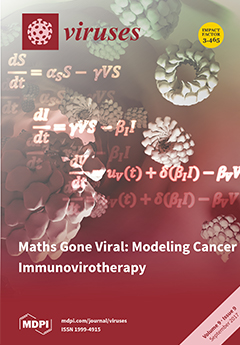
In the emerging era of biological therapies, renewed hope comes hand-in-hand with new challenges in understanding the pharmacokinetic properties of oncolytic viruses, and combinations thereof. Our ability to predict and manipulate the complex interplay between viral agents and the patient’s immune system will be crucial for the clinical success of these therapies. Mathematical modeling is expected to play a key role in such predictions and we hereby review the state-of-the-art of the field. View this paper
  • Issues are regarded as officially published after their release is announced to the table of contents alert mailing list.
  • You may sign up for e-mail alerts to receive table of contents of newly released issues.
  • PDF is the official format for papers published in both, html and pdf forms. To view the papers in pdf format, click on the "PDF Full-text" link, and use the free Adobe Reader to open them.
Order results
Result details
Section
Select all
Export citation of selected articles as:
159 KiB  
Editorial
Viruses of Microbes
by Laurent Debarbieux, Matthias Fischer and Tessa E. F. Quax
Viruses 2017, 9(9), 263; https://doi.org/10.3390/v9090263 - 20 Sep 2017
Cited by 2 | Viewed by 4121
Abstract
Viruses of microbes encompass all viruses that infect archaea, bacteria, and single-celled eukaryotes, especially algae and protozoa [...]
Full article
(This article belongs to the Special Issue Viruses of Microbes)
6199 KiB  
Article
PERK Signal-Modulated Protein Translation Promotes the Survivability of Dengue 2 Virus-Infected Mosquito Cells and Extends Viral Replication
by Jiun-Nan Hou, Tien-Huang Chen, Yi-Hsuan Chiang, Jing-Yun Peng, Tsong-Han Yang, Chih-Chieh Cheng, Eny Sofiyatun, Cheng-Hsun Chiu, Chuan Chiang-Ni and Wei-June Chen
Viruses 2017, 9(9), 262; https://doi.org/10.3390/v9090262 - 20 Sep 2017
Cited by 17 | Viewed by 5907
Abstract
Survival of mosquitoes from dengue virus (DENV) infection is a prerequisite of viral transmission to the host. This study aimed to see how mosquito cells can survive the infection during prosperous replication of the virus. In C6/36 cells, global protein translation was shut [...] Read more.
Survival of mosquitoes from dengue virus (DENV) infection is a prerequisite of viral transmission to the host. This study aimed to see how mosquito cells can survive the infection during prosperous replication of the virus. In C6/36 cells, global protein translation was shut down after infection by DENV type 2 (DENV2). However, it returned to a normal level when infected cells were treated with an inhibitor of the protein kinase RNA (PKR)-like ER kinase (PERK) signaling pathway. Based on a 7-Methylguanosine 5′-triphosphate (m7GTP) pull-down assay, the eukaryotic translation initiation factor 4F (eIF4F) complex was also identified in DENV2-infected cells. This suggests that most mosquito proteins are synthesized via canonical cap-dependent translation. When the PERK signal pathway was inhibited, both accumulation of reactive oxygen species and changes in the mitochondrial membrane potential increased. This suggested that ER stress response was alleviated through the PERK-mediated shutdown of global proteins in DENV2-infected C6/36 cells. In the meantime, the activities of caspases-9 and -3 and the apoptosis-related cell death rate increased in C6/36 cells with PERK inhibition. This reflected that the PERK-signaling pathway is involved in determining cell survival, presumably by reducing DENV2-induced ER stress. Looking at the PERK downstream target, α-subunit of eukaryotic initiation factor 2 (eIF2α), an increased phosphorylation status was only shown in infected C6/36 cells. This indicated that recruitment of ribosome binding to the mRNA 5′-cap structure could have been impaired in cap-dependent translation. It turned out that shutdown of cellular protein translation resulted in a pro-survival effect on mosquito cells in response to DENV2 infection. As synthesis of viral proteins was not affected by the PERK signal pathway, an alternate mode other than cap-dependent translation may be utilized. This finding provides insights into elucidating how the PERK signal pathway modulates dynamic translation of proteins and helps mosquito cells survive continuous replication of the DENV2. It was ecologically important for virus amplification in mosquitoes and transmission to humans. Full article
(This article belongs to the Special Issue Viral Subversion of Transcriptional Control)
Show Figures

Graphical abstract

2012 KiB  
Review
Mechanisms by which HPV Induces a Replication Competent Environment in Differentiating Keratinocytes
by Cary Moody
Viruses 2017, 9(9), 261; https://doi.org/10.3390/v9090261 - 19 Sep 2017
Cited by 59 | Viewed by 12855
Abstract
Human papillomaviruses (HPV) are the causative agents of cervical cancer and are also associated with other genital malignancies, as well as an increasing number of head and neck cancers. HPVs have evolved their life cycle to contend with the different cell states found [...] Read more.
Human papillomaviruses (HPV) are the causative agents of cervical cancer and are also associated with other genital malignancies, as well as an increasing number of head and neck cancers. HPVs have evolved their life cycle to contend with the different cell states found in the stratified epithelium. Initial infection and viral genome maintenance occurs in the proliferating basal cells of the stratified epithelium, where cellular replication machinery is abundant. However, the productive phase of the viral life cycle, including productive replication, late gene expression and virion production, occurs upon epithelial differentiation, in cells that normally exit the cell cycle. This review outlines how HPV interfaces with specific cellular signaling pathways and factors to provide a replication-competent environment in differentiating cells. Full article
(This article belongs to the Special Issue Expert Views on HPV Infection)
Show Figures

Figure 1

2255 KiB  
Article
A Recombinant HAV Expressing a Neutralization Epitope of HEV Induces Immune Response against HAV and HEV in Mice
by Kui Xiang, Yuri Kusov, Guan Ying, Wang Yan, Yi Shan, Wu Jinyuan, Yin Na, Zhou Yan, Li Hongjun and Sun Maosheng
Viruses 2017, 9(9), 260; https://doi.org/10.3390/v9090260 - 15 Sep 2017
Cited by 3 | Viewed by 4192
Abstract
Hepatitis A virus (HAV) and hepatitis E virus (HEV) are causative agents of acute viral hepatitis transmitted via the fecal–oral route. Both viruses place a heavy burden on the public health and economy of developing countries. To test the possibility that HAV could [...] Read more.
Hepatitis A virus (HAV) and hepatitis E virus (HEV) are causative agents of acute viral hepatitis transmitted via the fecal–oral route. Both viruses place a heavy burden on the public health and economy of developing countries. To test the possibility that HAV could be used as an expression vector for the development of a combination vaccine against hepatitis A and E infections, recombinant HAV-HEp148 was created as a vector to express an HEV neutralization epitope (HEp148) located at aa 459–606 of the HEV capsid protein. The recombinant virus expressed the HEp148 protein in a partially dimerized state in HAV-susceptible cells. Immunization with the HAV-HEp148 virus induced a strong HAV- and HEV-specific immune response in mice. Thus, the present study demonstrates a novel approach to the development of a combined hepatitis A and E vaccine. Full article
(This article belongs to the Section Viral Immunology, Vaccines, and Antivirals)
Show Figures

Figure 1

1029 KiB  
Article
LEDGF/p75 Deficiency Increases Deletions at the HIV-1 cDNA Ends
by Murilo T. D. Bueno, Daniel Reyes and Manuel Llano
Viruses 2017, 9(9), 259; https://doi.org/10.3390/v9090259 - 15 Sep 2017
Cited by 7 | Viewed by 3820
Abstract
Processing of unintegrated linear HIV-1 cDNA by the host DNA repair system results in its degradation and/or circularization. As a consequence, deficient viral cDNA integration generally leads to an increase in the levels of HIV-1 cDNA circles containing one or two long terminal [...] Read more.
Processing of unintegrated linear HIV-1 cDNA by the host DNA repair system results in its degradation and/or circularization. As a consequence, deficient viral cDNA integration generally leads to an increase in the levels of HIV-1 cDNA circles containing one or two long terminal repeats (LTRs). Intriguingly, impaired HIV-1 integration in LEDGF/p75-deficient cells does not result in a correspondent increase in viral cDNA circles. We postulate that increased degradation of unintegrated linear viral cDNA in cells lacking the lens epithelium-derived growth factor (LEDGF/p75) account for this inconsistency. To evaluate this hypothesis, we characterized the nucleotide sequence spanning 2-LTR junctions isolated from LEDGF/p75-deficient and control cells. LEDGF/p75 deficiency resulted in a significant increase in the frequency of 2-LTRs harboring large deletions. Of note, these deletions were dependent on the 3′ processing activity of integrase and were not originated by aberrant reverse transcription. Our findings suggest a novel role of LEDGF/p75 in protecting the unintegrated 3′ processed linear HIV-1 cDNA from exonucleolytic degradation. Full article
(This article belongs to the Special Issue Viruses and the DNA Damage Response)
Show Figures

Figure 1

1119 KiB  
Review
Geminiviruses and Plant Hosts: A Closer Examination of the Molecular Arms Race
by Shunmugiah V. Ramesh, Pranav P. Sahu, Manoj Prasad, Shelly Praveen and Hanu R. Pappu
Viruses 2017, 9(9), 256; https://doi.org/10.3390/v9090256 - 15 Sep 2017
Cited by 62 | Viewed by 11563
Abstract
Geminiviruses are plant-infecting viruses characterized by a single-stranded DNA (ssDNA) genome. Geminivirus-derived proteins are multifunctional and effective regulators in modulating the host cellular processes resulting in successful infection. Virus-host interactions result in changes in host gene expression patterns, reprogram plant signaling controls, disrupt [...] Read more.
Geminiviruses are plant-infecting viruses characterized by a single-stranded DNA (ssDNA) genome. Geminivirus-derived proteins are multifunctional and effective regulators in modulating the host cellular processes resulting in successful infection. Virus-host interactions result in changes in host gene expression patterns, reprogram plant signaling controls, disrupt central cellular metabolic pathways, impair plant’s defense system, and effectively evade RNA silencing response leading to host susceptibility. This review summarizes what is known about the cellular processes in the continuing tug of war between geminiviruses and their plant hosts at the molecular level. In addition, implications for engineered resistance to geminivirus infection in the context of a greater understanding of the molecular processes are also discussed. Finally, the prospect of employing geminivirus-based vectors in plant genome engineering and the emergence of powerful genome editing tools to confer geminivirus resistance are highlighted to complete the perspective on geminivirus-plant molecular interactions. Full article
(This article belongs to the Special Issue Geminiviruses)
Show Figures

Figure 1

5329 KiB  
Article
Characterization of vB_SauM-fRuSau02, a Twort-Like Bacteriophage Isolated from a Therapeutic Phage Cocktail
by Katarzyna Leskinen, Henni Tuomala, Anu Wicklund, Jenni Horsma-Heikkinen, Pentti Kuusela, Mikael Skurnik and Saija Kiljunen
Viruses 2017, 9(9), 258; https://doi.org/10.3390/v9090258 - 14 Sep 2017
Cited by 48 | Viewed by 9932
Abstract
Staphylococcus aureus is a commensal and pathogenic bacterium that causes infections in humans and animals. It is a major cause of nosocomial infections worldwide. Due to increasing prevalence of multidrug resistance, alternative methods to eradicate the pathogen are necessary. In this respect, polyvalent [...] Read more.
Staphylococcus aureus is a commensal and pathogenic bacterium that causes infections in humans and animals. It is a major cause of nosocomial infections worldwide. Due to increasing prevalence of multidrug resistance, alternative methods to eradicate the pathogen are necessary. In this respect, polyvalent staphylococcal myoviruses have been demonstrated to be excellent candidates for phage therapy. Here we present the characterization of the bacteriophage vB_SauM-fRuSau02 (fRuSau02) that was isolated from a commercial Staphylococcus bacteriophage cocktail produced by Microgen (Moscow, Russia). The genomic analysis revealed that fRuSau02 is very closely related to the phage MSA6, and possesses a large genome (148,464 bp), with typical modular organization and a low G+C (30.22%) content. It can therefore be classified as a new virus among the genus Twortlikevirus. The genome contains 236 predicted genes, 4 of which were interrupted by insertion sequences. Altogether, 78 different structural and virion-associated proteins were identified from purified phage particles by liquid chromatography-tandem mass spectrometry (LC-MS/MS). The host range of fRuSau02 was tested with 135 strains, including 51 and 54 Staphylococcus aureus isolates from humans and pigs, respectively, and 30 coagulase-negative Staphylococcus strains of human origin. All clinical S. aureus strains were at least moderately sensitive to the phage, while only 39% of the pig strains were infected. Also, some strains of Staphylococcus intermedius, Staphylococcus lugdunensis, Staphylococcus epidermidis, Staphylococcus haemolyticus, Staphylococcus saprophyticus and Staphylococcus pseudointer were sensitive. We conclude that fRuSau02, a phage therapy agent in Russia, can serve as an alternative to antibiotic therapy against S. aureus. Full article
(This article belongs to the Special Issue Hurdles for Phage Therapy (PT) to Become a Reality)
Show Figures

Figure 1

4565 KiB  
Article
Engineered Disease Resistance in Cotton Using RNA-Interference to Knock down Cotton leaf curl Kokhran virus-Burewala and Cotton leaf curl Multan betasatellite Expression
by Aftab Ahmad, Muhammad Zia-Ur-Rehman, Usman Hameed, Abdul Qayyum Rao, Ammara Ahad, Aneela Yasmeen, Faheem Akram, Kamran Shahzad Bajwa, Jodi Scheffler, Idrees Ahmad Nasir, Ahmad Ali Shahid, Muhammad Javed Iqbal, Tayyab Husnain, Muhammad Saleem Haider and Judith K. Brown
Viruses 2017, 9(9), 257; https://doi.org/10.3390/v9090257 - 14 Sep 2017
Cited by 16 | Viewed by 6084
Abstract
Cotton leaf curl virus disease (CLCuD) is caused by a suite of whitefly-transmitted begomovirus species and strains, resulting in extensive losses annually in India and Pakistan. RNA-interference (RNAi) is a proven technology used for knockdown of gene expression in higher organisms and viruses. [...] Read more.
Cotton leaf curl virus disease (CLCuD) is caused by a suite of whitefly-transmitted begomovirus species and strains, resulting in extensive losses annually in India and Pakistan. RNA-interference (RNAi) is a proven technology used for knockdown of gene expression in higher organisms and viruses. In this study, a small interfering RNA (siRNA) construct was designed to target the AC1 gene of Cotton leaf curl Kokhran virus-Burewala (CLCuKoV-Bu) and the βC1 gene and satellite conserved region of the Cotton leaf curl Multan betasatellite (CLCuMB). The AC1 gene and CLCuMB coding and non-coding regions function in replication initiation and suppression of the plant host defense pathway, respectively. The construct, , was transformed into cotton plants using the Agrobacterium-mediated embryo shoot apex cut method. Results from fluorescence in situ hybridization and karyotyping assays indicated that six of the 11 T1 plants harbored a single copy of the Vβ transgene. Transgenic cotton plants and non-transgenic (susceptible) test plants included as the positive control were challenge-inoculated using the viruliferous whitefly vector to transmit the CLCuKoV-Bu/CLCuMB complex. Among the test plants, plant Vβ-6 was asymptomatic, had the lowest amount of detectable virus, and harbored a single copy of the transgene on chromosome six. Absence of characteristic leaf curl symptom development in transgenic Vβ-6 cotton plants, and significantly reduced begomoviral-betasatellite accumulation based on real-time polymerase chain reaction, indicated the successful knockdown of CLCuKoV-Bu and CLCuMB expression, resulting in leaf curl resistant plants. Full article
(This article belongs to the Special Issue Geminiviruses)
Show Figures

Figure 1

9068 KiB  
Article
Identification of a Novel Inhibitor against Middle East Respiratory Syndrome Coronavirus
by Yaping Sun, Huaidong Zhang, Jian Shi, Zhe Zhang and Rui Gong
Viruses 2017, 9(9), 255; https://doi.org/10.3390/v9090255 - 14 Sep 2017
Cited by 28 | Viewed by 6739
Abstract
The Middle East respiratory syndrome coronavirus (MERS-CoV) was first isolated in 2012, and circulated worldwide with high mortality. The continual outbreaks of MERS-CoV highlight the importance of developing antiviral therapeutics. Here, we rationally designed a novel fusion inhibitor named MERS-five-helix bundle (MERS-5HB) derived [...] Read more.
The Middle East respiratory syndrome coronavirus (MERS-CoV) was first isolated in 2012, and circulated worldwide with high mortality. The continual outbreaks of MERS-CoV highlight the importance of developing antiviral therapeutics. Here, we rationally designed a novel fusion inhibitor named MERS-five-helix bundle (MERS-5HB) derived from the six-helix bundle (MERS-6HB) which was formed by the process of membrane fusion. MERS-5HB consists of three copies of heptad repeat 1 (HR1) and two copies of heptad repeat 2 (HR2) while MERS-6HB includes three copies each of HR1 and HR2. As it lacks one HR2, MERS-5HB was expected to interact with viral HR2 to interrupt the fusion step. What we found was that MERS-5HB could bind to HR2P, a peptide derived from HR2, with a strong affinity value (KD) of up to 0.24 nM. Subsequent assays indicated that MERS-5HB could inhibit pseudotyped MERS-CoV entry effectively with 50% inhibitory concentration (IC50) of about 1 μM. In addition, MERS-5HB significantly inhibited spike (S) glycoprotein-mediated syncytial formation in a dose-dependent manner. Further biophysical characterization showed that MERS-5HB was a thermo-stable α-helical secondary structure. The inhibitory potency of MERS-5HB may provide an attractive basis for identification of a novel inhibitor against MERS-CoV, as a potential antiviral agent. Full article
(This article belongs to the Section Animal Viruses)
Show Figures

Figure 1

1035 KiB  
Review
Immunopathogenesis of HPV-Associated Cancers and Prospects for Immunotherapy
by Sigrun Smola
Viruses 2017, 9(9), 254; https://doi.org/10.3390/v9090254 - 12 Sep 2017
Cited by 93 | Viewed by 14857
Abstract
Human papillomavirus (HPV) infection is a causative factor for various cancers of the anogenital region and oropharynx, and is supposed to play an important cofactor role for skin carcinogenesis. Evasion from immunosurveillance favors viral persistence. However, there is evidence that the mere presence [...] Read more.
Human papillomavirus (HPV) infection is a causative factor for various cancers of the anogenital region and oropharynx, and is supposed to play an important cofactor role for skin carcinogenesis. Evasion from immunosurveillance favors viral persistence. However, there is evidence that the mere presence of oncogenic HPV is not sufficient for malignant progression and that additional tumor-promoting steps are required. Recent studies have demonstrated that HPV-transformed cells actively promote chronic stromal inflammation and conspire with cells in the local microenvironment to promote carcinogenesis. This review highlights the complex interplay between HPV-infected cells and the local immune microenvironment during oncogenic HPV infection, persistence, and malignant progression, and discusses new prospects for diagnosis and immunotherapy of HPV-associated cancers. Full article
(This article belongs to the Special Issue Expert Views on HPV Infection)
Show Figures

Figure 1

2579 KiB  
Article
Analysis of Class I Major Histocompatibility Complex Gene Transcription in Human Tumors Caused by Human Papillomavirus Infection
by Steven F. Gameiro, Ali Zhang, Farhad Ghasemi, John W. Barrett, Anthony C. Nichols and Joe S. Mymryk
Viruses 2017, 9(9), 252; https://doi.org/10.3390/v9090252 - 10 Sep 2017
Cited by 31 | Viewed by 5117
Abstract
Oncoproteins from high-risk human papillomaviruses (HPV) downregulate the transcription of the class I major histocompatibility complex (MHC-I) antigen presentation apparatus in tissue culture model systems. This could allow infected or transformed cells to evade the adaptive immune response. Using data from over 800 [...] Read more.
Oncoproteins from high-risk human papillomaviruses (HPV) downregulate the transcription of the class I major histocompatibility complex (MHC-I) antigen presentation apparatus in tissue culture model systems. This could allow infected or transformed cells to evade the adaptive immune response. Using data from over 800 human cervical and head & neck tumors from The Cancer Genome Atlas (TCGA), we determined the impact of HPV status on the mRNA expression of all six MHC-I heavy chain genes, and the β2 microglobulin light chain. Unexpectedly, these genes were all expressed at high levels in HPV positive (HPV+) cancers compared with normal control tissues. Indeed, many of these genes were expressed at significantly enhanced levels in HPV+ tumors. Similarly, the transcript levels of several other components of the MHC-I peptide-loading complex were also high in HPV+ cancers. The coordinated expression of high mRNA levels of the MHC-I antigen presentation apparatus could be a consequence of the higher intratumoral levels of interferon γ in HPV+ carcinomas, which correlate with signatures of increased infiltration by T- and NK-cells. These data, which were obtained from both cervical and oral tumors in large human cohorts, indicates that HPV oncoproteins do not efficiently suppress the transcription of the antigen presentation apparatus in human tumors. Full article
(This article belongs to the Special Issue Viral Subversion of Transcriptional Control)
Show Figures

Figure 1

3767 KiB  
Article
Re-Assembly and Analysis of an Ancient Variola Virus Genome
by Chad Smithson, Jacob Imbery and Chris Upton
Viruses 2017, 9(9), 253; https://doi.org/10.3390/v9090253 - 08 Sep 2017
Cited by 13 | Viewed by 6327
Abstract
We report a major improvement to the assembly of published short read sequencing data from an ancient variola virus (VARV) genome by the removal of contig-capping sequencing tags and manual searches for gap-spanning reads. The new assembly, together with camelpox and taterapox genomes, [...] Read more.
We report a major improvement to the assembly of published short read sequencing data from an ancient variola virus (VARV) genome by the removal of contig-capping sequencing tags and manual searches for gap-spanning reads. The new assembly, together with camelpox and taterapox genomes, permitted new dates to be calculated for the last common ancestor of all VARV genomes. The analysis of recently sequenced VARV-like cowpox virus genomes showed that single nucleotide polymorphisms (SNPs) and amino acid changes in the vaccinia virus (VACV)-Cop-O1L ortholog, predicted to be associated with VARV host specificity and virulence, were introduced into the lineage before the divergence of these viruses. A comparison of the ancient and modern VARV genome sequences also revealed a measurable drift towards adenine + thymine (A + T) richness. Full article
(This article belongs to the Special Issue Smallpox and Emerging Zoonotic Orthopoxviruses: What Is Coming Next?)
Show Figures

Figure 1

4823 KiB  
Article
Ultrastructural Characterization of Membrane Rearrangements Induced by Porcine Epidemic Diarrhea Virus Infection
by Xingdong Zhou, Yingying Cong, Tineke Veenendaal, Judith Klumperman, Dongfang Shi, Muriel Mari and Fulvio Reggiori
Viruses 2017, 9(9), 251; https://doi.org/10.3390/v9090251 - 05 Sep 2017
Cited by 26 | Viewed by 5784
Abstract
The porcine epidemic diarrhea virus (PEDV) is a coronavirus (CoV) belonging to the α-CoV genus and it causes high mortality in infected sucking piglets, resulting in substantial losses in the farming industry. CoV trigger a drastic reorganization of host cell membranes [...] Read more.
The porcine epidemic diarrhea virus (PEDV) is a coronavirus (CoV) belonging to the α-CoV genus and it causes high mortality in infected sucking piglets, resulting in substantial losses in the farming industry. CoV trigger a drastic reorganization of host cell membranes to promote their replication and egression, but a detailed description of the intracellular remodeling induced by PEDV is still missing. In this study, we examined qualitatively and quantitatively, using electron microscopy, the intracellular membrane reorganization induced by PEDV over the course of an infection. With our ultrastructural approach, we reveal that, as most of CoV, PEDV initially forms double-membrane vesicles (DMVs) and convoluted membranes (CMs), which probably serve as replication/transcription platforms. Interestingly, we also found that viral particles start to form almost simultaneously in both the endoplasmic reticulum and the large virion-containing vacuoles (LVCVs), which are compartments originating from the Golgi, confirming that α-CoV assemble indistinguishably in two different organelles of the secretory pathway. Moreover, PEDV virons appear to have an immature and a mature form, similar to another α-CoV the transmissible gastroenteritis coronavirus (TGEV). Altogether, our study underlies the similarities and differences between the lifecycle of α-CoV and that of viruses belonging to other CoV subfamilies. Full article
(This article belongs to the Section Animal Viruses)
Show Figures

Graphical abstract

1216 KiB  
Article
Using Next Generation Sequencing to Identify and Quantify the Genetic Composition of Resistance-Breaking Commercial Isolates of Cydia pomonella Granulovirus
by Gianpiero Gueli Alletti, Annette J. Sauer, Birgit Weihrauch, Eva Fritsch, Karin Undorf-Spahn, Jörg T. Wennmann and Johannes A. Jehle
Viruses 2017, 9(9), 250; https://doi.org/10.3390/v9090250 - 04 Sep 2017
Cited by 20 | Viewed by 4830
Abstract
The use of Cydia pomonella granulovirus (CpGV) isolates as biological control agents of codling moth (CM) larvae is important in organic and integrated pome fruit production worldwide. The commercially available isolates CpGV-0006, CpGV-R5, and CpGV-V15 have been selected for the control of CpGV [...] Read more.
The use of Cydia pomonella granulovirus (CpGV) isolates as biological control agents of codling moth (CM) larvae is important in organic and integrated pome fruit production worldwide. The commercially available isolates CpGV-0006, CpGV-R5, and CpGV-V15 have been selected for the control of CpGV resistant CM populations in Europe. In infection experiments, CpGV-0006 and CpGV-R5 were able to break type I resistance and to a lower extent also type III resistance, whereas CpGV-V15 overcame type I and the rarely occurring type II and type III resistance. The genetic background of the three isolates was investigated with next generation sequencing (NGS) tools by comparing their nucleotide compositions to whole genome alignments of five CpGV isolates representing the known genetic diversity of the CpGV genome groups A to E. Based on the distribution of single nucleotide polymorphisms (SNPs) in Illumina sequencing reads, we found that the two isolates CpGV-0006 and CpGV-R5 have highly similar genome group compositions, consisting of about two thirds of the CpGV genome group E and one third of genome group A. In contrast, CpGV-V15 is composed of equal parts of CpGV genome group B and E. According to the identified genetic composition of these isolates, their efficacy towards different resistance types can be explained and predictions on the success of resistance management strategies in resistant CM populations can be made. Full article
(This article belongs to the Section Insect Viruses)
Show Figures

Figure 1

2611 KiB  
Article
SGS3 Cooperates with RDR6 in Triggering Geminivirus-Induced Gene Silencing and in Suppressing Geminivirus Infection in Nicotiana Benthamiana
by Fangfang Li, Yaqin Wang and Xueping Zhou
Viruses 2017, 9(9), 247; https://doi.org/10.3390/v9090247 - 04 Sep 2017
Cited by 22 | Viewed by 6951
Abstract
RNA silencing has an important role in defending against virus infection in plants. Plants with the deficiency of RNA silencing components often show enhanced susceptibility to viral infections. RNA-dependent RNA polymerase (RDRs) mediated-antiviral defense has a pivotal role in resistance to many plant [...] Read more.
RNA silencing has an important role in defending against virus infection in plants. Plants with the deficiency of RNA silencing components often show enhanced susceptibility to viral infections. RNA-dependent RNA polymerase (RDRs) mediated-antiviral defense has a pivotal role in resistance to many plant viruses. In RDR6-mediated defense against viral infection, a plant-specific RNA binding protein, Suppressor of Gene Silencing 3 (SGS3), was also found to fight against some viruses in Arabidopsis. In this study, we showed that SGS3 from Nicotiana benthamiana (NbSGS3) is required for sense-RNA induced post-transcriptional gene silencing (S-PTGS) and initiating sense-RNA-triggered systemic silencing. Further, the deficiency of NbSGS3 inhibited geminivirus-induced endogenous gene silencing (GIEGS) and promoted geminivirus infection. During TRV-mediated NbSGS3 or N. benthamiana RDR6 (NbRDR6) silencing process, we found that their expression can be effectively fine-tuned. Plants with the knock-down of both NbSGS3 and NbRDR6 almost totally blocked GIEGS, and were more susceptible to geminivirus infection. These data suggest that NbSGS3 cooperates with NbRDR6 against GIEGS and geminivirus infection in N. benthamiana, which provides valuable information for breeding geminivirus-resistant plants. Full article
(This article belongs to the Special Issue Geminiviruses)
Show Figures

Figure 1

1444 KiB  
Review
Die Another Day: Inhibition of Cell Death Pathways by Cytomegalovirus
by Wolfram Brune and Christopher E. Andoniou
Viruses 2017, 9(9), 249; https://doi.org/10.3390/v9090249 - 02 Sep 2017
Cited by 55 | Viewed by 8385
Abstract
Multicellular organisms have evolved multiple genetically programmed cell death pathways that are essential for homeostasis. The finding that many viruses encode cell death inhibitors suggested that cellular suicide also functions as a first line of defence against invading pathogens. This theory was confirmed [...] Read more.
Multicellular organisms have evolved multiple genetically programmed cell death pathways that are essential for homeostasis. The finding that many viruses encode cell death inhibitors suggested that cellular suicide also functions as a first line of defence against invading pathogens. This theory was confirmed by studying viral mutants that lack certain cell death inhibitors. Cytomegaloviruses, a family of species-specific viruses, have proved particularly useful in this respect. Cytomegaloviruses are known to encode multiple death inhibitors that are required for efficient viral replication. Here, we outline the mechanisms used by the host cell to detect cytomegalovirus infection and discuss the methods employed by the cytomegalovirus family to prevent death of the host cell. In addition to enhancing our understanding of cytomegalovirus pathogenesis we detail how this research has provided significant insights into the cross-talk that exists between the various cell death pathways. Full article
(This article belongs to the Special Issue Viral Infection and Apoptosis)
Show Figures

Figure 1

719 KiB  
Review
Epigenetic Alterations in Human Papillomavirus-Associated Cancers
by David Soto, Christine Song and Margaret E. McLaughlin-Drubin
Viruses 2017, 9(9), 248; https://doi.org/10.3390/v9090248 - 01 Sep 2017
Cited by 67 | Viewed by 7881
Abstract
Approximately 15–20% of human cancers are caused by viruses, including human papillomaviruses (HPVs). Viruses are obligatory intracellular parasites and encode proteins that reprogram the regulatory networks governing host cellular signaling pathways that control recognition by the immune system, proliferation, differentiation, genomic integrity, and [...] Read more.
Approximately 15–20% of human cancers are caused by viruses, including human papillomaviruses (HPVs). Viruses are obligatory intracellular parasites and encode proteins that reprogram the regulatory networks governing host cellular signaling pathways that control recognition by the immune system, proliferation, differentiation, genomic integrity, and cell death. Given that key proteins in these regulatory networks are also subject to mutation in non-virally associated diseases and cancers, the study of oncogenic viruses has also been instrumental to the discovery and analysis of many fundamental cellular processes, including messenger RNA (mRNA) splicing, transcriptional enhancers, oncogenes and tumor suppressors, signal transduction, immune regulation, and cell cycle control. More recently, tumor viruses, in particular HPV, have proven themselves invaluable in the study of the cancer epigenome. Epigenetic silencing or de-silencing of genes can have cellular consequences that are akin to genetic mutations, i.e., the loss and gain of expression of genes that are not usually expressed in a certain cell type and/or genes that have tumor suppressive or oncogenic activities, respectively. Unlike genetic mutations, the reversible nature of epigenetic modifications affords an opportunity of epigenetic therapy for cancer. This review summarizes the current knowledge on epigenetic regulation in HPV-infected cells with a focus on those elements with relevance to carcinogenesis. Full article
(This article belongs to the Special Issue Expert Views on HPV Infection)
Show Figures

Figure 1

2029 KiB  
Review
The Mouse Papillomavirus Infection Model
by Jiafen Hu, Nancy M. Cladel, Lynn R. Budgeon, Karla K. Balogh and Neil D. Christensen
Viruses 2017, 9(9), 246; https://doi.org/10.3390/v9090246 - 30 Aug 2017
Cited by 34 | Viewed by 8341
Abstract
The mouse papillomavirus (MmuPV1) was first reported in 2011 and has since become a powerful research tool. Through collective efforts from different groups, significant progress has been made in the understanding of molecular, virological, and immunological mechanisms of MmuPV1 infections in both immunocompromised [...] Read more.
The mouse papillomavirus (MmuPV1) was first reported in 2011 and has since become a powerful research tool. Through collective efforts from different groups, significant progress has been made in the understanding of molecular, virological, and immunological mechanisms of MmuPV1 infections in both immunocompromised and immunocompetent hosts. This mouse papillomavirus provides, for the first time, the opportunity to study papillomavirus infections in the context of a small common laboratory animal for which abundant reagents are available and for which many strains exist. The model is a major step forward in the study of papillomavirus disease and pathology. In this review, we summarize studies using MmuPV1 over the past six years and share our perspectives on the value of this unique model system. Specifically, we discuss viral pathogenesis in cutaneous and mucosal tissues as well as in different mouse strains, immune responses to the virus, and local host-restricted factors that may be involved in MmuPV1 infections and associated disease progression. Full article
(This article belongs to the Special Issue Expert Views on HPV Infection)
Show Figures

Graphical abstract

1387 KiB  
Review
Keratinocyte Differentiation-Dependent Human Papillomavirus Gene Regulation
by Sheila V. Graham
Viruses 2017, 9(9), 245; https://doi.org/10.3390/v9090245 - 30 Aug 2017
Cited by 66 | Viewed by 10776
Abstract
Human papillomaviruses (HPVs) cause diseases ranging from benign warts to invasive cancers. HPVs infect epithelial cells and their replication cycle is tightly linked with the differentiation process of the infected keratinocyte. The normal replication cycle involves an early and a late phase. The [...] Read more.
Human papillomaviruses (HPVs) cause diseases ranging from benign warts to invasive cancers. HPVs infect epithelial cells and their replication cycle is tightly linked with the differentiation process of the infected keratinocyte. The normal replication cycle involves an early and a late phase. The early phase encompasses viral entry and initial genome replication, stimulation of cell division and inhibition of apoptosis in the infected cell. Late events in the HPV life cycle include viral genome amplification, virion formation, and release into the environment from the surface of the epithelium. The main proteins required at the late stage of infection for viral genome amplification include E1, E2, E4 and E5. The late proteins L1 and L2 are structural proteins that form the viral capsid. Regulation of these late events involves both cellular and viral proteins. The late viral mRNAs are expressed from a specific late promoter but final late mRNA levels in the infected cell are controlled by splicing, polyadenylation, nuclear export and RNA stability. Viral late protein expression is also controlled at the level of translation. This review will discuss current knowledge of how HPV late gene expression is regulated. Full article
(This article belongs to the Special Issue Expert Views on HPV Infection)
Show Figures

Figure 1

2693 KiB  
Article
Key Determinants of Human α-Defensin 5 and 6 for Enhancement of HIV Infectivity
by Kimyata Valere, Wuyuan Lu and Theresa L. Chang
Viruses 2017, 9(9), 244; https://doi.org/10.3390/v9090244 - 29 Aug 2017
Cited by 7 | Viewed by 3721
Abstract
Defensins are antimicrobial peptides important for mucosal innate immunity. They exhibit a broad spectrum of activity against bacteria, viruses, and fungi. Levels of α-defensins are elevated at the genital mucosa of individuals with sexually transmitted infections (STIs). Somewhat paradoxically, human α-defensin 5 and [...] Read more.
Defensins are antimicrobial peptides important for mucosal innate immunity. They exhibit a broad spectrum of activity against bacteria, viruses, and fungi. Levels of α-defensins are elevated at the genital mucosa of individuals with sexually transmitted infections (STIs). Somewhat paradoxically, human α-defensin 5 and 6 (HD5 and HD6) promote human immunodeficiency virus (HIV) infectivity, and contribute to STI-mediated enhancement of HIV infection in vitro. Specific amino acid residues of HD5 and HD6 that are crucial for antimicrobial activities have been characterized previously; however, the key determinants of defensins responsible for enhancement of HIV infectivity are not known. Here, we have identified residues of HD5 and HD6 that are required for enhancement of HIV attachment and infection. Most of these residues are involved in hydrophobicity and self-association of defensins. Specifically, we found that mutant defensins L16A-HD5, E21me-HD5, L26A-HD5, Y27A-HD5, F2A-HD6, H27W-HD6, and F29A-HD6 significantly lost their ability to promote HIV attachment and infection. L29A mutation also reduced HIV infection-enhancing activity of HD5. Additionally, a number of mutations in charged residues variably affected the profile of HIV attachment and infectivity. One HD5 charged mutation, R28A, notably resulted in a 34–48% loss of enhanced HIV infectivity and attachment. These results indicate that defensin determinants that maintain high-ordered amphipathic structure are crucial for HIV enhancing activity. In a comparative analysis of the mutant defensins, we found that for some defensin mutants enhancement of HIV infectivity was associated with the reverse transcription step, suggesting a novel, HIV attachment-independent, mechanism of defensin-mediated HIV enhancement. Full article
(This article belongs to the Section Viral Immunology, Vaccines, and Antivirals)
Show Figures

Figure 1

1264 KiB  
Review
Regulation of Apoptosis during Flavivirus Infection
by Toru Okamoto, Tatsuya Suzuki, Shinji Kusakabe, Makoto Tokunaga, Junki Hirano, Yuka Miyata and Yoshiharu Matsuura
Viruses 2017, 9(9), 243; https://doi.org/10.3390/v9090243 - 28 Aug 2017
Cited by 58 | Viewed by 10280
Abstract
Apoptosis is a type of programmed cell death that regulates cellular homeostasis by removing damaged or unnecessary cells. Its importance in host defenses is highlighted by the observation that many viruses evade, obstruct, or subvert apoptosis, thereby blunting the host immune response. Infection [...] Read more.
Apoptosis is a type of programmed cell death that regulates cellular homeostasis by removing damaged or unnecessary cells. Its importance in host defenses is highlighted by the observation that many viruses evade, obstruct, or subvert apoptosis, thereby blunting the host immune response. Infection with Flaviviruses such as Japanese encephalitis virus (JEV), Dengue virus (DENV) and West Nile virus (WNV) has been shown to activate several signaling pathways such as endoplasmic reticulum (ER)-stress and AKT/PI3K pathway, resulting in activation or suppression of apoptosis in virus-infected cells. On the other hands, expression of some viral proteins induces or protects apoptosis. There is a discrepancy between induction and suppression of apoptosis during flavivirus infection because the experimental situation may be different, and strong links between apoptosis and other types of cell death such as necrosis may make it more difficult. In this paper, we review the effects of apoptosis on viral propagation and pathogenesis during infection with flaviviruses. Full article
(This article belongs to the Special Issue Viral Infection and Apoptosis)
Show Figures

Figure 1

275 KiB  
Perspective
Are We Prepared in Case of a Possible Smallpox-Like Disease Emergence?
by Victoria A. Olson and Sergei N. Shchelkunov
Viruses 2017, 9(9), 242; https://doi.org/10.3390/v9090242 - 27 Aug 2017
Cited by 20 | Viewed by 7464
Abstract
Smallpox was the first human disease to be eradicated, through a concerted vaccination campaign led by the World Health Organization. Since its eradication, routine vaccination against smallpox has ceased, leaving the world population susceptible to disease caused by orthopoxviruses. In recent decades, reports [...] Read more.
Smallpox was the first human disease to be eradicated, through a concerted vaccination campaign led by the World Health Organization. Since its eradication, routine vaccination against smallpox has ceased, leaving the world population susceptible to disease caused by orthopoxviruses. In recent decades, reports of human disease from zoonotic orthopoxviruses have increased. Furthermore, multiple reports of newly identified poxviruses capable of causing human disease have occurred. These facts raise concerns regarding both the opportunity for these zoonotic orthopoxviruses to evolve and become a more severe public health issue, as well as the risk of Variola virus (the causative agent of smallpox) to be utilized as a bioterrorist weapon. The eradication of smallpox occurred prior to the development of the majority of modern virological and molecular biological techniques. Therefore, there is a considerable amount that is not understood regarding how this solely human pathogen interacts with its host. This paper briefly recounts the history and current status of diagnostic tools, vaccines, and anti-viral therapeutics for treatment of smallpox disease. The authors discuss the importance of further research to prepare the global community should a smallpox-like virus emerge. Full article
(This article belongs to the Special Issue Smallpox and Emerging Zoonotic Orthopoxviruses: What Is Coming Next?)
1885 KiB  
Review
Investigations of Pro- and Anti-Apoptotic Factors Affecting African Swine Fever Virus Replication and Pathogenesis
by Linda K. Dixon, Pedro J. Sánchez-Cordón, Inmaculada Galindo and Covadonga Alonso
Viruses 2017, 9(9), 241; https://doi.org/10.3390/v9090241 - 25 Aug 2017
Cited by 45 | Viewed by 10505
Abstract
African swine fever virus (ASFV) is a large DNA virus that replicates predominantly in the cell cytoplasm and is the only member of the Asfarviridae family. The virus causes an acute haemorrhagic fever, African swine fever (ASF), in domestic pigs and wild boar [...] Read more.
African swine fever virus (ASFV) is a large DNA virus that replicates predominantly in the cell cytoplasm and is the only member of the Asfarviridae family. The virus causes an acute haemorrhagic fever, African swine fever (ASF), in domestic pigs and wild boar resulting in the death of most infected animals. Apoptosis is induced at an early stage during virus entry or uncoating. However, ASFV encodes anti-apoptotic proteins which facilitate production of progeny virions. These anti-apoptotic proteins include A179L, a Bcl-2 family member; A224L, an inhibitor of apoptosis proteins (IAP) family member; EP153R a C-type lectin; and DP71L. The latter acts by inhibiting activation of the stress activated pro-apoptotic pathways pro-apoptotic pathways. The mechanisms by which these proteins act is summarised. ASF disease is characterised by massive apoptosis of uninfected lymphocytes which reduces the effectiveness of the immune response, contributing to virus pathogenesis. Mechanisms by which this apoptosis is induced are discussed. Full article
(This article belongs to the Special Issue Viral Infection and Apoptosis)
Show Figures

Graphical abstract

9614 KiB  
Article
The Enigmatic Origin of Papillomavirus Protein Domains
by Mikk Puustusmaa, Heleri Kirsip, Kevin Gaston and Aare Abroi
Viruses 2017, 9(9), 240; https://doi.org/10.3390/v9090240 - 23 Aug 2017
Cited by 6 | Viewed by 6770
Abstract
Almost a century has passed since the discovery of papillomaviruses. A few decades of research have given a wealth of information on the molecular biology of papillomaviruses. Several excellent studies have been performed looking at the long- and short-term evolution of these viruses. [...] Read more.
Almost a century has passed since the discovery of papillomaviruses. A few decades of research have given a wealth of information on the molecular biology of papillomaviruses. Several excellent studies have been performed looking at the long- and short-term evolution of these viruses. However, when and how papillomaviruses originate is still a mystery. In this study, we systematically searched the (sequenced) biosphere to find distant homologs of papillomaviral protein domains. Our data show that, even including structural information, which allows us to find deeper evolutionary relationships compared to sequence-only based methods, only half of the protein domains in papillomaviruses have relatives in the rest of the biosphere. We show that the major capsid protein L1 and the replication protein E1 have relatives in several viral families, sharing three protein domains with Polyomaviridae and Parvoviridae. However, only the E1 replication protein has connections with cellular organisms. Most likely, the papillomavirus ancestor is of marine origin, a biotope that is not very well sequenced at the present time. Nevertheless, there is no evidence as to how papillomaviruses originated and how they became vertebrate and epithelium specific. Full article
(This article belongs to the Special Issue Expert Views on HPV Infection)
Show Figures

Figure 1

3232 KiB  
Review
Fighting Cancer with Mathematics and Viruses
by Daniel N. Santiago, Johannes P. W. Heidbuechel, Wendy M. Kandell, Rachel Walker, Julie Djeu, Christine E. Engeland, Daniel Abate-Daga and Heiko Enderling
Viruses 2017, 9(9), 239; https://doi.org/10.3390/v9090239 - 23 Aug 2017
Cited by 25 | Viewed by 9361
Abstract
After decades of research, oncolytic virotherapy has recently advanced to clinical application, and currently a multitude of novel agents and combination treatments are being evaluated for cancer therapy. Oncolytic agents preferentially replicate in tumor cells, inducing tumor cell lysis and complex antitumor effects, [...] Read more.
After decades of research, oncolytic virotherapy has recently advanced to clinical application, and currently a multitude of novel agents and combination treatments are being evaluated for cancer therapy. Oncolytic agents preferentially replicate in tumor cells, inducing tumor cell lysis and complex antitumor effects, such as innate and adaptive immune responses and the destruction of tumor vasculature. With the availability of different vector platforms and the potential of both genetic engineering and combination regimens to enhance particular aspects of safety and efficacy, the identification of optimal treatments for patient subpopulations or even individual patients becomes a top priority. Mathematical modeling can provide support in this arena by making use of experimental and clinical data to generate hypotheses about the mechanisms underlying complex biology and, ultimately, predict optimal treatment protocols. Increasingly complex models can be applied to account for therapeutically relevant parameters such as components of the immune system. In this review, we describe current developments in oncolytic virotherapy and mathematical modeling to discuss the benefit of integrating different modeling approaches into biological and clinical experimentation. Conclusively, we propose a mutual combination of these research fields to increase the value of the preclinical development and the therapeutic efficacy of the resulting treatments. Full article
(This article belongs to the Special Issue Mathematical Modeling of Viral Infections)
Show Figures

Graphical abstract

2218 KiB  
Article
The Response of Heterotrophic Prokaryote and Viral Communities to Labile Organic Carbon Inputs Is Controlled by the Predator Food Chain Structure
by Ruth-Anne Sandaa, Bernadette Pree, Aud Larsen, Selina Våge, Birte Töpper, Joachim P. Töpper, Runar Thyrhaug and Tron Frede Thingstad
Viruses 2017, 9(9), 238; https://doi.org/10.3390/v9090238 - 23 Aug 2017
Cited by 14 | Viewed by 5151
Abstract
Factors controlling the community composition of marine heterotrophic prokaryotes include organic-C, mineral nutrients, predation, and viral lysis. Two mesocosm experiments, performed at an Arctic location and bottom-up manipulated with organic-C, had very different results in community composition for both prokaryotes and viruses. Previously, [...] Read more.
Factors controlling the community composition of marine heterotrophic prokaryotes include organic-C, mineral nutrients, predation, and viral lysis. Two mesocosm experiments, performed at an Arctic location and bottom-up manipulated with organic-C, had very different results in community composition for both prokaryotes and viruses. Previously, we showed how a simple mathematical model could reproduce food web level dynamics observed in these mesocosms, demonstrating strong top-down control through the predator chain from copepods via ciliates and heterotrophic nanoflagellates. Here, we use a steady-state analysis to connect ciliate biomass to bacterial carbon demand. This gives a coupling of top-down and bottom-up factors whereby low initial densities of ciliates are associated with mineral nutrient-limited heterotrophic prokaryotes that do not respond to external supply of labile organic-C. In contrast, high initial densities of ciliates give carbon-limited growth and high responsiveness to organic-C. The differences observed in ciliate abundance, and in prokaryote abundance and community composition in the two experiments were in accordance with these predictions. Responsiveness in the viral community followed a pattern similar to that of prokaryotes. Our study provides a unique link between the structure of the predator chain in the microbial food web and viral abundance and diversity. Full article
(This article belongs to the Special Issue Marine Viruses 2016)
Show Figures

Figure 1

2134 KiB  
Review
The Epidemiology of African Swine Fever in “Nonendemic” Regions of Zambia (1989–2015): Implications for Disease Prevention and Control
by Edgar Simulundu, Caesar H. Lubaba, Juanita Van Heerden, Masahiro Kajihara, Liywalii Mataa, Herman Moses Chambaro, Yona Sinkala, Samuel Munalula Munjita, Hetron Mweemba Munang’andu, King Shimumbo Nalubamba, Kenny Samui, Girja Shanker Pandey, Ayato Takada and Aaron S. Mweene
Viruses 2017, 9(9), 236; https://doi.org/10.3390/v9090236 - 23 Aug 2017
Cited by 34 | Viewed by 8506
Abstract
African swine fever (ASF) is a highly contagious and deadly viral hemorrhagic disease of swine. In Zambia, ASF was first reported in 1912 in Eastern Province and is currently believed to be endemic in that province only. Strict quarantine measures implemented at the [...] Read more.
African swine fever (ASF) is a highly contagious and deadly viral hemorrhagic disease of swine. In Zambia, ASF was first reported in 1912 in Eastern Province and is currently believed to be endemic in that province only. Strict quarantine measures implemented at the Luangwa River Bridge, the only surface outlet from Eastern Province, appeared to be successful in restricting the disease. However, in 1989, an outbreak occurred for the first time outside the endemic province. Sporadic outbreaks have since occurred almost throughout the country. These events have brought into acute focus our limited understanding of the epidemiology of ASF in Zambia. Here, we review the epidemiology of the disease in areas considered nonendemic from 1989 to 2015. Comprehensive sequence analysis conducted on genetic data of ASF viruses (ASFVs) detected in domestic pigs revealed that p72 genotypes I, II, VIII and XIV have been involved in causing ASF outbreaks in swine during the study period. With the exception of the 1989 outbreak, we found no concrete evidence of dissemination of ASFVs from Eastern Province to other parts of the country. Our analyses revealed a complex epidemiology of the disease with a possibility of sylvatic cycle involvement. Trade and/or movement of pigs and their products, both within and across international borders, appear to have been the major factor in ASFV dissemination. Since ASFVs with the potential to cause countrywide and possibly regional outbreaks, could emerge from “nonendemic regions”, the current ASF control policy in Zambia requires a dramatic shift to ensure a more sustainable pig industry. Full article
(This article belongs to the Section Animal Viruses)
Show Figures

Figure 1

3716 KiB  
Article
Opposite Roles of RNase and Kinase Activities of Inositol-Requiring Enzyme 1 (IRE1) on HSV-1 Replication
by Airong Su, Huanru Wang, Yanlei Li, Xiaohui Wang, Deyan Chen and Zhiwei Wu
Viruses 2017, 9(9), 235; https://doi.org/10.3390/v9090235 - 23 Aug 2017
Cited by 21 | Viewed by 5521
Abstract
In response to the endoplasmic reticulum (ER) stress induced by herpes simplex virus type 1 (HSV-1) infection, host cells activate the unfolded protein response (UPR) to reduce the protein-folding burden in the ER. The regulation of UPR upon HSV-1 infection is complex, and [...] Read more.
In response to the endoplasmic reticulum (ER) stress induced by herpes simplex virus type 1 (HSV-1) infection, host cells activate the unfolded protein response (UPR) to reduce the protein-folding burden in the ER. The regulation of UPR upon HSV-1 infection is complex, and the downstream effectors can be detrimental to viral replication. Therefore, HSV-1 copes with the UPR to create a beneficial environment for its replication. UPR has three branches, including protein kinase RNA (PKR)-like ER kinase (PERK), inositol-requiring enzyme 1 (IRE1), and activated transcription factor 6 (ATF6). IRE1α is the most conserved branch of UPR which has both RNase and kinase activities. Previous studies have shown that IRE1α RNase activity was inactivated during HSV-1 infection. However, the effect of the two activities of IRE1α on HSV-1 replication remains unknown. Results in this study showed that IRE1α expression was up-regulated during HSV-1 infection. We found that in HEC-1-A cells, increasing RNase activity, or inhibiting kinase activity of IRE1α led to viral suppression, indicating that the kinase activity of IRE1α was beneficial, while the RNase activity was detrimental to viral replication. Further evidence showed that the kinase activity of IRE1α leads to the activation of the JNK (c-Jun N-terminal kinases) pathway, which enhances viral replication. Taken together, our evidence suggests that IRE1α is involved in HSV-1 replication, and its RNase and kinase activities play differential roles during viral infection. Full article
(This article belongs to the Section Viral Immunology, Vaccines, and Antivirals)
Show Figures

Figure 1

Previous Issue
Next Issue
Back to TopTop