

Sci. Pharm. 2026, 94(2), 44; https://doi.org/10.3390/scipharm94020044 - 29 May 2026
Abstract
Green synthesis of silver nanoparticles (CP-AgNPs) using Calotropis procera (CP) offers a sustainable approach to producing multifunctional therapeutic nanomaterials. This study aimed to synthesize CP-AgNPs and evaluate their antimicrobial, antioxidant, anti-inflammatory, and anticancer potential, with a focus on Helicobacter pylori and gastric cancer
[...] Read more.
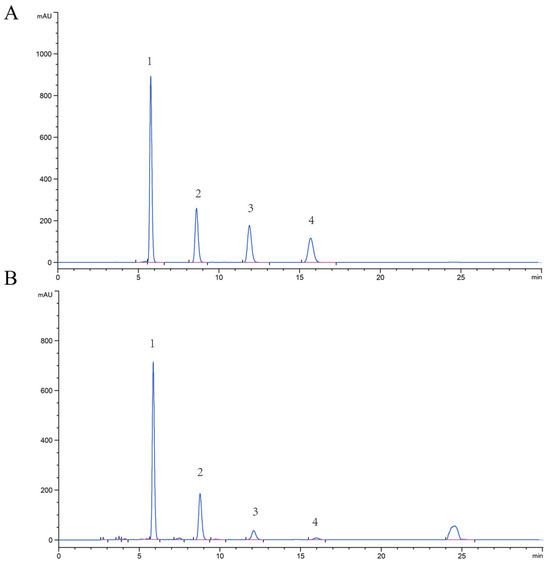
Green synthesis of silver nanoparticles (CP-AgNPs) using Calotropis procera (CP) offers a sustainable approach to producing multifunctional therapeutic nanomaterials. This study aimed to synthesize CP-AgNPs and evaluate their antimicrobial, antioxidant, anti-inflammatory, and anticancer potential, with a focus on Helicobacter pylori and gastric cancer cells. CP-AgNPs were prepared by phytochemical reduction using CP leaf extract and characterized by UV–Vis, XRD, FTIR, SEM, EDX, TEM, and Zeta. Antibacterial activity against H. pylori, time-kill kinetics, and SEM imaging of membrane damage were performed. Antioxidant (DPPH, ABTS) and anti-inflammatory assays, together with cytotoxicity studies in AGS cells (DAPI, AO/EtBr, and SEM), were also conducted. CP-AgNPs exhibited an SPR peak at 432 nm, face-centered cubic crystallinity, and spherical morphology (8–32 nm). They showed strong, dose-dependent antibacterial activity against H. pylori, surpassing metronidazole at higher doses. Time-kill assays and SEM confirmed membrane disruption. Antioxidant activity was notable (IC50: 40 µg/mL for DPPH; 60 µg/mL for ABTS). CP-AgNPs demonstrated significant anti-inflammatory effects and dose-dependent cytotoxicity in AGS cells, inducing apoptosis and morphological alterations. The broad biological activity of CP-AgNPs likely arises from the synergy between silver ions and CP phytochemicals. Their superior antibacterial effects, combined with antioxidant and anti-inflammatory properties, indicate strong therapeutic potential for gastric diseases. Anticancer activity in AGS cells suggests additional biomedical relevance, which may involve ROS-associated and apoptosis-related pathways, as suggested by previous studies. CP-AgNPs represent a promising natural nanoplatform for managing H. pylori infection, oxidative stress, inflammation, and gastric cancer, warranting further mechanistic and in vivo studies.
Full article


