Article

Development and Characterization of Melatonin-Loaded Glycerol–Gelatin-Based Vaginal Suppositories for Localized Delivery


by
Regina Julieta Delgadillo Hernández
1,
Gregorio Guadalupe Carbajal Arízaga
2,
José Alfonso Cruz Ramos
3,
Rodolfo Hernández Gutiérrez
1,
José Armando Hernández Díaz
1,4,
Ana Alejandra Arias García
1,
Norma Morales-Hernández
5,
José Nabor Haro-González
5,
Zaira Yunuen García Carvajal
1,* and
Moisés Martínez Velázquez
1,*
1
Biotecnología Médica y Farmacéutica, Centro de Investigación y Asistencia en Tecnología y Diseño del Estado de Jalisco, A.C. Av. Normalistas 800, Colonia Colinas de la Normal, Guadalajara 44270, Jalisco, Mexico
2
Departamento de Química, Universidad de Guadalajara, Marcelino García Barragán 1421, Guadalajara 44430, Jalisco, Mexico
3
Instituto Jalisciense de Cancerología, Puerto Guaymas 418, Colonia Miramar, Zapopan 45060, Jalisco, Mexico
4
Biotecnología Vegetal, Centro de Investigación y Asistencia en Tecnología y Diseño del Estado de Jalisco, A.C. Camino Arenero 1227, Colonia El Bajío del Arenal, Zapopan 45019, Jalisco, Mexico
5
Tecnología Alimentaria, Centro de Investigación y Asistencia en Tecnología y Diseño del Estado de Jalisco, A.C. Camino Arenero 1227, Colonia El Bajío del Arenal, Zapopan 45019, Jalisco, Mexico
*
Authors to whom correspondence should be addressed.
Sci. Pharm. 2026, 94(2), 31; https://doi.org/10.3390/scipharm94020031
Submission received: 19 February 2026 / Revised: 31 March 2026 / Accepted: 14 April 2026 / Published: 17 April 2026

Abstract

This research aimed to develop glycerol–gelatin vaginal suppositories loaded with melatonin to enhance the localized effects of antineoplastic agents. The solubility of melatonin in different solvents was determined, and glycofurol, which is approved for pharmaceutical use, presented the highest solubilizing capacity. Furthermore, the cytotoxicity of melatonin incorporated into suppositories against HeLa cells was evaluated using MTT assays, individually and in combination with cisplatin. The results indicate that melatonin enhances the cytotoxic effects of cisplatin. The optimal formulation obtained from an experimental design was 33% gelatin, 1% PVA, 1% PEG 6000, 10% glycerol, 15% glycofurol, and 40% water. To ensure that the vaginal suppositories presented the necessary physical properties for optimal handling and application, tests were performed to determine weight uniformity, texture, surface features and disintegration time. Vaginal suppositories weighted around 1.43 g, showed Young’s modulus values of 7389.6 N/m2 and hardness around 1100 gf, and they disintegrated after 30 min at pH 4.2. Additionally, for in vitro melatonin release, FTIR and XRD tests confirmed the presence of melatonin in the formulation. It is concluded that the developed vaginal suppositories can be explored as potential vehicles for localized delivery of melatonin to the tumor site to enhance therapeutic outcomes.

1. Introduction

Cervical cancer is considered a public health problem. Worldwide, it is among the four types of cancer with the highest incidence and mortality. In 2022, 661,021 new cases and 348,189 deaths were recorded [1], with the human papillomavirus (HPV) being the main risk factor. Current treatments for this type of cancer include systemic chemotherapy, surgical ablation, external beam radiation, and brachytherapy. However, some of these clinical treatments often present high toxicity and severe side effects. Furthermore, their invasive nature and high cost may lead to their discontinuation, which could favor the development of resistance in cancer cells [2]. The use of adjuvants alongside primary treatments enhances their efficacy and the treatment outcomes of patients. Melatonin, a hormone primarily produced by the pineal gland, possesses a variety of properties, including antioxidant activity and regulation of the immune system [3]. Moreover, it has been utilized in the treatment of various disorders, such as insomnia [4], diabetes [5], and neurodegenerative [6] and infectious diseases [3]. Regarding the relationship between melatonin and cancer, a considerable number of reviews summarize the anticancer effects, which encompass inhibition of tumor cell proliferation and migration, inhibition of angiogenesis, and induction of tumor cell apoptosis. Moreover, melatonin inhibits tumor development through mechanisms such as epigenetic regulation, metabolic reprogramming, modulation of immune responses and regulation of signaling pathways [7,8,9,10,11,12,13]. Further, melatonin has long been proposed as a combination therapy agent. A series of studies have demonstrated that treatment with melatonin increases the sensitivity of human cervical cancer cells to apoptosis induced by the antineoplastic agent cisplatin [14,15]. Consequently, this approach has the potential to enhance the effectiveness of existing therapeutic interventions while concomitantly reducing their adverse effects [16,17].
Vaginal delivery of chemotherapeutic drugs to the cervix offers several advantages over systemic administration, such as direct delivery to the tumor site, a lower dose being required, and a reduction in systemic side effects [18,19,20,21].
Currently, the most common method of melatonin administration is via oral solid forms; however, studies have demonstrated that this route exhibits low bioavailability [22]. Then, when highly localized doses are required, other routes of administration, such as vaginal, rectal, or pulmonary, are preferred, since they improve bioavailability and significantly reduce systemic and gastrointestinal side effects [23]. Moreover, the administration of the substance through mucosal membranes, such as vaginal and rectal, circumvents the first-pass hepatogastrointestinal metabolism associated with the oral route. A recent study investigated the pharmacokinetics and safety of melatonin when administered intravenously, intravesically, rectally, transdermally, and vaginally to healthy female volunteers, demonstrating the safety of these routes of administration [22]. This research aimed to formulate and characterize a melatonin-loaded intravaginal device and to evaluate its chemotherapy-enhancing effect in vitro. To achieve this, the optimal solvent of melatonin was identified. Then, melatonin was incorporated into a glycerol–gelatin matrix as a vaginal suppository, and the physical and chemical properties of this pharmaceutical form were assessed and the cytotoxic effect was verified. This innovative approach endeavors to contribute to the development of less invasive and more effective treatments for cervical cancer, thus promoting the health of affected patients.

2. Materials and Methods

2.1. Reagents

Gelatin was purchased from Fermont (Monterrey, Mexico), polyvinyl alcohol (PVA, molecular weight ~89 kDa and hydrolysis degree ~99.8%), glycerol, and propylene glycol were obtained from ALDROPAZ (Guadalajara, Mexico), melatonin (MEL, chemically produced, 98% purity) was purchased from SHQ (Querétaro, Mexico), ethanol was obtained from AZ (Guadalajara, Mexico), polyethylene glycol (PEG) 6000 wt, dimethyl sulfoxide (DMSO), glycofurol, Dulbecco’s Modified Eagle’s Medium (DMEM) high-glucose medium, and 3-(4,5-dimethyl-2-thiazolyl)-2,5-diphenyl-2 H-tetrazolium bromide (MTT) were purchased from Sigma Aldrich (St. Louis, MO, USA). 100X penicillin–streptomycin–neomycin (PSN) antibiotic mixture was obtained from Invitrogen (Carlsbad, CA, USA). Fetal bovine serum (FBS) was purchased from Gibco (Grand Island, NY, USA).

2.2. Simulated Vaginal Fluid

One liter of simulated vaginal fluid was prepared according to Owen and Katz [24], which included sodium chloride (3.51 g), potassium hydroxide (1.40 g), calcium hydroxide (0.222 g), bovine albumin (0.018 g), lactic acid (2.0 g), glacial acetic acid (1.0 g), glycerin (0.16 g), urea (0.40 g), dextrose (5.00 g), and water. The solution was adjusted to pH 4.2 using hydrochloric acid.

2.3. Melatonin Solubility

Melatonin solubility was evaluated in different dispersing media: water, ethanol, propylene glycol, glycofurol, and DMSO, identifying the dissolution and precipitation points. Ascending concentrations of melatonin (from 0 to 100 mg) were prepared in 1 mL of each solvent in 1.5 mL Eppendorf tubes via vortexing for two minutes. The samples were then shaken at 300 rpm at 35 °C for 20 min.

2.4. Cell Culture

The HeLa cervical cancer cell line was cultured in high-glucose DMEM medium supplemented with 10% fetal bovine serum (FBS) and 1% penicillin–streptomycin–neomycin (PSN) antibiotic mixture at 37 °C, 80% relative humidity, and 5% CO2.

Cellular Viability

In vitro cytotoxic activity assays were performed using the MTT reagent. Approximately 1 × 10⁴ cells per well were seeded on 96-well microplates, with 100 μL of culture medium. A stock solution of melatonin was prepared with glycofurol (400 mM), which was used to obtain dilutions of 10, 5, 2.5, 1.25, 0.65, 0.31, and 0.15 mM. In addition, the toxicity of the vehicle was evaluated under the same conditions, testing the following concentrations: 2.5, 1.25, 0.63, 0.3, 0.16, 0.08, and 0.04 volume %. Another experiment was designed in which melatonin was applied serially, as in the previous treatment, but at the IC50 of cisplatin, with incubation for 24 h. The experiments were performed in quadruplicate for each concentration in two independent experiments. After 24 h, 10 μL of MTT (5 mg/mL in PBS) was added, and the mixture was incubated for another 3 h. The medium was then removed, and the formazan salts were solubilized with DMSO. The absorbance of the samples was measured at 570 nm in a microplate reader (Bio-Rad, Hercules, CA, USA). Cell viability was calculated as follows:
C e l l   v i a b i l i t y ( % ) = S a m p l e a b s C o n t r o l a b s × 100

2.5. Formulation and Manufacturing of Vaginal Suppositories

Vaginal suppositories were prepared by following the protocol of Abass et al. [23], with modifications. Several formulations with varying ingredient proportions were prepared, to optimize the physical and functional properties of suppositories (see Table S1). Final formulation contained 33% gelatin, 1% PVA, 1% PEG 6000, 10% glycerol, 15% glycofurol, and 40% water. Initially, PVA suspension was prepared at 5 wt%. PVA powder was dissolved in distilled water at 90 °C under magnetic stirring for 1 h. The PEG suspension was prepared at 5 wt%. PEG powder was dissolved in deionized water under mechanical stirring at 45 °C for 1 h. The melatonin solution was prepared by adding 25 mg of melatonin to 300 μL of glycofurol.
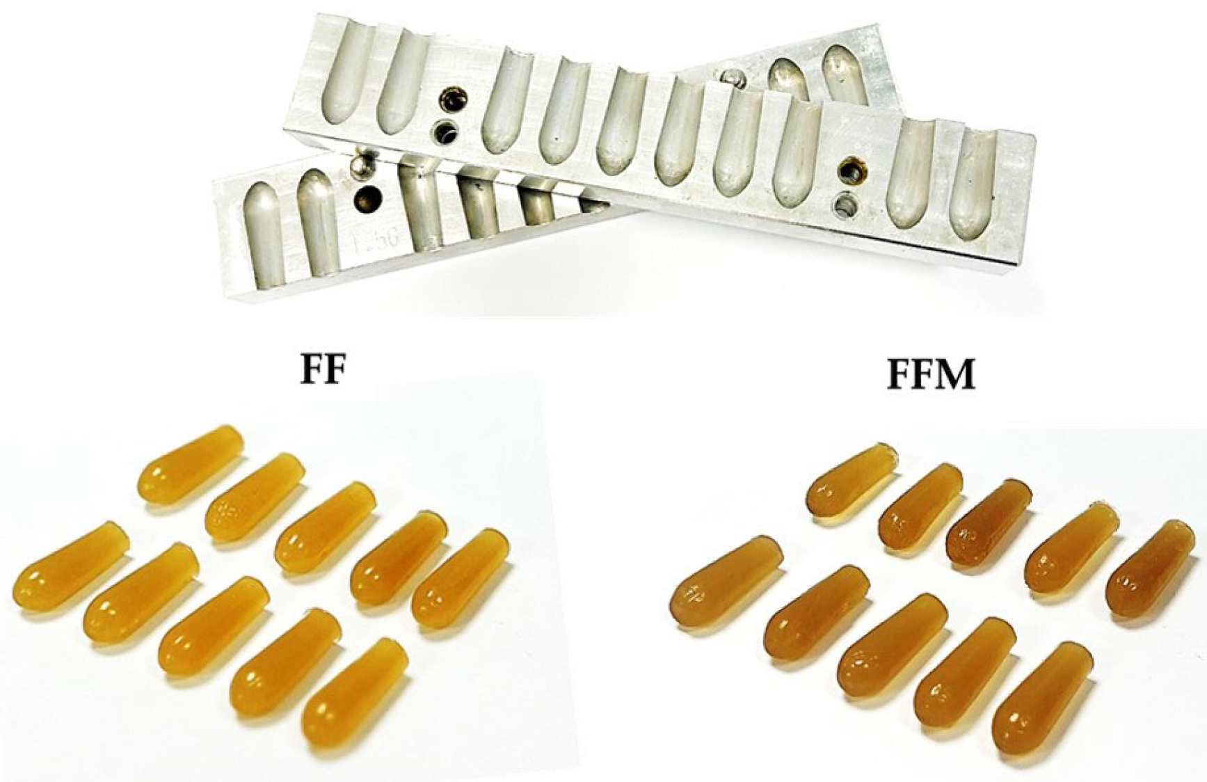
The vaginal suppositories were prepared as follows: Step 1: In a 10 mL beaker, 650 mg of gelatin powder was placed, and 200 μL of distilled water was added to hydrate the gelatin partially. Step 2: Then, 350 mg of PVA suspension, 350 mg of PEG suspension, and 180 mg of glycerol were added at room temperature. Polymer suspensions were accurately weighed using an analytical balance (ATX224, Cobos Precision, Barcelona, Spain). Step 3: The polymer mixture was transferred to a water bath (60 °C) and maintained under magnetic stirring at 6000 rpm for 5–10 min until a homogeneous mixture was obtained. It was then allowed to cool to 37 °C. Step 4: Previously solubilized melatonin (25 mg in 300 μL glycofurol) was added to the mixture under constant magnetic stirring until complete homogenization. Step 5: The mixture was immediately poured into metal suppository molds with a capacity of 1.5 g (bullet-shaped suppository mold, ZBMZB, with 8.99 mm diameter and 24 mm high holes) and stored at 4 °C for 24 h. The molds were previously coated with a thin layer of glycerol to facilitate demolding. The suppositories were removed from the molds and stored at 4 °C.

2.6. Characterization of Vaginal Suppositories

2.6.1. Weight Uniformity Test

The weight uniformity test was conducted according to the European Pharmacopoeia 2.9.5. A set of 25 vaginal suppositories with or without melatonin was weighed with an analytical balance (ATX224, Cobos Precision, Barcelona, Spain), and the average weight and standard deviation were determined [25].

2.6.2. Texture Analysis

Texture analysis was performed using a texture analyzer TA-XT Plus (Stable Micro Systems Co., Ltd., Surrey, UK) equipped with a cylindrical probe of 50 mm in diameter. The analyses conducted included the determination of Young’s modulus and texture profile analysis (TPA). The suppositories were stored at 4 °C until analysis. Before testing, suppositories were equilibrated at 20 °C for 10 min to ensure homogeneous evaluation conditions.
Young’s modulus was determined by analyzing the linear region of the stress–strain curve. The stress (σ) was calculated as the ratio between the applied force (F) and the cross-sectional area (A) of the specimen (σ = F/A). The suppositories were subjected to controlled deformation in the range of 15–90%. The linear portion of the stress–strain curve was fitted with a straight-line equation, and the slope was taken as Young’s modulus (E), which represents the stiffness of the material. A higher modulus indicates greater rigidity, whereas lower values correspond to more flexible materials [26].
Texture profile analysis (TPA) was performed through two consecutive compression cycles under the following test conditions: pre-test speed of 0.5 mm s−1, test speed of 0.5 mm s−1, post-test speed of 100 mm s−1, trigger force of 0.05 N, strain of 40%, strain time of 5 s, and a load cell capacity of 5 kg. The parameters evaluated were hardness, springiness, cohesiveness, and resilience. Eight replicates were performed, and results were expressed as the mean ± standard deviation [26,27].

2.6.3. Determination of Disintegration Time

The test was conducted by placing three suppositories separately in 25 mL of simulated vaginal fluid (SVF) in 50 mL tubes, which were placed in a shaking incubator (LUZEREN, THZ-100, Tlajomulco de Zuñiga, Mexico) at 37 °C and 130 rpm. The disintegration time was recorded as soon as the suppositories began to melt or completely disintegrate.

2.6.4. In Vitro Release Test

The in vitro release test was conducted using a shaking incubator (LUZEREN, THZ-100, Tlajomulco de Zuñiga, Mexico). Three suppositories were placed separately in tubes containing 15 mL of simulated vaginal fluid (SVF), at a temperature of 37 °C and an agitation speed of 130 rpm. Samples of 15 mL were withdrawn at different time intervals (0, 15, 30, 60, 120, and 180 min). After each interval, samples were centrifuged at 1000 rpm for 2 min. The extracted volume was replaced with fresh SVF to maintain a constant volume throughout the test. The removed SVF was filtered through a 0.45 µm filter and analyzed using UV–vis spectrometry (NanoDrop 2000 Spectrophotometer, Thermo Scientific, Waltham, MA, USA). The release of melatonin was quantified by measuring the absorbance at 280 nm. Background readings were obtained from samples without melatonin, and the experiments were conducted in triplicate.

2.6.5. Fourier Transform Infrared (FTIR) Spectroscopy Analysis

FTIR spectroscopy was used to analyze the chemical composition in the formulations. For this purpose, sagittal cuts were made in the vaginal suppositories to obtain 0.5 × 0.5 cm slides, which were placed over the diamond window of the attenuated total reflectance (ATR) device of a Thermo Scientific spectrometer, model is5 (Thermo Scientific, Waltham, MA, USA). Spectra were collected in the transmittance mode by averaging eight scans, with a spectral resolution of 4 cm−1.

2.6.6. X-Ray Diffraction (XRD) Analysis

X-ray diffraction was performed using a Panalytical Empyrean diffractometer (Panalytical, Malvern, UK) with a copper anode tube emitting CuKα radiation = 0.154 nm. The slides extracted from the suppositories for the FTIR analysis were placed over the aluminum holder where the surface of the slides matched the standard height recommended by the supplier. Readings were conducted with a step of 0.02 degrees at a rate of 30 s per step.

2.6.7. Scanning Electron Microscopy (SEM) Analysis

SEM analysis was performed on 1 mm thick slices obtained from the vaginal suppositories using a scalpel. The slices were placed on a carbon tape and coated with a gold film. The analysis was performed with a high-resolution JEOL microscope, model JSM IT710 (JEOL, Peabody, MA, USA), operated at a 15 kV under high vacuum. The images at 25× magnification were processed using ImageJ v.1.53 to measure the hollow diameter. Eighty measurements were taken to construct the size plots.

2.7. Anticancer Evaluation of Melatonin-Loaded Vaginal Suppositories

The cytotoxic effects of melatonin-loaded vaginal suppositories (FFM), melatonin-free suppositories (FF) and simulated vaginal fluid (SVF) on HeLa cells were evaluated via MTT assays as described in the Cellular viability procedure. Briefly, 1 × 10⁴ cells per well were seeded on 96-well microplates. Prior to treatment, each vaginal suppository was dissolved in 2 mL of SVF in a beaker, using a shaking incubator at 180 rpm and 37 °C for 30 min. The samples were disinfected by incubation for 5 min in a crosslinker equipment, followed by exposure to ultraviolet light for 10 min in a biosafety cabinet. The samples were serially diluted in DMEM medium to obtain a range of melatonin concentrations from 0.15 to 10 mM or the equivalent volume of the suppository without melatonin and the SVF, and applied to cells. Additionally, melatonin-containing samples were applied in combination with the IC50 of cisplatin. The experiments were performed in quadruplicate for each concentration in two independent experiments. After 24 h, 10 μL of MTT (5 mg/mL in PBS) was added, and the mixture was incubated for another 3 h. The medium was then removed, and the formazan salts were solubilized with DMSO. Finally, the absorbance of the samples was measured at 570 nm in a microplate reader (Bio-Rad, Hercules, CA, USA).

2.8. Statistical Analysis

The data were processed statistically using ANOVA (GraphPad Prism 9 software, (GraphPad Software Inc., San Diego, CA, USA). Post hoc tests and p-values for each study are indicated in the relevant figures or tables.

3. Results and Discussion

3.1. Melatonin Solubility

The solubility of the active pharmaceutical ingredient (API) is a crucial parameter in the design of vaginal formulations because it directly affects the drug’s absorption into vaginal tissue, influencing bioavailability and treatment effectiveness [19]. In designing the proposed melatonin-loaded formulation, selecting an appropriate solubilizer excipient for melatonin that is mucosally tolerated and physicochemically stable was considered essential. Melatonin solubility was evaluated in water, ethanol, propylene glycol, dimethyl sulfoxide (DMSO) and glycofurol (tetrahydrofurfuryl alcohol polyethylene glycol ether). As shown in Table 1, there were clear differences in the melatonin solubilizing capacity of each solvent. Melatonin solubility was observed to be highest in DMSO and glycofurol (up to 100 mg/mL), followed by ethanol (up to 70 mg/mL), and propylene glycol (up to 30 mg/mL). Melatonin was not soluble in water at any concentration. A recent study showed that the solubility of melatonin in propylene glycol was only 3.8 mg/mL, while it was 11.1 mg/mL in glycofurol [28]. Our findings strongly demonstrate that DMSO and glycofurol dissolve the highest amount of melatonin, and that solutions are homogeneous and stable. Gylcofurol is considered a non-toxic solvent and it is classified as a Class 1 solvent by the FDA [29]. Glycofurol is a semi polar solvent agent widely used as a biocompatible solvent in pharmaceutical formulations [30], and for gel formation [29]. The use of glycofurol as a solvent in pharmaceutical formulations seems to favor stability formulation over time, which contributes favorably to the shelf life of the final product [28,31] and has excellent compatibility with a wide range of active ingredients and excipients, allowing its incorporation into multicomponent systems without compromising the functionality or integrity of the delivery system [32,33]. Based on these characteristics, glycofurol was incorporated as a key component in the developed vaginal formulation, allowing for the optimization of the solubility, stability, and compliance with the safety regulations of the proposed delivery system for melatonin.

3.2. Anticancer Activity

The next step in the design of the vaginal formulation was to evaluate the cytotoxic activity of melatonin dissolved in different dispersion media on the HeLa cervical cancer cell line. The cytotoxic effect of the vehicles was also assessed. The results showed that at concentrations of 1.25 mM and above, melatonin exerted significant cytotoxicity against HeLa cells (Figure 1A and Figures S1–S3). However, it was also observed that all the vehicles used, except ethanol, carry considerable cellular toxicity.
Then, by choosing the vehicle with the highest melatonin-dissolving capacity, the proportion of solvent in the vaginal formulation is reduced.
Additionally, the chemotherapy-enhancing capacity of melatonin was evaluated by applying a combined treatment of melatonin plus the IC50 of cisplatin (43.29 µM, Figure S4) in HeLa cells for 24 h. As shown in Figure 1B, melatonin significantly potentiates the cytotoxic effects of cisplatin at concentrations of 1.25 to 10 mM. In light of these results, it would be highly relevant to evaluate lower concentrations of cisplatin in combination with melatonin in future trials to determine the nature of drug interaction and identify optimal combinations to balance therapeutic efficacy with reduction of cisplatin-related side effects.
Our results are consistent with the findings reported by Pariente et al. [14] and Chen et al. [15]. Cisplatin exerts its cytotoxic effects through the covalent binding of platinum to the purine bases guanine and adenine in DNA. This covalent binding leads to intra- and interstrand cross-links, which cause strand breaks, interfere with DNA repair mechanisms, and induce apoptosis in cancer cells [34]. Regarding melatonin, it exerts its cytotoxic action in cancer cells by inhibiting signaling pathways such as JNK [15] and NF-κB [35]. It can also suppress aerobic glycolysis (Warburg-type metabolism), leading to cell death [36].

3.3. Formulation and Manufacturing of Vaginal Suppositories

Suppositories have traditionally utilized lipophilic bases that melt at human body temperature. Zetner et al. [22] reported fatty suppositories loaded with 25 mg of melatonin but did not mention formulation composition. However, the authors demonstrated that melatonin vaginal administration was safe, and only mild transient adverse effects were observed. Although vaginal bioavailability varied among healthy females, a mean bioavailability of 97.8% was observed using this administration route.
Otherwise, gelatin-based suppositories have minimal effects on tissue osmolality, so they do not draw additional fluid from the underlying mucous membrane. This makes them useful for vaginal application. Moreover, this kind of suppository is very popular due to the ease use and provides complete release of the active ingredients in vaginal fluids [37]. Polyvinyl alcohol (PVA) and polyethylene glycol (PEG) also have properties suitable for vaginal formulations. They are chemically stable, nonirritating, miscible in water and mucous secretions, and widely available. Thus, it is possible to manufacture gelatin-based suppositories with a wide range of firmness, dissolving rates, and melting points.
For the formulation and manufacturing of vaginal suppositories, the following criteria were considered: (a) the attributes of the initial raw material (primarily that it is approved for human use); (b) homogeneous incorporation of dissolved melatonin in glycerol-gelatin suppositories; (c) a solid suppository-like form for potential easy insertion; (d) physical integrity at room temperature; (e) homogeneous weight variation (no more than 10%), and (f) a suitable disintegration time (no more than 30 min) in simulated vaginal fluid (SVF). During the initial development stage, different vaginal suppository compositions were formulated to optimize the physical and functional performance (Table S1). The formulations were evaluated using disintegration time, hardness, and weight uniformity tests, which are critical parameters for ensuring stability, handling, and proper release of the active ingredient. Some of the formulations presented an appropriate morphology, with well-formed suppositories. However, several of them exhibited an overly soft consistency, making them prone to deformation at temperatures above 30 °C [38]. This thermal behavior indicated low structural strength, compromising the integrity of the suppository during handling and potentially during intravaginal administration. As a result, these formulations were discarded because they did not meet the minimum requirements for mechanical strength and thermal stability, which are essential to guarantee both product safety and reproducibility in clinical applications. The final formulation (FF) contained 33% gelatin, 1% PVA, 1% PEG 6000, 10% glycerol, 15% glycofurol, and 40% water, and was chosen and developed in compliance with the necessary parameters for proper application and handling, discarding those that did not meet the established requirements. This formulation was achieved by modifying the excipient concentrations and selecting them based on the disintegration time obtained from the corresponding test. The developed vaginal suppositories had a solid and uniform consistency, were oval-shaped, yellow in color, and weighed approximately 1.5 g each. In this case, these are hydrophilic glycerol-gelatin-based vaginal suppositories. By eye, the melatonin-loaded vaginal suppositories (FFM) appeared slightly darker than the unloaded ones (Figure 2).

3.4. Characterization of Vaginal Suppositories

3.4.1. Weight Uniformity Test

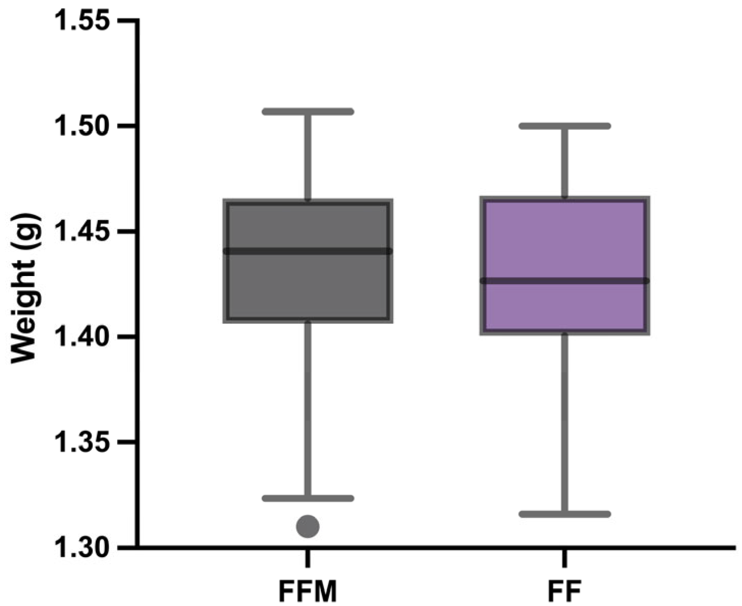
Weight uniformity is an essential parameter in the quality control of individual solid dosage forms. For this test, the suppositories from each batch were individually weighed, comparing their mass with the average weight of the sample. The results were evaluated according to the criteria established by the Mexican Pharmacopoeia (FEUM), the British Pharmacopeia (BP), and the United States Pharmacopeia (USP), which establish acceptable ranges of percentage variation in unitary solid products (less than 10% of the individual mass deviates from the average mass by 5%). Vaginal suppositories satisfactorily met the tolerance limits, with values of 1.432 ± 0.048 g for FFM (loaded with melatonin) and 1.429 ± 0.046 g for FF (without melatonin). Similar mean and SD values suggest that the inclusion of melatonin into a vaginal formulation did not affect the final weight, demonstrating mass uniformity (Figure 3). The vaginal suppositories analyzed remained within the permitted range, confirming the consistency of the manufacturing process and the reliability of the batches produced [39].

3.4.2. Texture Analysis

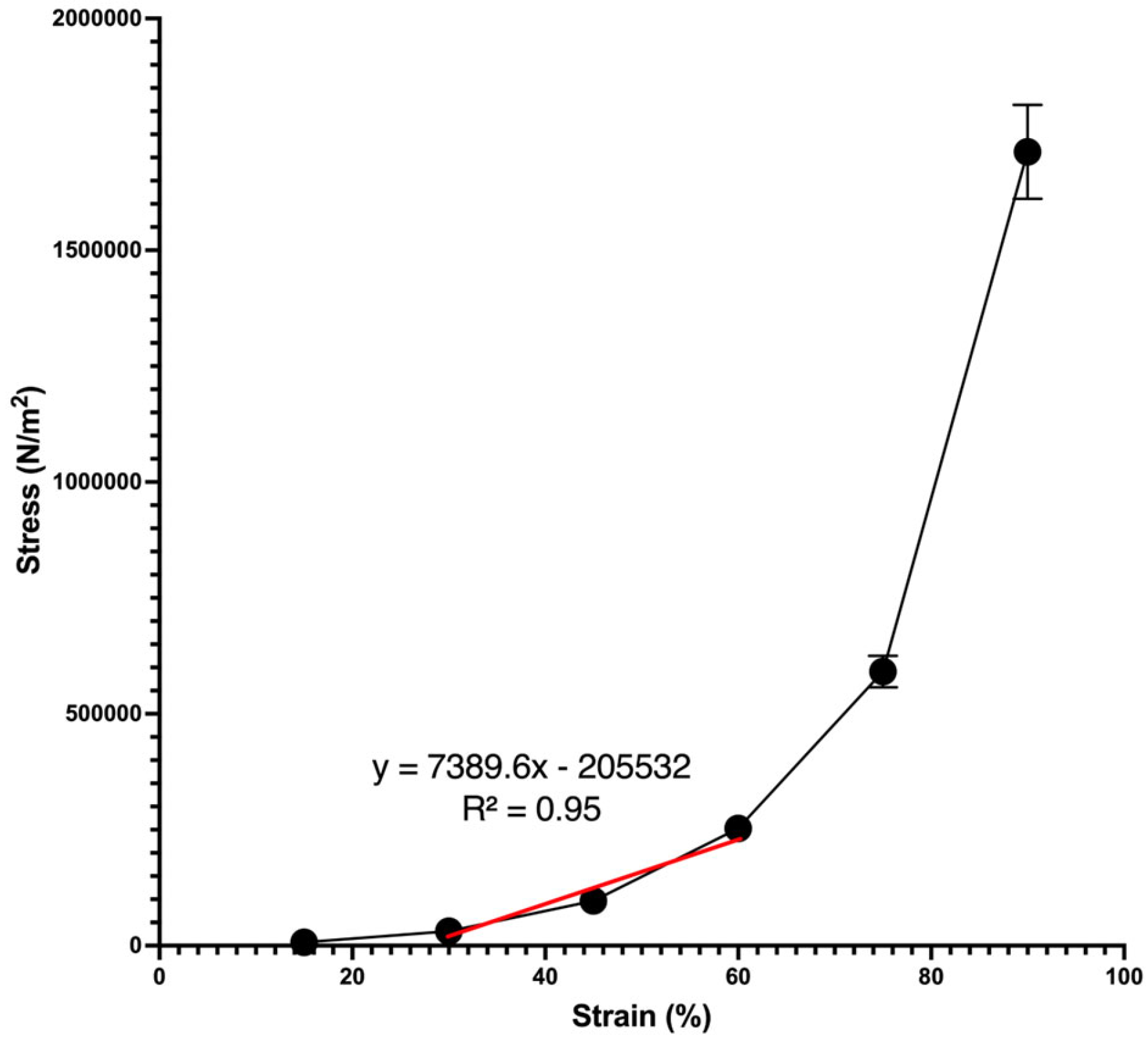
The mechanical properties of the vaginal suppositories were determined using Young’s modulus (Figure 4) and texture profile analysis (TPA) (Table 2), performed after tempering the samples to 20 °C for 10 min. Young’s modulus, obtained from the slope of the linear portion of the curve (Figure 4), showed values of 7389.6 N/m2 (R2 = 0.95), indicating that this material is not very rigid and very resistant to deformation. The suppositories exhibited an elastic response at the initial stages of deformation (15–45%). As the stress increased, the response force also increased. However, neither yield strength nor a plastic region is observed within the evaluated range.
TPA has been widely employed to characterize the mechanical behavior of pharmaceutical gels and semisolid systems [40]. Hardness is a critical parameter for vaginal suppositories and ovules, ensuring that they can withstand packaging, transportation, and insertion without breaking, while also softening appropriately in the body to release the drug effectively [41]. Ideal hardness maintains the integrity of the dosage form, prevents irritation, facilitates administration, and influences drug release. However, as the disinfection procedure uses UV light, the relationship between exposure time and mechanical properties should be examined in further studies. In contrast, gels should ideally have lower hardness to ease application on mucosal tissues, such as the vaginal mucosa [42]. In our study, hardness values were 1129.9 ± 153.6 gf for FF and 1091.9 ± 69.0 gf for FFM, suggesting that melatonin incorporation did not substantially compromise structural integrity. According to the FTIR spectra, physical interactions between gelatin and melatonin may involve weak hydrogen bonds, van der Waals forces, or dipole–dipole interactions. Our results align with those from Chanu et al. [43], who reported that gelatin sponges crosslinked with fructose and loaded with melatonin exhibited weak mechanical properties and readily dissolved in acidic environments (HCl 0.1 N). Likewise, Özgüney and Kardhiqi [40] reported suppositories prepared by the cold method using poloxamer 407, poloxamer 188 and various amounts of different bioadhesive polymers with similar compression values but more adhesive. Similarly, the obtained suppositories were softer than those reported for gelatin–glycerin and gelatin–glycerin-PEG 400 [27].
Springiness in the suppositories is a good combination of firmness and softness, which translates into comfort within the body while remaining firm enough for easy insertion, making it easier to locate [41,44]. The values obtained here showed slightly higher springiness in FF (1.365 ± 0.232) than in FFM (1.218 ± 0.225), suggesting that drug-free formulations are more elastic and recover their height more effectively after deformation; however, there is no statistically significant difference between them.
Cohesiveness, although often more conceptual than a strictly defined parameter, reflects the degree to which a material withstands mechanical disruption. It represents the internal forces binding particles together, preventing fracture and maintaining integrity [40]. In this study, both FF (0.939 ± 0.008) and FFM (0.936 ± 0.006) showed similar levels of cohesiveness, confirming that melatonin does not alter the internal stability of the matrix. High cohesiveness is desirable to ensure that suppositories retain their shape and structural integrity. Hence, the observed balance suggests a well-designed system [45,46].
Resilience measures the degree and speed of recovery after deformation [47]. In suppositories, resilience ensures stability during storage, resistance to breakage upon handling and insertion, and functional integrity until administration. Ideally, suppositories should also melt or dissolve at body temperature, be non-toxic, non-irritating, and release their active ingredient without affecting absorption [48,49]. In our study, FF (0.807 ± 0.012) demonstrated higher resilience than FFM (0.782 ± 0.012), suggesting that the drug-free formulation recovers energy more rapidly. This significant reduction in resilience for FFM could be linked to interactions between melatonin and the gelatin matrix that mildly restrict elastic recovery.
The TPA results confirm that the incorporation of melatonin did not significantly affect the mechanical performance of the suppositories. Drug-free and drug-loaded formulations exhibited moderate hardness, high cohesiveness, and adequate resilience. These attributes ensure that the formulations are structurally suitable for handling and administration, while remaining sufficiently elastic to prevent brittle failure and support patient comfort. In future studies, the effect of the final application temperature should be considered, as it can lead to significant changes in the structure of the suppositories. Likewise, applying oscillatory rheology and testing alternative excipient ratios (e.g., PEG 600 percentage) would further clarify how the formulation and application variables influence the mechanical and functional behavior of these systems.

3.4.3. Determination of Disintegration Time

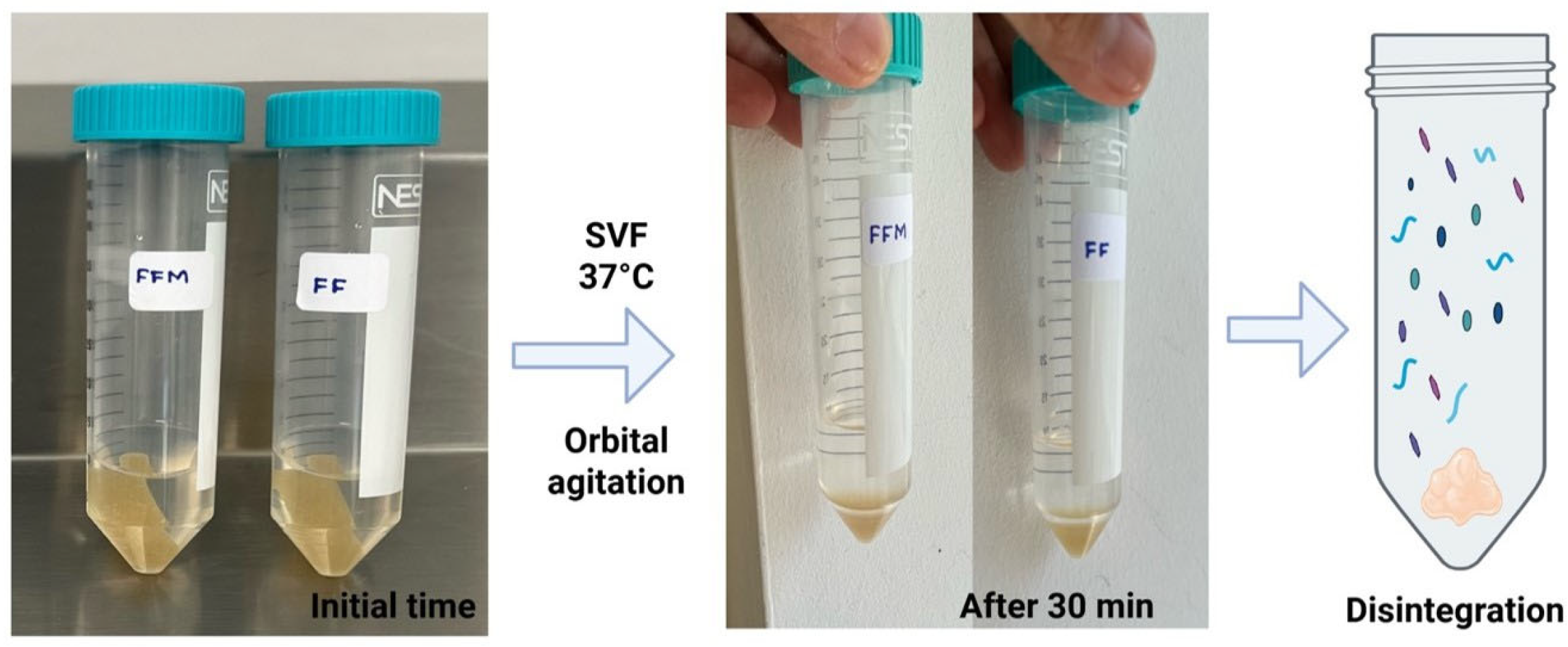
The disintegration time of suppositories containing melatonin was 30 min in the simulated vaginal fluid (SVF) at pH 4.2. A swelling was observed in the vaginal suppositories at five minutes. After 27 min, the original shape changed, indicating loss of structural integrity, and it completely disintegrated at 30 min, transforming into a gel-like mass (Figure 5). Drug-free vaginal suppositories disintegrated in similar time spans, suggesting that melatonin did not affect the disintegration properties of the delivery vehicle.
Cabral-Romero et al. [50] prepared vaginal ovules loaded with bismuth lipophilic nanoparticles and cetylpyridinium chloride using glycerinated gelatin as a vehicle. The authors observed complete disintegration of loaded and drug-free formulations at 19 min, demonstrating that drug incorporation into the vehicle did not affect the disintegration properties of the delivery vehicle.

3.4.4. In Vitro Release Test

We studied the controlled release of the melatonin entrapped within the structure of the suppository. The drug-release pattern depends on many factors, including morphology, drug binding affinity, and the rate of hydration of the polymeric material, among others [51]. The primary mechanisms governing controlled drug release from a polymer matrix are typically diffusion of the drug through the matrix and the dissolution (or erosion) of the polymeric matrix itself, which controls the matrix’s structure over time. These processes can act together, with diffusion influencing the rate at which the drug moves out of the matrix and dissolution/erosion dictating the eventual breakdown and release of the embedded drug [52].
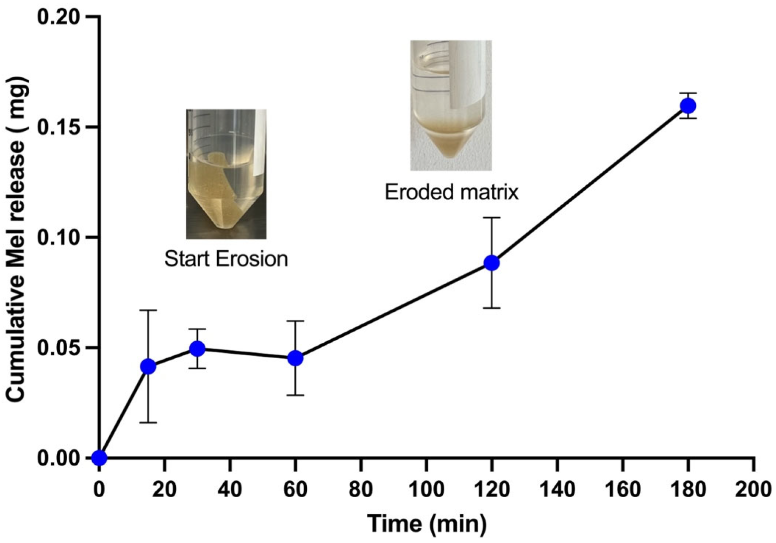
According to the experimental release patterns (Figure 6), rapid release kinetics are observed for melatonin entrapped in the polymeric network of the suppository. Subsequently, the concentration of melatonin released in the SVF decreases, followed by an increase over the same time. This phenomenon is attributed to the rapid erosion of the matrix and the presence of eroded and sedimented particles. This will eventually allow melatonin to diffuse into the release medium, resulting in an increase in melatonin concentration. At acid pH, a gelatin-glycerol matrix’s response is primarily driven by gelatin’s polyampholyte nature and glycerol’s plasticizing effect [53,54]. Gelatin’s positively charged amino groups become protonated, affecting its network structure. The matrix will typically weaken, decrease in gel strength and modulus, and become more flexible or even begin to dissolve [55,56]. The disintegration of the suppository, the weak molecular interactions between melatonin and the polymer matrix (see below, FTIR, XRD and SEM analyses), as well as the pH and salts in the SVF will promote the release of melatonin into the environment.

3.4.5. Fourier Transform Infrared (FTIR) Spectroscopy Analysis

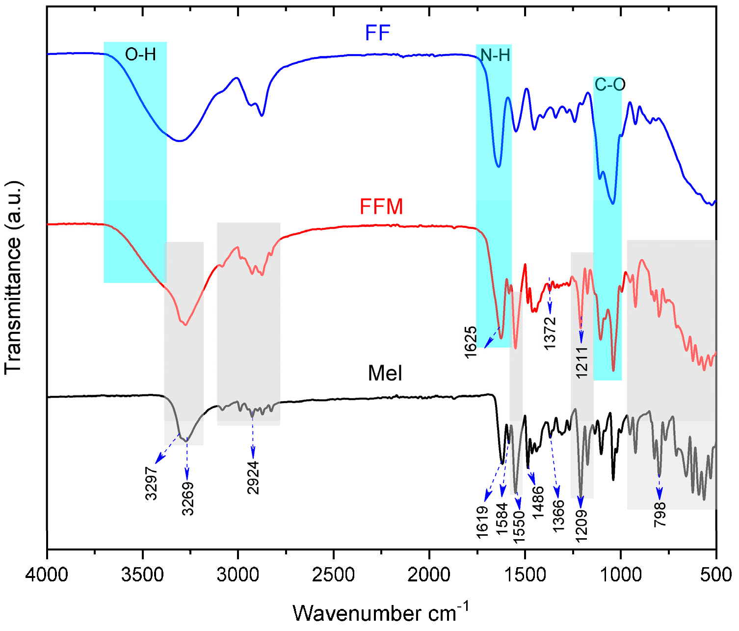
The infrared spectrum of melatonin (Figure 7) is composed of a profile fully described elsewhere [57,58]. Briefly, we note the bands at 3297 and 3269 cm−1 corresponding to the N–H stretching of the indolic ring, a set of low-intensity bands between 3000 and 2700 cm−1 to vibrations of C–H bonds, while the signal at 1619 cm−1 was mainly related to the C=O stretching of the amide group. The bands at 1584 and 1486 cm−1 are assigned to C=C and C–N stretching modes, along with N–H bending. The signal at 1366 cm−1 was attributed to C–N stretching and the symmetric deformation of the -CH3 group. A strong band was detected at 1209 cm−1, assigned to a combination of C–OCH3 stretching and C–C–O bending, also associated with N–H bending and C–N stretching vibrations. Finally, the band at 798 cm−1 corresponds to the out-of-plane deformation of C–H bonds in the indolic aromatic ring. The most intense bands in this spectrum also appear in the vaginal suppository loaded with melatonin (FFM) (blue shadows), indicating that melatonin is present; however, some bands show slight widening and shifting. Shifts were detected in the bands at 1629–1625 cm−1, 1366–1372 cm−1, and 1209–1211 cm−1. These changes could be attributed to variations in the chemical environment of melatonin when interacting with the polymeric matrix, mainly through hydrogen bonding and other molecular interactions that alter vibrational energies and consequently modify the position and intensity of the bands. These results suggest that, although melatonin maintains the chemical structure within the formulation, the incorporation into the polymeric matrix generates specific molecular interactions that confirm its integration into the system. In turn, the FTIR spectrum profile of the FF matrix with intense bands in 3600, 1630, and 1050 cm−1 from O–H, N–H, and C–O stretching modes is clearly present in the FFM suppository, indicating the presence of the gelatin.
These findings are consistent with previous studies, in which the incorporation of melatonin into polymeric systems such as chitosan revealed similar shifts, allowing a comparison with our results [59]. Furthermore, such band shifts are not limited to polymer systems, since interactions of melatonin with other molecules, such as sericin and resveratrol, have also been reported to induce vibrational modifications [60].

3.4.6. X-Ray Diffraction (XRD) Analysis

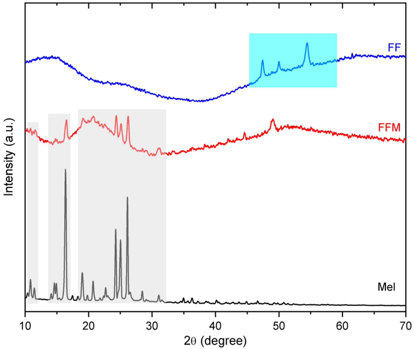
X-ray diffraction (XRD) analysis was employed to understand the crystallinity of the components in the formulation loaded with melatonin (FFM). The diffractogram of melatonin exhibited well-defined peaks, confirming its highly crystalline nature (Figure 8). The profile matched the observed data in pure melatonin [59,61,62]. The peaks with the highest intensities are marked with a shadow region, and they are also present in the FFM sample, indicating that melatonin molecules aggregate within the solidified suppository and form microcrystals. These crystals were not visible to the naked eye, and a plausible explanation is the small size and low content of melatonin, which is in agreement with the low relative intensity of the shadowed peaks in the FFM sample.
Regarding the FF matrix, the XRD profile is composed of wide and low-intensity signals from 10 to 40 (2θ) degrees, indicating disorganization among macromolecular chains as expected in gelatin [63]. However, sharp reflections appeared (shadowed region); signals at high angles can be associated with intramolecular organized atoms in polymers or short molecules, as the additives used to prepare FF. When the matrix is combined with melatonin, the network reorganizes in a more amorphous network since the low signals at 15 and 20 degrees disappear and form a halo centered at 21 degrees in FFM. Additionally, the three reflections from PVA disappear, suggesting that melatonin microcrystals disaggregate and disorganize the gelatin and PVA network when embedded in the matrix. Such disorganization of the polymeric gelatin chains represents a favorable energetic condition for the release of melatonin since a crystalized or more organized gelatin matrix requires additional energy for disorganization of the network and then release the drug. In fact, gelatin with 36% crystallinity requires 18 J/g for a melting while below 1% crystallinity requires less than 1 J/g [64]. Moreover, the release of melatonin is favored by a weak interaction with the gelatin matrix as most of the bands in the infrared spectrum does not become wider.

3.4.7. SEM Analysis

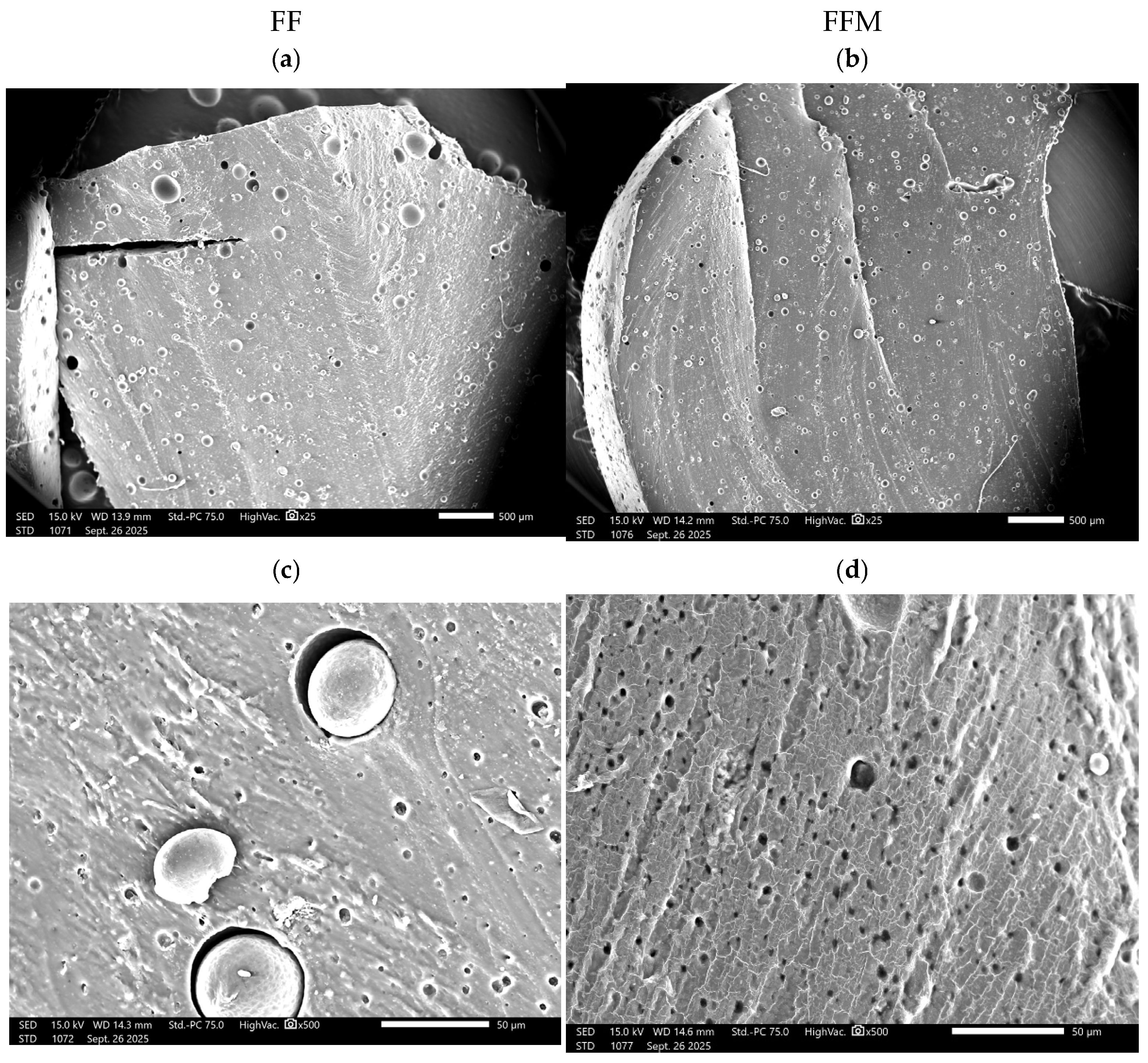
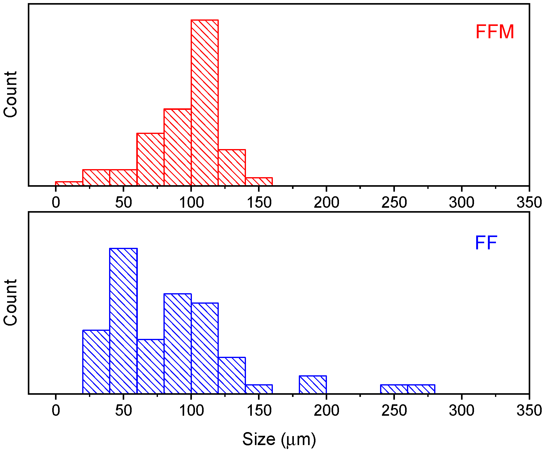
At the first inspection at 25× magnification, the FF and FFM samples exhibit spherical cavities and, in some cases, are hollow (Figure 9a,b). The size distribution (Figure 10) evidenced that the FF sample contains larger cavities, reaching diameters above 200 µm.
At 500× magnification, the FF cavities contain a spherical material, clearly separated from the matrix, suggesting weak interfacial bonding between components (Figure 9c). Clean holes in the matrix and smooth surface of the filler are commonly observed when the adhesion between both components is weak or incompatible [65,66] and leads to a decrease in the mechanical performance [66]. In the FFM image, the cavities are evenly distributed and of regular size (Figure 9d), indicating that the matrix is more organized, as inferred from the XRD analysis, which reinforces the assumption that gelatin and PVA chains interpenetrate when melatonin is added, then the uniform pores and low crystallinity enhance the release of the drug in the present experiment. The formation of voids warrants further investigation; understanding their origin, volume, number, and distribution would facilitate the advanced design of drug delivery systems, as demonstrated by the release of verapamil from porous gelatin, in which zero-order release kinetics have even been achieved [67].

3.5. Anticancer Evaluation of Melatonin-Loaded Vaginal Suppositories

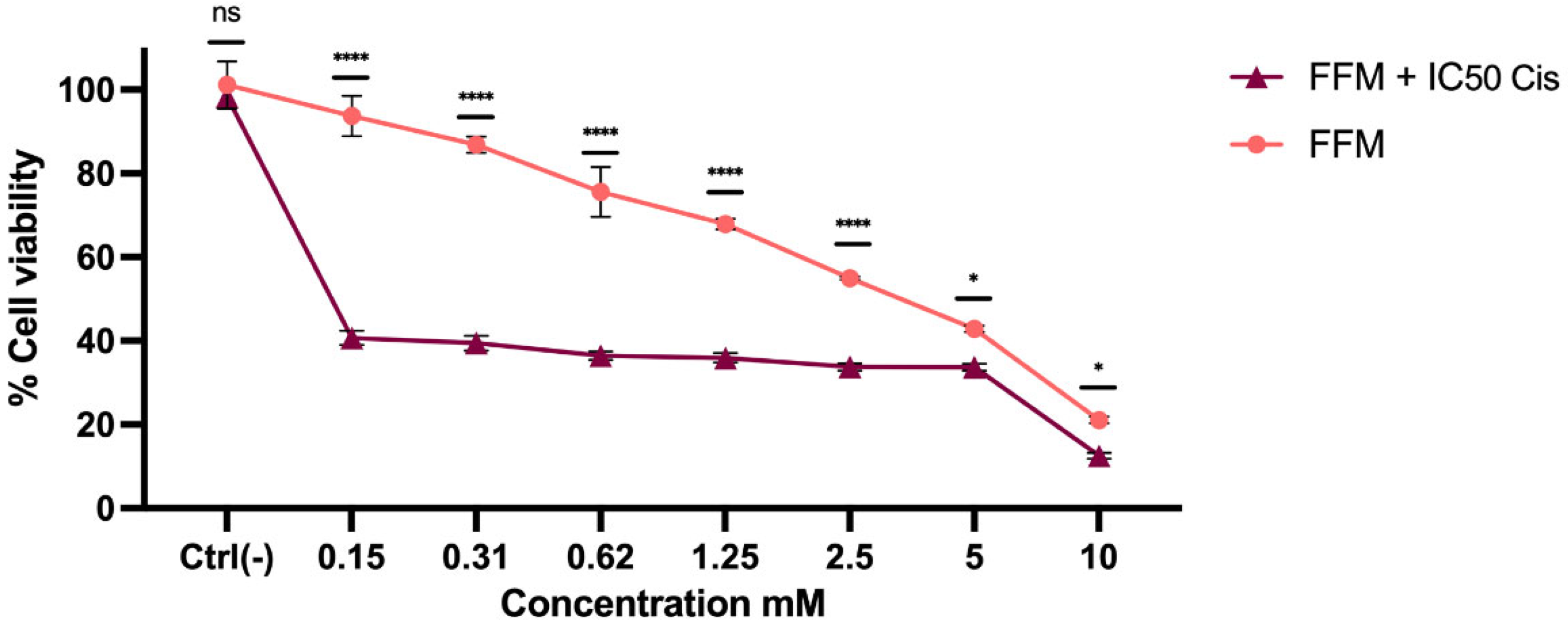
Finally, we sought to determine whether melatonin incorporated into vaginal suppositories maintained its cytotoxic effect against HeLa cells, after its release into a simulated vaginal fluid. The results indicate that melatonin exerted concentration-dependent cytotoxic effects on HeLa cells. Furthermore, its chemotherapy-enhancing effect was confirmed, after release from the vaginal suppository (Figure 11). Vaginal suppositories without melatonin (FF) exhibited lower cytotoxicity, likely attributable to the presence of glycofurol, whereas the SVF sample showed slight toxicity in HeLa cells (Supplementary Figures S5 and S6). Our findings suggest that the vaginal suppositories are functional, as the incorporated and released melatonin maintains its biological activity. This reinforces the feasibility of using them as a vehicle for localized delivery of melatonin via the vaginal route. We envision two clinical scenarios for the administration of cisplatin and melatonin. In the first, cisplatin would be administered intravenously, as is routine, while melatonin would be administered intravaginally through the suppositories. The second scenario would be the co-administration of melatonin and cisplatin in the same suppository. In both scenarios, melatonin is proposed to enhance the cytotoxic effects of cisplatin, although both approaches require further experimental validation.
In summary, the choice of solvent used for melatonin solubilization, along with the formulation components and the preparation process, demonstrates the feasibility of manufacturing. The suppositories obtained have a homogeneous appearance and weight and do not disintegrate upon manipulation, demonstrating reproducibility across batches. Furthermore, FTIR indicates the presence of non-covalent interactions that would facilitate the diffusion of melatonin into the external environment. Both XRD and SEM demonstrate that melatonin was incorporated homogeneously and that the manufacturing process had no impact, representing a significant advance compared to conventional lipophilic formulations. This approach expands the application possibilities of melatonin in pharmaceutical products.

4. Conclusions

In this study, the solubility of melatonin in different solvents was determined, which allowed us to select the best solvent for incorporation in liquid form, with glycofurol being the most suitable option. Furthermore, the cytotoxicity of melatonin, in its free form and when incorporated into vaginal suppositories, was evaluated in HeLa cells, both individually and in combination with the oncotherapeutic drug cisplatin. Based on these results, and in agreement with other research, it is suggested that melatonin has the ability to enhance the effects of antineoplastic drugs. The final vaginal formulation was obtained after optimizing the excipients until acceptable consistency and parameters were achieved. It contained 33% gelatin, 1% PVA, 1% PEG 6000, 10% glycerol, 15% glycofurol, and 40% water. To validate these characteristics, disintegration and hardness tests were performed, ensuring that the vaginal suppositories exhibited the physical properties necessary for optimal handling and application. Furthermore, FTIR and XRD analyses confirmed the presence of melatonin after its incorporation into the formulation. These parameters are essential for quality control of formulations, guaranteeing their efficacy and safety. The developed vaginal suppositories can be explored as a potential vehicle to obtain higher drug loading and improved dissolution of the poorly water-soluble melatonin, offering local delivery at the tumor site, and enhancing therapeutic outcomes. To advance to a translational design, determination of drug permeation would be needed to define systemic or local application and to conduct bioavailability and clearance assays. Among the limitations of our study, one outstanding issue to be resolved is how to maximize melatonin solubility in glycofurol to reduce its potential cellular toxicity. Another pending issue is ensuring the sterility of the developed formulations and extending the shelf life, possibly by adding preservative agents. It is also important to conduct in vivo functional studies of the developed vaginal suppositories.

Supplementary Materials

The following supporting information can be downloaded at: https://www.mdpi.com/article/10.3390/scipharm94020031/s1, Figure S1: Cytotoxic activity in HeLa cells after treatment with melatonin dissolved in DMSO and DMSO alone, at different concentrations for 24 h; Figure S2: Cytotoxic activity in HeLa cells after treatment with melatonin dissolved in propylene glycol and propylene glycol alone, at different concentrations for 24 h; Figure S3: Cytotoxic activity in HeLa cells after treatment with melatonin dissolved in ethanol and ethanol alone, at different concentrations for 24 h; Figure S4: Cytotoxic activity in HeLa cells after treatment with cisplatin at different concentrations for 24 h; Figure S5: Cytotoxic activity in HeLa cells after treatment with vaginal suppositories without melatonin (FF) after 24 h; Figure S6: Cytotoxic activity in HeLa cells after treatment with simulated vaginal fluid (SVF) after 24 h. Table S1: Optimization of the vaginal suppository formulations.

Author Contributions

Conceptualization and investigation: G.G.C.A., J.A.C.R., R.H.G., Z.Y.G.C. and M.M.V.; methodology: R.J.D.H., J.A.H.D., A.A.A.G., N.M.-H. and J.N.H.-G.; formal analysis and data curation: R.J.D.H., G.G.C.A., J.A.C.R., J.A.H.D., A.A.A.G., J.N.H.-G., Z.Y.G.C. and M.M.V.; resources: G.G.C.A., J.A.C.R., R.H.G., N.M.-H., Z.Y.G.C. and M.M.V.; writing—original draft preparation: R.J.D.H., J.A.H.D., A.A.A.G. and J.N.H.-G.; writing—review and editing: G.G.C.A., Z.Y.G.C. and M.M.V.; supervision, project administration and funding acquisition: Z.Y.G.C. and M.M.V. All authors have read and agreed to the published version of the manuscript.

Funding

This research was funded by Secretaría de Ciencia, Humanidades, Tecnología e Innovación (SECIHTI), grant number CBF2023-2024-2164.

Institutional Review Board Statement

Not applicable.

Informed Consent Statement

Not applicable.

Data Availability Statement

The raw data supporting the conclusions of this article will be made available by the corresponding authors upon request.

Conflicts of Interest

The authors declare no conflicts of interest.

References

  1. Bray, F.; Laversanne, M.; Sung, H.; Ferlay, J.; Siegel, R.L.; Soerjomataram, I.; Jemal, A. Global cancer statistics 2022: GLOBOCAN estimates of incidence and mortality worldwide for 36 cancers in 185 countries. CA Cancer J. Clin. 2024, 74, 229–263. [Google Scholar] [CrossRef] [Scilit]
  2. Lin, X.; Kong, D.; Chen, Z. Editorial: Chemo-Radiation-Resistance in Cancer Therapy. Front. Pharmacol. 2022, 13, 904063. [Google Scholar] [CrossRef] [Scilit] [PubMed]
  3. Maestroni, G.J.M. Role of Melatonin in Viral, Bacterial and Parasitic Infections. Biomolecules 2024, 14, 356. [Google Scholar] [CrossRef] [Scilit] [PubMed]
  4. Poza, J.J.; Pujol, M.; Ortega-Albás, J.J.; Romero, O. Melatonin in sleep disorders. Neurologia 2022, 37, 575–585. [Google Scholar] [CrossRef] [Scilit]
  5. Espino, J.; Pariente, J.A.; Rodríguez, A.B. Role of melatonin on diabetes-related metabolic disorders. World J. Diabetes 2011, 2, 82–91. [Google Scholar] [CrossRef] [Scilit] [PubMed]
  6. Kołodziejska, R.; Woźniak, A.; Bilski, R.; Wesołowski, R.; Kupczyk, D.; Porzych, M.; Wróblewska, W.; Pawluk, H. Melatonin—A Powerful Antioxidant in Neurodegenerative Diseases. Antioxidants 2025, 14, 819. [Google Scholar] [CrossRef] [Scilit]
  7. Cao, Y.; Zhang, H.; Chen, X.; Li, C.; Chen, J. Melatonin: A natural guardian in cancer treatment. Front. Pharmacol. 2025, 16, 1617508. [Google Scholar] [CrossRef] [Scilit]
  8. Wang, L.; Wang, C.; Choi, W.S. Use of Melatonin in Cancer Treatment: Where Are We? Int. J. Mol. Sci. 2022, 23, 3779. [Google Scholar] [CrossRef] [Scilit]
  9. Reiter, R.J.; Rosales-Corral, S.A.; Tan, D.-X.; Acuna-Castroviejo, D.; Qin, L.; Yang, S.-F.; Xu, K. Melatonin, a full service anti-cancer agent: Inhibition of initiation, progression and metastasis. Int. J. Mol. Sci. 2017, 18, 843. [Google Scholar] [CrossRef] [Scilit]
  10. Favero, G.; Moretti, E.; Bonomini, F.; Reiter, R.J.; Rodella, L.F.; Rezzani, R. Promising antineoplastic actions of melatonin. Front. Pharmacol. 2018, 9, 1086. [Google Scholar] [CrossRef] [Scilit]
  11. Bhattacharya, S.; Patel, K.K.; Dehari, D.; Agrawal, A.K.; Singh, S. Melatonin and its ubiquitous anticancer effects. Mol. Cell. Biochem. 2019, 462, 133–155. [Google Scholar] [CrossRef] [Scilit]
  12. Talib, W.H.; Alsayed, A.R.; Abuawad, A.; Daoud, S.; Mahmod, A.I. Melatonin in cancer treatment: Current knowledge and future opportunities. Molecules 2021, 26, 2506. [Google Scholar] [CrossRef] [Scilit]
  13. Mehrzadi, S.; Pourhanifeh, M.H.; Mirzaei, A.; Moradian, F.; Hosseinzadeh, A. An updated review of mechanistic potentials of melatonin against cancer: Pivotal roles in angiogenesis, apoptosis, autophagy, endoplasmic reticulum stress and oxidative stress. Cancer Cell Int. 2021, 21, 188. [Google Scholar] [CrossRef] [Scilit] [PubMed]
  14. Pariente, R.; Pariente, J.A.; Rodríguez, A.B.; Espino, J. Melatonin sensitizes human cervical cancer HeLa cells to cisplatin- induced cytotoxicity and apoptosis: Effects on oxidative stress and DNA fragmentation. J. Pineal Res. 2016, 60, 55–64. [Google Scholar] [CrossRef] [Scilit] [PubMed]
  15. Chen, L.; Liu, L.; Li, Y.; Gao, J. Melatonin increases human cervical cancer HeLa cells apoptosis induced by cisplatin via inhibition of JNK/Parkin/mitophagy axis. In Vitro Cell. Dev. Biol. 2018, 54, 1–10. [Google Scholar] [CrossRef] [Scilit]
  16. Fernández, B.I. Melatonin Increases the Cytotoxicity of Radiotherapy and Cisplatin in a Head and Neck Cancer Model by Stimulating Mitochondrial ROS Generation, Apoptosis, and Autophagy; Universidad de Granada: Granada, Spain, 2019. [Google Scholar]
  17. Abbaszadeh, A.; Haddadi, G.H.; Haddadi, Z. Melatonin Role in Ameliorating Radiation-induced Skin Damage: From Theory to Practice (A Review of Literature). J. Biomed. Phys. Eng. 2017, 7, 127–136. [Google Scholar] [PubMed]
  18. Major, I.; Mcconville, C. Vaginal drug delivery for the localised treatment of cervical cancer. Drug Deliv. Transl. Res. 2017, 7, 817–828. [Google Scholar] [CrossRef] [Scilit]
  19. Osmałek, T.; Froelich, A.; Jadach, B.; Tatarek, A.; Gadzinski, P.; Falana, A.; Gralinska, K.; Ekert, M.; Puri, V.; Wrotynska-Barczynska, J.; et al. Recent Advances in Polymer-Based Vaginal Drug Delivery Systems. Pharmaceutics 2021, 13, 884. [Google Scholar] [CrossRef] [Scilit]
  20. Saha, I.; Halder, J.; Rajwar, T.K.; Mahanty, R.; Pradhan, D.; Dash, P.; Das, C.; Rai, V.K.; Kar, B.; Ghosh, G.; et al. Novel Drug Delivery Approaches for the Localized Treatment of Cervical Cancer. AAPS PharmSciTech 2024, 25, 85. [Google Scholar] [CrossRef] [Scilit]
  21. Wang, X.; Liu, S.; Guan, Y.; Ding, J.; Ma, C.; Xie, Z. Vaginal drug delivery approaches for localized management of cervical cancer. Adv. Drug Deliv. Rev. 2021, 174, 114–126. [Google Scholar] [CrossRef] [Scilit]
  22. Zetner, D.; Andersen, L.P.K.; Alder, R.; Jessen, M.L.; Tolstrup, A.; Rosenberg, J. Pharmacokinetics and Safety of Intravenous, Intravesical, Rectal, Transdermal, and Vaginal Melatonin in Healthy Female Volunteers: A Cross-Over Study. Pharmacology 2021, 106, 169–176. [Google Scholar] [CrossRef] [Scilit]
  23. Abass, H.; Kamel, R.; Abdelbary, A. Metronidazole Bioadhesive Vaginal Suppositories: Formulation, In Vitro and In Vivo Evaluation. Int. J. Pharm. Pharm. Sci. 2012, 4, 344–353. [Google Scholar]
  24. Owen, D.H.; Katz, D.F. A Vaginal Fluid Simulant. Contraception 1999, 59, 91–95. [Google Scholar] [CrossRef] [Scilit] [PubMed]
  25. Hargoli, S.; Farid, J.; Azarmi, S.H.; Ghanbarzadeh, S.; Zakeri-Milani, P. Preparation and In Vitro Evaluation of Naproxen Suppositories. Indian J. Pharm. Sci. 2013, 75, 143–148. [Google Scholar] [CrossRef] [PubMed]
  26. Moreno Valtierra, M.; Urue Corral, A.; Jiménez-Avalos, J.A.; Barbosa Avalos, E.; Dávila-Rodriguez, J.; Morales Hernández, N.; Comas-García, M.; Toriz González, G.; Oceguera-Villanueva, A.; Cruz-Ramos, J.A.; et al. Patterned PVA Hydrogels with 3D Petri Dish® Micro-Molds of Varying Topography for Spheroid Formation of HeLa Cancer Cells: In Vitro Assessment. Gels 2024, 10, 518. [Google Scholar] [CrossRef] [Scilit]
  27. Jadach, B.; Otworowska, M. Formulation and Evaluation of Clotrimazole Mucoadhesive Vaginal Globules. Gels 2024, 10, 716. [Google Scholar] [CrossRef] [Scilit]
  28. Zetner, D.; Rosenberg, J. Solubility and stability of melatonin in propylene glycol, glycofurol, and dimethyl sulfoxide [version 1; peer review: 2 approved with reservations]. F1000Research 2020, 9, 85. [Google Scholar] [CrossRef] [Scilit]
  29. Sobel, D.; Ramasubramanian, B.; Sawhney, P.; Parmar, K. Preparation of PLGA Microspheres Using the Non-Toxic Glycofurol as Polymer Solvent by a Modified Phase Inversion Methodology. Polymers 2024, 16, 434. [Google Scholar] [CrossRef] [Scilit]
  30. Boongird, A.; Nasongkla, N.; Hongeng, S.; Sukdawong, N.; Sa-Nguanruang, W.; Larbcharoensub, N. Biocompatibility study of glycofurol in rat brains. Exp. Biol. Med. 2011, 236, 77–83. [Google Scholar] [CrossRef] [Scilit]
  31. Barakat, N.S. Evaluation of Glycofurol-Based Gel as a New Vehicle for Topical Application of Naproxen. AAPS PharmSciTech 2010, 11, 1138–1146. [Google Scholar] [CrossRef] [Scilit]
  32. Rowe, R.C.; Sheskey, P.J.; Quinn, M.E. (Eds.) Handbook of Pharmaceutical Excipients, 6th ed.; Pharmaceutical Press: London, UK; American Pharmacists Association: Washington, DC, USA, 2009. [Google Scholar]
  33. Allen, L.V.J.; Ansel, H.C. (Eds.) Ansel’s Pharmaceutical Dosage Forms and Drug Delivery Systems, 10th ed.; Lippincott Williams & Wilkins: Philadelphia, PA, USA, 2014; ISBN 9781451188769. [Google Scholar]
  34. Dasari, S.; Tchounwou, P.B. Cisplatin in cancer therapy: Molecular mechanisms of action. Eur. J. Pharmacol. 2014, 740, 364–378. [Google Scholar] [CrossRef] [Scilit]
  35. Minocha, T.; Das, M.; Rai, V.; Verma, S.S.; Awasthee, N.; Gupta, S.C.; Haldar, C.; Yadav, S.K. Melatonin induces apoptosis and cell cycle arrest in cervical cancer cells via inhibition of NF–κB pathway. Inflammopharmacology 2022, 30, 1411–1429. [Google Scholar] [CrossRef] [Scilit]
  36. Mihanfar, A.; Yousefi, B.; Azizzadeh, B.; Majidinia, M. Interactions of melatonin with various signaling pathways: Implications for cancer therapy. Cancer Cell Int. 2022, 22, 420. [Google Scholar] [CrossRef] [Scilit] [PubMed]
  37. Bassi, A.; Sharma, G.; Deol, P.K.; Madempudi, R.S.; Kaur, I.P. Preclinical Potential of Probiotic-Loaded Novel Gelatin–Oil Vaginal Suppositories: Efficacy, Stability, and Safety Studies. Gels 2023, 9, 244. [Google Scholar] [CrossRef] [Scilit] [PubMed]
  38. Delgadillo-Hernández, R.J.; García-Carvajal, Z.Y.; Carbajal-Arízaga, G.G.; Cruz-Ramos, J.A.; Hernández-Gutiérrez, R.; Martínez-Velázquez, M. Formulación vaginal a base de melatonina para el tratamiento de cáncer cervicouterino. Enfoques Transdiscipl. Cienc. Soc. 2025, 3, 277–292. [Google Scholar]
  39. Mahaguna, V.; McDermott, J.M.; Zhang, F.; Ochoa, F. Investigation of Product Quality Between Extemporaneously Compounded Progesterone Vaginal Suppositories and an Approved Progesterone Vaginal Gel. Drug Dev. Ind. Pharm. 2004, 30, 1069–1078. [Google Scholar] [CrossRef] [Scilit]
  40. Özgüney, I.; Kardhiqi, A. Properties of bioadhesive ketoprofen liquid suppositories: Preparation, determination of gelation temperature, viscosity studies and evaluation of mechanical properties using texture analyzer by 4 x 4 factorial design. Pharm. Dev. Technol. 2014, 19, 968–975. [Google Scholar] [CrossRef] [Scilit]
  41. Zaveri, T.; Primrose, R.J.; Surapaneni, L.; Ziegler, G.R.; Hayes, J.E. Firmness Perception Influences Women’s Preferences for Vaginal Suppositories. Pharmaceutics 2014, 6, 512–529. [Google Scholar] [CrossRef] [Scilit]
  42. Jones, D.S.; Irwin, C.R.; Woolfson, A.D.; Djokic, J.; Adams, V. Physicochemical Characterization and Preliminary in Vivo Efficacy of Bioadhesive, Semisolid Formulations Containing Flurbiprofen for the Treatment of Gingivitis. J. Pharm. Sci. 1999, 88, 592–598. [Google Scholar] [CrossRef] [Scilit]
  43. Chanu, N.R.; Bhattacharya, K.; Marbaniang, D.; Pal, P.; Ray, S.; Mazumder, B. Evaluation of a novel melatonin-loaded gelatin sponge as a wound dressing. J. Vasc. Nurs. 2022, 40, 2–10. [Google Scholar] [CrossRef] [Scilit]
  44. Guthrie, K.M.; Dunsiger, S.; Vargas, S.E.; Fava, J.L.; Shaw, J.G.; Rosen, R.K.; Kiser, P.F.; Kojic, E.M.; Friend, D.R.; Katz, D.F. Perceptibility and the “Choice Experience”: User Sensory Perceptions and Experiences Inform Vaginal Prevention Product Design. AIDS Res. Hum. Retroviruses 2016, 32, 1022–1030. [Google Scholar] [CrossRef] [Scilit] [PubMed]
  45. Ma, C.Y.; Nguyen, T.T.H.; Gajjar, P.; Styliari, I.D.; Hammond, R.B.; Withers, P.J.; Murnane, D.; Roberts, K.J. Predicting the Strength of Cohesive and Adhesive Interparticle Interactions for Dry Powder Inhalation Blends of Terbutaline Sulfate with α-Lactose Monohydrate. Mol. Pharm. 2023, 20, 5019–5031. [Google Scholar] [CrossRef] [Scilit] [PubMed]
  46. Ham, A.S.; Buckheit, R.W., Jr. Designing and developing suppository formulations for anti-HIV drug delivery. Ther. Deliv. 2017, 8, 805–817. [Google Scholar] [CrossRef] [Scilit] [PubMed]
  47. Kusjuriansah, K.; Rodhiyah, M.; Syifa, N.A.; Luthfianti, H.R.; Waresindo, W.X.; Hapidin, D.A.; Suciati, T.; Edikresnha, D.; Khairurrijal, K. Composite Hydrogel of Poly (vinyl alcohol) Loaded by Citrus hystrix Leaf Extract, Chitosan, and Sodium Alginate with In Vitro Antibacterial and Release Test. ACS Omega 2024, 9, 13306–13322. [Google Scholar] [CrossRef] [Scilit]
  48. Ansari, M.T.; Srivastava, D.; Ali, O.A.M.A.; Hau, T.W.; Sami, F.; Hasnain, M.S. Chapter 17—Advances in semisolid dosage form. In Dosage Forms, Formulation Developments and Regulations; Nayak, A.K., Sen, K.K., Eds.; Academic Press: New York, NY, USA, 2024; pp. 519–542. [Google Scholar]
  49. Belniak, P.; Swiader, K.; Szumilo, M.; Hyla, A.; Poleszak, E. Comparison of physicochemical properties of suppositories containing starch hydrolysates. Saudi Pharm. J. 2017, 25, 365–369. [Google Scholar] [CrossRef] [Scilit]
  50. Cabral-Romero, C.; Hernández-Delgadillo, R.; Torres-Betancourt, J.A.; García-Cuellar, C.M.; Sánchez-Pérez, Y.; Solis-soto, J.M.; Meester, I.; Pineda-Aguilar, N.; Nakagoshi-Cepeda, S.E.; Cauich-Rodríguez, J.V.; et al. Vaginal Ovule Loaded with Bismuth Lipophilic Nanoparticles and Cetylpyridinium Chloride Inhibits Human Cervical Carcinoma and Candida albicans Growth. J. Funct. Biomater. 2024, 15, 206. [Google Scholar] [CrossRef] [Scilit]
  51. Kamaly, N.; Yameen, B.; Wu, J.; Farokhzad, O.C. Degradable Controlled-Release Polymers and Polymeric Nanoparticles: Mechanisms of Controlling Drug Release. Chem. Rev. 2016, 116, 2602–2663. [Google Scholar] [CrossRef] [Scilit]
  52. Adepu, S.; Ramakrishna, S. Controlled Drug Delivery Systems: Current Status and Future Directions. Molecules 2021, 26, 5905. [Google Scholar] [CrossRef] [Scilit]
  53. Watanabe, M.; Li, H.; Yamamoto, M.; Horinaka, J.; Tabata, Y.; Flake, A.W. Addition of glycerol enhances the flexibility of gelatin hydrogel sheets; application for in utero tissue engineering. J. Biomed. Mater. Res. 2021, 109, 921–931. [Google Scholar] [CrossRef] [Scilit]
  54. Yunoki, S.; Mogi, A.; Mizuno, K.; Nagakawa, Y.; Hiraoka, Y. Plasticizer-gelatin mixed solutions as skin protection materials with flexible-film-forming capability. Heliyon 2024, 10, e25441. [Google Scholar] [CrossRef] [Scilit]
  55. Goudie, K.J.; McCreath, S.J.; Parkinson, J.A.; Davidson, C.M.; Liggat, J.J. Investigation of the influence of pH on the properties and morphology of gelatin hydrogels. J. Polym. Sci. 2023, 61, 2316–2332. [Google Scholar] [CrossRef] [Scilit]
  56. Bhardwaj, N.; Tashfeen, A.M.; Maitra, J. Tailoring Gelatin Films: Functionality, Stability, and Beyond Biodegradability. Biopolymers 2025, 116, e23645. [Google Scholar] [CrossRef] [Scilit] [PubMed]
  57. Gunasekaran, S.; Balaji, R.A.; Kumaresan, S.; Anand, G.; Srinivasan, S. Experimental and theoretical investigations of spectroscopic properties of N-acetyl-5-methoxytryptamine. Can. J. Anal. Sci. Spectrosc. 2008, 53, 149–162. [Google Scholar]
  58. Singh, G.; Abbas, J.M.; Dev, S.D.; Sachdeva, R.; Rai, B.; Tripathi, S.K.; Prakash, S.; Sathe, V.; Saini, G.S.S. Vibrational and electronic spectroscopic studies of melatonin. Spectrochim. Acta Part A Mol. Biomol. Spectrosc. 2014, 118, 73–81. [Google Scholar] [CrossRef] [Scilit]
  59. Topal, B.; Çetin Altindal, D.; Gümüsderelioglu, M. Melatonin/HPβCD complex: Microwave synthesis, integration with chitosan scaffolds and inhibitory effects on MG-63CELLS. Int. J. Pharm. 2015, 496, 801–811. [Google Scholar] [CrossRef] [Scilit]
  60. Aghaz, F.; Asadi, Z.; Sajadimajd, S.; Kashfi, K.; Arkan, E.; Rahimi, Z. Codelivery of resveratrol melatonin utilizing pH responsive sericin based nanocarriers inhibits the proliferation of breast cancer cell line at the different pH. Sci. Rep. 2023, 13, 11090. [Google Scholar] [CrossRef] [Scilit]
  61. Al-Omary, F.A.M.; Melatonin. Comprehensive Profile, 1st ed.; Elsevier Inc.: Amsterdam, The Netherlands, 2013; Volume 38, ISBN 9780124076914. [Google Scholar]
  62. Borrego-sánchez, A.; Muñoz-santiburcio, D.; Viseras, C.; Hernández-laguna, A.; Sainz-Díaz, C.I. Melatonin/nanoclay hybrids for skin delivery. Appl. Clay Sci. 2022, 218, 106417. [Google Scholar] [CrossRef] [Scilit]
  63. Islam, A.A.; Rahman, A.F.M.M.; Iftekhar, S.; Salem, K.S.; Sultana, N.; Bari, L. Morphology, Thermal Stability, Electrical, and Mechanical Properties of Graphene Incorporated Poly (vinyl alcohol)-Gelatin Nanocomposites. Int. J. Compos. Mater. 2016, 6, 172–182. Available online: http://article.sapub.org/10.5923.j.cmaterials.20160606.02.html (accessed on 19 February 2026).
  64. Aguirre-Alvarez, G.; Pimentel-González, D.J.; Campos-Montiel, R.G.; Foster, T.; Hill, S.E. The effect of drying temperature on mechanical properties of pig skin gelatin films. CyTA J. Food 2011, 9, 243–249. [Google Scholar] [CrossRef] [Scilit]
  65. Maras, M.M. Tensile and flexural strength cracking behavior of geopolymer composite reinforced with hybrid fibers. Arab. J. Geosci. 2021, 14, 2258. [Google Scholar] [CrossRef] [Scilit]
  66. Kumar, P.; Maiti, T.K.; Maurya, A.K.; Adwani, P. Effect of alkali treated groundnut shell powder in the basalt fiber reinforced polyethylene hybrid composites: A sustainable approach towards green composites. Polym. Compos. 2023, 44, 3952–3963. [Google Scholar] [CrossRef] [Scilit]
  67. Chen, G.L.; Hao, W.H. Factors affecting zero-order release kinetics of porous gelatin capsules. Drug Dev. Ind. Pharm. 1998, 24, 557–562. [Google Scholar] [CrossRef] [Scilit]
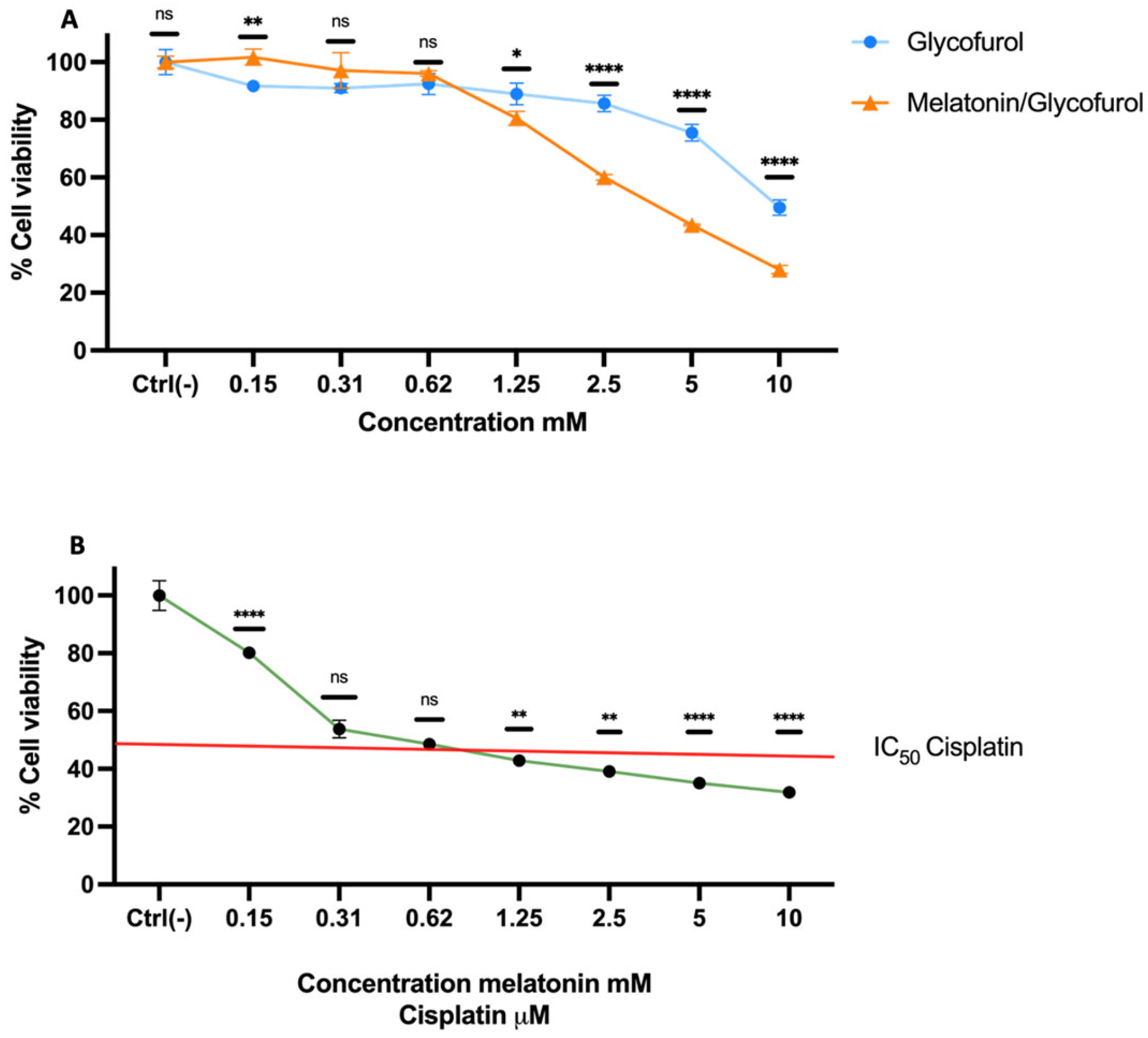
Figure 1. Cytotoxic activity in HeLa cells. (A) Treatments with melatonin dissolved in glycofurol or glycofurol alone, at different concentrations for 24 h. Data are presented as mean ± SD for each group. To compare the different treatments, statistical significance was calculated by one-way analysis of variance (ANOVA) followed by post hoc Tukey test. Asterisks indicate significant differences between treatments at * p < 0.01, ** p < 0.001, **** p < 0.0001. ns = not significant. (B) Combined treatment with different concentrations of melatonin along with IC50 cisplatin for 24 h. Data are presented as mean ± SD. Statistical significance was calculated by one-way ANOVA followed by post hoc Dunnett’s test to compare all test samples against positive control (cisplatin). Asterisks indicate significant differences at ** p < 0.003, **** p < 0.0001. ns = not significant.
Scipharm 94 00031 g001
Figure 2. Manufacturing of vaginal suppositories. FF—Final formulation; FFM—Final formulation loaded with melatonin.
Scipharm 94 00031 g002
Figure 3. Vaginal suppositories loaded with melatonin (FFM) had the same mass uniformity as the drug-free vaginal suppositories (FF) (n = 25). Bars and error bars, mean ± SD, CV% and maximum and minimum values.
Scipharm 94 00031 g003
Figure 4. Stress–strain curve used to estimate Young’s modulus.
Scipharm 94 00031 g004
Figure 5. Vaginal suppositories loaded with melatonin (FFM) and drug-free vaginal suppositories (FF) at the initial time and after 30 min in the simulated vaginal fluid (SVF).
Scipharm 94 00031 g005
Figure 6. In vitro cumulative release profiles of melatonin from vaginal suppositories based on gelatin.
Scipharm 94 00031 g006
Figure 7. The FTIR spectra of melatonin (Mel), the vaginal suppository base (final formulation, FF), and the vaginal suppository loaded with melatonin (FFM).
Scipharm 94 00031 g007
Figure 8. X-ray diffraction pattern of melatonin (Mel), vaginal suppository base (final formulation, FF), and vaginal suppository loaded with melatonin (FFM).
Scipharm 94 00031 g008
Figure 9. SEM micrographs collected with 25× magnification from (a) FF and (b) FFM samples, and at 500× magnification from (c) FF and (d) FFM samples.
Scipharm 94 00031 g009
Figure 10. Size distribution of hollows in the FF and FFM samples.
Scipharm 94 00031 g010
Figure 11. Cytotoxic activity in HeLa cells after treatment with melatonin-loaded vaginal suppositories alone (FFM) or combined with IC50 cisplatin (FFM + IC50 Cis) after 24 h. Both samples were previously dissolved in SVF. Data are presented as mean ± SD for each group. To compare the different treatments, statistical significance was calculated by one-way ANOVA followed by post-hoc Tukey test. Asterisks indicate significant differences between treatments at * p < 0.01, **** p < 0.0001. ns = not significant.
Scipharm 94 00031 g011
Table 1. Solubility of melatonin using different solvents.
SolventMelatonin (mg/mL)
102030405060708090100
WaterNSNSNSNSNSNSNSNSNSNS
Ethanol+++++++NSNSNS
Propylene glycol+++NSNSNSNSNSNSNS
DMSO++++++++++
Glycofurol++++++++++
+ = Soluble; NS = Non-soluble.
Table 2. Texture profile analyses (TPAs) of the vaginal suppositories.
FFM
Loaded with Melatonin
FF
Drug-Free
Hardness (gf)1091.953±69.0101121.620±109.505
Springiness1.218±0.2251.369±0.212
Cohesiveness0.936±0.0060.939±0.008
Resilience0.782±0.012 *0.807±0.012 *
Asterisk indicates significant differences between samples at * p < 0.0001.
Disclaimer/Publisher’s Note: The statements, opinions and data contained in all publications are solely those of the individual author(s) and contributor(s) and not of MDPI and/or the editor(s). MDPI and/or the editor(s) disclaim responsibility for any injury to people or property resulting from any ideas, methods, instructions or products referred to in the content.