Article

Green-Synthesized Silver Nanoparticles Derived from Calotropis procera as a Multifunctional Nanotherapeutic Platform Targeting Helicobacter pylori, Oxidative Stress, Inflammation, and Gastric Cancer


by
Mounishwaran Kamalesan
1,2,
Mohanraj Raja
1,2,
Rameshkumar Neelamegam
3,
Muthukalingan Krishnan
4,
Kayalvizhi Nagarajan
1,* and
Douglas J. H. Shyu
2,*
1
Department of Zoology, Periyar University, Salem 636011, Tamil Nadu, India
2
Department of Biological Science and Technology, National Pingtung University of Science and Technology, Pingtung 912301, Taiwan
3
Amity Institute of Biotechnology, Amity University, Mumbai 410206, Maharashtra, India
4
Central University of Tamil Nadu, Thiruvarur 610005, Tamil Nadu, India
*
Authors to whom correspondence should be addressed.
Sci. Pharm. 2026, 94(2), 44; https://doi.org/10.3390/scipharm94020044
Submission received: 23 April 2026 / Revised: 22 May 2026 / Accepted: 27 May 2026 / Published: 29 May 2026

Abstract

Green synthesis of silver nanoparticles (CP-AgNPs) using Calotropis procera (CP) offers a sustainable approach to producing multifunctional therapeutic nanomaterials. This study aimed to synthesize CP-AgNPs and evaluate their antimicrobial, antioxidant, anti-inflammatory, and anticancer potential, with a focus on Helicobacter pylori and gastric cancer cells. CP-AgNPs were prepared by phytochemical reduction using CP leaf extract and characterized by UV–Vis, XRD, FTIR, SEM, EDX, TEM, and Zeta. Antibacterial activity against H. pylori, time-kill kinetics, and SEM imaging of membrane damage were performed. Antioxidant (DPPH, ABTS) and anti-inflammatory assays, together with cytotoxicity studies in AGS cells (DAPI, AO/EtBr, and SEM), were also conducted. CP-AgNPs exhibited an SPR peak at 432 nm, face-centered cubic crystallinity, and spherical morphology (8–32 nm). They showed strong, dose-dependent antibacterial activity against H. pylori, surpassing metronidazole at higher doses. Time-kill assays and SEM confirmed membrane disruption. Antioxidant activity was notable (IC50: 40 µg/mL for DPPH; 60 µg/mL for ABTS). CP-AgNPs demonstrated significant anti-inflammatory effects and dose-dependent cytotoxicity in AGS cells, inducing apoptosis and morphological alterations. The broad biological activity of CP-AgNPs likely arises from the synergy between silver ions and CP phytochemicals. Their superior antibacterial effects, combined with antioxidant and anti-inflammatory properties, indicate strong therapeutic potential for gastric diseases. Anticancer activity in AGS cells suggests additional biomedical relevance, which may involve ROS-associated and apoptosis-related pathways, as suggested by previous studies. CP-AgNPs represent a promising natural nanoplatform for managing H. pylori infection, oxidative stress, inflammation, and gastric cancer, warranting further mechanistic and in vivo studies.

1. Introduction

Nanotechnology has emerged as one of the fastest-growing therapeutic areas in medicine owing to its broad biomedical applications, cost-effectiveness, and reduced environmental impact, and it holds potential to yield new drug candidates and alternative therapies [1]. Metallic nanoparticles, in particular, silver nanoparticles, have attracted considerable interest because of their high surface-to-volume ratio, increased bioreactivity, and demonstrated efficacy in drug delivery, antimicrobial therapy, and cancer treatment [2]. Among the various synthesis approaches, green synthesis is gaining prominence because it avoids hazardous chemicals and employs biological extracts from plants, bacteria, fungi, and algae, making it safer and more sustainable than conventional chemical methods.
Globally, gastric cancer remains one of the leading causes of cancer-related deaths. According to the World Health Organization (WHO), cancer causes almost 9.6 million deaths every year, with gastric cancer accounting for around 783,000 deaths in 2018 alone; nearly 70% of these deaths occurred in low- and middle-income countries [3]. In addition to cancer, infectious diseases continue to cause major morbidity and mortality worldwide, largely because of the emergence of antibiotic-resistant pathogens. Among these, Helicobacter pylori, a Gram-negative, microaerophilic bacterium that colonizes the gastric mucosa, infects more than half of the world’s population [4]. This pathogen is directly associated with chronic gastritis, peptic ulcer disease, mucosa-associated lymphoid tissue (MALT) lymphoma, and gastric adenocarcinoma [5]. Significantly, H. pylori has developed resistance to many commonly used antibiotics, including clarithromycin and metronidazole, which seriously jeopardizes current treatment strategies [3]. Finding alternative antimicrobials that can overcome antibiotic resistance and target H. pylori biofilms is therefore a key research priority.
Green-synthesized nanoparticles are particularly promising, because plant-derived phytochemicals not only reduce metal ions but also act as capping and stabilizing agents and contribute additional biological activities such as antioxidant, anti-inflammatory, and anticancer effects [6,7]. Recent studies further highlight the multifunctional therapeutic potential of plant-mediated nanoparticles in antimicrobial, antioxidant, anti-inflammatory, and anticancer applications [8]. Plants of the genus Calotropis and, in particular, Calotropis procera, contain a variety of bioactive metabolites, including flavonoids, terpenoids, tannins, alkaloids, and phenolic compounds, that can enhance nanoparticle stability and therapeutic efficacy. The surface charge of nanoparticles and their colloidal stability are crucial physicochemical factors affecting their dispersion behavior, aggregation, and performance in biomedical applications. As a result, zeta potential analysis is frequently used to assess the stability of nanoparticles that have been biosynthesized [9]. Despite numerous reports of plant-mediated AgNPs, comprehensive investigations that integrate antibacterial, antioxidant, anti-inflammatory, and anticancer evaluations of AgNPs derived from C. procera remain scarce.
To the best of our understanding, this research stands as one of the limited thorough examinations of green-synthesized silver nanoparticles derived from Calotropis procera for diverse biomedical applications targeting Helicobacter pylori and gastric cancer. In contrast to earlier studies that mainly concentrated on singular biological activities, the current investigation encompasses physicochemical characterization, antibacterial, antibiofilm, antioxidant, anti-inflammatory, and anticancer assessments utilizing AGS gastric cancer cells, thereby underscoring the multifunctional therapeutic capabilities of CP-AgNPs.
Overall, the growing burden of H. pylori infection, oxidative stress, inflammation, and gastric cancer underscores the need for safe, multifunctional therapies. To address this gap, the present study focuses on the green synthesis of CP-AgNPs from C. procera leaf extract and evaluates their physicochemical properties and multifunctional biomedical potential. Specifically, we synthesize and characterize CP-AgNPs and assess their antibacterial, antioxidant, anti-inflammatory, and anticancer activities, with the aim of supporting the development of CP-AgNPs as a promising natural platform for nanotherapeutics.

2. Materials and Methods

2.1. Synthesis of Silver Nanoparticles

Fresh and healthy leaves of Calotropis procera were taken, washed thoroughly with distilled water to remove any dust particles which might have accumulated, and dried in the shade for a few days. The dried leaves were finely ground and used in water extraction. Approximately 10 g of powdery leaves were dissolved in 100 mL of distilled water at a temperature of 60 to 70 °C for 20 min and then filtered with a Whatman No. 1 strip of filter paper. The resulting yellow-colored filtrate was stored at 4 °C and used as a reducing and stabilizing agent for the synthesis of nanoparticles.
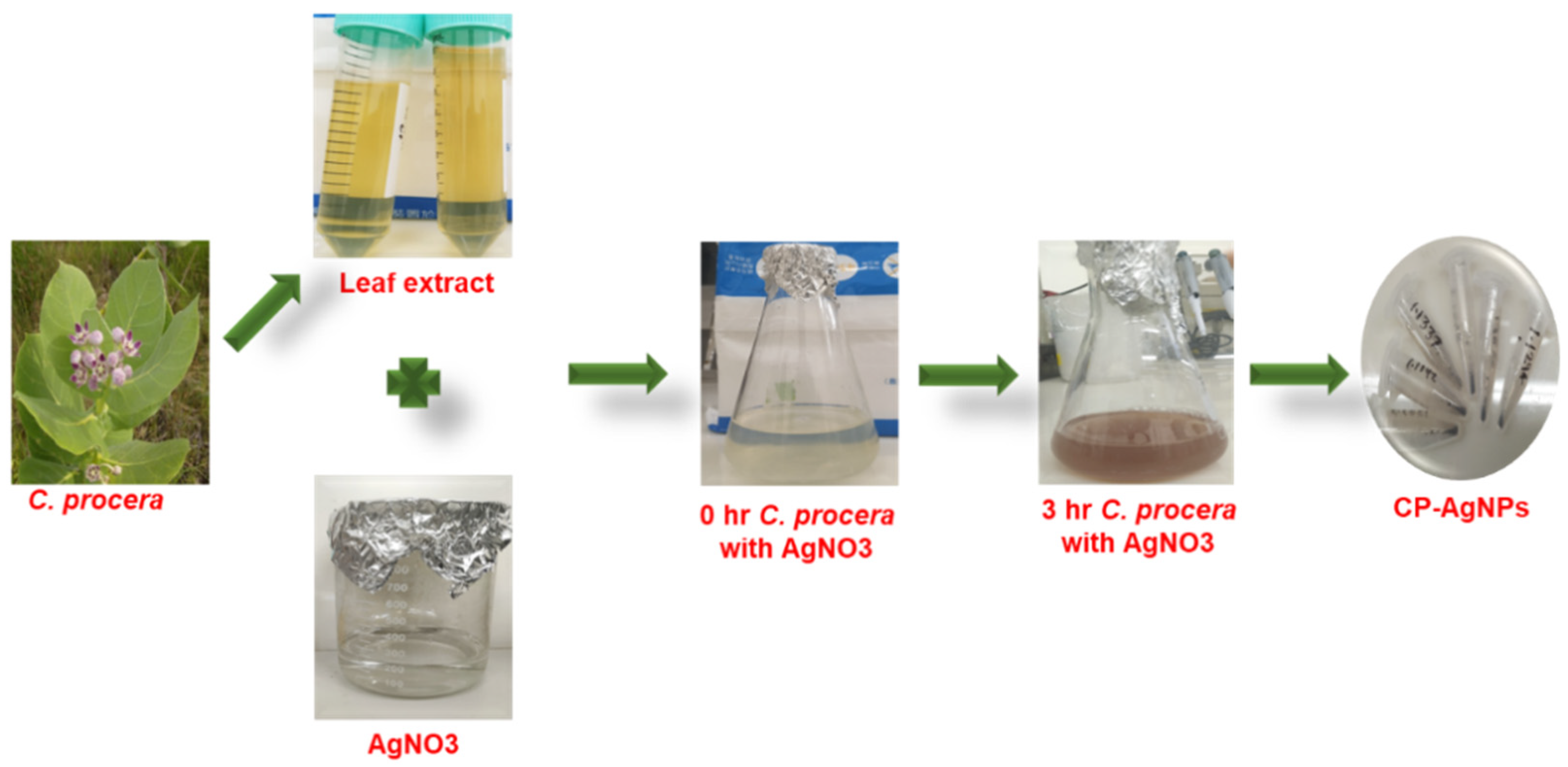
To biosynthesize silver nanoparticles, 10 mL of C. procera leaf extract was added by drop-wax to 90 mL of freshly prepared solution of 1 mM silver nitrate (AgNO3) in a continuous stirring motion. The reaction mixture was incubated at room temperature under dark conditions in order to avoid the silver ion being photoactivated. A gradual color change from pale yellow to red–brown over 3 h was observed, indicating that Ag+ ions were reduced to silver nanoparticles and CP-AgNPs were successfully formed. The synthesized nanoparticles were centrifuged at 12,000 rpm for 15 min, washed three times with distilled water to remove the unsaturated phytochemicals, and dried at 60 °C to obtain a purified AgNP powder for further characterization (Figure 1).
Visual color change was further confirmed by UV–visible spectrophotometry, and the synthesis protocol is based on established principles for green nanofabrication, where plant biomolecules such as flavonoids, phenols, and terpenoids act as reducing and stabilizing factors in the formation of nanoparticles [2]. This phytochemical approach improves the stability, biocompatibility and biological activity of the nanoparticles compared to chemically synthesized AgNPs.

2.2. Characterization

The physicochemical properties of the CP-AgNPs synthesized have been analyzed by several complementary techniques. UV–visible spectroscopy (300–700 nm) has been used for the first time to monitor nanoparticle formation by detecting the characteristic surface plasmon resonance peaks associated with AgNPs [2,10]. The crystalline structure of the dry nanoparticles was investigated by X-ray diffraction (XRD) with Cu Kα irradiation (λ = 1.5406 azide) over a 2 years range of 20 to 80 °C, confirming the cubic (fcc) nature of metallic silver. Functional groups involved in the reduction and stabilization of nanoparticles have been identified by Fourier-transform infrared spectroscopy (FTIR) recording from 400 to 4000 cm−1, allowing for detection of the vibratory bands O-H, C-C, N-N, and C-O of phytochemical-derived nanoparticles [11]. Surface morphology and particle size distribution were evaluated by scanning electron microscopy (SEM) after gold-spatter coating of samples, and elemental composition was determined by energy-dispersive X-ray (EDX) spectroscopy by using a Hitachi 4500 instrument (Hitachi, Ltd., Tokyo, Japan), confirming the presence and purity of silver [12]. In addition, high-resolution transmission electron microscopy (TEM) has been performed by depositing nanoparticle suspensions on a copper plate and imaging the film on a sphere to visualize the morphology of the nanoparticles and their size at the nanoscale. The zeta potential of CP-AgNPs was assessed to gauge the surface charge of the nanoparticles and their colloidal stability. The suspension of biosynthesized nanoparticles was examined with a zeta potential analyzer following standard operating procedures, and the zeta potential value was noted in millivolts (mV) [7,13]. These combined analyses provided a comprehensive structural, chemical, and morphological validation of the synthesis of CP-AgNPs.

2.3. Antibacterial Activity

The antibacterial activity of the synthesized CP-AgNPs against Helicobacter pylori was evaluated by diffusion agar using standardized protocols with minor modifications. Helicobacter pylori strain ATCC 26695 was used in this study. The bacterial strain was obtained from the American Type Culture Collection (ATCC, Manassas, VA, USA). Fresh cultures of H. pylori were cultured on brain–hepatitis (BHI) agar supplemented with 10% fetal bovine serum under microaerobic conditions (5% O2, 10% CO2, and 85%N2) at 37 °C for 48 to 72 h. The bacterial suspension was prepared in sterile saline and adjusted to a McFarland standard of 0.5 McFarland (1 × 10⁵ CFU/mL). Mueller–Hinton agar plates enriched with 5% sheep-derived blood were uniformly swabbed with bacterial inoculum, and wells with a diameter of 6 mm were prepared using a sterile cork borer. Each well was filled with 50 mL of CP-AgNP solutions at 25, 50, and 75 µg/mL, with metronidazole as the positive control and sterile distilled water as the negative control. The plates were incubated under microaerophilic conditions at 37 °C for 48 to 72 h, and the antibacterial effect was assessed by measuring the zone of inhibition (ZOI) around each well in millimeters. All tests were performed in triplicate, and the results were averaged to give an average ± SD. Agar diffusion tests are widely used to assess the antimicrobial efficacy of biosynthesized nanoparticles, particularly against drug-resistant pathogens such as H. pylori [14,15,16]. This method provides a reliable preliminary evaluation of inhibition of bacterial growth by nanoparticles.

2.4. Time-Kill Assay

The kinetic time-killing test was carried out to assess the bactericidal activity of green-synthesized silver nanoparticles (CP-AgNPs) against Helicobacter pylori. The test followed the standard procedures described for kinetic studies of antimicrobials with modifications suitable for nanoparticle testing [1,17,18]. Briefly, clinical isolates of H. pylori were prepared in Brucella broth enriched with 10% fetal bovine serum (FBS) and cultured on Columbia agar supplemented with 5% sheep blood at 37 °C under microaerobic conditions (10% CO2, 5% O2, and 85% N2).
CP-AgNPs synthesized from the leaf extract of Calotropis procera were freshly prepared at final concentrations of 25, 50, 75, and 100 µg/mL, with metronidazole (10 mg/mL) as the positive control and untreated cultures as the negative control. For each condition, 1 mL of the standardized bacterial suspension was mixed in sterile glass tubes with the respective concentrations of CP-AgNPs and incubated under microaerobic conditions at 37 °C. At predetermined time points (0, 6, 12, 18, 24, 30, and 36 h), 100 mL of aliquots were withdrawn, serially diluted, and seeded onto Columbian blood agar plates. The plates were incubated for 72 h and colony-forming units (CFU/mL) were counted to evaluate bacterial viability. The decrease in the log CFU over time was calculated in order to determine the bacteriostatic or bactericidal effect.
In order to ensure reliability, all trials were performed in triplicate, and the results were expressed as mean ± standard deviation. The bactericidal endpoint was interpreted as a reduction in CFU > 3-log10 compared to the original inoculum [19]. The test design and concentration range are based on recent recommendations for the assessment of the kinetic antibacterial activity of H. pylori against biosynthesized AgNPs [20,21].

2.5. Biofilm Formation and Inhibition Assay

The formation of biofilms and inhibition of green synthetic CP-AgNPs were evaluated using a standard 96-well microtiter plate crystal violet test. Briefly, Helicobacter pylori cultures were grown overnight in enriched broth diluted to a concentration of 1 × 10⁵ CFU per well on sterile flat bottom polystyrene plates of 96 wells, and then 20 mL of CP-AgNP suspensions (final concentrations: 25, 50, and 75 µg/mL) and controls were added. The plates were statically incubated at 37 °C under microaerophilic conditions for 24 to 48 h to allow biofilm formation. After incubation, the planktonic cells were gently removed, the wells were washed three times with phosphate-buffered saline (PBS) to remove the non-essential bacteria, and the biofilms were fixed for 15 to 20 min with 0.1% (w + v) crystal violet. The excess was washed with sterile water, the plates were air dried, and the bound color was dissolved in 95% ethanol (or 33% acetic acid) for 10 to 15 min. The absorbance of the dissolved crystal violet was measured at 590–600 nm by microplate readers and the biomass of the biofilm was quantified by comparing the treated wells with untreated control wells. This follows the recently standardized protocols for the assessment of antibiofilm substances by microtiter plates, stressing reproducibility, minimum technical requirements, and quantitative biomass estimation by the CV [22,23,24].

2.6. SEM Analysis of Antibiofilm Activity

The inhibition of Helicobacter pylori by the green-synthesized silver nanoparticles (CP-AgNPs) in the biofilm was assessed using scanning electron microscopy (SEM). Briefly, H. pylori was cultured in enriched broth and allowed to mature in sterile glass coverslips for 48 h under microaerobic conditions at concentrations of 25, 50, and 75 mL of CP-AgNPs, respectively, using untreated coverslips as controls. After treatment, the non-adherent cells were gently washed with phosphate-buffered saline (PBS), and the remaining adhering biofilm was fixed at 4 °C for 2 h with 2.5% glutaraldehyde. The samples were then dried with a series of graduated ethanol grades (30%, 50%, 70%, and 90%), air dried, attached to aluminum rods, and covered with gold foil. Finally, the structure of the biofilm and the bacterial morphology were examined by high resolution SEM (usually 10 to 15 kV). This approach allows for visualization of the structural changes in biofilm biomass, bacterial aggregates, and cell membrane integrity and provides qualitative evidence for antibacterial activity against bacterial biofilms (e.g., against MDR pathogens and mixed biofilms) [25,26].

2.7. DAPI and AO/EtBr Staining in AGS Cells

The human gastric adenocarcinoma cell line AGS was used for the anticancer study. AGS cells were maintained under standard culture conditions for subsequent experimental analysis. The gastric cancer cells were seeded at 1 × 10⁵ cells per well on 12 wells (on sterile glass plates) in a humidified incubator with 5% CO2 at 37 °C and allowed to attach overnight to a medium of RPMI-1640 supplemented with 10% fetal bovine serum (Gibco, Grand Island, NY, USA). After 24 h, cells were treated with greens-synthesized CP-AgNPs at concentrations corresponding to the experimentally determined IC50 (<40–50 µg/mL and higher than 100 µg/mL) for 24 h; untreated cells were used as negative control.

2.7.1. DAPI Staining

After treatment, the medium was removed, and the cells were washed gently with sterile phosphate-buffered saline (PBS, pH 7.4) to remove any unwanted or loose cell adhesion. The cells on the coverslips were then fixed in PBS with 4% paraformaldehyde for 15 min at room temperature and washed with three PBS drops. The fixed cells were incubated in PBS for 10 min in darkness with 1 mL of DAPI (4′6-diamidino-2-phenylindole). After coloring, the excess dye was removed by washing twice with PBS. The coated plates were mounted on microscopic slides with an anti-fade medium, and the stained nuclei were examined under a fluorescence microscope (excitation ~350 nm and emission ~460 nm). Nuclear morphology was evaluated for apoptotic markers such as chromatin condensation, nuclear fragmentation, and apoptotic bodies.

2.7.2. AO/EtBr (Acridine Orange/Ethidium Bromide) Dual Staining

In parallel wells, after 24 h treatment with acridine orange (AO) and PBS washing, the cells were stained with freshly prepared acridine orange and ethidium bromide (EtBr) mixtures. Typically, 100 µg of AO and 100 µg of EtBr stock solutions (in PBS) are diluted to working concentrations. Cells are incubated in the dark for 2 to 5 min at room temperature with the mixture of AO and EtBr. Immediately afterwards, the cells were examined under fluorescence microscopy with suitable filters for AO (green fluorescence; excitation ~490 nm, emission ~530 nm) and EGT (red fluorescence; excitation ~520–540 nm, emission ~600–620 nm). Live cells with intact membranes show uniform green fluorescence (AO), early apoptotic cells show a clear green-to-yellow–green nuclei, and late apoptotic or necrotic cells show a red-to-orange fluorescence as a result of uptake of EtBr and nuclear chromatin modification.

2.8. Anti-Inflammatory Activity

2.8.1. Membrane Protection

Membrane protection was evaluated using a method based on the hypoprotonation-induced hemolysis adapted from standard membrane stabilization protocols. Healthy volunteers who had abstained from NSAIDS for 2 weeks before the trial were recruited to donate blood, which was collected from EDTA tubes and centrifuged at a rate of 1500 times for 10 min; the plasma was then removed, and the pooled erythrocytes were washed three times with phosphate-depleted saline (PBS, pH 7.4). The 10% (v/v) erythrocyte suspension was prepared in hydrogenated PBS. Test samples of CP-AgNPs were prepared in PBS at final concentrations of 20, 40, 60, 80, and 100 µg/mL. For each test well (or tube), 0.5 mL of the test sample, 1.0 mL of the hypotonic solution (0.36% w/v NaCl), and 0.5 mL of the suspension of 10% RBC were mixed. For the negative control, substitute 0.5 mL PBS for the test sample; for the positive control, use 0.5 mL diclofenac sodium (a standard anti-inflammatory medicine) in the same concentration. The mixture is incubated at 37 °C for 30 min and then centrifuged at a rate of 1500× g for 10 min [27,28]. The absorption of the supernatant was measured at 540 nm. The percentage of membrane protection (% of hemolytic inhibition) was calculated as follows:
A n t i i n f l a m m a t o r y   a c t i v i t y % = A b s o r b a n c e   ( C o n t r o l ) A b s o r b a n c e ( S a m p l e ) A b s o r b a n c e   ( C o n t r o l ) × 100
Membrane stabilization (%) = [(Absorbance of control − Absorbance of sample)/Absorbance of control] × 100
All treatments were carried out in duplicate and data reported as mean ± SD. The statistical significance between the groups was determined using a one-way ANOVA, followed by a post hoc Tukey t test; p < 0.05 was considered significant [29].

2.8.2. Inhibition of Protein Denaturation

Inhibition of protein denaturation was measured using the BSA thermal denaturation test, slightly modified from the published protocols. Prepare 0.4% (w/v) of BSA in PBS (pH 7.4). Mix 0.5 mL of CP-AgNPs in desired concentrations (20 to 100 µg/mL) with 0.4 mL of BSA solution. Prepare a negative control by mixing 0.5 mL PBS with 0.5 mL BSA and a positive control by mixing a standard solution of anti-inflammatory drugs (such as diclofenac sodium) with BSA. The mixture is incubated at 37 °C for 15 min and then heated at 70 °C for 5 min to induce denaturation. After cooling to room temperature, the absorbance shall be measured by spectrophotometry at 660 nm [28]. The percentage of the inhibition of protein denaturation is calculated as follows:
P r o t e i n   d e n a t u r a t i o n   i n h i b i t i o n   ( % ) = A b s o r b a n c e   ( C o n t r o l ) A b s o r b a n c e ( S a m p l e ) A b s o r b a n c e   ( C o n t r o l ) × 100
Protein denaturation inhibition (%) = [(Absorbance of control − Absorbance of sample)/Absorbance of control] × 100
Perform all tests three times and submit data as mean ± SD. Statistical differences shall be analyzed by means of a one-way ANOVA with appropriate post hoc testing (p < 0.05) [30,31].

2.9. Antioxidant Activity

2.9.1. DPPH Radical Scavenging Assay

The antioxidant potential of the biosynthetic catabolite catalase (CP-AgNPs) silver nanoparticles has been assessed by 2,2-diphenyl-1-pyrazyl (DPH) radical scavenger assay, according to established protocols with minor changes [11,32]. The 0.1 mM DPPH solution has been prepared in methanol and is shielded from light. Different concentrations of CP-AgNPs (10 to 100 µg/mL) have been prepared in methanol. For each reaction, 1 mL of DPPH solution was mixed with 1 mL of each concentration of the sample, vortexed, and incubated at room temperature for 30 min in the dark [33]. To ensure that the DPPH radicals are fully interacted with, the radicals are fully absorbed. The control was prepared by mixing 1 mL of DPPH solution with 1 mL of methanol, using, as the standard reference antioxidant, 10–100 mg ascorbic acid [34]. After incubation, the absorbance was measured by UV–visible spectrophotometry at 517 nm.
The DPPH radical scavenging percentage was calculated using:
DPPH scavenging assay (%) = (Control Absorbance − (Sample Absorbance − Blank Absorbance))/(Control Absorbance) × 100
The IC50 value (the concentration required for 50% DPPH radical scavenging test) was determined by nonlinear regression plot of the percentage of DPPH radical scavenging test by concentration [35]. All the trials were performed in triplicate, and the data were expressed as mean ± standard deviation.

2.9.2. ABTS Radical Scavenging Assay

The antioxidant activity of Calotropis procera green synthetic silver nanoparticles (CP-AgNPs) was evaluated by the ABTS•+ radical scavenging test using a modified standard procedure. Briefly, the ABTS stock solution (7 mM) was prepared and mixed with 2.45 mM potassium sulphate in a 1:1 ratio and incubated for 12 to 16 h at room temperature to produce the radical cation of ABTS. The resulting ABTS+ solution was diluted with ethanol to obtain a 734 nm absorbance of 0.700 ± 0.02. The various concentrations of CP-AgNPs (20 to 100 µg/mL) were mixed with 1 mL of the ABTS solution and incubated at room temperature for 10 min. Absorption was measured by 734 nm UV–visible spectrophotometer with ethanol as a blank. Ascorbic acid was used as the standard antioxidant [36]. The percentage of radical scavenging activity has been calculated using the following equation:
ABTS scavenging assay percentage = (Absorbance control − Absorbance treated)/(Absorbance control) × 100
This method is widely used because of its sensitivity to both hydrophilic and lipophilic antioxidants and its utility in assessing the electron donor potential of nanoparticle systems.

2.10. Statistical Analysis

The statistical analysis was conducted by means of a one-way variance analysis (ANOVA). SPSS 17.0 software was used for statistical analysis. Three replicates of each experiment and test were performed (n = 3). The average of the three values are reported in each of the cases.

3. Results and Discussion

3.1. Physicochemical Characterization

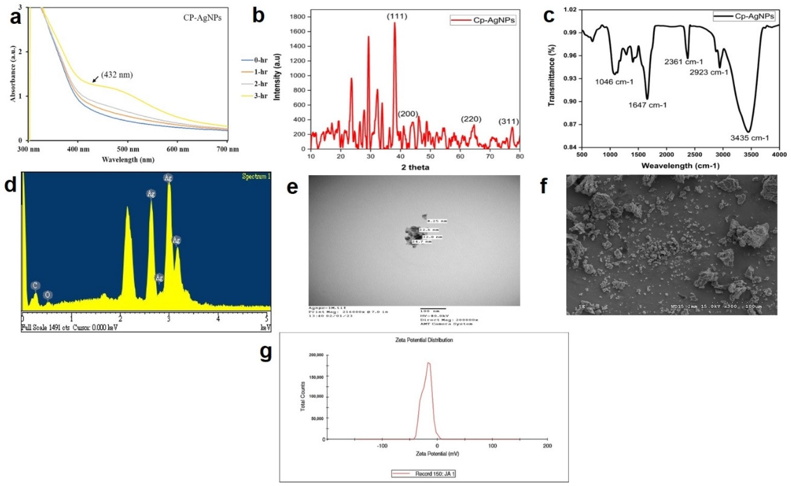
The green synthesis of silver nanoparticles (AgNPs) using Calotropis procera leaf extract was confirmed by spectroscopic and microscopic analyses. UV–visible spectroscopy showed a distinct surface plasmon resonance (SPR) peak at 432 nm after 3 h (Figure 2a), indicating the effective reduction of Ag+ to metallic Ag0 and successful nanoparticle formation. This SPR position is consistent with previous reports on plant-derived AgNPs [37] and confirms that the biosynthetic process is efficient.
X-ray diffraction (XRD) analysis (Figure 2b) revealed strong diffraction peaks at 38.05°, 44.23°, 64.41°, and 76.66° (2θ), corresponding to the (111), (200), (220), and (311) planes of the face-centered cubic silver structure; this confirms the crystalline nature of the synthesized nanoparticles and agrees with earlier studies [38]. Fourier-transform infrared (FTIR) spectroscopy (Figure 2c) showed absorbance bands at 3435, 2923, 2361, 1647, and 1046 cm−1. These bands are attributable to O–H/N–H, C–H, possible CO2/overtone, C=O/C=C, and C–O vibrations, respectively, suggesting that phytochemicals in the extract participate in reduction and act as stabilizing (capping) agents. This biological capping prevents aggregation and enhances long-term stability [39].
Morphological examination by SEM (Figure 2e) and TEM (Figure 2f) showed mostly spherical nanoparticles with sizes ranging from 8.25 to 32.0 nm, with many particles in the 20–30 nm range, consistent with typical plant-synthesized AgNPs [12,37]. Energy-dispersive X-ray (EDX) analysis (Figure 2d) produced a strong silver signal at ~2.7–3.1 keV, representing 54.32% of the detected elemental content, indicating high silver purity and minimal contamination [12].
Taken together, these characterization results confirm that Calotropis procera leaf extract acts as an effective reducing and stabilizing agent, producing well-defined, crystalline, spherical AgNPs with the desired nanoscale dimensions. The observed physicochemical properties are consistent with the literature on biogenic AgNPs and highlight the potential of C. procera as a sustainable source for nanoparticle synthesis [38]. Zeta potential analysis (Figure 2g) revealed that CP-AgNPs possessed a net surface charge of −19 mV, indicating moderate colloidal stability of the biosynthesized nanoparticles. The negative zeta potential may be attributed to phytochemical constituents from Calotropis procera acting as capping and stabilizing agents on the nanoparticle surface. Such surface charge may reduce nanoparticle aggregation through electrostatic repulsion, thereby contributing to suspension stability and supporting potential biomedical applications. The structural stability, high purity, and size uniformity of the CP-AgNPs synthesized indicate their suitability for downstream biomedical applications, including antibacterial, antioxidant, and anticancer uses.

3.2. Antibacterial Activity Assay

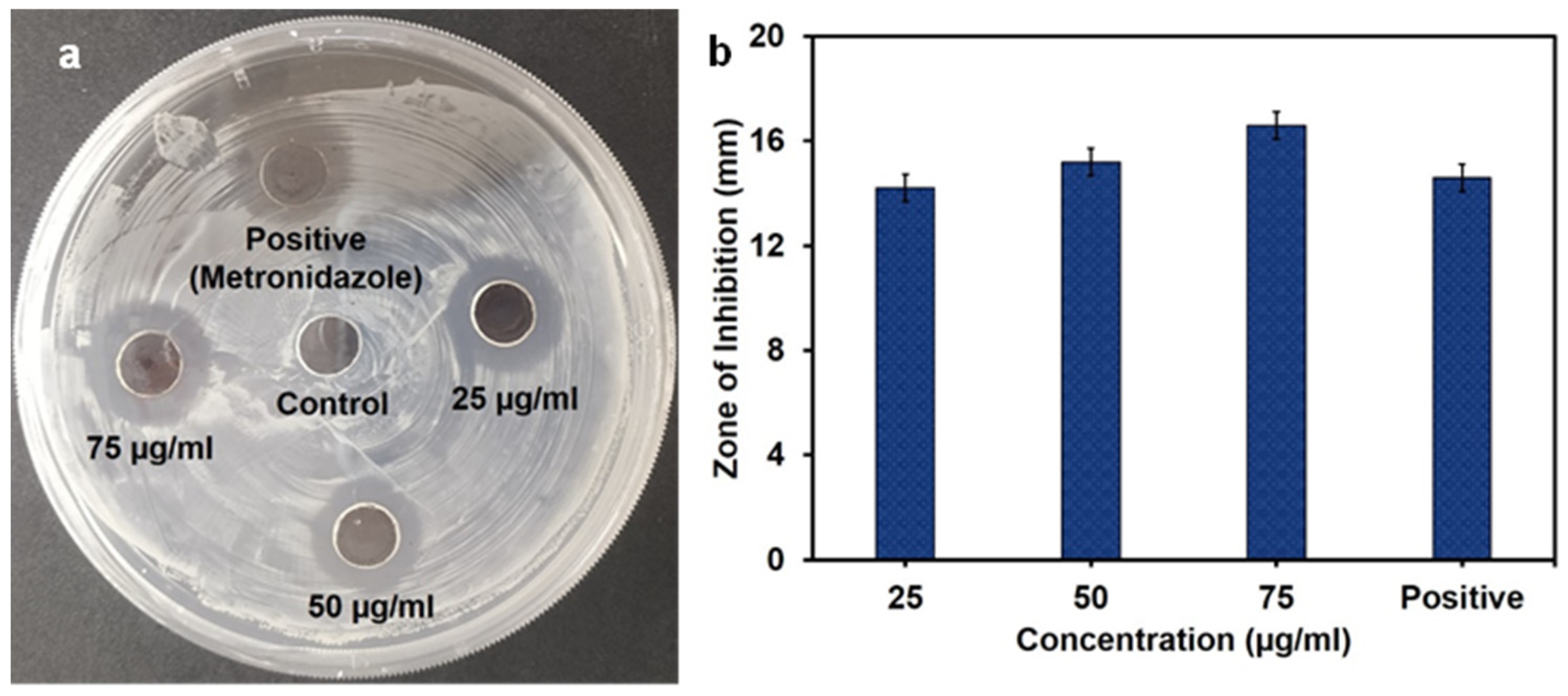
Green-synthesized silver nanoparticles (CP-AgNPs) derived from Calotropis procera leaf extract exhibited clear concentration-dependent antibacterial activity against Helicobacter pylori. At 25 µg/mL, the nanoparticles produced a zone of inhibition (ZOI) of 14.2 ± 0.45 mm, which increased to 16.6 ± 0.55 mm at 50 µg/mL. Notably, the antimicrobial activity at the highest concentration exceeded that of the standard antibiotic metronidazole (14.6 ± 0.55 mm), suggesting that the CP-AgNPs have greater bactericidal potency under the tested conditions (Figure 3). This enhanced activity demonstrates the efficacy of C. procera phytochemicals in facilitating the formation of biologically active silver nanoparticles, consistent with recent reports of the strong antimicrobial activity of plant-derived AgNPs against pathogenic bacteria, including H. pylori [40].
The concentration-dependent antibacterial trend observed here aligns with previous findings that green-synthesized AgNPs display potent inhibitory activity due to their small size, spherical morphology, and high surface reactivity, which increase interactions with bacterial membranes [41]. The higher efficacy of CP-AgNPs compared with metronidazole may arise from multiple mechanisms, including the disruption of membrane integrity, induction of oxidative stress, and interference with essential cellular processes. In addition, recent studies of AgNP-based antimicrobials highlight promising activity against antibiotic-resistant H. pylori, supporting the potential of nanoparticle-based therapies to combat gastrointestinal pathogens [42].

3.3. Time-Kill Assay

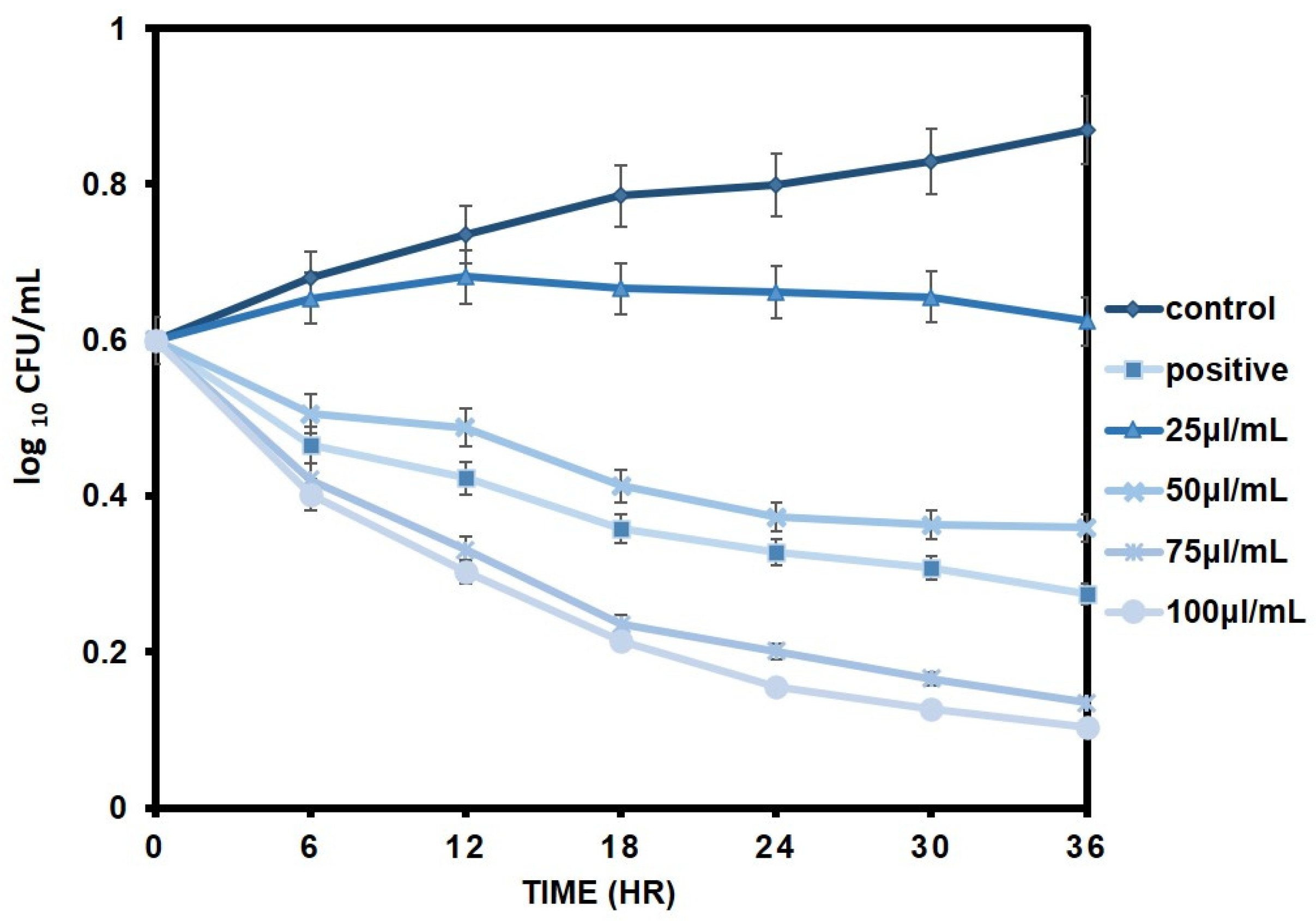
In a time-killed assay, CP-AgNPs had a clear concentration- and time-dependent bactericidal effect on H. pylori. At lower concentrations (25 and 50 µg/mL), the viability of the bacteria gradually declined, but not to the point of complete eradication. In contrast, treatment with 75 µg/mL and especially 100 µg/mL resulted in a significant decrease in viable cell counts and almost complete elimination within 36 h, which indicates strong antibacterial activity. These findings indicate that CP-AgNPs maintain their antimicrobial activity over time with prolonged exposure, which is in line with previous reports that AgNPs derived from plants show dose- and time-dependent killing kinetics against H. pylori (Figure 4) [43]. The increased bactericidal activity of CP-AgNPs is probably due to synergistic effects between silver nanoparticles and bioactive phytochemicals present in the Calotropis extract. Silver nanoparticles have been known to disrupt the bacterial cell membranes, induce the production of reactive oxygen species (ROS), interfere with enzyme functions (e.g., urease, which is critical for the survival of H. pylori), and induce DNA damage, ultimately leading to cell death [44].
The enhanced bactericidal effect of CP-AgNPs likely reflects synergistic interactions between the silver nanoparticles and bioactive phytochemicals in the C. procera extract. AgNPs are known to disrupt bacterial membrane integrity, induce reactive oxygen species (ROS) production, interfere with key enzymes (for example, urease, which is critical for H. pylori survival), and cause DNA damage, ultimately leading to cell death. Moreover, green-synthesized AgNPs have been reported to retain activity under pH conditions similar to that of the gastric environment, supporting their potential therapeutic relevance [45]. These results support the potential of green-synthesized AgNPs as an alternative or adjunct to conventional antibiotics for treating H. pylori, particularly in the context of rising antibiotic resistance. Based on the predefined bactericidal criterion (≥3 log10 CFU/mL reduction relative to the initial inoculum), CP-AgNPs at higher concentrations (75 and 100 µg/mL) exhibited bactericidal activity, whereas lower concentrations showed comparatively reduced killing effects.

3.4. Biofilm Inhibition Assay

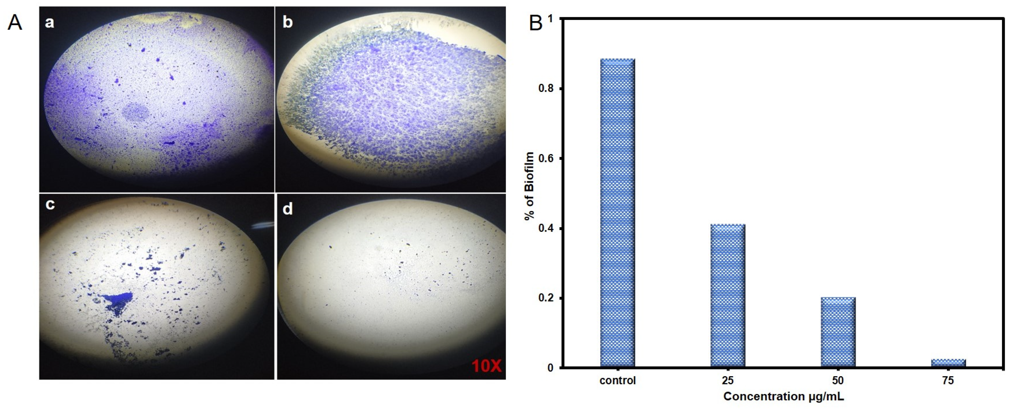
Crystal violet staining showed clear concentration-dependent inhibition of H. pylori biofilm formation after treatment with CP-AgNPs synthesized from the leaf extract of Calotropis procera. In untreated controls (Figure 5A(a)), thick and uniform biofilm coverage was observed, reflecting the strong innate ability of H. pylori to form structured biofilms that increase its survival and resistance to antibiotics. Treatment with 25 µg/mL CP-AgNPs (Figure 5A(b)) resulted in a noticeable decrease in biofilm density, although significant clumping and deposition of extracellular polymers were still evident. At 50 µg/mL (Figure 5A(c)), bacterial adherence and EPS formation were markedly reduced, resulting in a disrupted, sparse biofilm structure with scattered cell aggregates. The highest tested concentration, 75 µg/mL (Figure 5A(d)), produced a dramatic decrease in the biofilm biomass with only minimal residual staining, indicating that adherence and maturation of the biofilm were almost completely suppressed.
The dose-dependent reduction in biofilm formation is consistent with AgNPs’ ability to penetrate EPS layers, damage bacterial membranes, inhibit quorum sensing, and interfere with genes involved in adhesion and biofilm stability. Recent studies have confirmed that green-synthesized AgNPs effectively inhibit H. pylori biofilms by inducing oxidative stress, blocking urease activity, and disrupting signaling pathways involved in biofilm formation [43,44,45]. These results indicate that CP-AgNPs have a strong antibacterial potential against H. pylori and suggest they are a promising alternative or adjunct for treating persistent, resistant infections. The crystal violet assay provided qualitative and semi-quantitative evidence of concentration-dependent biofilm inhibition by CP-AgNPs; however, metabolic activity assays (e.g., XTT reduction) and image-based quantitative biofilm measurements were beyond the scope of the present study.

3.5. SEM Analysis of Antibiofilm Effect

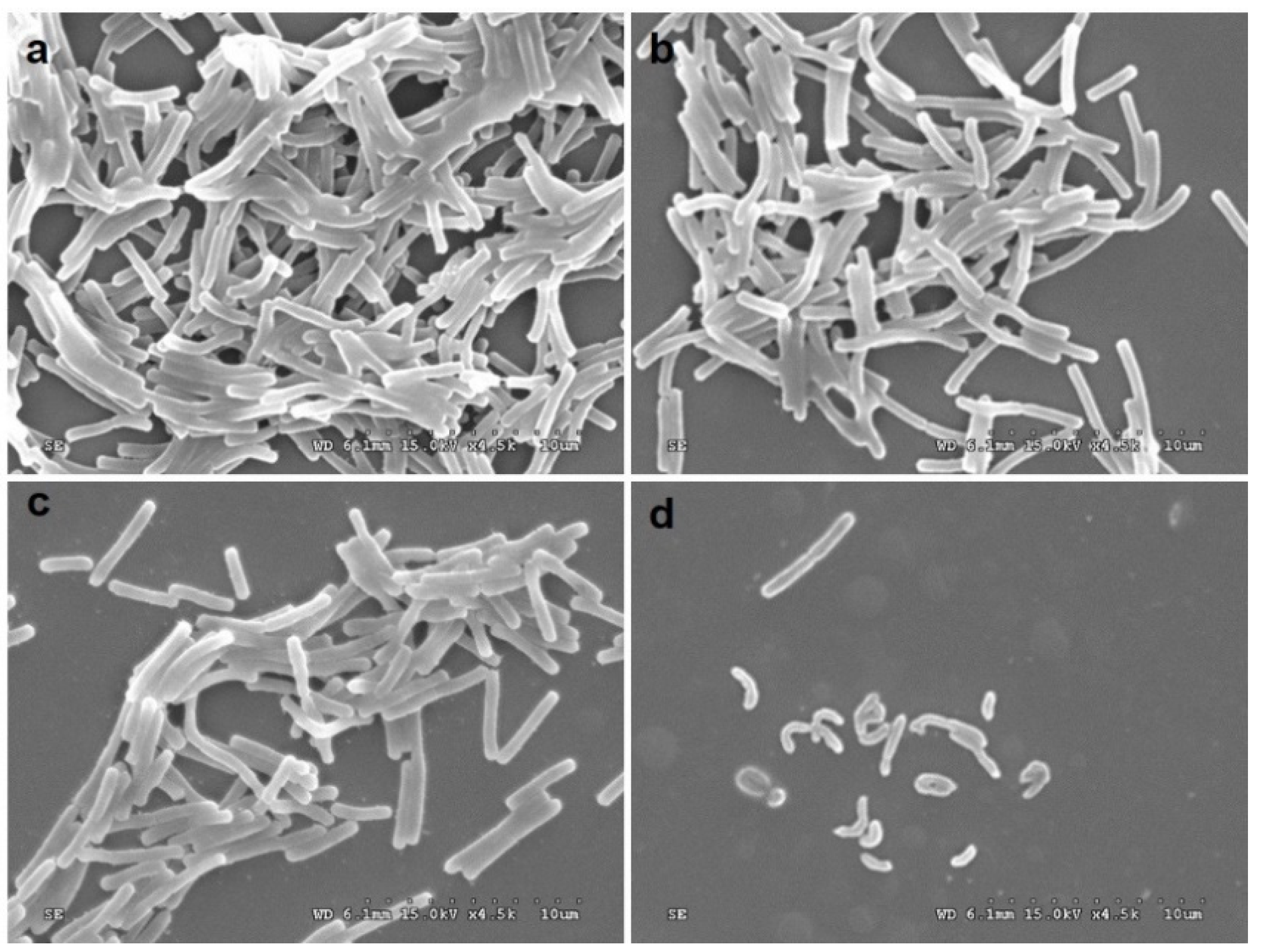
SEM images clearly showed dose-dependent antibiofilm activity and morphological damage to Helicobacter pylori following treatment with Calotropis procera leaf extract-based CP-AgNPs. The untreated control (Figure 6a) displayed a dense, multilayered biofilm with well-defined helical and rod-shaped bacteria densely aggregated in a mature extracellular matrix. Exposure to CP-AgNPs at 25 µg/mL (Figure 6b) caused slight structural degradation, reduced clumping, and early signs of membrane deformation compared with the compact, homogeneous control biofilm. At 50 µg/mL (Figure 6c), the biofilm network was markedly degraded, with fewer adherent cells, elongated cell bodies with rough surfaces, and collapsed bacterial structures, suggesting progressive nanoparticle-induced damage. The highest concentration tested, 75 µg/mL (Figure 6d), resulted in a dramatic reduction in bacterial density, severe cell-shape distortion, and pronounced cell shrinkage and fragmentation, demonstrating the potent antibacterial and bactericidal activity of CP-AgNPs.
These morphological changes are consistent with known AgNP mechanisms, including membrane rupture, the induction of oxidative stress, and disruption of biofilm matrix integrity, which together impair the adhesion and viability of H. pylori. Similar SEM-documented damage, like cell wall collapse, loss of helical morphology, and biofilm degradation, has been reported for plant-derived silver nanoparticles against clinical isolates of H. pylori, reinforcing the potential of green-synthesized AgNPs as effective antibiofilm agents [46]. Overall, the SEM results confirm that CP-AgNPs exert a potent, concentration-dependent antibacterial and morphological damaging effect against H. pylori, supporting their promise as an alternative or adjunctive treatment.

3.6. Anticancer Activity

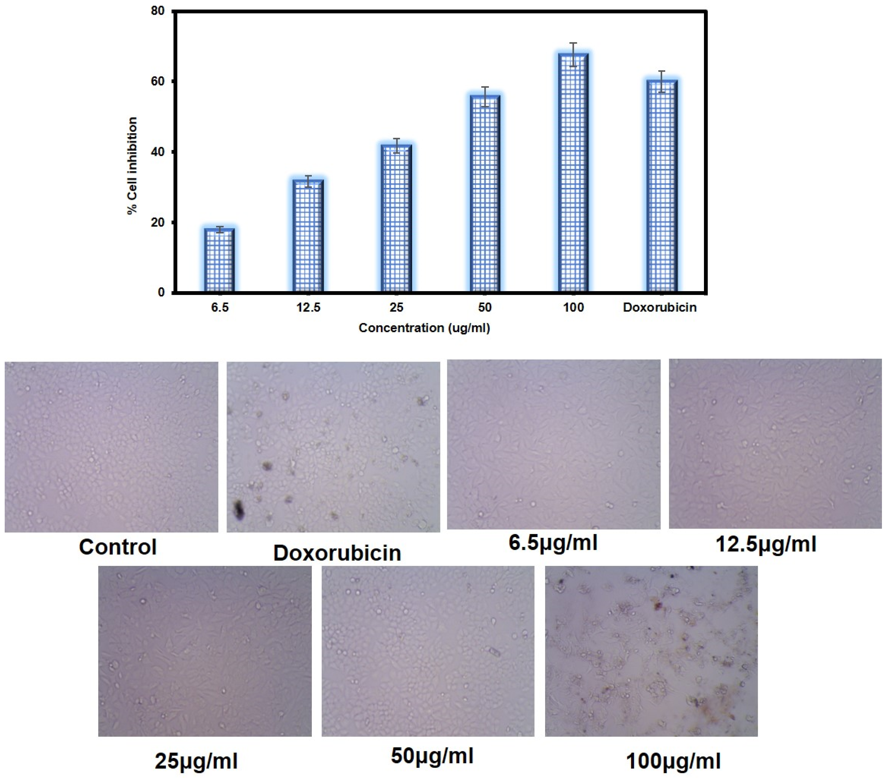
The anticancer activity of CP-AgNPs against AGS gastric carcinoma cells showed a clear concentration-dependent reduction in cell viability, with an IC50 value of 40.26 µg/mL, indicating strong cytotoxic potency (Figure 7).
Lower concentrations (6.5–25 µg/mL) produced mild to moderate inhibition, while concentrations near the IC50 (40–50 µg/mL) caused pronounced cytotoxicity and a marked loss of cellular metabolic activity. The highest concentration (100 µg/mL) induced nearly complete inhibition, consistent with the extensive morphological deterioration observed by microscopy. AGS cells treated with the nanoparticles exhibited typical apoptotic features, including cell shrinkage, membrane blebbing, reduced confluency, and the formation of cellular debris, which increased with the nanoparticle dose. The potent cytotoxic effect of CP-AgNPs may be attributed to the synergistic actions of Calotropis procera phytochemicals and silver nanoparticles, which are known to induce ROS generation, mitochondrial depolarization, DNA damage, and apoptotic signaling. Comparative studies have shown that green-synthesized AgNPs exert strong antiproliferative effects against gastric and other tumor cells by inducing ROS-mediated apoptosis and cell-cycle arrest, supporting the mechanistic basis of the present findings [47,48]. Overall, the results confirm that CP-AgNPs have significant anticancer potential against AGS cells, with an IC50 of 40.26 µg/mL, and underscore their promise as an alternative or adjunctive treatment for gastric cancer.

3.7. DAPI Staining and AO/EtBr Dual Staining

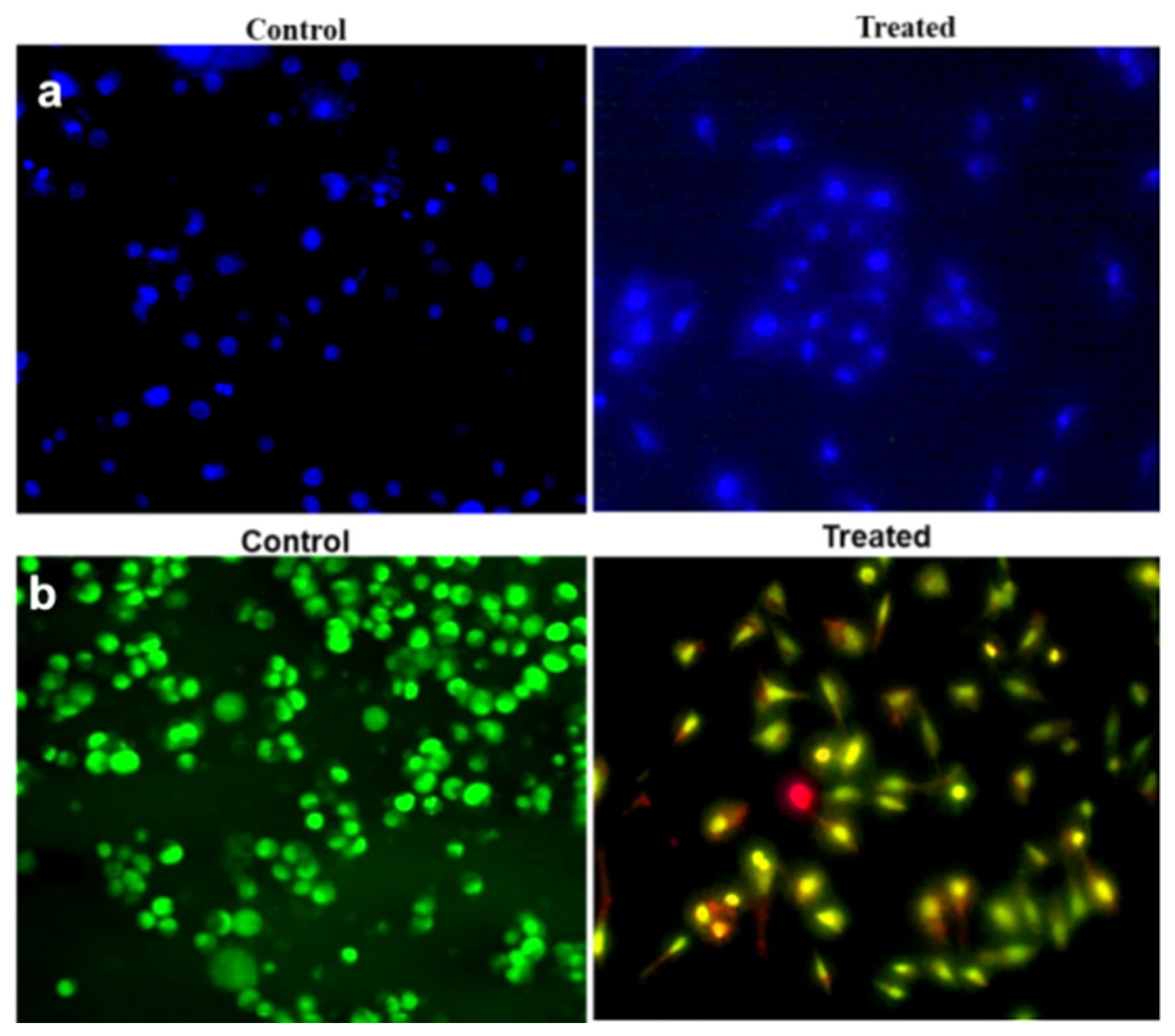
DAPI staining (Figure 8a) showed a significant increase in nuclear condensation and fragmentation in AGS cells treated with CP-AgNPs at IC50 concentrations (46 µg/mL), suggesting chromatin damage associated with apoptosis. Compared with untreated controls, which displayed uniformly distributed, intact nuclei, treated cells exhibited brightly fluorescing, compacted, and fragmented nuclei, consistent with early and intermediate apoptotic events. These observations agree with recent reports that AgNPs synthesized from medicinal plants induce nuclear damage and chromatin condensation in gastric cancer cells via ROS-mediated apoptotic signaling pathways [27,28].
Similarly, acridine orange/ethidium bromide (AO/EtBr) double staining (Figure 8b) showed a clear shift from predominantly green (viable) AGS cells in the control group to a mixture of yellow, orange, and red fluorescence in cells treated with CP-AgNPs at IC50 concentrations (50.55 µg/mL). Yellow or orange fluorescence indicates early apoptosis, while red fluorescence indicates late apoptosis or necrosis due to extensive membrane damage. This progressive loss of membrane integrity is consistent with previous findings that bio-based AgNPs can penetrate tumor cell membranes, increase oxidative stress, and induce apoptosis followed by secondary necrosis in a dose-dependent manner [49,50].
Combined fluorescence staining results support the strong anticancer effects induced by CP-AgNPs in AGS cells through apoptosis-dominant pathways. These findings are consistent with the literature on nanotherapeutic agents, which report that phyto-mediated AgNPs can induce mitochondrial dysfunction, upregulate pro-apoptotic markers, and suppress tumor cell proliferation in a concentration-dependent manner [51,52].

3.8. Anti-Inflammatory

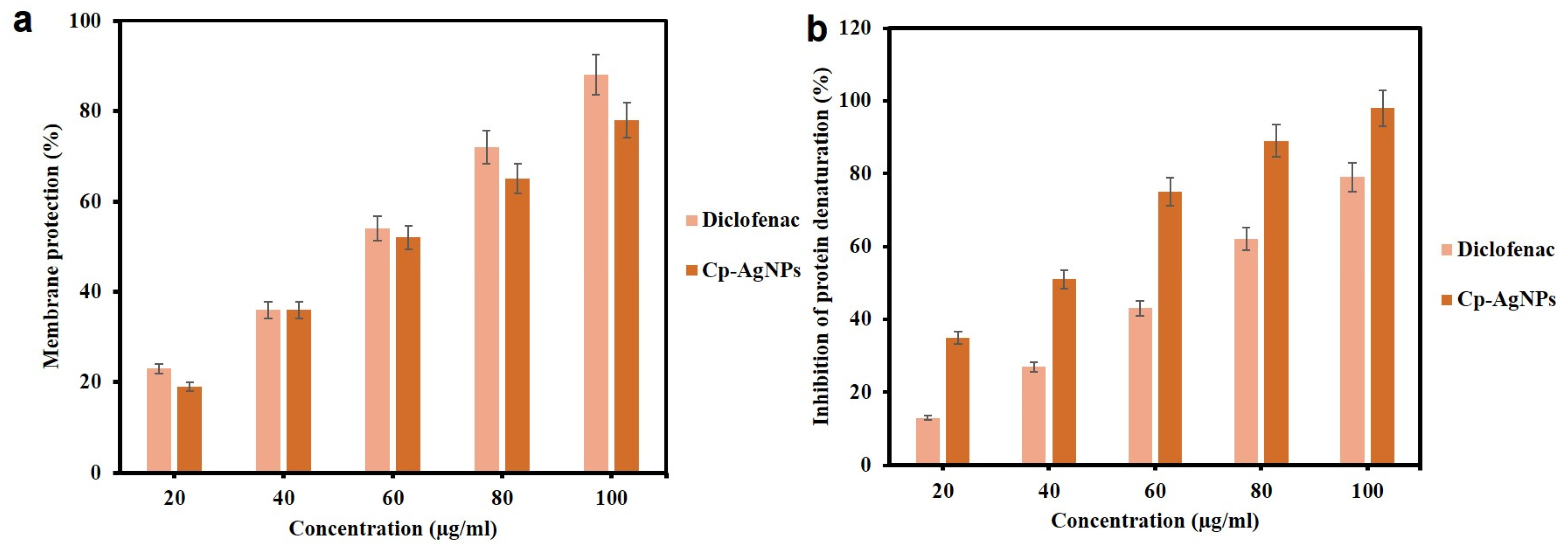
The anti-inflammatory potential of CP-AgNPs was assessed by membrane protection and protein denaturation assays. In the membrane protection test (Figure 9a), CP-AgNPs showed a clear concentration-dependent increase in protective activity. The membrane stabilization rose from 28% at 20 µg/mL to 92% at 100 µg/mL, indicating that CP-AgNPs strongly prevent erythrocyte lysis under heat-induced stress. This activity was comparable to that of the standard drug diclofenac, suggesting effective anti-inflammatory potential. The observed membrane stabilization is likely due to interactions between phytochemicals bound to the nanoparticles (including flavonoids, terpenoids, and phenols) and erythrocyte membranes, thereby enhancing structural integrity and resistance to denaturation. These phytochemicals are known for potent free-radical scavenging and anti-inflammatory properties that may synergize with the AgNP metallic core to enhance bioactivity [7].
Similarly, in the protein denaturation assay (Figure 9b), CP-AgNPs showed strong inhibitory activity, increasing from 35% inhibition at 20 µg/mL to 98% inhibition at 100 µg/mL, and CP-AgNPs demonstrated a concentration-dependent ability to stabilize membranes and prevent protein denaturation, displaying effects similar to the positive control diclofenac. Protein denaturation is a key feature of inflammatory pathology, and compounds that inhibit this process are considered effective anti-inflammatory agents. The potent inhibition by CP-AgNPs suggests that the plant-encapsulated nanoparticles may prevent protein denaturation, likely via hydrogen bonding, which stabilizes protein structure and prevents heat-induced aggregation. Similar findings have been reported for green-synthesized metal nanoparticles, which exhibit anti-inflammatory and membrane-stabilizing effects by inhibiting inflammatory mediators and preventing protein degradation [9,30]. While diclofenac served as a positive control, a direct statistical comparison between CP-AgNPs and diclofenac under concentration-matched conditions was not conducted in this study, indicating the need for further research. Overall, the concentration-dependent membrane protection and inhibition of protein denaturation indicate that CP-AgNPs possess strong anti-inflammatory potential, attributable to synergistic effects between silver nanoparticles and the rich phytochemical profile of C. procera, supporting their promise as candidates for natural anti-inflammatory therapeutics.

3.9. Antioxidant Activity of CP-AgNPs

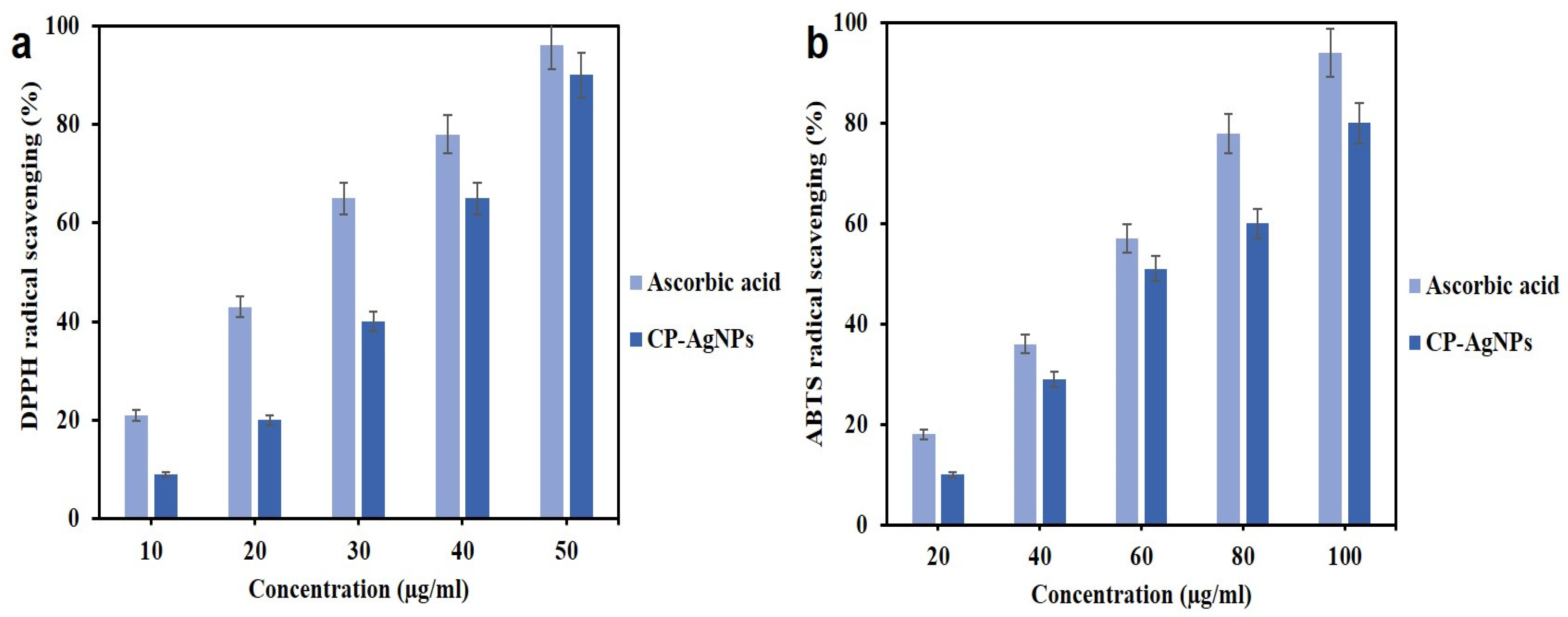
The antioxidant activity of CP-AgNPs derived from Calotropis procera leaf extract was evaluated by DPPH and ABTS radical scavenging assays, which revealed a potent, concentration-dependent free-radical neutralizing capacity. In the DPPH assay, CP-AgNPs showed progressively increasing scavenging activity with increasing concentration and achieved a particularly high radical-quenching effect at the highest tested dose. The calculated IC50 of 40 µg/mL indicates strong electron donor capability and confirms potent antioxidant activity (Figure 10a). This enhanced activity exceeds that of the pure plant extract and can be attributed to phytoconstituents (e.g., polyphenols, flavonoids, and reducing sugars) that act as biogenic stabilizers and antioxidants during nanoparticle formation, thereby improving electron transfer efficiency and facilitating rapid DPPH neutralization. These findings are consistent with recent studies reporting higher DPPH scavenging activity for biogenic AgNPs due to their nanoscale size, high surface-to-volume ratio, and synergistic interactions between metallic silver and plant functional groups [53,54,55].
Similarly, the ABTS·+ radical scavenging assay showed a clear dose-dependent increase in antioxidant potency, with scavenging percentages ranging from 15% at 20 µg/mL to 78% at 100 µg/mL (Figure 10b). Although ascorbic acid exhibited slightly higher activity, CP-AgNPs retained a strong ABTS·+ neutralizing potential, further demonstrating robust antioxidant properties. The observed effect is attributed to a plant-derived capping layer on the nanoparticles that enhances electron donation and stabilizes reactive species. Recent evidence supports that AgNPs synthesized using plant extracts consistently exhibit high ABTS activity due to synergistic redox interactions between biomolecules and the metal core [7]. This is relevant for biomedical, nutraceutical, and pharmaceutical applications where the mitigation of oxidative stress is important.
Earlier research has indicated encouraging biological effects of silver nanoparticles synthesized through plant methods; however, the majority concentrated on specific applications. Consequently, a comparative evaluation with prior studies was conducted to emphasize the multifunctional capabilities and uniqueness of the current CP-AgNPs investigation (Table 1).
Although CP-AgNPs demonstrated multifunctional antibacterial, antioxidant, anti-inflammatory, and anticancer activities, the present study primarily provides biological and phenotypic evidence rather than direct mechanistic validation. Specific mechanistic investigations, including ROS quantification, apoptosis signaling pathways, inflammatory mediator profiling, and molecular pathway analyses, were beyond the scope of the current study and warrant further investigation to establish a unified mechanistic framework underlying CP-AgNP bioactivity.

4. Conclusions

In this study, silver nanoparticles (CP-AgNPs) synthesized from the leaf extract of Calotropis procera were produced using a green phytochemical reduction process and thoroughly characterized for the nanoscale morphology, stability, and adsorption of bioactive phytochemicals. CP-AgNPs exhibited potent antibacterial activity against Helicobacter pylori, as demonstrated by low MIC and MBC values, rapid killing in the time-kill assay, and strong inhibition of biofilm formation. SEM images revealed extensive structural damage to H. pylori cells and the disruption of the biofilm matrix, confirming that CP-AgNPs impair membrane integrity and inhibit bacterial adhesion and colonization.
Beyond antimicrobial effects, the nanoparticles showed strong antioxidant activity in DPPH and ABTS assays, indicating robust electron-donating and radical-neutralizing properties. CP-AgNPs also demonstrated significant anti-inflammatory potential by stabilizing erythrocyte membranes and inhibiting protein denaturation, suggesting usefulness in mitigating inflammation-related damage. Importantly, the nanoparticles exhibited pronounced anticancer activity against AGS tumor cells, with dose-dependent cytotoxicity, apoptotic nuclear condensation (DAPI), altered membrane permeability (AO/EtBr), and SEM-confirmed apoptotic morphology.
Overall, the results identify CP-AgNPs as a multifunctional nanomaterial with strong antibacterial, antioxidant, anti-inflammatory, and anticancer properties. Their broad activity profile highlights the potential of CP-AgNPs as promising natural therapeutic candidates for treating H. pylori infection, oxidative stress-related disorders, inflammatory conditions, and gastric cancer. Further in vivo and molecular studies are recommended to elucidate precise mechanisms and to support the translation into biomedical and pharmaceutical applications.

Author Contributions

Conceptualization, M.K. (Mounishwaran Kamalesan), K.N. and D.J.H.S.; methodology, M.K. (Mounishwaran Kamalesan), K.N. and M.R.; formal analysis, R.N.; investigation, M.K. (Mounishwaran Kamalesan); resources, K.N. and D.J.H.S.; data curation, K.N. and M.K. (Muthukalingan Krishnan); writing—original draft preparation, M.K. (Mounishwaran Kamalesan), K.N. and M.R.; writing—review and editing, R.N. and D.J.H.S.; supervision, K.N. and D.J.H.S. All authors have read and agreed to the published version of the manuscript.

Funding

This research received no external funding.

Institutional Review Board Statement

Not applicable.

Informed Consent Statement

Not applicable.

Data Availability Statement

The original contributions presented in this study are included in the article. Further inquiries can be directed to the corresponding authors.

Acknowledgments

Mounishwaran Kamalesan was supported by the University Research Fellowship (PU/AD-3/URF/020811/2021, dated: 17 December 2021), Periyar University, Salem, Tamilnadu, India. Mounishwaran Kamalesan and Mohanraj Raja were also supported by the 108-111 TEEP@AsiaPlus, provided by the Ministry of Education, Taiwan. Kayalvizhi Nagarajan (CO-PI) acknowledges ANRF-PAIR for providing consumables (ANRF/F/5329/2025-2026, dated: 9 July 2025).

Conflicts of Interest

The authors declare no conflicts of interest.

References

  1. Meher, A.; Tandi, A.; Moharana, S.; Chakroborty, S.; Mohapatra, S.S.; Mondal, A.; Chandra, P. Silver nanoparticle for biomedical applications: A review. Hybrid Adv. 2024, 6, 100184. [Google Scholar] [CrossRef] [Scilit]
  2. Rădulescu, D.-M.; Surdu, V.-A.; Ficai, A.; Ficai, D.; Grumezescu, A.-M.; Andronescu, E. Green synthesis of metal and metal oxide nanoparticles: A review of the principles and biomedical applications. Int. J. Mol. Sci. 2023, 24, 15397. [Google Scholar] [CrossRef] [Scilit]
  3. Kasahun, G.G.; Demoz, G.T.; Desta, D.M. Primary resistance pattern of Helicobacter pylori to antibiotics in adult population: A systematic review. Infect. Drug Resist. 2020, 13, 1567–1573. [Google Scholar] [CrossRef] [Scilit] [PubMed]
  4. Wu, X.; Wu, D.; Cui, G.; Lee, K.H.; Yang, T.; Zhang, Z.; Chen, Z. Association between biofilm formation and structure and antibiotic resistance in H. pylori. Infect. Drug Resist. 2024, 17, 2501–2512. [Google Scholar] [CrossRef] [Scilit] [PubMed]
  5. Flores-Treviño, S.; Mendoza-Olazarán, S.; Bocanegra-Ibarias, P.; Maldonado-Garza, H.J.; Garza-González, E. Helicobacter pylori drug resistance: Therapy changes and challenges. Expert Rev. Gastroenterol. Hepatol. 2018, 12, 819–827. [Google Scholar] [CrossRef] [Scilit]
  6. Shanmuganathan, R.; Karuppusamy, I.; Saravanan, M.; Muthukumar, H.; Ponnuchamy, K.; Ramkumar, V.S.; Pugazhendhi, A. Synthesis of silver nanoparticles and their biomedical applications—A comprehensive review. Curr. Pharm. Des. 2019, 25, 2650–2660. [Google Scholar] [CrossRef] [Scilit]
  7. Asif, M.; Yasmin, R.; Asif, R.; Ambreen, A.; Mustafa, M.; Umbreen, S. Green synthesis of silver nanoparticles (AgNPs), structural characterization, and their antibacterial potential. Dose-Response 2022, 20, 15593258221088709. [Google Scholar] [CrossRef] [Scilit]
  8. Wahnou, H.; Limami, Y.; Oudghiri, M. Flavonoids and flavonoid-based nanoparticles for osteoarthritis and rheumatoid arthritis management. BioChem 2024, 4, 38–61. [Google Scholar] [CrossRef] [Scilit]
  9. Hassan, M.H.; Ismail, M.A.; Moharram, A.M.; Shoreit, A.A. Phytochemical and antimicrobial of latex serum of Calotropis procera and its silver nanoparticles against some reference pathogenic strains. J. Ecol. Health Environ. 2017, 5, 65–75. [Google Scholar] [CrossRef] [Scilit]
  10. Mahto, M.; Priya, S.; Thakur, S.; Paramanik, P.; Verma, R.; Mahato, P.; Bera, A. A comprehensive review of green synthesis, characterization and biomedical applications of silver nanoparticle synthesized using plant extracts. World J. Adv. Res. Rev. 2024, 22, 1950–1977. [Google Scholar] [CrossRef] [Scilit]
  11. Mohan, A.; Rajendran, S.; Palani, N.; Syed Altaf, R.R.; Mendonce, K.C.; Nithya, T.G.; Venkatesan, P.; Nagendran, S.; Surya, P.; Srinivasan, M.; et al. Sustainable synthesis of iron oxide nanoparticles using phytochemicals: Mechanisms, functionalization strategies, applications and future perspectives. Mater. Sci. Eng. 2025, 9, 36–54. [Google Scholar]
  12. Dhar, S.A.; Chowdhury, R.A.; Das, S.; Nahian, M.K.; Islam, D.; Gafur, M.A. Plant-mediated green synthesis and characterization of silver nanoparticles using Phyllanthus emblica fruit extract. Mater. Today Proc. 2021, 42, 1867–1871. [Google Scholar] [CrossRef] [Scilit]
  13. Yadav, M.; Gaur, N.; Wahi, N.; Singh, S.; Kumar, K.; Amoozegar, A.; Sharma, E. Phytochemical-assisted fabrication of biogenic silver nanoparticles from Vitex negundo: Structural features, antibacterial activity, and cytotoxicity evaluation. Colloids Interfaces 2025, 9, 55. [Google Scholar] [CrossRef] [Scilit]
  14. Hernández-Díaz, J.A.; Garza-García, J.J.; Zamudio-Ojeda, A.; León-Morales, J.M.; López-Velázquez, J.C.; García-Morales, S. Plant-mediated synthesis of nanoparticles and their antimicrobial activity against phytopathogens. J. Sci. Food Agric. 2021, 101, 1270–1287. [Google Scholar] [CrossRef] [Scilit] [PubMed]
  15. Pop, R.; Tăbăran, A.F.; Ungur, A.P.; Negoescu, A.; Cătoi, C. Helicobacter pylori-induced gastric infections: From pathogenesis to novel therapeutic approaches using silver nanoparticles. Pharmaceutics 2022, 14, 1463. [Google Scholar] [CrossRef] [Scilit]
  16. Sampath, S.; Madhavan, Y.; Muralidharan, M.; Sunderam, V.; Lawrance, A.V.; Muthupandian, S. A review on algal mediated synthesis of metal and metal oxide nanoparticles and their emerging biomedical potential. J. Biotechnol. 2022, 360, 92–109. [Google Scholar] [CrossRef] [Scilit] [PubMed]
  17. Humphries, R.; Bobenchik, A.M.; Hindler, J.A.; Schuetz, A.N. Overview of changes to the Clinical and Laboratory Standards Institute performance standards for antimicrobial susceptibility testing, M100. J. Clin. Microbiol. 2021, 59, e00213-21. [Google Scholar] [CrossRef] [Scilit] [PubMed]
  18. Li, X.; Robinson, S.M.; Gupta, A.; Saha, K.; Jiang, Z.; Moyano, D.F.; Sahar, A.; Riley, M.A.; Rotello, V.M. Functional gold nanoparticles as potent antimicrobial agents against multi-drug-resistant bacteria. ACS Nano 2014, 8, 10682–10686. [Google Scholar] [CrossRef] [Scilit]
  19. Petersen, P.J.; Labthavikul, P.; Jones, C.H.; Bradford, P.A. In vitro antibacterial activities of tigecycline in combination with other antimicrobial agents determined by chequerboard and time-kill kinetic analysis. J. Antimicrob. Chemother. 2006, 57, 573–576. [Google Scholar] [CrossRef] [Scilit]
  20. Adeniji, O.O.; Ojemaye, M.O.; Okoh, A.I. Antibacterial activity of metallic nanoparticles against multidrug-resistant pathogens isolated from environmental samples: Nanoparticles/antibiotic combination therapy and cytotoxicity study. ACS Appl. Bio Mater. 2022, 5, 4814–4826. [Google Scholar] [CrossRef] [Scilit]
  21. Hashmat, D.; Faisal, U.; Choudry, Z.; Safdar, J.; Nasir, A.; Javed, A. Green synthesis of nanoparticles: Mechanisms, characterization, and emerging applications. Res. Med. Sci. Rev. 2025, 3, 898–909. [Google Scholar] [CrossRef]
  22. Hathroubi, S.; Servetas, S.L.; Windham, I.; Merrell, D.S.; Ottemann, K.M. Helicobacter pylori biofilm formation and its potential role in pathogenesis. Microbiol. Mol. Biol. Rev. 2018, 82, e00001-18. [Google Scholar] [CrossRef] [Scilit]
  23. Hernández-Venegas, P.A.; Martínez-Martínez, R.E.; Zaragoza-Contreras, E.A.; Domínguez-Pérez, R.A.; Reyes-López, S.Y.; Donohue-Cornejo, A.; Espinosa-Cristóbal, L.F. Bactericidal activity of silver nanoparticles on oral biofilms related to patients with and without periodontal disease. J. Funct. Biomater. 2023, 14, 311. [Google Scholar] [CrossRef] [Scilit] [PubMed]
  24. Fahmy, N.F.; Abdel-Kareem, M.M.; Ahmed, H.A.; Helmy, M.Z.; Mahmoud, E.A.R. Evaluation of the antibacterial and antibiofilm effect of mycosynthesized silver and selenium nanoparticles and their synergistic effect with antibiotics on nosocomial bacteria. Microb. Cell Fact. 2025, 24, 6. [Google Scholar] [CrossRef] [Scilit]
  25. Elshaer, S.; Shaaban, M.I. Antibiofilm activity of biosynthesized silver and copper nanoparticles using Streptomyces S29. AMB Express 2023, 13, 139. [Google Scholar] [CrossRef] [Scilit]
  26. Kamer, A.M.A.; El Maghraby, G.M.; Shafik, M.M.; Al-Madboly, L.A. Silver nanoparticle with potential antimicrobial and antibiofilm efficiency against multiple drug resistant, extensive drug resistant Pseudomonas aeruginosa clinical isolates. BMC Microbiol. 2024, 24, 277. [Google Scholar] [CrossRef] [Scilit] [PubMed]
  27. Kumarasamy, R.V.; Natarajan, P.M.; Mathivanan, I.; Balasubramaniam, M.; SN, S.; Prabhu, D.; Mironescu, I.D. Sustainable biogenic synthesis of silver nanoparticles for oral cancer: A comprehensive review. Front. Nanotechnol. 2025, 7, 1648900. [Google Scholar] [CrossRef] [Scilit]
  28. Xu, Z.; Feng, Q.; Wang, M.; Zhao, H.; Lin, Y.; Zhou, S. Green biosynthesized silver nanoparticles with aqueous extracts of Ginkgo biloba induce apoptosis via mitochondrial pathway in cervical cancer cells. Front. Oncol. 2020, 10, 575415. [Google Scholar] [CrossRef] [Scilit]
  29. Khashan, A.A.; Dawood, Y.; Khalaf, Y.H. Green chemistry and anti-inflammatory activity of silver nanoparticles using an aqueous curcumin extract. Results Chem. 2023, 5, 100913. [Google Scholar] [CrossRef] [Scilit]
  30. Iheme, C.I.; Mogbonjubola, M.A.; Iwuchukwu, B.K.; Alisi, P.N.; Nkwoada, A.U.; Okpara, C.J.; Ujowundu, C.O. Eco-benign phytochemically-functionalized Ag-core Fe-shell bimetallic nanoparticles for targeted antioxidant and anti-inflammatory drug delivery. BMC Chem. 2025, 19, 277. [Google Scholar] [CrossRef] [Scilit]
  31. Afreen, A.; Ahmed, R.; Mehboob, S.; Tariq, M.; Alghamdi, H.A.; Zahid, A.A.; Hasan, A. Phytochemical-assisted biosynthesis of silver nanoparticles from Ajuga bracteosa for biomedical applications. Mater. Res. Express 2020, 7, 075404. [Google Scholar] [CrossRef] [Scilit]
  32. Singh, M.P.; Shabir, S.; Deopa, A.S.; Raina, S.R.; Bantun, F.; Jalal, N.A.; Vamanu, E. Synthesis of green engineered silver nanoparticles through Urtica dioica: Inhibition of microbes and alleviation of cellular and organismal toxicity in Drosophila melanogaster. Antibiotics 2022, 11, 1690. [Google Scholar] [CrossRef] [Scilit]
  33. Bedlovičová, Z.; Strapáč, I.; Baláž, M.; Salayová, A. A brief overview on antioxidant activity determination of silver nanoparticles. Molecules 2020, 25, 3191. [Google Scholar] [CrossRef] [Scilit]
  34. Khan, M.F.; Khan, M.A. Plant-derived metal nanoparticles (PDMNPs): Synthesis, characterization, and oxidative stress-mediated therapeutic actions. Future Pharmacol. 2023, 3, 252–295. [Google Scholar] [CrossRef] [Scilit]
  35. Qubtia, M.; Ghumman, S.A.; Noreen, S.; Hameed, H.; Noureen, S.; Kausar, R.; Bin Jardan, Y.A. Evaluation of plant-based silver nanoparticles for antioxidant activity and promising wound-healing applications. ACS Omega 2024, 9, 12146–12157. [Google Scholar] [CrossRef] [Scilit] [PubMed]
  36. Jain, A.; Jangid, T.; Jangir, R.N.; Bhardwaj, G.S. A comprehensive review on the antioxidant properties of green synthesized nanoparticles: In vitro and in vivo insights. Free Radic. Antioxid. 2024, 14, 2. [Google Scholar] [CrossRef] [Scilit]
  37. Ajitha, B.; Reddy, Y.A.K.; Reddy, P.S. Green synthesis and characterization of silver nanoparticles using Lantana camara leaf extract. Mater. Sci. Eng. C 2015, 49, 373–381. [Google Scholar] [CrossRef] [Scilit]
  38. Murthy, H.A.; Desalegn Zeleke, T.; Ravikumar, C.R.; Anil Kumar, M.R.; Nagaswarupa, H.P. Electrochemical properties of biogenic silver nanoparticles synthesized using Hagenia abyssinica medicinal plant leaf extract. Mater. Res. Express 2020, 7, 055016. [Google Scholar] [CrossRef] [Scilit]
  39. Bhavi, S.M.; Thokchom, B.; Abbigeri, M.B.; Bhat, S.S.; Singh, S.R.; Joshi, P.; Yarajarla, R.B. Green synthesis, characterization, antidiabetic, antioxidant and antibacterial applications of silver nanoparticles from Syzygium caryophyllatum (L.) Alston leaves. Process Biochem. 2024, 145, 89–103. [Google Scholar] [CrossRef] [Scilit]
  40. Sampath, G.; Govarthanan, M.; Rameshkumar, N.; Vo, D.V.N.; Krishnan, M.; Sivasankar, P.; Kayalvizhi, N. Eco-friendly biosynthesis metallic silver nanoparticles using Aegle marmelos (Indian bael) and its clinical and environmental applications. Appl. Nanosci. 2023, 13, 663–674. [Google Scholar] [CrossRef] [Scilit]
  41. Nagime, P.V.; Singh, S.; Shaikh, N.M.; Gomare, K.S.; Chitme, H.; Abdel-Wahab, B.A.; Bakir, M.B. Biogenic fabrication of silver nanoparticles using Calotropis procera flower extract with enhanced biomimetic attributes. Materials 2023, 16, 4058. [Google Scholar] [CrossRef] [Scilit] [PubMed]
  42. Hochvaldova, L.; Posselt, G.; Wessler, S.; Kvítek, L.; Panáček, A. Implications of silver nanoparticles for H. pylori infection: Modulation of CagA function and signaling. Front. Cell. Infect. Microbiol. 2024, 14, 1419568. [Google Scholar] [CrossRef] [Scilit]
  43. Amin, M.; Hameed, S.; Ali, A.; Anwar, F.; Shahid, S.A.; Shakir, I.; Khan, S.A. Green synthesis of silver nanoparticles: Structural features and in vivo and in vitro therapeutic effects against Helicobacter pylori induced gastritis. Bioinorg. Chem. Appl. 2014, 2014, 135824. [Google Scholar] [CrossRef] [Scilit]
  44. Gurunathan, S.; Jeong, J.K.; Han, J.W.; Zhang, X.F.; Park, J.H.; Kim, J.H. Multidimensional effects of biologically synthesized silver nanoparticles in Helicobacter pylori, Helicobacter felis, and human lung (L132) and lung carcinoma A549 cells. Nanoscale Res. Lett. 2015, 10, 35. [Google Scholar] [CrossRef] [Scilit]
  45. Tănase, C.; Berta, L.; Coman, N.A.; Roșca, I.; Man, A.; Toma, F.; Mare, A. Investigation of in vitro antioxidant and antibacterial potential of silver nanoparticles obtained by biosynthesis using beech bark extract. Antioxidants 2019, 8, 459. [Google Scholar] [CrossRef] [Scilit]
  46. Zolfaghari, M.; Yadegar, A.; Rezaei, A.; Kazemi, M.; Fazeli, H.; Tabesh, E.; Karbasizade, V. Evaluation of antimicrobial and antibiofilm activities of peptide Impatiens balsamina-M1 and zinc oxide nanoparticles against Helicobacter pylori. Sci. Rep. 2026, 16, 1723. [Google Scholar] [CrossRef] [Scilit]
  47. Almutairi, B.O.; Ali, D.; Almarzoug, M.H.; Asiri, A.M.; Alkahtani, S.; Alarifi, S. Impact of green-synthesized copper silver nanoparticles against human colon cancer cells utilizing Sargassum muticum extract in terms of cytotoxicity and apoptosis. Nat. Prod. Commun. 2024, 19, 1934578X241299692. [Google Scholar] [CrossRef] [Scilit]
  48. Balkrishna, A.; Kumar, A.; Arya, V.; Rohela, A.; Verma, R.; Nepovimova, E.; Kuca, K. Phytoantioxidant functionalized nanoparticles: A green approach to combat nanoparticle-induced oxidative stress. Oxid. Med. Cell. Longev. 2021, 2021, 3155962. [Google Scholar] [CrossRef] [Scilit]
  49. Bethu, M.S.; Netala, V.R.; Domdi, L.; Tartte, V.; Janapala, V.R. Potential anticancer activity of biogenic silver nanoparticles using leaf extract of Rhynchosia suaveolens: An insight into the mechanism. Artif. Cells Nanomed. Biotechnol. 2018, 46, 104–114. [Google Scholar] [CrossRef] [Scilit] [PubMed]
  50. Dey, A.; Manna, S.; Chattopadhyay, S.; Mondal, D.; Chattopadhyay, D.; Raj, A.; Roy, S. Azadirachta indica leaves mediated green synthesized copper oxide nanoparticles induce apoptosis through activation of TNF-α and caspases signaling pathway against cancer cells. J. Saudi Chem. Soc. 2019, 23, 222–238. [Google Scholar] [CrossRef] [Scilit]
  51. Nguyen, P.H.; Nguyen, T.K.O.; Khieu, T.T.; Le, T.N.T.; Trinh, T.H.; Le Mong, T.; Le, T.T.H. Biogenic silver nanoparticles from Croton tonkinensis extract: Inhibition of gastric cancer cells via cell cycle arrest and ROS-mediated apoptosis. J. Agric. Food Res. 2025, 23, 102171. [Google Scholar] [CrossRef] [Scilit]
  52. Antony, S.; Chanthini, K.M.P. Biogenically synthesized Moringa oleifera leaf extract-derived silver nanoparticles exhibit potent antimicrobial and anticancer effects via oxidative stress and apoptotic pathways in AGS gastric cancer cells. Biocatal. Agric. Biotechnol. 2025, 67, 103674. [Google Scholar] [CrossRef] [Scilit]
  53. Varadharajan, S.; Pandian, P. Antioxidant, antimicrobial, and potential biomedical applications of green synthesized silver nanoparticles using Aloe barbadensis root. J. Environ. Nanotechnol. 2025, 14, 97–104. [Google Scholar] [CrossRef] [Scilit]
  54. Singh, R.; Singh, R.; Parihar, P.; Mani, J.V. Green synthesis of silver nanoparticles using Solanum sisymbriifolium leaf extract: Characterization and evaluation of antioxidant, antibacterial and photocatalytic degradation activities. Process Biochem. 2024, 143, 337–352. [Google Scholar] [CrossRef] [Scilit]
  55. Jabeen, S.; Qureshi, R.; Ikram, M.; Omar, A.A.; Hussain, M.; Elnaggar, N.; Oraby, H.F. Phyto-fabrication and characterization of silver nanoparticles using Withania somnifera: Investigating antioxidant potential. Green Process. Synth. 2023, 12, 20230167. [Google Scholar] [CrossRef] [Scilit]
Figure 1. Systematic representation of the green synthesis of silver nanoparticles using Calotropis procera leaf extract.
Scipharm 94 00044 g001
Figure 2. Physicochemical characterization of (CP-AgNPs) (a) UV–visible spectra, (b) X-ray diffraction, (c) Fourier-transform infrared spectroscopy, (d) energy-dispersive X-ray, (e) transmission electron microscopy, (f) scanning electron microscopy, and (g) zeta potential distribution.
Scipharm 94 00044 g002
Figure 3. (a) Agar well diffusion assay showing the antibacterial activity of CP-AgNPs against H. pylori at 25, 50, and 75 µg/mL, compared with metronidazole and control (DMSO). (b) The corresponding bar graph illustrates a clear concentration-dependent increase in the zone of inhibition.
Scipharm 94 00044 g003
Figure 4. Time-kill kinetics of CP-AgNPs against H. pylori showing a concentration-dependent reduction in viable bacterial counts over 36 h. Higher nanoparticle concentrations (75 and 100 µg/mL) exhibited rapid and sustained bactericidal activity compared to the positive control (metronidazole) and untreated control, and data are presented as mean ± SD of triplicate experiments (n = 3).
Scipharm 94 00044 g004
Figure 5. Crystal violet-stained microscopy images showing the inhibitory effect of CP-AgNPs on H. pylori biofilm formation: (A): (a) untreated control exhibiting dense biofilm, (b) 25 µg/mL showing reduced clustering, (c) 50 µg/mL showing disrupted biofilm structure, and (d) 75 µg/mL showing near-complete inhibition at 10× magnification. (B): Quantitative assessment of biofilm biomass (% biofilm) showing concentration-dependent inhibition of H. pylori biofilm following CP-AgNP treatment.
Scipharm 94 00044 g005
Figure 6. SEM micrographs showing the antibiofilm effect of CP-AgNPs on H. pylori: (a) untreated control displaying dense, intact rod-shaped cells, (b) 25 µg/mL showing partial disruption, (c) 50 µg/mL showing severe structural damage and reduced clustering, and (d) 75 µg/mL revealing extensive deformation and collapse of bacterial cells.
Scipharm 94 00044 g006
Figure 7. Dose-dependent cytotoxicity of CP-AgNPs against AGS gastric cancer cells showing increasing percentage of cell inhibition from 6.5 to 100 µg/mL (IC50 = 40.26 µg/mL). Microscopy images correspondingly display progressive morphological damage, cell shrinkage, and loss of adherence with increasing nanoparticle concentration.
Scipharm 94 00044 g007
Figure 8. Fluorescence microscopy images of AGS cells treated with CP-AgNPs showing (a) DAPI staining with bright, condensed nuclei indicating apoptosis and (b) AO/EtBr dual staining differentiating viable (green), early apoptotic (yellow), and late apoptotic/necrotic cells (red), confirming nanoparticle-induced cell death.
Scipharm 94 00044 g008
Figure 9. Anti-inflammatory activity of CP-AgNPs showing (a) concentration-dependent membrane stabilization (%) and (b) inhibition of protein denaturation (%) compared with diclofenac. CP-AgNPs exhibit strong anti-inflammatory potential, with higher protective and inhibitory effects at increased concentrations.
Scipharm 94 00044 g009
Figure 10. Antioxidant activity of CP-AgNPs showing (a) DPPH and (b) ABTS radical scavenging percentages at different concentrations compared with ascorbic acid. Both assays demonstrate dose-dependent scavenging efficiency, confirming strong free-radical neutralizing potential of the nanoparticles.
Scipharm 94 00044 g010
Table 1. Comparative analysis of the present study with previously reported plant-mediated silver nanoparticles targeting Helicobacter pylori, inflammation, oxidative stress, and gastric cancer.
Plant SourceNanoparticleModel UsedMajor FindingsStudy
Calotropis proceraAgNPsPathogenic bacteriaAntimicrobial activity[10]
Biogenic AgNPsAgNPsH. pylori + cancer cellsAntibacterial and cytotoxic effects[11]
Moringa oleiferaAgNPsAGS cellsAnticancer activity[13]
Calotropis proceraCP-AgNPsH. pylori + AGS cellsAntibacterial, antibiofilm, antioxidant, anti-inflammatory, anticancerThis study
Disclaimer/Publisher’s Note: The statements, opinions and data contained in all publications are solely those of the individual author(s) and contributor(s) and not of MDPI and/or the editor(s). MDPI and/or the editor(s) disclaim responsibility for any injury to people or property resulting from any ideas, methods, instructions or products referred to in the content.