Article

Synthesis, Characterization, Antioxidant and Antimicrobial Potentials of Novel Organometallic Compounds Derived from Quercetin


by
Orlando Maia Barboza
1,
Luan Henrique Santos Barreto
1,
Felipe dos Santos Mendes
1,
Ivana Ferreira Simões
1,
Luís Filipe Gomes Santos
2,
Carlos Fernando da Silva Ferreira
1,
Luís Guilherme dos Santos de Sant’Anna
1,
Tainá Santos Lima
1,
Kaique Souza Santos de Jesus
1,
Saul Vislei Simões da Silva
1,3,
Victor Pena Ribeiro
4,
Silvia Lima Costa
3,
Gustavo Souza dos Santos
1,
Lourdes Cardoso de Souza Neta
2 and
Aníbal de Freitas Santos Júnior
1,2,*
1
Life Sciences Department (DCV), University of Bahia State (UNEB), Salvador 40150-000, Bahia, Brazil
2
Exact and Earth Sciences (DCET), University of Bahia State (UNEB), Salvador 40150-000, Bahia, Brazil
3
Laboratory of Neurochemistry and Cell Biology, Department of Biochemistry and Biophysics, Federal University of Bahia (UFBA), Salvador 40231-300, Bahia, Brazil
4
School of Pharmaceutical Sciences of Ribeirão Preto, University of São Paulo (FCFRP-USP), Ribeirão Preto 14040-900, São Paulo, Brazil
*
Author to whom correspondence should be addressed.
Sci. Pharm. 2026, 94(2), 26; https://doi.org/10.3390/scipharm94020026
Submission received: 10 December 2025 / Revised: 29 January 2026 / Accepted: 23 March 2026 / Published: 27 March 2026

Abstract

Quercetin, one of the most abundant flavonoids in nature, has attracted the attention of many researchers due to its chemical and biological properties. A series of metal–quercetin complexes (Cu2+, Co2+, Zn2+, Sn2+, Al3+, Cd2+ and Mg2+) were synthesized and systematically characterized by Fourier transform infrared spectroscopy (FTIR), UV-visible spectroscopy (UV–Vis) and nuclear magnetic resonance (NMR). These analyses confirmed that the complexes predominantly form through coordination with the 4-carbonyl group and adjacent phenolic hydroxyls. This induces measurable shifts in the ν(C=O), ν(O–H), and π→π* transition bands relative to free quercetin. The antioxidant capacity of the complexes was evaluated using 2,2-Diphenyl-1-Picrylhydrazyl (DPPH) radical scavenging method, 2,2′-Azinobis(3-Ethylbenzothiazoline-6-Sulfonic Acid) (ABTS)+ radical activity, and Ferric Reducing Antioxidant Power (FRAP) assay. Several complexes exhibited higher radical scavenging efficiency than quercetin, with inhibition percentages exceeding 80% in the DPPH and ABTS•+ assays. Others showed reduced activity due to the masking of redox-active hydroxyl groups during metal coordination. FRAP results corroborated these trends, indicating metal-dependent modulation of reducing power. Antimicrobial evaluation revealed that selected complexes were more active than free quercetin, particularly against Staphylococcus aureus and Candida spp., with minimum inhibitory concentrations (MICs) ranging from 75–250 μg mL−1. Overall, metal complexation significantly alters the electronic structure and biological behavior of quercetin, highlighting the potential of metal–flavonoid complexes as multifunctional antioxidants and antimicrobials.

1. Introduction

Natural products, generally secondary metabolites, are produced (with signaling or defense functions), extracted and isolated from plants, animals, microorganisms and marine organisms. These products have been continuously studied and serve as important resources for the discovery of new drugs. Among the various secondary metabolites, polyphenols stand out for their chemical and biological potential, which has been widely described in the literature. Quercetin (3,3′,4′,5,7-pentahydroxyflavone) is one of the most abundant flavonoids in nature and has attracted the attention of many researchers due to its chemical and biological properties. It is a member of the flavonol family, which are compounds with the 3-hydroxyflavone structure [1]. Quercetin is considered the main flavonoid found in foods with diverse pharmacological activities. It has anticancer and induces apoptosis through downregulating P4HA2, antiallergic, antiviral, antiprotozoal, antimicrobial, cardiovascular, anti-inflammatory effects [2,3,4]. The anti-inflammatory effect has been the subject of studies as the efficacy of quercetin has been investigated in patients with severe symptoms of the disease, including Severe Acute Respiratory Syndrome Coronavirus 2 (SARS-CoV-2) [5,6].
The bioavailability of quercetin in the gastrointestinal tract can vary widely, but averages about 20% [7]. Furthermore, the low rate of gastrointestinal absorption, rapid metabolism, and rapid clearance result in reduced bioavailability in vivo, which limits their application in pharmaceuticals and functional foods [8]. The interaction of flavonoids with metal ions can alter the antioxidant properties and also the biological effects of flavonoids. It is suggested that the biological activity of an organic ligand can be enhanced when coordinated or mixed with a suitable metal ion due to its ability to act as a free radical acceptor [9]. Thus, the scope of chemistry related to metal complexation has become increasingly evident.
Organometallic compounds containing metals such as iron (Fe), arsenic (As), and silver (Ag) have been synthesized. However, their applications may be limited due to their ease of oxidation and toxicity. Ong and Gasser [10] highlighted that organometallic compounds are increasingly advancing and gaining importance in the field of medicine, from the antimicrobial organoarsenics that have been prominently used throughout history to the ferrocene-containing compounds that have been investigated for various diseases such as malaria or cancer. In the last three decades, inorganic medicinal chemistry has become relevant in the development of possible metal-based drugs for the treatment of various diseases [11]. Since the inclusion of cisplatin in cancer therapy, the design of new metal complexes with pharmacological effects has become an important approach for the treatment of these and other pathologies, such as parasitic diseases, bacterial diseases, and Alzheimer’s disease [12].
The inclusion of a metal center in the structure of an organic molecule (bioactive ligand) can lead to improved pharmacological properties. The bioactive metal-ligand interaction would increase the bioavailability of the organic molecule by modifying the solubility, lipophilicity, and stability in biological systems, allowing therapeutic targets to be achieved more efficiently. Thus, index therapy can be improved by increasing biological activity, decreasing toxicity, or both. Furthermore, the coordination of a bioactive ligand network can achieve metal-drug synergism through dual or multiple mechanisms of action [13].
The aim of this work is to obtain novel organometallic complexes, resulting from structural modifications of the bioflavonoid quercetin with metals (Cu2+, Co2+, Zn2+, Sn2+, Al3+, Cd2+ and Mg2+), through chemically sustainable synthetic routes, and to evaluate their antioxidant and antimicrobial potentials. This study is relevant due to the innovative potential of the synthesized compounds, which can be used in various pharmaceutical practices. They can contribute to permeability and absorption studies (in vitro, in vivo, and ex vivo), dissolution assays, drug delivery systems, and bioequivalence studies of quercetin and its derivatives by evaluating potential increases in bioavailability. The compounds can also serve as a basis for in silico studies of potential drug candidates with antimicrobial and anti-inflammatory properties.

2. Materials and Methods

2.1. Reagents and Materials

The reagents were purchased from commercial sources (Sigma Aldrich®, St. Louis, MO, USA; Neon®, São Paulo, Brazil; and Êxodo Científica®), Sumaré, São Paulo, Brazil) and were used without further purification: Quercetin ≥ 95% (117-39-5), 2,2-bipyridine (366-18-7), triethylamine (121-44-8), methanol 99.8% (67-56-1) (MeOH), ethanol 99% (64-17-5) (EtOH), dimethylacetamide 99% (127-19-5) (DMA), dimethyl sulfoxide 99% (67-68-5) (DMSO), and diethyl ether 99% (60-29-7). Cobalt(II) chloride 99% (7791-13-1) (Cl2Co·6H2O), Copper(II) nitrate trihydrate 99% [Cu(NO3)2·3H2O] (10031-43-3), Magnesium(II) chloride hexahydrate 99.0% (MgCl2·6H2O) (7791-18-6), Zinc(II) chloride 97% P.A (ZnCl2) (7646-85-7), Cadmium(II) iodide 99% (CdI2) (7790-80-9), Aluminum(III) chloride 99% (7446-70-0) (AlCl3), and Tin(II)chloride dihydrate 99% (SnCl2•2H2O) (10025-69-1).

2.2. Synthesis of the Metal-Quercetin Complexes

In this study, a series of organometallic compounds were synthesized through complexation with the metal salts Cu2+, Co2+, Zn2+, Sn2+, Al3+, Cd2+, and Mg2+, resulting in seven metal complexes. The quercetin-metal complexes were synthesized using methods described in previous literature [14,15,16,17,18]. In a 50 cm−3 two-necked round-bottomed flask provide with electromagnetic stirrer, thermometer. Added quercetin•2H2O (0.17 g; 0.01 moL) in MeOH (20 mL) stirred until solid quercetin was completely dissolved within 15 min the color of the solution was light yellow then quickly added solid 0.02 moL of metal (CuSO4•5H2O, MgSO4•2.H2O, ZnCl2, CoCl2•6H2O, AlCl3, CdI2 and SnCl2•2H2O) in the reaction mixture, the time varied according to the metallic salt used, for CuSO4•5H2O, MgSO4•2.H2O, AlCl3 and CdI2 it was 1.5 h; for SnCl2•2H2O it was 6 h; and for CoCl2 and ZnCl2 it was 3 h. After stirring the reaction mixture was filtered, and the filtrate was slowly evaporated at room temperature for 10 days. There was no further purification, the complexes and the complexes analyzed by FTIR that did not demonstrate modification were discarded.

2.3. Characterization of the Metal-Quercetin Complexes

2.3.1. Fourier Transform Infrared Spectroscopy (FTIR) Analysis

The absorption spectra in the infrared range, from 4000 to 650 cm−1, were obtained with an FTIR spectrophotometer Cary 630 FTIR Spectrometer, brand Agilent Technologies® (Santa Clara, CA, USA), with 4 scans, resolution of 4 cm−1, the crystal used was Diamond ATR (Attenuated Total Reflectance). The software used to produce the graphs was Origin Pro 8.

2.3.2. Nuclear Magnetic Resonance (NMR) Analysis

The NMR experiments were carried out on a Bruker (TX, USA) Avance III 500 spectrometer (11.75 T), operating at a frequency of 500.13 MHz for the hydrogen nucleus; equipped with a three-channel inverse detection probe 1H, 13C and XBB, Broad Band Inverse Detection (BBI) for the analysis of liquid samples in 5 mm diameter tubes. The experiments were performed at 25 °C (298 K), using dimethyl sulfoxide (DMSO) as solvent. Topspin 3.5 software (Bruker BioSpin) was used to operate and process the experiments. Amix Viewer software, version 3.9.15, was used for chemometric analyses.

2.3.3. UV/Visible Spectroscopy Analysis

UV-Vis analysis was performed on a Cary UV-Vis 60 from Agilent Technologies® (Santa Clara, CA, USA) in the range of 190 to 1000 nm. To solve the complexes, 5 mg of the synthesized complexes were dissolved in 100 mL of methanol in quartz cuvettes containing 3 mL of the solution.

2.4. Antioxidant Potentials

2.4.1. 2,2-Diphenyl-1-Picrylhydrazyl (DPPH) Radical Scavenging Method

DPPH assays were conducted as proposed by Brand-Williams; Cuvelier and Berset [19]. It was necessary to prepare a solution containing 2.4 mg of DPPH radical, weighed in 100 mL of methanol. However, due to the sensitivity of the analytical balance, it was necessary to weigh 24 mg (0.024 g) in 100 mL of methanol and dilute it 10 times to obtain a concentration of 60 mM. From the synthesized product, dilutions with concentrations of 1000, 500, 250, 100, 50, and 25 μg mL−1 were prepared, with 3 sample dilutions in each dilution: 0.1 mL of the extract + 3.9 mL of the 60 micromolar stock of DPPH. Then, after homogenization, the reading was carried out on the spectrophotometer, in three cuvettes, in the dark, and after 30 min, at 517 nm, after observing the stabilization of the absorbance, and the values obtained were noted. The procedure was also carried out in triplicate with a negative standard, i.e., the DPPH solution with methanol only, respecting the same quantity, noting the final absorbance and the absorbance after waiting 30 min. The absorbance of the blank (methanol only) was also measured without waiting 30 min.

2.4.2. Antioxidant 2,2′-Azinobis(3-Ethylbenzothiazoline-6-Sulfonic Acid) (ABTS)+ Radical Activity

ABTS assays were conducted as proposed by Re et al. [20]. A mass of 192 mg (0.192 g) of ABTS was dissolved in 50 mL of distilled water in a volumetric flask, followed by homogenization and transfer to a labeled amber glass bottle. The solution was stored under refrigeration for up to one month. Then, 189.2 mg (0.1892 g) of potassium persulfate was dissolved in distilled water and made up to 5 mL in a volumetric flask. The solution was then homogenized and transferred to an amber glass bottle. To perform the test, dilutions of the products obtained at concentrations of 1000, 500, 250, 100, 50, and 25 μg mL−1 were prepared in triplicate. To perform the assay, 30 μL of each extract was transferred to 3.0 mL of ABTS•+ radical. Vortexing of the tubes in the presence of ABTS and products was necessary for better homogenization. After stirring, it was necessary to wait 6 min for the reaction between the radical and the product obtained to occur. The reading was made on a Femto (São Paulo, Brazil), model 700 Plus spectrophotometer, at a wavelength of 734 nm, in the dark, according to the equation:
ABTS•+ Absorbance (%) = (1 − A0/A1) × 100; where A0 is the absorbance of the control and A1 is the absorbance of the sample or standard. All assays were performed in triplicate. IC50 values were calculated and expressed as the mean ± SD, in µM.

2.4.3. Ferric Reducing Antioxidant Power (FRAP) Assay

FRAP assays were conducted as proposed by Benzie and Strain [21]. The FRAP reagent was prepared by combining 25 mL of 0.3 M acetate buffer, 2.5 mL of 10 mM TPTZ solution and 2.5 mL of 20 mM ferric chloride aqueous solution and used immediately after preparation. For the test, dilutions of the products obtained at concentrations of 1000, 500, 250, 100, 50, and 25 μg mL−1 were prepared in triplicate. In a dark environment, a 90 µL aliquot of each dilution of the extract was transferred to test tubes, to which 270 µL of distilled water was added, mixed with 2.7 mL of FRAP reagent, homogenized in a tube shaker and kept in a water bath at 37 °C. After 30 min, the prepared mixture was read (595 nm) and the FRAP reagent was used as a blank to calibrate the spectrophotometer.

2.5. Antimicrobial Potentials

Minimum inhibitory concentrations (MICs) were determined for quercetin and metal complexes using the 96-well plate microdilution method (SPL Life Sciences®, Gyeonggi-do, Korea). The studies were performed according to the recommendations of the Clinical and Laboratory Standards Institute (CLSI) and the European Committee on Antimicrobial Susceptibility Testing (EUCAST). Briefly, 100 μL of Mueller-Hinton broth (Merck®, Darmstadt, Germany) was added to each well and serial dilutions of each substance were performed to obtain concentrations in the range of 1–1000 μg mL−1.
The bacterial cell suspension was adjusted to the turbidity of the McFarland 0.5 standard (108 cells/mL), and for Candida, 5 × 10³ cells/mL was confirmed by hemocytometric counting [22,23]. The MICs, minimum bactericidal concentration (MBC) and minimum fungicidal concentration (MFC) of each compound were determined by the microdilution method using 96-well plates. The wells without substances were used as positive controls, while the controls with substances were used as negative controls. An inoculum of each microbial cell suspension was added to the wells and the plates were incubated at 35 °C for 24 h. At the end of this incubation, the colony counts on the blood agar medium were used to determine Minimum inhibitory concentrations (MIC), Minimum bactericidal concentration (MBC), and Minimum fungicidal concentration (MFC). Analyses were repeated three times.

3. Results and Discussion

3.1. Synthesis of Metal Complexes

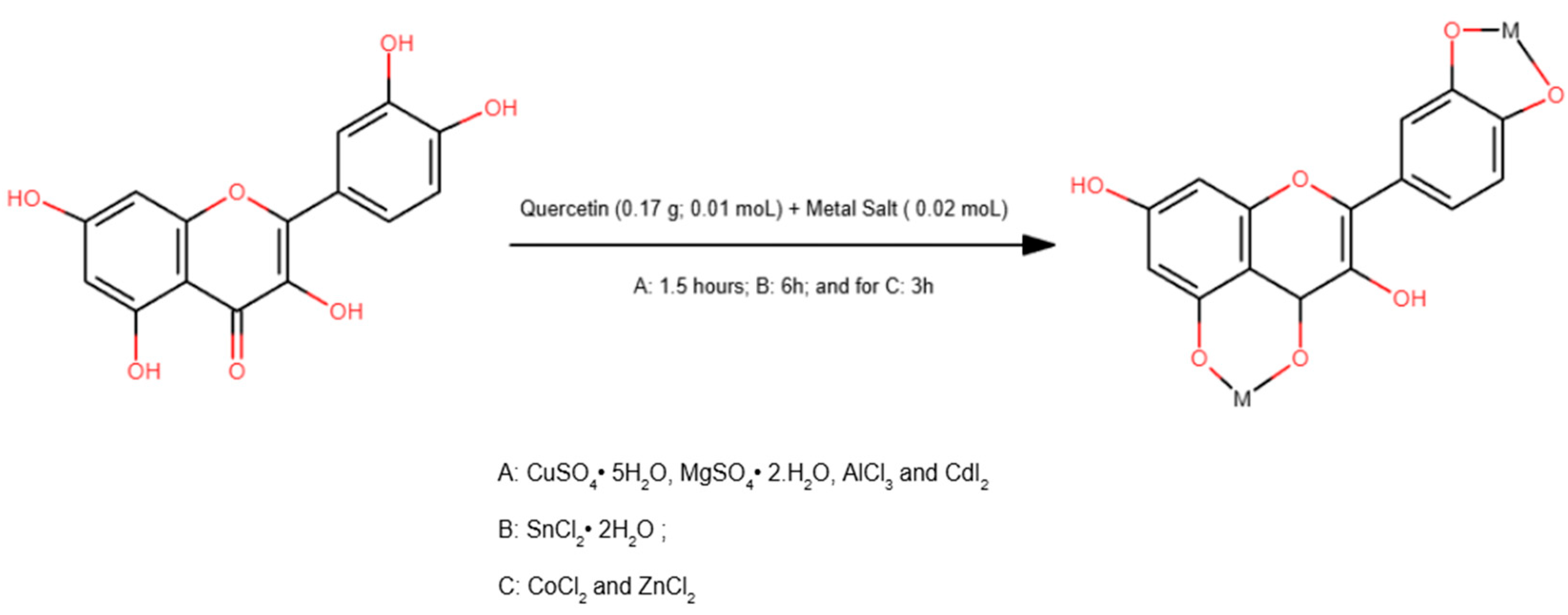
The complexation of quercetin with Cu2+, Co2+, Zn2+, Sn2+, Al3+, Cd2+, and Mg2+ ions produced seven metal-quercetin complexes. These complexes’ structures can be explained based on spectroscopic evidence and established coordination chemistry principles. While the lack of single-crystal X-ray diffraction (XRD) data prevents definitive structural determination, the proposed coordination modes are supported by Fourier-transform infrared (FTIR), ultraviolet–visible (UV–Vis), and nuclear magnetic resonance (NMR) analyses. There is also strong consistency with previously reported quercetin–metal systems. Therefore, the structures discussed herein should be regarded as hypothetical yet chemically plausible models [24,25,26].
Flavonoids such as quercetin preferentially coordinate metal ions through the 4-oxo and 5-hydroxyl groups, forming stable chelate rings that significantly alter their physicochemical and biological properties [27]. Quercetin contains multiple oxygen donor sites that can coordinate metal ions. These sites include the 3-hydroxyl/4-carbonyl (O3/O4), 5-hydroxyl/4-carbonyl (O5/O4), and catechol moiety at the B ring (O3′/O4′). The chelation site involving the C4 carbonyl group and the C5 hydroxyl group is widely recognized as thermodynamically favorable due to the formation of stable five-membered chelate rings [28,29]. Spectroscopic data from this study suggest that complexation primarily occurs at this pharmacologically relevant site, leading to substantial electronic redistribution within the flavonoid framework.
Coordination involving the hydroxyl groups at the 3′ and 4′ positions of the B ring was also inferred, particularly for metal ions with high affinity for catechol-type ligands. Although this site is generally considered of lower pharmacological importance compared to the C-ring chelation site, its participation in metal binding has been extensively reported [13,15,30]. Indeed, depending on the nature of the metal ion and experimental conditions, quercetin–metal complexation has been described at the O3/O4, O4/O5, and O3′/O4′ positions [14,18,31].
The propensity of specific hydroxyl groups to participate in metal coordination is closely related to their acidity, since binding typically occurs through deprotonated hydroxyl oxygens. Structural considerations suggest that the hydroxyl group at position 3 is more acidic than that at position 5 due to its proximity to the carbonyl group and the heterocyclic character of ring C [32,33]. These features enhance electron delocalization and facilitate deprotonation, favoring coordination at the O3/O4 site. Nevertheless, several experimental studies have demonstrated that coordination at the O5/O4 site is also highly favorable, particularly for divalent and trivalent metal ions [32,34,35].
Literature data further corroborate these coordination preferences. Ren et al. demonstrated that iron–quercetin complexes preferentially form via the 3-OH/4-C=O site, with binding strength following the order 3–4 > 4–5 > 3′–4′ [30]. Similar trends have been reported for nickel, magnesium, calcium, and zinc complexes, supporting the idea that quercetin can adopt different coordination modes depending on the metal ion and stoichiometry [14,36,37,38]. In complexes containing one metal ion per quercetin molecule, chelation through the O3/O4 site generally predominates. However, higher metal-to-ligand ratios may promote additional coordination involving the B-ring hydroxyl groups.
The synthetic routes employed in this study (Figure 1) were selected or adapted to favor complexation at established coordination sites. Although literature-based methods have been successfully applied to various complexes, modifications to reaction times were necessary for the Cu(II), Al(III), and Zn(II) systems to effectively chelate. These adaptations are consistent with previous reports emphasizing quercetin’s sensitivity to solvent polarity, temperature, and metal salt reactivity [13,39,40]. It should be noted that the synthesis route for the Al(III)–quercetin complex was not described by the aforementioned authors and was tested for the first time in this study.
Overall, inserting metal ions into the quercetin structure induces significant changes to its electronic and coordination environment, primarily through chelation of carbonyl and phenolic oxygen atoms. While definitive structural elucidation awaits XRD analysis, the proposed structures are strongly supported by spectroscopic evidence and extensive precedent in the literature [39,40]. These hypothetical structural models provide a sound basis for interpreting the physicochemical and biological properties of the synthesized metal–quercetin complexes.

3.2. FTIR Characterization of Metal Complexes

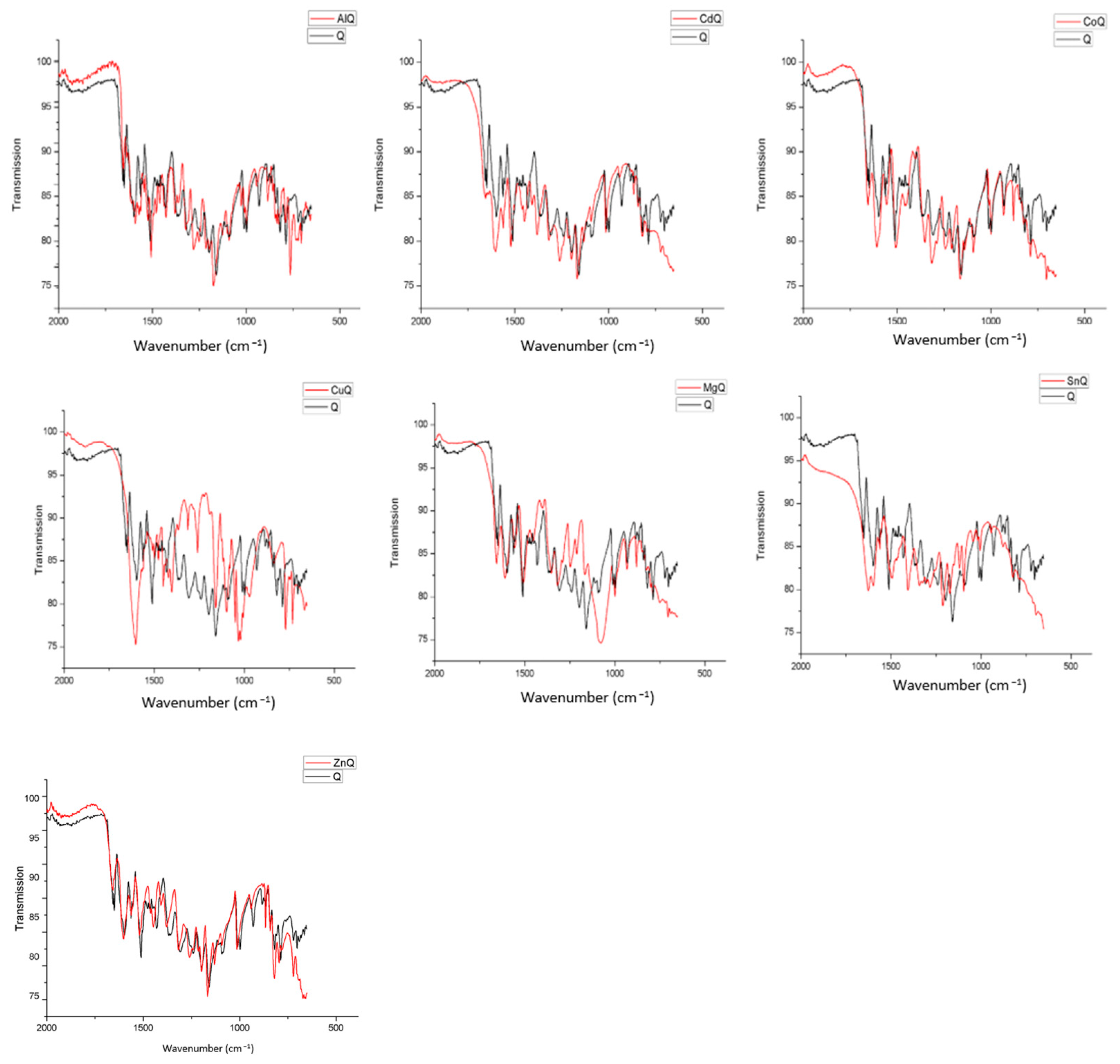
The FTIR spectrum of free quercetin shows well-defined absorption bands that are characteristic of flavonoids. A broad ν(O–H) stretching band is observed in the 3400–3200 cm−1 region, which arises from phenolic hydroxyl groups involved in intra- and intermolecular hydrogen bonding. The strong absorption near 1660 cm−1 is attributed to the ν(C=O) stretching vibration of the γ-pyrone ring, while bands in the 1600–1500 cm−1 region correspond to aromatic ν(C=C) stretching modes. Additional absorptions in the 1300–1000 cm−1 range are attributed to ν(C–O) stretching vibrations of phenolic and ether groups. These functional groups are well known to influence both metal coordination and antioxidant activity in quercetin-based systems [33,41,42].
Upon complexation with metal ions, systematic and metal-dependent spectral changes are observed, confirming coordination through oxygen donor atoms. In the Al(III)–quercetin complex, the ν(O–H) stretching band decreases in intensity and shifts slightly toward lower wavenumbers. This indicates the involvement of phenolic hydroxyl groups in metal binding. Concurrently, the ν(C=O) stretching band shifts downwards more clearly than in free quercetin, confirming the participation of the carbonyl oxygen in coordination. These features are consistent with the formation of a bidentate chelate involving the 3-OH and 4-C=O sites, as has been widely reported for Al(III)–quercetin complexes [42,43,44].
The FTIR spectrum of the Cd(II)–quercetin complex exhibits more pronounced modifications. Significant broadening and attenuation of the ν(O–H) band are observed, together with a marked shift in the ν(C=O) band to lower wavenumbers. Noticeable changes in the 1300–1000 cm−1 region, associated with ν(C–O) vibrations, further support coordination via phenolic and carbonyl oxygen atoms. These spectral features suggest strong metal-ligand interactions and reduced availability of free hydroxyl groups, consistent with prior research on Cd(II)–quercetin complexes [17,45,46].
Moderate but distinct spectral changes are detected for the Co(II)–quercetin complex. The ν(O–H) band shows reduced intensity, while the ν(C=O) stretching vibration undergoes a slight downshift, indicating coordination through the carbonyl group and at least one hydroxyl function. In contrast, the aromatic ν(C=C) bands remain largely unchanged, suggesting preservation of the flavonoid π-electron system. Similar FTIR behavior has been reported for Co(II)–quercetin complexes and is associated with the partial retention of redox-active sites [16,29,47].
More pronounced spectral alterations are observed in the Cu(II)–quercetin complex. A substantial decrease in the intensity of the ν(O–H) stretching band, together with a clear shift in the ν(C=O) band to lower wavenumbers, indicates strong coordination involving both the phenolic and carbonyl oxygen atoms. Additional changes in the 1200–1000 cm−1 region further confirm chelation. These findings are consistent with the strong affinity of Cu(II) for oxygen donor ligands and with literature reports describing significant electronic reorganization in Cu(II)–quercetin complexes, often correlated with altered antioxidant behavior [18,44,48].
In contrast, the Mg(II)–quercetin complex exhibits comparatively subtle spectral changes. The ν(O–H) band remains visible, although slightly shifted and less intense than in free quercetin, while only a minor displacement of the ν(C=O) band is observed. Overall, the FTIR profile of MgQ closely resembles that of the free ligand, suggesting weaker and more ionic metal–ligand interactions. This behavior is characteristic of Mg(II)–flavonoid complexes and has been associated with the preservation or enhancement of antioxidant activity due to the retention of free phenolic hydroxyl groups [15,49].
Significant spectral changes are also observed upon coordination with Sn(II). The ν(O–H) band becomes broader and shifts toward lower wavenumbers, indicating the strong involvement of phenolic hydroxyl groups and suggesting partial or complete deprotonation. The ν(C=O) stretching band shows a noticeable downshift compared to free quercetin, which provides strong evidence for coordination through the carbonyl oxygen. Alterations in the 1200–1000 cm−1 region, attributed to ν(C–O) vibrations, further support chelation via oxygen donor atoms. According to Dehghan and Khoshkam [17], the strong interaction between Sn(II) and phenolic groups results in stable complexes, though it reduces antioxidant activity by occupying redox-active hydroxyl sites [17,49].
In comparison, the Zn(II)–quercetin complex exhibits more subtle spectral modifications. The ν(O–H) band remains broad with slightly reduced intensity, indicating the partial involvement of hydroxyl groups without extensive deprotonation, a behavior typical of Zn(II) as a moderate Lewis acid forming relatively labile metal–oxygen bonds [14]. A slight shift in the ν(C=O) stretching frequency suggests the carbonyl group participates in chelation, likely through bidentate coordination involving the 3-OH/4-C=O or 5-OH/4-C=O sites, as is commonly observed in Zn(II)–flavonoid complexes [49]. Changes in the 1300–1100 cm−1 region associated with ν(C–O) vibrations further confirm complex formation. Importantly, Zn(II)’s relatively mild coordination preserves some of the free phenolic hydroxyl groups. This explains why antioxidant activity is maintained or enhanced in DPPH and ABTS assays for ZnQ, as documented extensively in the literature [14].
Overall, the FTIR results unequivocally confirm the formation of quercetin–metal complexes and demonstrate that coordination predominantly occurs through phenolic hydroxyl and carbonyl groups. The extent of spectral perturbation depends heavily on the nature of the metal ion. Cu(II), Cd(II), and Sn(II) induce more pronounced modifications, while Mg(II) and Zn(II) produce milder effects. These observations are consistent with the reported structure-coordination relationships in quercetin–metal systems (Figure 2) and provide a robust spectroscopic framework for interpreting differences in antioxidant behavior.

3.3. NMR Analysis of Metal Complexes

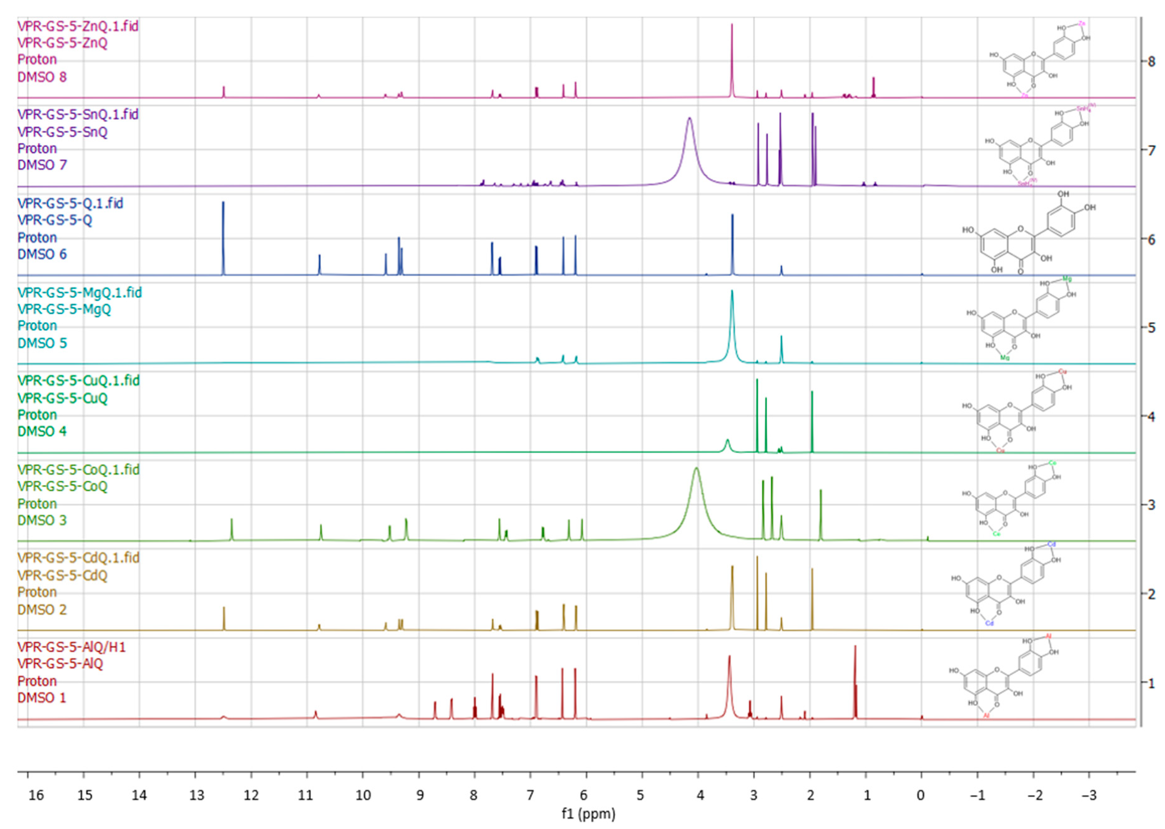
In the 1H NMR spectra, free quercetin exhibited signals in the 6–9 ppm range (Figure 3). These signals were attributed to the aromatic protons of the hydroxylated flavonoid structure of quercetin, which is consistent with classical spectral assignments for flavonoids [50,51]. These signals were absent in the metal–quercetin complexes, indicating coordination through phenolic hydroxyl groups and leading to deprotonation and metal insertion. All complexes exhibited significant spectral changes compared to free quercetin, confirming coordination. SnQ and CoQ displayed singlets around 4 ppm, which were assigned to protons involved in metal-oxygen interactions. This evidence suggests the direct participation of hydroxyl groups in complex formation. In contrast, signals near 3 ppm were preserved in the other complexes and were attributed to non-coordinating hydroxyl groups at C-3 and C-7. This finding is consistent with previous reports on flavonoid systems, in which these positions do not participate in chelation [50]. The singlet observed at approximately 12 ppm in free quercetin and in the complexes CoQ, CdQ, and ZnQ was assigned to a proton engaged in strong intramolecular hydrogen bonding with the 4-oxo group. This demonstrates the preservation of the carbonyl functionality in complexes where this moiety is not involved in metal coordination [52,53].
These observations are consistent with the findings of Ghosh et al. [15], who reported the disappearance of the 3-OH signal and chemical shift perturbations in the Mg–quercetin complex due to deprotonation and increased electronic conjugation upon formation of the complex. Similarly, Halevas et al. [14] demonstrated that the protons of ring A in the Zn–quercetin complex remained largely unaffected, while significant changes occurred in the B-ring proton signals. This confirms preferential coordination through the 3′,4′-dihydroxy system. From a broader perspective, systematic NMR studies of flavonoid–metal complexes indicate that, depending on the nature of the metal ion and the coordination environment, quercetin preferentially coordinates metal ions via the phenolic hydroxyls of ring B and/or the 5-OH/4-oxo chelation site. This leads to hydroxyl signal suppression, chemical shift perturbations, and electronic reorganization of the flavonoid framework, as established in foundational works and coordination chemistry studies [52,53]. The signals obtained by NMR were:
Q 1H NMR (400 MHz, DMSO) δ 12.50, 10.78, 9.59, 9.36, 9.31, 7.69, 7.69, 7.56, 7.56, 7.54, 7.54, 6.90, 6.88, 6.42, 6.41, 6.20, 6.19, 3.85, 3.39, 2.52, 2.51, 2.51, 2.50, 2.50, 1.20.
AlQ RMN H’ 1H NMR (400 MHz, DMSO) δ 12.49, 10.85, 9.63, 9.35, 8.72, 8.71, 8.70, 8.43, 8.41, 8.02, 8.01, 8.00, 7.99, 7.98, 7.97, 7.68, 7.68, 7.57, 7.56, 7.56, 7.54, 7.53, 7.51, 7.51, 7.50, 7.50, 7.50, 7.49, 7.48, 7.48, 7.20, 6.94, 6.91, 6.89, 6.49, 6.43, 6.43, 6.20, 6.20, 6.00, 5.99, 5.99, 3.85, 3.44, 3.11, 3.09, 3.09, 3.08, 3.07, 3.06, 3.05, 3.04, 2.94, 2.78, 2.52, 2.51, 2.51, 2.50, 2.50, 2.17, 2.09, 1.96, 1.21, 1.19, 1.17, −0.01.
CdQ 1H NMR (400 MHz, DMSO) δ 12.48, 10.78, 9.59, 9.59, 9.35, 9.30, 7.68, 7.68, 7.56, 7.55, 7.54, 7.53, 6.90, 6.87, 6.41, 6.40, 6.19, 6.18, 3.85, 3.41, 3.40, 3.40, 3.39, 3.39, 3.38, 2.94, 2.78, 2.52, 2.51, 2.51, 2.50, 2.50, 1.96, 1.21, −0.01.
CoQ 1H NMR (400 MHz, DMSO) δ 12.35, 10.75, 9.52, 9.23, 9.22, 7.56, 7.55, 7.45, 7.44, 7.43, 7.42, 6.79, 6.77, 6.32, 6.31, 6.08, 6.08, 4.03, 2.84, 2.68, 2.51, 1.81, 1.12, −0.11.
CuQ 1H NMR (400 MHz, DMSO) δ 7.92, 7.91, 7.90, 7.89, 7.86, 7.86, 7.84, 7.68, 7.67, 7.56, 7.55, 7.54, 7.53, 7.53, 7.50, 7.43, 7.41, 7.40, 7.40, 7.23, 7.10, 6.97, 6.94, 6.90, 6.89, 6.88, 6.87, 6.78, 6.75, 6.73, 6.68, 6.67, 6.66, 6.66, 6.64, 6.63, 6.62, 6.48, 6.47, 6.47, 6.46, 6.45, 6.44, 6.44, 6.43, 6.43, 6.42, 6.42, 6.41, 6.19, 6.19, 6.18, 6.17, 6.16, 6.16, 3.85, 3.48, 2.95, 2.79, 2.61, 2.57, 2.55, 2.54, 2.52, 2.51, 2.51, 2.50, 2.50, 1.96, 1.91, 1.23, 0.00.
MgQ 1H NMR (400 MHz, DMSO) δ 10.83, 9.23, 7.91, 7.77, 7.76, 6.88, 6.86, 6.42, 6.18, 3.39, 2.95, 2.79, 2.51, 1.96, 1.23, 0.88, 0.87, 0.85, 0.00.
SnQ 1H NMR (400 MHz, DMSO) δ 8.26, 7.85, 7.84, 7.81, 7.17, 7.04, 6.96, 6.94, 6.66, 6.65, 6.64, 6.64, 6.63, 6.63, 6.46, 6.44, 6.43, 6.42, 6.42, 4.15, 2.93, 2.77, 2.55, 2.53, 2.52, 2.51, 2.51, 2.50, 1.95, 1.90.
ZnQ 1H NMR (400 MHz, DMSO) δ 12.49, 10.79, 9.60, 9.36, 9.31, 7.68, 7.68, 7.66, 7.56, 7.55, 7.54, 7.53, 6.90, 6.88, 6.42, 6.41, 6.19, 6.19, 4.33, 3.85, 3.40, 3.10, 3.08, 3.07, 2.94, 2.78, 2.52, 2.51, 2.51, 2.50, 2.50, 2.09, 1.96, 1.43, 1.42, 1.41, 1.41, 1.40, 1.39, 1.39, 1.39, 1.37, 1.37, 1.36, 1.36, 1.35, 1.34, 1.34, 1.33, 1.32, 1.31, 1.30, 1.30, 1.29, 1.29, 1.28, 1.27, 1.27, 1.26, 1.25, 1.22, 1.19, 1.17, 1.15, 0.88, 0.86, 0.84, −0.01.

3.4. UV-Vis Characterization of Metal Complexes

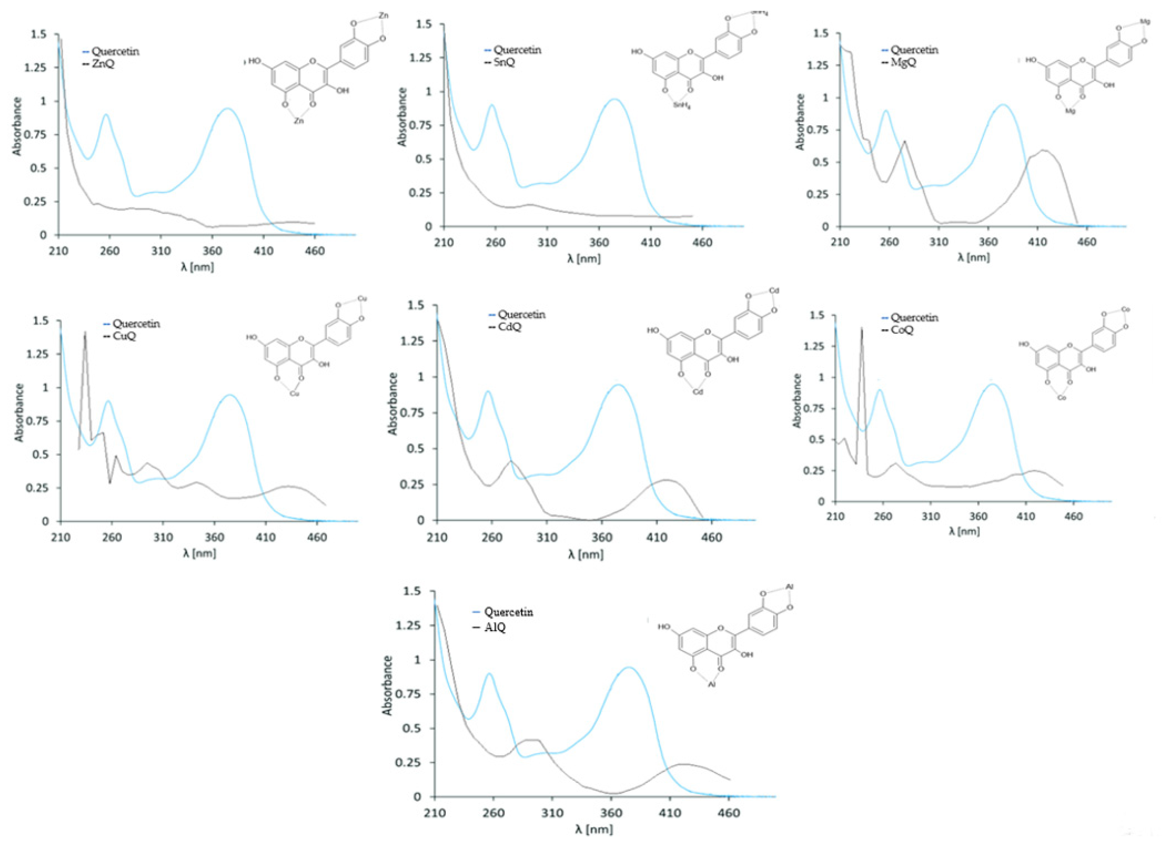
Flavonoids exhibit absorption bands in the ultraviolet (UV) and visible (Vis) regions that arise from electronic π–π* transitions [54]. The UV–Vis spectrum of quercetin in ethanol exhibits two characteristic absorption bands at 256 and 374 nm. These bands are attributed to the benzoyl (rings A/B) and cinnamoyl (rings B/C) systems, respectively. The spectra of the synthesized complexes (Figure 4) showed significant shifts in the 375 nm band for complexes A, B, C, E, F, and G. However, complex D retained both quercetin bands, which shifted bathochromically to 270 and 410 nm.
Flavonoids typically exhibit two absorption bands in the UV–Vis region: Band I (320–385 nm) is associated with the cinnamoyl system of ring B. Band II (240–280 nm) is related to the benzoyl system of ring A. Both bands originate from π → π* electronic transitions. Upon metal coordination, these bands undergo bathochromic shifts, indicating modifications in electronic structure and conjugation [52]. Metal binding to quercetin preferentially involves the 3-OH and 4-oxo groups because of the higher acidity and chelating ability of the 3-hydroxyl proton. The 3′,4′-dihydroxy groups may coordinate a second metal ion. The 5-OH group generally does not participate because of its lower acidity and steric hindrance after initial complexation [47]. Comparative UV–Vis studies with related flavonoids support this coordination pattern [30].
Several metal–quercetin complexes have been reported to exhibit consistent bathochromic and hypo- or hyperchromic effects. Zn2+ coordination induces a significant red shift in Band I to approximately 428 nm, which is attributed to increased conjugation via chelation through the 3-OH/4-oxo moiety [13,14]. Similarly, Sn2+ complexation produces a gradual red shift in Band I and the appearance of a new absorption band near 443 nm, which confirms coordination at the 3-hydroxyl and 4-carbonyl groups [16].
Similar spectral behavior has been observed in other flavonoid-metal systems. For example, Halevas et al. [14] reported bathochromic and hyperchromic shifts in curcumin-metal complexes, which are indicative of extended π-electron delocalization upon coordination [43]. Bukhari et al. [16] observed a red shift in the absorption maximum from ~407 to ~427 nm in Co2+–quercetin complexes, accompanied by a visible color change. This further supports conjugation extension induced by metal binding.
Overall, the consistent bathochromic shifts observed across different metal–quercetin complexes provide strong spectroscopic evidence of coordination-driven electronic reorganization. The 3-OH/4-oxo chelation site plays a dominant role in modulating quercetin’s optical and physicochemical properties.

3.5. Antioxidant Potential Assessment

Quercetin has significant antioxidant potential due to its ability to neutralize free radicals due to the presence of multiple phenolic hydroxyls [33]. We sought to evaluate the antioxidant potential of compounds synthesized between quercetin and metals (Cu2+, Co2+, Zn2+, Sn2+, Al3+, Cd2+ and Mg2+). The 3′ and 4′ ortho-dihydroxyl groups are the major contributors to the antioxidant activity of flavonoids. These catechol moieties form ortho-semiquinone radicals which are highly stabilized by electron delocalization and intramolecular hydrogen bonding. The combination of the C2 = C3 and 4-C- -O groups in the C ring also aids in the delocalization of the π-electrons in the B ring. This in turn influences the dissociation of the phenolic hydroxyl groups as well as the stability of the phenoxy radicals formed on the B ring [55]. The meta-hydroxyl groups present in the A ring are less important than the dihydroxy groups on the B ring, which are more readily oxidized [44]. Thus, the most important factor to consider in the design of metal-flavonoid complexes with high antioxidant activity is the coordination site between the flavonoid structure and the central metal ion. It is important that the flavonoids have the 3′-OH and 4′-OH groups in their structure and that the coordination is preferably at the 5-OH, 4-C-OH and/or 3-OH groups.
In classic antioxidant assays (DPPH, ABTS, and FRAP), the analytical response reflects specific reaction mechanisms, such as hydrogen donation, electron transfer, or reducing power. The presence of metals and alcoholic/phenolic groups alters these mechanisms, yet does not invalidate the assay. It is important to note that these tests do not measure “absolute antioxidant activity,” but rather the ability to interact with a specific chemical reagent. Therefore, when a metal complex exhibits lower activity than free quercetin, it does not indicate a failure of the method. Rather, it suggests that coordination has reduced the availability of hydrogen donor sites or altered the kinetics of electron transfer [19,20,21,48]. The use of IC50 in antioxidant assays is preferred because these methods evaluate functional responses that are dependent on both dose and time, and involve multiple, non-stoichiometric mechanisms. Thus, IC50 provides a robust, reproducible, and widely accepted comparative measure of antioxidant efficiency [38,56].

3.5.1. DPPH Radical Scavenging Method

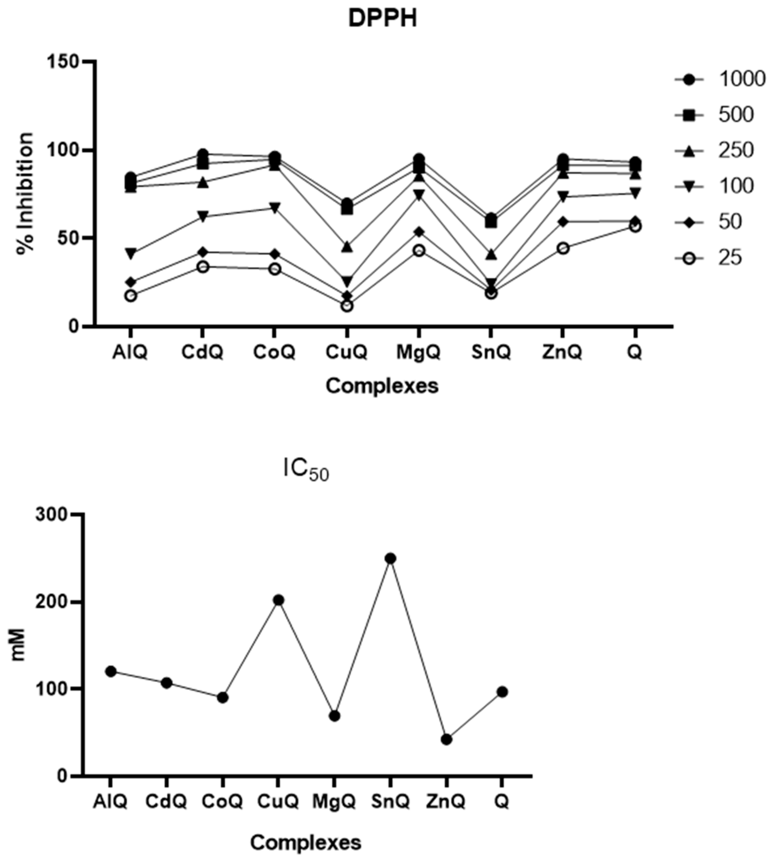
As shown in Figure 5, the results confirm the high antioxidant capacity of standard quercetin. At the highest evaluated concentration, quercetin inhibited 92.88% of the DPPH radical, which is consistent with its well-established radical scavenging properties. This strong activity is associated with quercetin’s molecular structure, particularly the presence of 3′,4′-dihydroxyl groups on the B ring. These groups play a central role in hydrogen atom transfer reactions with the DPPH radical and electron donation processes [57,58]. As expected, the radical scavenging efficiency of free quercetin increased in a dose- and time-dependent manner, reflecting its high intrinsic antioxidant potential.
Metal complexation significantly altered this behavior and enhanced the antioxidant activity of the complexes relative to free quercetin in several cases. The CdQ, MgQ, and ZnQ complexes exhibited antioxidant activities comparable to or greater than quercetin’s, with inhibition values of 96.0%, 94.3%, and 95.2%, respectively. Similarly, the AlQ and ZnQ complexes demonstrated high antioxidant activity with inhibition values of 86.02% and 95.2%, respectively, at 1000 μM. These results suggest that coordination with specific metal ions may favor hydrogen atom or electron transfer processes through electronic redistribution or stabilization of radical intermediates, thereby enhancing DPPH scavenging efficiency. Similar enhancements have been reported for Co(II), Fe(II), and Cu(II) quercetin complexes, which displayed lower IC50 values and higher scavenging efficiencies than free quercetin in DPPH assays [15,16,17]. Notably, Co(II)-quercetin exhibited an IC50 value of 2.01 ± 0.25 μM, which is significantly lower than the value for quercetin (3.88 ± 0.06 μM). This confirms that, under certain conditions, metal coordination can enhance antiradical activity.
In contrast, the SnQ and CuQ complexes exhibited significantly reduced antioxidant activity compared to the positive control, underscoring the pivotal role of the coordinated metal ion in shaping the overall antioxidant behavior. This reduction is consistent with previous reports indicating that quercetin–Sn(II) and quercetin–Tb(III) complexes exhibit weaker DPPH scavenging activity than free quercetin due to the suppression of hydrogen-donating sites during coordination [17,57]. Spectrophotometric analyses after the DPPH reaction corroborate these observations because metal coordination mainly occurs through phenolic hydroxyl and carbonyl groups. These groups are directly responsible for quercetin’s antioxidant activity. Depending on the metal, such coordination can stabilize radical intermediates or reduce the availability of free hydroxyl groups, thereby diminishing antioxidant efficiency.
The present results are largely consistent with previously reported data. Dehghan and Khoshkam [17] reported that the SnQ complex inhibited approximately 40% of DPPH, which is consistent with the low activity observed here. Bukhari et al. [16] showed that the CoQ complex achieved around 80% inhibition at 700 μM, while free quercetin inhibited approximately 90% of the radical. These values are comparable to those obtained in this study. Ghosh et al. [15] reported 55% inhibition for the MgQ complex after 30 min at 700 μM. Notably, the MgQ complex synthesized in this study exhibited superior performance (approximately 85% inhibition). This suggests that the synthetic route and coordination environment can substantially influence antioxidant outcomes. However, the lower activity observed here (86.34% at 700 μM) further emphasizes the sensitivity of DPPH scavenging to synthesis conditions and metal–ligand interactions when compared to the results of Halevas et al. [18], who reported approximately 95% inhibition for the CuQ complex.
IC50 values further support these trends. The ZnQ complex exhibited the lowest IC50 value (42.36 ± 7.98 μM), which is even lower than the value for the standard compound quercetin (96.81 ± 8.11 μM). This finding confirms the ZnQ complex’s superior antioxidant efficiency. Conversely, SnQ and CuQ displayed the highest IC50 values (249.9 ± 8.33 μM and 202.2 ± 10.66 μM, respectively), indicating reduced radical scavenging capacity. These results show that metal complexation can enhance or reduce quercetin’s antioxidant activity depending on the metal ion’s nature.
As reported by De Giovani et al. 2004 [27], metal coordination modifies the redox behavior of flavonoids, which may enhance radical scavenging activity by stabilizing the oxidized flavonoid species. From a mechanistic perspective, quercetin’s antioxidant activity is primarily associated with the 3′-OH and 4′-OH groups of ring B. These groups are directly involved in hydrogen atom transfer reactions with DPPH. Additionally, intramolecular hydrogen bonding involving the 3-OH group promotes coplanarity of the flavonoid rings, favoring electron delocalization and stabilization of radical species [58,59,60]. However, metal coordination can modify this electronic distribution by engaging hydroxyl groups or altering redox properties. This can either enhance radical stabilization or limit hydrogen donation, as observed for Cd(II), Co(II), Fe(II), Tb(III), and Sn(II) complexes [16,61]. Overall, these results reinforce the concept that metal ions can positively or negatively modulate quercetin’s physicochemical and antioxidant properties, depending on their coordination behavior and redox characteristics.

3.5.2. Antioxidant 2,2′-Azinobis(3-Ethylbenzothiazoline-6-Sulfonic Acid) (ABTS)+ Radical Activity

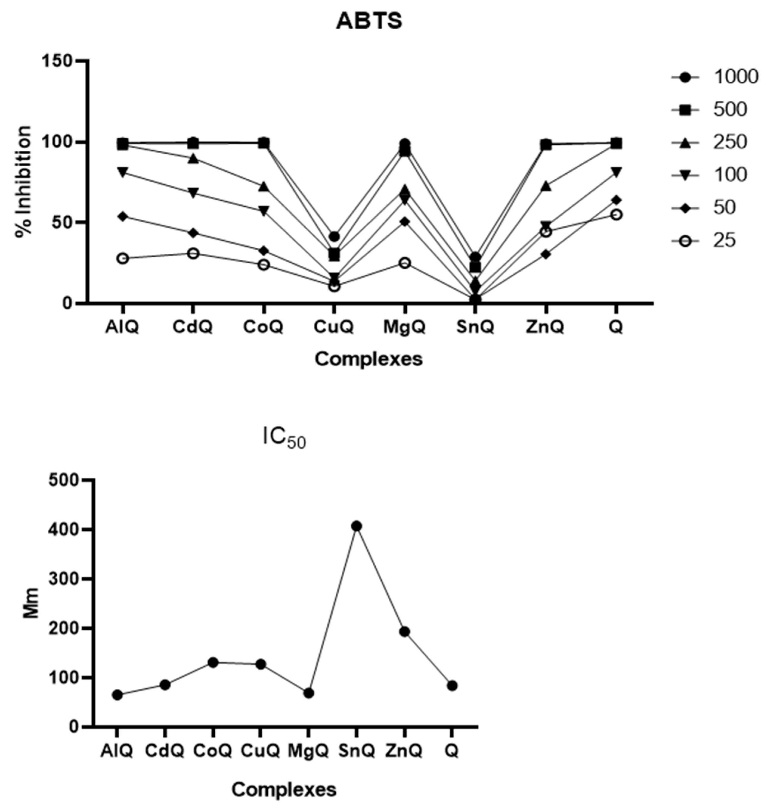
The ABTS•+ radical scavenging assay revealed that most of the synthesized quercetin–metal complexes had high antioxidant capacity, especially at higher concentrations (Figure 6). The CdQ complex exhibited complete ABTS•+ inhibition, achieving 100% scavenging; meanwhile, free quercetin (Q) demonstrated nearly identical inhibition at 99.7%. Similarly, AlQ (99.86%), CoQ (99.85%), MgQ (98.52%), and ZnQ (99.5%) exhibited antioxidant activity comparable to quercetin’s at the commonly used concentration of 1000 μg mL−1, suggesting that coordination with these metals largely preserves the flavonoid’s intrinsic radical scavenging properties. However, the CuQ and SnQ complexes exhibited reduced antioxidant activity, with inhibition values of 49.86% and 28.3%, respectively. This suggests that complexation with these metals negatively affects quercetin’s antioxidant performance. concentration-dependent analysis further supported these observations.
A concentration-dependent analysis further supported these observations. All complexes exhibited strong ABTS•+ scavenging activity at the highest tested concentrations (1000 and 500 μg mL−1), whereas free quercetin demonstrated the most potent inhibition across the entire evaluated concentration range (1000–25 μg mL−1). This behavior underscores the potent antioxidant capacity of quercetin at the molecular level and suggests that metal coordination exerts a more significant impact on radical scavenging efficiency at lower concentrations.
These findings are consistent with previous studies on quercetin–metal complexes evaluated using the ABTS assay. For example, studies on the quercetin-Tb(III) complex revealed a concentration-dependent decrease in ABTS•+ absorbance at 734 nm for free quercetin and the complex, indicating effective antiradical activity. However, free quercetin consistently exhibited superior scavenging efficiency compared to the quercetin–Tb(III) complex. This indicates that metal coordination reduces, but does not eliminate, the ABTS•+ radical scavenging capacity of quercetin [61]. A similar trend was reported for the quercetin–cadmium complex: the percentage of ABTS•+ scavenging efficiency was consistently lower than that of free quercetin across the studied concentration range. This reduced activity has been attributed to the quercetin–Cd(II) complex’s higher oxidation potential relative to free quercetin, which limits its ability to donate electrons or hydrogen atoms to the ABTS radical [62].
A review of the literature revealed limited reports employing the ABTS•+ assay for quercetin–metal complexes. Dehghan and Khoshkam [17] reported approximately 55% ABTS•+ inhibition for the SnQ complex at 700 μM. In the present study, however, SnQ exhibited significantly lower scavenging efficiency (approximately 24%) under comparable conditions. This finding confirms its consistently poor antioxidant performance and emphasizes the strong dependence of ABTS•+ scavenging activity on the nature of the coordinated metal ion and the resulting electronic structure of the complex.
Although CdQ achieved complete inhibition of ABTS•+ at the highest tested concentration, analysis of scavenging efficiency as a function of concentration revealed that its antioxidant activity was inferior to that of free quercetin. The superior performance of quercetin can be attributed to its multiple hydroxyl groups with low oxidation potential, which facilitate efficient hydrogen atom transfer and electron donation. In contrast, coordination with Cd(II) and other metal ions, such as Tb(III), can increase the flavonoid framework’s oxidation potential and restrict the availability of hydroxyl groups, thereby reducing radical scavenging efficiency [61,62,63].
The IC50 values obtained from the ABTS assay quantitatively support these trends. The SnQ complex exhibited the highest IC50 value (407.30 ± 19.77 μg mL−1), confirming its weak radical scavenging capacity. Conversely, AlQ (65.12 ± 11.12 μg mL−1) and MgQ (68.77 ± 3.44 μg mL−1) exhibited the lowest IC50 values among the synthesized complexes, which were approximately 20 μg mL−1 lower than the value for free quercetin (83.98 ± 12.22 μg mL−1). This indicates enhanced antioxidant efficiency. CdQ (85.79 ± 9.32 μg mL−1) had an IC50 value similar to quercetin’s, consistent with previous reports indicating that cadmium coordination partially suppresses ABTS•+ scavenging activity relative to the free ligand [62]. CoQ, CuQ, and ZnQ displayed intermediate IC50 values, reflecting their moderate antioxidant performance.
Overall, these results demonstrate that metal complexation can modulate the ABTS•+ scavenging activity of quercetin in different ways. Coordination with certain metal ions, such as Al3+ and Mg2+, enhances antioxidant efficiency. However, coordination with other metal ions, particularly Sn2+, Cu2+, Cd2+, and Tb3+, tends to reduce radical scavenging capacity by increasing oxidation potential and limiting electron transfer. These findings align well with previously reported structure–activity relationships for quercetin–metal complexes, confirming that the observed antioxidant behavior in the ABTS assay depends heavily on metal identity and coordination-induced electronic effects.

3.5.3. Ferric Reducing Antioxidant Power (FRAP) Assay

We employed the ferric reducing antioxidant power (FRAP) assay to evaluate the antioxidant potential of quercetin (Q) and its metal complexes further, providing additional information on their electron-donating capacity. The FRAP assay is based on antioxidants’ ability to reduce the Fe(III)-TPTZ complex to Fe(II)-TPTZ under acidic conditions. This reaction forms an intense blue complex with a maximum absorption wavelength of 593 nm. The amount of Fe(II) generated is directly proportional to antioxidant activity [57]. Consistent with previous reports on quercetin-Tb(III) complexes [61], which demonstrated that metal coordination can modulate, but not suppress, the reducing ability of quercetin, the present results suggest that FRAP activity depends strongly on concentration and metal identity.
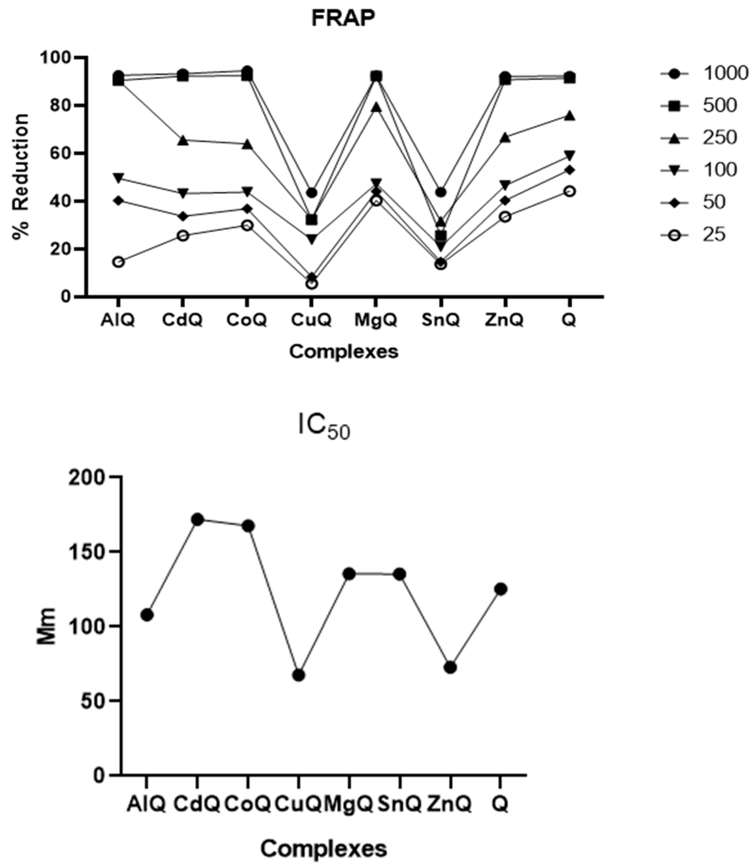
At the highest tested concentration (1000 μg mL−1), free quercetin exhibited an inhibition profile similar to those of the AlQ, CdQ, CoQ, MgQ, and ZnQ complexes. This indicates that complexation with these metals largely preserves quercetin’s ferric reducing ability. A similar trend was previously observed for the quercetin–Tb(III) complex, which retained measurable reducing power but displayed lower activity than free quercetin [61]. This is consistent with the idea that electron transfer is partially attenuated upon chelation. In contrast, the CuQ and SnQ complexes exhibited the weakest antioxidant activity, suggesting that coordination with these metal ions interferes more strongly with electron donation.
At the lowest evaluated concentration, free quercetin inhibited approximately 48% of the FRAP complex, demonstrating the highest reducing capacity of all the tested compounds. This behavior is consistent with previous studies on flavonoids that demonstrated quercetin has the strongest ferric reducing ability and apigenin has the weakest [57]. Structural comparisons suggest that having a single hydroxyl group on the B ring, as with apigenin and chrysin, has a minimal impact on antioxidant activity because of its reduced electron-donating capability. In contrast, kaempferol’s (Kae) higher ferric reducing ability relative to apigenin’s (Api) has been attributed to the presence of the 3-OH group, which highlights the importance of hydroxyl substitution patterns [57]. MgQ exhibited the second-highest activity, with approximately 40% inhibition. The remaining complexes displayed negligible reducing ability at low concentrations, emphasizing the dominant contribution of free hydroxyl groups to electron donation.
At higher concentrations, all synthesized complexes except SnQ and CuQ exhibited inhibition values approaching 100%, which is comparable to the performance of free quercetin (Figure 7). These results are similar to those observed for quercetin–Tb(III) [61], where an increased effective concentration partially compensated for coordination-induced electronic effects, enabling the complex to approach the reducing performance of free quercetin. Overall, these results suggest that, when sufficient concentration is available to overcome chelation-related electronic constraints, metal complexation generally preserves and sometimes enhances the antioxidant potential of quercetin.
From a structure–activity perspective, quercetin’s strong ferric reducing ability has been attributed to its multiple hydroxyl groups, especially the 3-OH group, which plays a critical role in electron donation. Comparative FRAP analyses have shown that quercetin’s ferric reducing capacity exceeds that of apigenin (Api), chrysin (Chr), and kaempferol (Kae) by 1.06, 1.07, and 0.28 mmol Fe2+ L−1, respectively. This confirms that hydroxyl substituents significantly enhance the FRAP response of flavonoid compounds [63]. However, coordination to metal ions, such as Tb(III), may partially limit the availability of these hydroxyl and carbonyl sites for redox reactions, thereby modulating FRAP activity [61].
Overall, the FRAP results corroborate the findings from the DPPH and ABTS•+ assays and align with prior studies on quercetin-Tb(III) complexes [61], demonstrating that metal complexation can influence quercetin’s antioxidant behavior differently. Certain metal ions preserve or enhance quercetin’s ferric reducing capacity, while others significantly attenuate it. This underscores the critical roles of hydroxyl substitution patterns, metal identity, and coordination environment in determining antioxidant efficiency.

3.6. Antimicrobial Potential Assessment

This study revealed the antibacterial and antifungal properties of the tested metal and quercetin combination (Table 1). However, the activity of these compounds varied considerably. In general, these compounds exhibited greater activity against fungi than bacteria. The following trend was observed in microbial susceptibility to the compounds: C. glabrata > S. aureus > B. cereus.
The metal–quercetin complexes exhibited antibacterial and antifungal activity, demonstrating substantial variability among compounds and microorganisms (Table 1). Generally, the antifungal efficacy was higher than the antibacterial efficacy, and the order of microbial susceptibility was C. glabrata > S. aureus > B. cereus. CdQ and CoQ exhibited the most pronounced antibacterial effects, inhibiting P. aeruginosa and E. coli at minimum inhibitory concentrations (MIC) of 250 μg mL−1, while AlQ, CdQ, and MgQ were more active than free quercetin against S. aureus with MIC values of 250, 250, and 125 μg mL−1, respectively. Furthermore, CdQ exhibited bacteriostatic activity against B. cereus at concentrations above 250 μg mL−1.
Fungal strains showed greater sensitivity than bacterial strains, particularly Candida spp. and C. glabrata, which has been reported to be highly susceptible to metal-based antimicrobial agents due to its membrane composition and metal ion uptake mechanisms, showed the highest sensitivity. Of the complexes, CoQ exhibited the most promising antifungal profile, inhibiting C. albicans at concentrations above 75 μg mL−1 and C. glabrata at concentrations above 250 μg mL−1. AlQ and CdQ were active against C. albicans and C. glabrata, respectively, at concentrations above 250 μg mL−1. Although they outperformed free quercetin, none of the complexes matched the inhibitory potency of fluconazole.
The antimicrobial action of flavonoids is widely associated with their ability to interact with microbial cell envelopes. Scalbert [64] demonstrated that flavonoids and related polyphenols can bind to cell wall proteins and membrane-associated enzymes, which leads to structural destabilization and impaired cellular function. Cushnie and Lamb [65] further demonstrated that flavonoids disrupt cytoplasmic membranes by increasing their permeability, inducing the leakage of essential ions and promoting the aggregation of bacterial cells. This membrane damage results in depolarization and the collapse of cellular energy metabolism, which is consistent with observations of bacteriostatic and bactericidal effects [66]. Pawlikowska-Pawlega et al. [67] clarified that these effects require an initial interaction of flavonoids with the polar head groups of phospholipids, followed by penetration into hydrophobic membrane regions. This explains why fungi with sterol-rich membranes are more susceptible.
Metal complexation further amplifies these effects by altering the flavonoid’s physicochemical properties. According to Chohan and Supuran [68], chelation increases lipophilicity and facilitates transmembrane transport, enabling metal–flavonoid complexes to reach intracellular targets more efficiently. Singh et al. [69] demonstrated that metal–flavonoid complexes interfere with microbial respiration and inhibit protein and nucleic acid synthesis, leading to irreversible growth arrest. Additionally, Sumra and Chohan [70] and Yasmeen et al. [71] showed that chelation reduces metal ion polarity through partial charge sharing with donor atoms and π-electron delocalization within the chelate ring. This enhances membrane permeability and biological activity. Consequently, metal complexes can function as both activated carriers of bioactive ligands and intrinsically cytotoxic species, which explains their consistently higher antimicrobial efficacy compared to free quercetin.
Investigating organometallic complexes with flavonoids, particularly quercetin–metal complexes, is more than just a chemical curiosity. Altering the geometry of the molecule can modify its physicochemical properties, such as increasing lipophilicity and consequently its ability to cross cell membranes. The goal of this study was to synthesize, characterize, and evaluate the antioxidant and antimicrobial potential of organometallic compounds derived from quercetin. Innovations in the pharmaceutical field are essential for developing metal-based drugs to treat cancer, infectious diseases, and other illnesses. In the cosmetics sector, organometallic complexes can be used as preservatives, pigments, and delivery agents for active ingredients, ensuring greater penetration of the skin in a controlled and effective manner. Thus, this work contributes to the fields of chemistry, pharmacy, cosmetics, and nutrition by exploring the potential of these metal-flavonoid complexes.

4. Conclusions

This study demonstrates that quercetin is a versatile ligand that can form stable coordination compounds with metal ions of different charges and electronic properties, including Cu(II), Co(II), Zn(II), Sn(II), Al(III), Cd(II), and Mg(II). Despite the need for adjustments in selected synthetic protocols, the successful synthesis of all seven complexes highlights the strong sensitivity of quercetin–metal complexation to reaction parameters such as solvent, stoichiometry, and metal salt reactivity.
FTIR, NMR, and UV–Vis analyses confirmed quercetin’s coordination with metal ions. This was evidenced by shifts in the O–H, C=O, and C–O bands; changes in the NMR chemical environment; and bathochromic shifts in the UV–Vis spectra. Together, these results suggest the formation of metal–quercetin complexes.
The antioxidant evaluation revealed that metal coordination modulates the redox behavior of quercetin in a metal-dependent manner. Notably, the Zn(II)–, Mg(II)–, and Al(III)–quercetin complexes exhibited antioxidant activities equal to or greater than free quercetin. This indicates that coordination with redox-inactive or weakly interacting metal ions can preserve and enhance the flavonoid’s radical-scavenging capacity. This enhancement is attributed to limited perturbation of phenolic hydroxyl groups and favorable electronic delocalization within the quercetin framework, as supported by spectroscopic analyses. These findings reinforce the potential of selected metal–quercetin complexes as improved antioxidant systems compared to the parent molecule.
The biological response depended heavily on the metal coordination, with certain complexes exhibiting notable antibacterial or antifungal effects, particularly against Candida species. These results suggest that metal coordination enhances quercetin’s bioactivity by improving its interaction with microbial cells. However, its efficacy remains lower than that of standard antifungal drugs. This study proposes evaluating the synthesis, characterization, and preliminary testing of organometallic compounds derived from quercetin. However, based on the results obtained, we suggest expanding the bioassays to include cytotoxicity, antitumor, and acute and chronic toxicity tests as future prospects. Overall, this study clearly shows that coordination with metal ions significantly changes the electronic structure and functional properties of quercetin.

Author Contributions

O.M.B., L.H.S.B. and A.d.F.S.J., conceptualization, resources, experimental data acquisition, methodology, validation, formal analysis, writing—original draft, and writing—review & editing; I.F.S., F.d.S.M., C.F.d.S.F., L.G.d.S.d.S., T.S.L., K.S.S.d.J., L.F.G.S., S.V.S.d.S., V.P.R. and G.S.d.S., experimental data acquisition, methodology, formal analysis, writing—original draft, and writing—review & editing; A.d.F.S.J., L.C.d.S.N. and S.L.C., supervision, validation, and writing—original draft; A.d.F.S.J., conceptualization, resources, funding acquisition, supervision, writing—original draft, and writing—review & editing. All authors have read and agreed to the published version of the manuscript.

Funding

This research was funded by “Conselho Nacional de Desenvolvimento Científico e Tecnológico” (CNPq, Brasília, Brazil) (406331/2023-5). This study was also financed in part by “Coordenação de Aperfeiçoamento de Pessoal de Nível Superior” (CAPES, Brasília, Brazil)—Finance Code 001.

Institutional Review Board Statement

Not applicable.

Informed Consent Statement

Not applicable.

Data Availability Statement

The datasets used and/or analyzed during the current study are available from the corresponding author on reasonable request.

Acknowledgments

The authors are grateful to the University of Bahia State (UNEB), “Conselho Nacional de Desenvolvimento Científico e Tecnológico” (CNPq, Brasília, Brazil) and “Coordenação de Aperfeiçoamento de Pessoal de Nível Superior” (CAPES, Brasília, Brazil) for financial support and grants.

Conflicts of Interest

The authors declare no conflicts of interest.

Abbreviations

The following abbreviations are used in this manuscript:
ABTS2,2′-azino-bis(3-ethylbenzothiazoline-6-sulfonic acid)
DPPH1,1-diphenyl-2-picrylhydrazyl
FRAPFerric Reducing Antioxidant Power
FTIRFourier Transform Infrared Spectroscopy
MBCMinimum bactericidal concentration
MFCMinimum fungicidal concentration
MICMinimum inhibitory concentrations
NMRNuclear Magnetic Resonance
TPTZ2,4,6-Tris(2-Pyridyl)-S-Triazine
UV-VISUV/visible spectroscopy

References

  1. Jan, R.; Khan, M.; Asaf, S.; Asif, S.; Kim, K.M. Bioactivity and therapeutic potential of kaempferol and quercetin: New insights for plant and human health. Plants 2022, 11, 2623. [Google Scholar] [CrossRef]
  2. da Silva, S.V.S.; Barboza, O.M.; Souza, J.T.; Soares, É.N.; dos Santos, C.C.; Pacheco, L.V.; Santos, I.P.; Magalhães, T.B.d.S.; Soares, M.B.P.; Guimarães, E.T.; et al. Structural design, synthesis and antioxidant, antileishmania, anti-inflammatory and anticancer activities of a novel quercetin acetylated derivative. Molecules 2021, 26, 6923. [Google Scholar] [CrossRef]
  3. Batiha, G.E.S.; Beshiby, A.M.; Ikram, M.; Mulla, Z.; El-Hack, M.; Taha, A.; Algammal, A.; Elewa, Y.H. The pharmacological activity, biochemical properties, and pharmacokinetics of the major natural polyphenolic flavonoid: Quercetin. Foods 2020, 9, 374. [Google Scholar] [CrossRef]
  4. Zhang, J.; Guo, J.; Qian, Y.; Yu, L.; Ma, J.; Gu, B.; Tang, W.; Li, Y.; Li, H.; Wu, W. Quercetin induces apoptosis through downregulating P4HA2 and inhibiting the PI3K/Akt/mTOR axis in hepatocellular carcinoma cells: An in vitro study. Cancer Rep. 2025, 8, e70220. [Google Scholar] [CrossRef] [PubMed]
  5. Athira, N.D.; James, T.J. Computational screening of phytocompounds from Moringa oleifera leaf as potential inhibitors of SARS-CoV-2 Mpro. Res. Sq. 2020, 3, 1–14. [Google Scholar]
  6. Saeedi-Boroujeni, A.; Mahmoudian-Sani, M.R. Anti-inflammatory potential of quercetin in COVID-19 treatment. J. Inflamm. 2021, 18, 3. [Google Scholar] [CrossRef] [PubMed]
  7. Aghababaei, F.; Hadidi, M. Recent advances in potential health benefits of quercetin. Pharmaceuticals 2023, 16, 1020. [Google Scholar] [CrossRef]
  8. Cai, J.; Nelson, K.C.; Wu, M.; Sternberg, P., Jr.; Jones, D.P. Oxidative damage and protection of the retinal pigment epithelium. Prog. Retin. Eye Res. 2000, 19, 205–221. [Google Scholar] [CrossRef]
  9. El-Gammal, O.; Shawky, F.; Rezk, G.; El-Bindary, A.S. Synthesis, characterization, catalytic, DNA binding and antibacterial activities of Co(II), Ni(II) and Cu(II) complexes with new Schiff base ligand. J. Mol. Liq. 2021, 326, 115223. [Google Scholar] [CrossRef]
  10. Ong, Y.C.; Gasser, G. Organometallic compounds in drug discovery: Past, present and future. Drug Discov. Today Technol. 2020, 37, 117–124. [Google Scholar] [CrossRef]
  11. Gambino, D.; Otero, L. Design of prospective antiparasitic metal-based compounds including selected organometallic cores. Inorg. Chim. Acta 2017, 472, 58–75. [Google Scholar] [CrossRef]
  12. Liu, Z.; Zhou, T.; Ziegler, A.C.; Dimitrion, P.; Zuo, L. Oxidative stress in neurodegenerative diseases: From molecular mechanisms to clinical applications. Oxid. Med. Cell. Longev. 2017, 2017, 2525967. [Google Scholar] [CrossRef]
  13. Selvaraj, S.; Krishnaswamy, S.; Devashya, V.; Sethuraman, S.; Krishnan, U.M. Flavonoid–metal ion complexes: A novel class of therapeutic agents. Med. Res. Rev. 2014, 34, 677–702. [Google Scholar] [CrossRef]
  14. Halevas, E.; Mavroidi, B.; Maria, P.; Hatzidimitriou, A. Structurally characterized zinc complexes of flavonoids chrysin and quercetin with antioxidant potential. Inorg. Chim. Acta 2021, 523, 120407. [Google Scholar] [CrossRef]
  15. Ghosh, N.; Chakraborty, T.; Mallick, S.; Mana, S.; Singha, D.; Ghosh, B.; Roy, S. Synthesis, characterization and study of antioxidant activity of quercetin–magnesium complex. Spectrochim. Acta A Mol. Biomol. Spectrosc. 2015, 151, 807–813. [Google Scholar] [CrossRef] [PubMed]
  16. Bukhari, S.; Memon, S.; Mahroof-Tahir, M.; Bhanger, M.I. Synthesis, characterization and investigation of antioxidant activity of cobalt–quercetin complex. J. Mol. Struct. 2008, 892, 39–46. [Google Scholar] [CrossRef]
  17. Dehghan, G.; Khoshkam, Z. Tin(II)–quercetin complex: Synthesis, spectral characterisation and antioxidant activity. Food Chem. 2012, 131, 422–426. [Google Scholar] [CrossRef]
  18. Halevas, E.; Mavroidi, B.; Antonoglou, O.; Kessissoglou, D.P.; Psomas, G. Copper(II) complexes with flavonoids: Synthesis, structure and antioxidant activity. J. Inorg. Biochem. 2020, 206, 111018. [Google Scholar]
  19. Brand-Williams, W.; Cuvelier, M.E.; Berset, C. Use of a free radical method to evaluate antioxidant activity. Food Sci. Technol. 1995, 28, 25–30. [Google Scholar] [CrossRef]
  20. Re, R.; Pellegrini, N.; Proteggente, A.; Pannala, A.; Yang, M.; Rice-Evans, C. Antioxidant activity applying an improved ABTS radical cation decolorization assay. Free Radic. Biol. Med. 1999, 26, 1231–1237. [Google Scholar] [CrossRef]
  21. Benzie, I.F.; Strain, J.J. The ferric reducing ability of plasma (FRAP) as a measure of antioxidant power. Anal. Biochem. 1996, 239, 70–76. [Google Scholar] [CrossRef] [PubMed]
  22. ISO 20776-1:2006; Clinical Laboratory Testing and In Vitro Diagnostic Test Systems—Susceptibility Testing of Infectious Agents and Evaluation of Performance of Antimicrobial Susceptibility Test Devices—Part 1: Reference Method for Testing the In Vitro Activity of Antimicrobial Agents Against Rapidly Growing Aerobic Bacteria Involved in Infectious Diseases. International Organization for Standardization: Geneva, Switzerland, 2006.
  23. EUCAST. Breakpoint Tables for Interpretation of MICs and Zone Diameters; The European Committee on Antimicrobial Susceptibility Testing: Denmark, Europe, 2019. [Google Scholar]
  24. Bukhari, S.B.; Memon, S.; Mahroof-Tahir, M.; Bhanger, M.I. Synthesis, characterization and antioxidant activity of metal–quercetin complexes. Spectrochim. Acta A Mol. Biomol. Spectrosc. 2008, 71, 1901–1906. [Google Scholar] [CrossRef]
  25. Da Silva, M.M.; Oliveira, M.C.; Santos, R.H.A.; Cunha, E.F.F.; Ramalho, T.C. Structural modification of quercetin by acetylation and evaluation of antioxidant activity. J. Braz. Chem. Soc. 2014, 25, 1186–1194. [Google Scholar]
  26. Kostyuk, V.A.; Potapovich, A.I.; Strigunova, E.N.; Kostyuk, T.V.; Afanas’ev, I.B. Experimental evidence that flavonoid metal complexes may act as prooxidants. Arch. Biochem. Biophys. 2004, 428, 204–208. [Google Scholar] [CrossRef]
  27. Souza, R.F.V.; De Giovani, W.F. Antioxidant properties of complexes of flavonoids with metal ions. Redox Rep. 2004, 9, 97–104. [Google Scholar] [CrossRef]
  28. Mathiyalagan, S.; Mandal, B.K. Stability comparison of quercetin and its metal complexes and their biological activity. Biointerface Res. Appl. Chem. 2021, 11, 7890–7902. [Google Scholar]
  29. Afanas’ev, I.B.; Dcrozhko, A.I.; Brodskii, A.V.; Kostyuk, V.A.; Potapovitch, A.I. Chelating and free radical scavenging mechanisms of inhibitory action of rutin and quercetin. Biochem. Pharmacol. 1989, 38, 1763–1769. [Google Scholar] [CrossRef] [PubMed]
  30. Ren, J.; Meng, S.; Lekka, C.E.; Kaxiras, E. Complexation of flavonoids with iron: Structure and optical signatures. J. Phys. Chem. B. 2008, 112, 1845–1850. [Google Scholar] [CrossRef]
  31. Kargar, H.; Adabi Ardakani, A.; Munawar, K.S.; Ashfaq, M.; Tahir, M.N. Synthesis and characterization of quercetin Schiff base metal complexes. J. Mol. Struct. 2012, 1029, 217–225. [Google Scholar]
  32. Lee, Y.J.; Kim, H.J.; Seo, S.Y.; Park, Y.H.; Kim, J.S. Synthesis and neuroprotective effects of quercetin derivatives. Bioorg. Med. Chem. 2010, 18, 779–789. [Google Scholar]
  33. Rice-Evans, C.; Miller, N.; Paganga, G. Structure–antioxidant activity relationships of flavonoids and phenolic acids. Free Radic. Biol. Med. 1996, 20, 933–956. [Google Scholar] [CrossRef]
  34. Leopoldini, M.; Russo, N.; Toscano, M. The molecular basis of working mechanism of natural polyphenolic antioxidants. Food Chem. 2011, 125, 288–306. [Google Scholar] [CrossRef]
  35. Nijveldt, R.J.; van Nood, E.; van Hoorn, D.E.C.; Boelens, P.G.; van Norren, K.; van Leeuwen, P.A.M. Flavonoids: A review of probable mechanisms of action and potential applications. Am. J. Clin. Nutr. 2001, 74, 418–425. [Google Scholar] [CrossRef]
  36. Altundag, H.; Tuzen, M.; Soylak, M. Spectroscopic characterization of quercetin and its metal complexes. J. Mol. Struct. 2015, 1097, 43–50. [Google Scholar]
  37. Khater, M.; Ravishankar, D.; Greco, F.; Osbor, H.M. Metal complexes of flavonoids: Their synthesis, characterization and enhanced antioxidant and anticancer activities. Future Med. Chem. 2019, 11, 2845–2867. [Google Scholar] [CrossRef]
  38. Psomas, G.; Kessissoglou, D.P. Metal–flavonoid complexes: Structure–activity relationships. Coord. Chem. Rev. 2013, 257, 1928–1939. [Google Scholar]
  39. Patra, A.K.; Mukherjee, S. Spectroscopic characterization of metal–quercetin complexes and their biological relevance. J. Inorg. Biochem. 2012, 108, 89–96. [Google Scholar]
  40. Hider, R.C.; Liu, Z.D.; Khodr, H.H. Metal chelation of polyphenols: Implications for antioxidant activity. Free Radic. Res. 2001, 35, 285–299. [Google Scholar]
  41. Pietta, P.G. Flavonoids as antioxidants. J. Nat. Prod. 2000, 63, 1035–1042. [Google Scholar] [CrossRef]
  42. Cornard, J.P.; Merlin, J.-C. Complexes of aluminium(III) with quercetin: Spectroscopic characterization and structural determination. J. Inorg. Biochem. 2002, 92, 19–27. [Google Scholar] [CrossRef]
  43. Cornard, J.-P.; Dangleterre, L.; Lapouge, C. Computational and spectroscopic investigation of Al(III)–quercetin complexes. J. Phys. Chem. A 2005, 109, 10044–10051. [Google Scholar] [CrossRef]
  44. Mira, L.; Fernandez, M.T.; Santos, M.; Rocha, R.; Florêncio, M.H.; Jennings, K.R. Interactions of flavonoids with iron and copper ions. Free Radic. Res. 2002, 36, 1199–1208. [Google Scholar] [CrossRef]
  45. Kumbhar, A.S.; Padhye, S.; Ross, D.; Raut, S.; Dahanukar, S. Cadmium–flavonoid complexes: Synthesis, characterization and biological evaluation. J. Inorg. Biochem. 2001, 84, 181–190. [Google Scholar]
  46. Kumar, S.; Pandey, A.K. Chemistry and biological activities of flavonoids. Sci. World J. 2013, 2013, 162750. [Google Scholar] [CrossRef]
  47. Bukhari, S.B.; Memon, S.; Bhanger, M.I. Antioxidant activity and interaction of flavonoids with transition metals. J. Mol. Struct. 2009, 921, 1–7. [Google Scholar]
  48. Perron, N.R.; Brumaghim, J.L. A review of the antioxidant mechanisms of polyphenol compounds related to iron binding. Cell Biochem. Biophys. 2009, 53, 75–100. [Google Scholar] [CrossRef] [PubMed]
  49. Trouillas, P.; Calliste, C.A.; Allais, D.P.; Simon, A.; Marfak, A.; Delage, C.; Duroux, J.-L. Antioxidant, anti-inflammatory, and antiproliferative properties of sixteen water plant extracts. Food Chem. 2003, 80, 399–407. [Google Scholar] [CrossRef]
  50. Ahmedova, A.; Paradowska, K.; Wawer, I. H-1, C-13 MAS NMR and DFT GIAO study of quercetin and its complex with Al(III) in solid state. J. Inorg. Biochem. 2012, 110, 27–35. [Google Scholar] [CrossRef]
  51. Vlček, A. Mechanistic roles of metal-to-ligand charge-transfer excited states in organometallic photochemistry. Coord. Chem. Rev. 1998, 177, 219–256. [Google Scholar] [CrossRef]
  52. Markham, K.R. Techniques of Flavonoid Identification; Academic Press: London, UK, 1982. [Google Scholar]
  53. Mabry, T.J.; Markham, K.R.; Thomas, M.B. The Systematic Identification of Flavonoids; Springer: New York, NY, USA, 1970. [Google Scholar]
  54. Jurd, L.; Geissman, T. Absorption spectra of metal complexes of flavonoid compounds. J. Org. Chem. 1956, 21, 1395–1401. [Google Scholar] [CrossRef]
  55. Bors, W.; Heller, W.; Michel, C.; Saran, M. Flavonoids as antioxidants. Methods Enzymol. 1990, 186, 343–355. [Google Scholar]
  56. Kostyuk, V.A.; Potapovich, A.I.; Strigunova, E.N.; Kostyuk, T.V.; Afanas’ev, I.B. Coordination Chemistry of Flavonoids and Their Antioxidant Activity. J. Inorg. Biochem. 2001, 85, 193–202. [Google Scholar]
  57. Chen, Y.H.; Yang, Z.S.; Wen, C.C.; Chang, Y.S.; Wang, B.C.; Hsiao, C.A.; Shih, T.L. Evaluation of the structure–activity relationship of flavonoids. Food Chem. 2012, 134, 717–724. [Google Scholar] [CrossRef] [PubMed]
  58. Hanasaki, Y.; Ogawa, S.; Fukui, S. Correlation between active oxygen scavenging and antioxidative effects of flavonoids. Free Radic. Biol. Med. 1994, 16, 845–850. [Google Scholar] [CrossRef]
  59. Liu, Y.; Guo, M. Studies on transition metal–quercetin complexes. Spectrochim. Acta A Mol. Biomol. Spectrosc. 2015, 137, 207–215. [Google Scholar]
  60. Cotelle, N.; Bernier, J.L.; Hénichart, J.P.; Catteau, J.P.; Gaydou, E.; Wallet, J.C. Scavenger and antioxidant properties of synthetic flavones. Free Radic. Biol. Med. 1992, 13, 211–219. [Google Scholar] [CrossRef]
  61. Dolatabadi, J.E.N.; Mokhtarzadeh, A.; Ghareghoran, S.M.; Dehghan, G. Synthesis, characterization and antioxidant property of quercetin–Tb(III) complex. J. Mol. Struct. 2014, 1072, 105–112. [Google Scholar]
  62. Xu, D.; Hu, M.J.; Wang, Y.Q.; Cui, Y.L. Antioxidant activities of quercetin and its complexes. Molecules 2019, 24, 1123. [Google Scholar] [CrossRef] [PubMed]
  63. Wang, Q.; Zhao, H.; Zhu, M.; Gao, L.; Cheng, N.; Cao, W. Spectroscopy characterization, theoretical study and antioxidant activities of flavonoids–Pb(II) complexes. J. Mol. Struct. 2020, 1209, 127919. [Google Scholar] [CrossRef]
  64. Scalbert, A. Antimicrobial properties of tannins and flavonoids. Phytochemistry 1991, 30, 3875–3883. [Google Scholar] [CrossRef]
  65. Cushnie, T.P.T.; Lamb, A.J. Antimicrobial activity of flavonoids. Int. J. Antimicrob. Agents 2005, 26, 343–356. [Google Scholar] [CrossRef]
  66. Lambert, P.A. Cell membrane damage and antibacterial agents. J. Appl. Microbiol. 2002, 92, 46S–54S. [Google Scholar] [CrossRef] [PubMed]
  67. Pawlikowska-Pawlega, B.; Gruszecki, W.I.; Misiak, L.; Paduch, R.; Piersiak, T.; Zarzyka, B.; Pawelec, J.; Gawron, A. Modification of membranes by flavonoids. Biochim. Biophys. Acta Biomembr. 2007, 1768, 2195–2204. [Google Scholar]
  68. Chohan, Z.H.; Supuran, C.T. Metal binding and antimicrobial activity of flavonoid complexes. J. Enzyme Inhib. Med. Chem. 2008, 23, 240–251. [Google Scholar] [CrossRef] [PubMed]
  69. Singh, K.; Barwa, M.S.; Tyagi, P. Synthesis, characterization and antimicrobial activity of metal–flavonoid complexes. Eur. J. Med. Chem. 2007, 42, 394–404. [Google Scholar] [CrossRef]
  70. Sumrra, S.H.; Chohan, Z.H. Metal based new triazoles: Synthesis and antibacterial activities. Spectrochim. Acta A Mol. Biomol. Spectrosc. 2012, 98, 53–61. [Google Scholar] [CrossRef] [PubMed]
  71. Yasmeen, S.; Sumrra, S.H.; Akram, M.S.; Chohan, Z.H. Antimicrobial metal-based thiophene derived compounds. J. Enzyme Inhib. Med. Chem. 2017, 32, 106–112. [Google Scholar] [CrossRef]
Figure 1. Hypothetical structural formulas of the synthesized complexes.
Scipharm 94 00026 g001
Figure 2. FTIR spectra of synthesized all synthesized complexes.
Scipharm 94 00026 g002
Figure 3. NMR spectra of synthesized complexes with quercetin.
Scipharm 94 00026 g003
Figure 4. UV-Vis spectra of synthesized complexes with quercetin.
Scipharm 94 00026 g004
Figure 5. Inhibition of DPPH by metal complexes and IC50 values.
Scipharm 94 00026 g005
Figure 6. Inhibition and IC50 of antioxidant activity by the ABTS•+ test.
Scipharm 94 00026 g006
Figure 7. Inhibition and IC50 of antioxidant activity by the FRAP test.
Scipharm 94 00026 g007
Table 1. Antimicrobial activity (μg mL−1) of the complexes against bacterial strains.
ComplexesGram (−)
P. aeruginosa
ATCC 27583
Gram (−)
E. coli
ATCC 25922
Gram (+)
S. aureus
ATCC 12600
Gram (+)
B. cereus
ATCC 14579
C. albicans
ATCC 10231
C. glabrata
ATCC 2001
AlQ>500>500>250>500>250>500
CdQ>500>500>250>250>500>250
CoQ>250>500>500>500>250>75
CuQ>500>500>500>500>500>500
MgQ>500>500>125>500>500>500
SnQ>500>500>500>500>500>500
ZnQ>500>500>500>500>500>500
Q>500>500>500>500>500>500
Gentamicin/
Fluconazole
7.57.57.57.57.57.5
DMSO>500>500>500>500>500>500
Disclaimer/Publisher’s Note: The statements, opinions and data contained in all publications are solely those of the individual author(s) and contributor(s) and not of MDPI and/or the editor(s). MDPI and/or the editor(s) disclaim responsibility for any injury to people or property resulting from any ideas, methods, instructions or products referred to in the content.