Skip to Content

Neurology International

Neurology International is an international, peer-reviewed, open access journal which provides an advanced forum for studies related to all aspects of neurology and neuroscience, published monthly online by MDPI (since Volume 12, Issue 3 - 2020). The Panhellenic Federation of Alzheimer's Disease and Related Disorders (PFADRD) is affiliated with Neurology International and its members receive discounts on the article processing charges.

Get Alerted

Add your email address to receive forthcoming issues of this journal.

All Articles (1,028)

  • Review
  • Open Access

INPP5D, which encodes Src homology 2 domain-containing inositol 5-phosphatase 1 (SHIP1), has emerged as an Alzheimer’s disease (AD)-associated gene with strong links to microglial biology. As a hematopoietic-enriched lipid phosphatase, SHIP1 converts phosphatidylinositol-3,4,5-trisphosphate to phosphatidylinositol-3,4-bisphosphate, thereby regulating phosphoinositide-dependent receptor signaling, membrane remodeling, phagocytosis, vesicle trafficking, and inflammatory responses. In AD, microglia are chronically exposed to amyloid-β, lipid-rich debris, damaged synapses, complement-tagged structures, and inflammatory mediators. These substrates require coordinated uptake, endolysosomal processing, autophagic adaptation, and controlled inflammatory output. Current data place INPP5D/SHIP1 at a step after receptor engagement, where microglial uptake must be coupled to vesicular routing and lysosomal degradation. When this coupling fails, engulfed amyloid, lipid debris, or synaptic material may accumulate in stressed endolysosomal compartments, promoting defective autophagy, NLRP3 inflammasome activation, and sustained cytokine release. Although current data support a primarily microglial or myeloid-centered role for INPP5D in the brain, altered microglial states may secondarily affect the neurovascular unit through cytokine release, complement activation, oxidative stress, and impaired amyloid or lipid clearance. This review summarizes the molecular function of INPP5D/SHIP1 in AD-related microglial signaling and discusses its implications for phagocytosis, endolysosomal stress, inflammasome activation, therapeutic targeting, and microglia–vascular communication.

Neurol. Int.

29 September 2026

Working model of INPP5D/SHIP1-mediated regulation of microglial substrate handling in Alzheimer’s disease (AD). In the AD brain, microglia are exposed to amyloid-β (Aβ) aggregates, lipid-rich material derived from myelin and apoptotic cells, and complement-tagged synaptic elements. These substrates engage receptor pathways including TREM2–DAP12, CD33, and complement receptors, which converge on phosphoinositide-dependent signaling. INPP5D encodes the lipid phosphatase SHIP1, which converts phosphatidylinositol-3,4,5-trisphosphate (PIP3) to phosphatidylinositol-3,4-bisphosphate [PI(3,4)P2] and thereby modifies membrane-associated signaling, phagocytic uptake, and intracellular cargo routing. Efficient delivery of internalized material to lysosomes supports substrate degradation and limits inflammatory output. By contrast, defective vesicular trafficking or insufficient degradative capacity promotes cargo retention, lysosomal stress, and activation of the NLRP3 inflammasome, followed by inflammatory output. These responses may contribute to synaptic dysfunction and loss, persistent neuroinflammation, and secondary neurovascular dysfunction, including blood–brain barrier dysfunction and vascular inflammation.
  • Article
  • Open Access

Correlation Between Magnetic Resonance Imaging Biomarkers and Blood Biomarkers in Acute Ischemic Stroke

  • Samirah A. Alameer,
  • Reem T. Alturki and
  • Meaad M. Almusined
  • + 1 author

Background: Acute ischemic stroke (AIS) is a major cause of mortality. Despite substantial advances in stroke diagnosis, investigation, and therapeutic management, the need for improved prognostic assessment remains an important clinical challenge. This study aimed to evaluate the role of combining magnetic resonance imaging (MRI)-derived and blood biomarkers in predicting functional outcomes in AIS at 3 months. Methods: A retrospective observational cohort study used data from Prince Mohd bin Naser Hospital in Saudi Arabia to integrate clinical variables and MRI-derived and blood biomarkers from adult patients with AIS. Results: The primary outcome, 3-month functional status, was assessed by the modified Rankin Scale (mRS) (good outcome: mRS < 3; poor outcome: mRS ≥ 3). The corrected analytical cohort included 100 patients. Among the 100 patients (median age: 61 years; male: 75.0%), 67 and 33 had good and poor 3-month outcomes, respectively. Compared with the good outcome group, the poor outcome group had significantly higher National Institutes of Health Stroke Scale (NIHSS) score at admission, which was the strongest discriminator between the two groups, and a trend toward a lower lesion’s mean apparent diffusion coefficient (ADC) that did not reach statistical significance. Multivariable analysis showed the best predictive performance (area under the curve (AUC): 0.934; cross-validated AUC: 0.906) with the base model consisting of admission NIHSS, lesion’s mean ADC, neutrophil percentage, and red cell distribution width; this cross-validated performance was not statistically distinguishable from a comparator model using NIHSS alone (CV-AUC: 0.898). Correlation analyses showed weak-to-moderate relationships between MRI-derived and blood biomarkers, with no correlations remaining significant after false discovery rate correction. Conclusions: Admission NIHSS score was the most powerful and consistent predictor of the 3-month outcome, while the incremental value of the multimarker model over NIHSS alone requires confirmation in larger, externally validated cohorts.

Neurol. Int.

27 September 2026

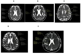
Representative apparent diffusion coefficient (ADC) map images demonstrating region-of-interest (ROI) placement for quantitative ADC measurement in five patients with acute ischemic stroke. Panels (A–E) show circular ROIs positioned within ischemic lesions to obtain quantitative measurements, including mean ADC, standard deviation, and ROI area. Uniform image dimensions were maintained across panels to facilitate visual comparison.
  • Review
  • Open Access

Descending pain modulation is a major determinant of pain processing across physiological and pathological conditions. Although descending pain modulation has long been recognized as bidirectional, capable of both inhibiting and facilitating nociceptive transmission, recent advances now allow it to be examined with far greater anatomical, cellular, neurochemical, and functional precision. Circuit tracing, optogenetic and chemogenetic manipulation, molecular profiling, connectomics, and human neuroimaging have expanded classical brainstem models, revealing a modulatory network in which inhibitory and facilitatory influences are dynamically recruited according to physiological and pathological state. In this narrative review, we revisit descending pain modulation across acute, inflammatory, neuropathic, and nociplastic pain states. We discuss canonical pathways, including the periaqueductal gray–rostral ventromedial medulla axis and locus coeruleus–spinal noradrenergic projections, together with the established dorsal reticular nucleus system and more recently identified descending circuits. We examine how circuit organization, neurochemical diversity, and local and long-range network interactions shape modulatory output across pain conditions. Attention is given to state-dependent plasticity and to the progressive reorganization of descending systems during pain chronification. In this context, reduced inhibitory flexibility and enhanced facilitatory bias emerge as recurrent, but not uniform, features of chronic pain. We also consider how human experimental paradigms, pharmacological approaches, and neuroimaging findings can be interpreted in light of these mechanistic advances, while acknowledging the limits of translating circuit-specific animal findings into integrated human measures. By framing descending pain modulation as a distributed and dynamically reconfigurable network rather than as a unitary analgesic system, this review highlights its relevance to pain phenotyping, treatment stratification, and mechanism-informed therapeutic strategies.

Neurol. Int.

26 September 2026

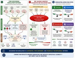
Circuit organization of descending pain modulation. Descending pain control is represented as a distributed, projection-specific, and state-dependent network spanning forebrain, brainstem, and spinal levels. Prefrontal/infralimbic, anterior cingulate, sensorimotor, amygdalar, and hypothalamic inputs convey contextual, affective, motivational, autonomic, and action-related information to brainstem modulatory systems. The periaqueductal gray (PAG) acts as a heterogeneous integrative hub that recruits the rostral ventromedial medulla (RVM), locus coeruleus (LC), dorsal reticular nucleus (DRt/SRD), and additional projection-defined descending routes. Within the RVM, OFF-, ON-, and NEUTRAL-cell populations illustrate the coexistence of inhibitory, facilitatory, and context-dependent microcircuit outputs. LC–spinal noradrenergic projections are shown as predominantly inhibitory through spinal α2-adrenoceptor mechanisms, whereas RVM ON-cell and DRt/SRD outputs contribute to facilitatory gain. The DRt/SRD is represented as a facilitatory and integrative node involved in salience-dependent amplification, homotopic facilitation, and DNIC/CPM-related network configurations. At the spinal dorsal horn, convergent descending influences dynamically regulate inhibitory and facilitatory gain imposed on nociceptive afferent input. Green arrows indicate predominantly inhibitory influences, red arrows predominantly facilitatory influences, purple dashed arrows projection-defined or emerging routes, blue arrows nociceptive afferent input, and black arrows structural or integrative projections. The functional sign of a pathway is not fixed, but depends on receptor subtype, projection target, behavioral context, and pain state. CPM, conditioned pain modulation; DNIC, diffuse noxious inhibitory controls. This is an author-derived conceptual schematic and should not be interpreted as an empirically validated pathway map. The figure was generated with the assistance of SciFig.AI (https://scifig.ai) and ChatGPT and was subsequently reviewed, edited, and refined by the authors through successive rounds of revision to ensure scientific accuracy, conceptual coherence, and consistency with the literature.
  • Case Report
  • Open Access

Background: Unlike acute basilar artery occlusion, non-acute vertebrobasilar occlusion has limited evidence for intervention and requires individualized risk assessment. Case Presentation: A 62-year-old woman developed progressive vertigo, diplopia, dysarthria, and gait ataxia over ten days. Dual antiplatelet therapy was started after posterior circulation ischemia was suspected, but symptoms continued to progress. MRI showed acute to subacute left cerebellar infarction, and CTA showed absent antegrade basilar opacification with collateral distal filling. DSA showed distal left vertebral artery occlusion, retrograde basilar filling through posterior communicating artery collaterals, and hypoplastic right vertebral artery. Endovascular recanalization was performed after multidisciplinary review and informed consent. Intraluminal crossing, stepwise angioplasty, and stent reconstruction restored antegrade basilar flow without angiographic complications. NIHSS improved from 5 to 2 at discharge, and mRS was 1 at 30 days. No follow-up vascular imaging was available. Conclusions: This case illustrates technical feasibility in selected non-acute vertebrobasilar occlusion, but does not establish medical treatment failure, causal recovery, generalizable safety, or long-term durability.

Neurol. Int.

25 September 2026

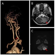
Baseline neuroimaging showing posterior circulation ischemia and vertebrobasilar occlusive disease. (A) Three-dimensional CT angiography demonstrates absent antegrade basilar artery opacification with distal posterior circulation filling through collateral pathways. (B) Axial T2-weighted MRI demonstrates signal abnormality in the left cerebellar hemisphere, consistent with subacute ischemic injury (arrow). (C) Diffusion-weighted imaging demonstrates restricted diffusion in the corresponding left cerebellar region, consistent with acute-to-subacute infarction (arrow).

Highly Accessed Articles

News & Conferences

Latest Issues

Open for Submission

Journal Sections

XFacebookLinkedIn
Neurol. Int. - ISSN 2035-8377