


Metabolites 2026, 16(5), 345; https://doi.org/10.3390/metabo16050345 - 20 May 2026
Abstract
Background/Objectives: The dysregulation of thiol metabolites is strongly linked to hepatocellular carcinoma (HCC) pathogenesis. However, quantifying these highly polar and oxidation-prone thiols in clinical serum samples via conventional liquid chromatography–mass spectrometry (LC-MS) remains challenging due to their poor sensitivity and reproducibility. Methods
[...] Read more.
Background/Objectives: The dysregulation of thiol metabolites is strongly linked to hepatocellular carcinoma (HCC) pathogenesis. However, quantifying these highly polar and oxidation-prone thiols in clinical serum samples via conventional liquid chromatography–mass spectrometry (LC-MS) remains challenging due to their poor sensitivity and reproducibility. Methods: We developed a sensitive and robust iodoacetamine-alkyne (IAM) derivatization–based LC-MS method for quantification of seven trans-sulfuration pathway thiols in human serum. Results: IAM derivatization markedly improved the method’s specificity due to enhanced chromatographic retention and diagnostic MS/MS fragments containing both the alkyne tag and analyte backbone. Sensitivity increased 33-to-160-fold versus underivatized analytes, with limits of detection of 0.02–0.1 nM. All analytes exhibited good linearity, acceptable precision with intra-day and inter-day relative standard deviations in the range of 1.2–13.8%, and high recovery from 88.6% to 102.9%. Conclusions: From the thiol quantification in human serum from 40 HCC patients and 40 healthy controls, it was found that levels of cysteine, homocysteine, glutathione, and cysteinylglycine were significantly lower in HCC patients (p < 0.05). A two-variable logistic regression model using cysteine and cysteinylglycine achieved 90.0% specificity and 80.0% sensitivity for robust HCC discrimination between HCC patients and healthy controls to some extent, with an area under the receiver operating characteristic curve of 0.88 (95% confidence interval: 0.792–0.968).
Full article
(This article belongs to the Special Issue Derivatization Techniques in Mass Spectrometry: Unlocking the Low-Abundance Metabolome)
►
Show Figures
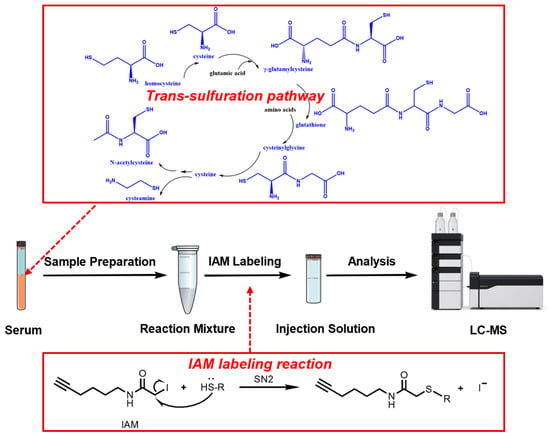
Figure 1


