


Cells 2026, 15(13), 1141; https://doi.org/10.3390/cells15131141 (registering DOI) - 23 Jun 2026
Abstract
►
Show Figures
The multifaceted oncogenic role of teratocarcinoma-derived growth factor 1 (TDGF1) in colon cancer remains incompletely understood. Through integrative bioinformatic and functional analyses, we identified a novel competing endogenous RNA (ceRNA) axis wherein the long non-coding RNA OLMALINC directly sponges hsa-miR-3614-5p, leading to the
[...] Read more.
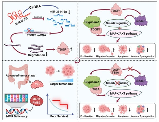
The multifaceted oncogenic role of teratocarcinoma-derived growth factor 1 (TDGF1) in colon cancer remains incompletely understood. Through integrative bioinformatic and functional analyses, we identified a novel competing endogenous RNA (ceRNA) axis wherein the long non-coding RNA OLMALINC directly sponges hsa-miR-3614-5p, leading to the derepression of TDGF1. This OLMALINC/miR-3614-5p/TDGF1 axis promoted colon cancer cell proliferation, migration, invasion, and anti-apoptosis in vitro, whereas TDGF1 knockdown significantly suppressed tumor growth in vivo. Mechanistically, TDGF1 co-activated oncogenic signaling via the Thr88-dependent Nodal/Smad2 cascade and the Glypican-1-mediated MAPK/AKT pathway. Beyond cell-autonomous effects, transcriptomic and single-cell analyses revealed that elevated TDGF1 correlates with an immunosuppressive microenvironment, characterized by reduced immune infiltration and altered LGALS9-CD44 malignant-T cell communication. Clinically, high TDGF1 expression in a tissue microarray cohort was significantly associated with advanced T stage, reduced expression of specific mismatch repair proteins (MLH1/PMS2), and poor overall survival. Collectively, this study delineates the OLMALINC/miR-3614-5p/TDGF1 regulatory circuit and establishes TDGF1 as a multifaceted driver of tumor progression, highlighting its potential as a prognostic biomarker and therapeutic target in colon cancer.
Full article
Graphical abstract


