1. Introduction
Localized prostate cancer is an informative disease model for studying autophagy as a stress-response program because tissue exposure, lesion biology, redox tone, androgen signaling, lysosomal function, and short-term clinical trajectories can still be examined before metastatic selection and multi-line therapy obscure interpretation. In this clinical window, autophagy should be interpreted not as a simple marker of tumor suppression or tumor support, but as a dynamic organelle-centered process that can preserve homeostasis, support adaptation, reveal blocked clearance, or expose stress-overload vulnerability. This clinical setting is heterogeneous and includes patients managed through active surveillance or curative-intent approaches such as radical prostatectomy, external-beam radiotherapy, brachytherapy, and, depending on risk category, androgen-deprivation-based strategies. Radiotherapy is particularly relevant for a stress-response framework because localized low- and intermediate-risk disease can be treated with different fractionation schedules, and moderate hypofractionated radiotherapy has been described as a valid alternative strategy supported by randomized clinical evidence [
1]. Reviews have addressed autophagy genes, autophagy in cancer, and prostate-cancer-specific autophagy contexts [
2,
3,
4,
5], whereas separate syntheses have discussed nutraceutical, soy/isoflavone, and flavonoid-associated activity in prostate cancer models [
6,
7,
8]. Human intervention studies have also assessed green tea catechins, genistein, and polyphenol-rich interventions in localized or high-risk prostate settings [
9,
10,
11,
12,
13,
14]. However, most syntheses describe pathways and compounds more readily than they define when an autophagy-associated readout is biologically interpretable.
The central contribution of this review is therefore not another catalog of autophagy-modulating compounds. It is a state-based, flux-aware interpretive framework that treats autophagy as the organizing principle of a broader stress-response network in localized prostate cancer. Conceptually, the review reframes autophagy-associated readouts as disease-context-dependent states; methodologically, it defines the minimum assays needed to assign meaning to LC3, p62/SQSTM1, lysosomal, and redox signals; translationally, it links those states to clinically accessible windows such as HGPIN, active surveillance, and pre-prostatectomy studies [
15,
16,
17].
That gap matters because autophagy in prostate cancer is not a directional biomarker. Depending on the androgenic context, nutrient availability, mitochondrial stress, and lysosomal function, the same machinery can limit oxidative damage and preserve tissue quality control, or maintain malignant fitness under hypoxia, therapy pressure, and metabolic strain [
2,
3,
4,
5,
18,
19,
20,
21]. A rigorous analysis must therefore decipher the biological state reflected in the readout rather than merely confirming that an autophagy-associated marker has changed.
Flavonoid-associated studies make this interpretive problem difficult to ignore. They are often presented as examples of antioxidants or autophagy-inducing chemoprevention. Yet, many reports rely on parent aglycones that do not reflect the conjugated species measured in human circulation and tissues, use concentrations unlikely to be reached in the prostate, or interpret LC3 and p62/SQSTM1 changes without flux resolution. Under these conditions, an apparently favorable signal may reflect adaptive redox buffering, non-canonical ATG8 lipidation, lysosomal congestion, or a broader organelle-stress program rather than productive macroautophagy.
Unlike previous reviews that discuss autophagy pathways or flavonoid activity as largely separate topics, this review addresses a broader disease-relevant problem in autophagy biology: when can an autophagy-associated readout generated under stress be assigned biological meaning in localized prostate cancer? Previous conceptual, prostate-focused, and autophagy methodology literature provides the background for this question [
2,
3,
4,
6,
8,
15,
16,
22]. We do not aim to rank flavonoids as therapeutic candidates. Instead, flavonoid-associated responses are used here as methodological probes within a broader stress-response framework. LC3, p62/SQSTM1, and puncta formation require flux-aware interpretation before they can be linked to productive autophagy [
15,
16,
23]. ROS modulation may instead reflect adaptive buffering or stress amplification, depending on the biological context [
24,
25,
26,
27,
28]. Lysosomal congestion and blocked clearance require lysosomal competence assays [
29,
30,
31,
32,
33,
34], whereas ferritinophagy-linked stress and organelle-overload claims require pathway-specific controls [
35,
36,
37]. In this sense, flavonoids are illustrative perturbations within a broader stress-response framework that also includes metabolic stress, androgen-pathway manipulation, inflammatory cues, radiation, targeted therapy, and other disease-relevant conditions that alter autophagy-associated readouts without necessarily inducing productive degradation.
This positioning also clarifies the novelty relative to existing reviews of prostate cancer autophagy. Prior reviews have summarized autophagy machinery, therapeutic resistance, androgen signaling, and prostate cancer progression [
2,
3,
17,
38]. The present review builds on that literature but shifts the unit of interpretation from pathways or compounds to evidence-supported autophagy-associated states. The advance is therefore the integration of flux verification, lysosomal competence, redox burden, exposure realism, and localized-disease context into a single decision framework.
This framework directly addresses autophagy as a stress-response program by integrating oxidative burden and disease-stage context in localized prostate cancer [
4,
9,
18,
19,
20,
21,
28]. It also incorporates lysosomal competence and selective autophagy as central interpretive determinants, because static changes in LC3 or p62/SQSTM1 cannot distinguish productive flux from blocked clearance without flux-aware validation [
15,
16,
23] and assessment of lysosomal function [
29,
30,
31,
32,
33]. In addition, mitophagy, ferritinophagy, p62/SQSTM1-NRF2 signaling, and ferroptosis-aware controls are included as stress-related modules that help classify whether the observed response reflects homeostatic control, adaptive tumor survival, blocked clearance, or stress-overload vulnerability. The p62/SQSTM1-NRF2 axis is supported mainly by redox-adaptation literature [
19,
20,
39,
40,
41], whereas ferritinophagy and ferroptosis-aware interpretation require dedicated iron- and lipid-peroxidation controls [
35,
36,
42,
43,
44].
This state-based framework is intended to improve both mechanistic interpretation and translational study design. First, it separates autophagosome-marker movement from productive flux [
15,
16,
23]; second, it places lysosomal function and redox burden at the center of autophagy interpretation; third, it uses flavonoid-associated perturbations as methodological stress tests rather than therapeutic rankings; and fourth, it links each autophagy-associated state to candidate vulnerabilities that can be examined in clinically anchored settings such as high-risk precursor lesions, active surveillance cohorts, and pre-prostatectomy studies [
7,
9,
10,
11,
12,
13,
14]. Tissue exposure and pharmacodynamic plausibility should then be evaluated using metabolite recovery and evidence from prostate tissue pharmacology [
45,
46,
47,
48,
49,
50]. By doing so, this review positions localized prostate cancer as a disease-relevant model in which autophagy, stress adaptation, organelle crosstalk, and translational relevance can be interpreted together.
1.1. Scope, Literature Selection Strategy, and Evidence Hierarchy
This review focuses on interpretation rather than exhaustive cataloging or quantitative effect estimation. Literature was identified through PubMed, Scopus, and Web of Science, with emphasis on studies published between 2010 and early 2026 that were most informative for localized prostate cancer, autophagic flux, lysosomal competence, flavonoid metabolism, redox signaling, and stress adaptation. Autophagy and prostate cancer background searches were anchored in prostate- and cancer-autophagy literature [
2,
3,
8]. In contrast, flux-, lysosomal-competence-, and ferritinophagy-related searches were anchored in the autophagy-monitoring and organelle-stress literature [
15,
16,
29,
30,
43]. Priority search concepts included prostate cancer, localized disease, HGPIN, active surveillance, pre-prostatectomy, flavonoids, catechins, isoflavones, autophagy, flux, lysosome, proteostasis, mitophagy, ferritinophagy, ferroptosis, and disease models.
Search and selection boundaries. The final search was completed in early 2026. Searches combined prostate cancer terms (localized prostate cancer, HGPIN, ASAP, active surveillance, pre-prostatectomy) with autophagy and organelle terms (autophagic flux, LC3, SQSTM1, lysosome, TFEB, mitophagy, ferritinophagy, ferroptosis, NRF2) and exposure terms (flavonoids, catechins, EGCG, isoflavones, genistein, daidzein, equol, apigenin, luteolin, quercetin). Studies were prioritized when they clarified disease-stage context, tissue exposure, flux-aware interpretation, lysosomal function, organelle crosstalk, or translationally interpretable endpoints. Extra-prostate studies were retained only when they informed a specific mechanistic module not yet resolved in localized prostate cancer and are interpreted here as hypothesis-generating rather than confirmatory.
Study selection was guided by the interpretive question rather than by compound identity alone. When several reports addressed the same mechanism, priority was given to studies that provided the strongest evidence tier: human tissue-linked or pathology-linked localized prostate cancer data first, broader prostate cancer data second, prostate-specific preclinical studies with flux-aware assays third, and extra-prostate mechanistic studies only when they clarified a module that remains unresolved in localized disease. Studies were not used for central interpretation when they relied only on supraphysiologic exposure, static LC3 or p62/SQSTM1 changes, or non-prostate models without a clear mechanistic bridge. In such cases, the evidence was retained only as hypothesis-generating support [
17,
24,
38,
45,
46,
51,
52,
53,
54,
55,
56,
57,
58]. When evidence quality was otherwise comparable, studies with clearer disease-stage context, explicit exposure reporting, and functional flux or lysosomal assays were prioritized over purely descriptive reports.
This review was designed as a structured narrative synthesis rather than a systematic review because its main objective is interpretive rather than quantitative. No meta-analysis, formal risk-of-bias scoring, or PRISMA-style study selection flow was performed. This should be considered a limitation of scope rather than an attempt to claim systematic completeness. The field of autophagy in localized prostate cancer is heterogeneous in experimental models, exposure conditions, biomarker panels, disease windows, and endpoint definitions. Under these conditions, a pooled or strictly comparative analysis would risk treating biologically distinct readouts as equivalent. Therefore, the purpose of this review is not to estimate an overall effect size or to rank interventions, but to define conceptual and methodological criteria required to assign biological meaning to autophagy-associated marker changes.
A narrative framework is particularly appropriate here because the central problem is cross-disciplinary. Interpreting autophagy-associated readouts requires integrating autophagic flux, lysosomal competence, redox biology, mitochondrial quality control, iron handling, the realism of pharmacological exposure, and prostate cancer disease stage. The evidence was therefore organized hierarchically, prioritizing studies that link clinically relevant prostate cancer settings with tissue pharmacology, flux-aware readouts, lysosomal function, and translationally interpretable endpoints. This approach allows the review to identify recurring failure modes and propose practical standards for future studies.
Priority was given to clinically informative settings, such as HGPIN, ASAP, active surveillance, and pre-prostatectomy studies, because these designs offer the clearest opportunity to connect interventions, tissue biology, and clinically anchored short-term trajectories [
7,
9,
10,
11,
12,
13,
14]. Human studies were considered especially informative when oral exposure could be linked to prostate tissue pharmacology, biomarker modulation, or pathology-relevant endpoints [
46,
47,
48,
49,
50,
54].
To make interpretation more transparent, the evidence discussed throughout this review is weighed hierarchically. The highest tier includes human intervention studies with tissue or pathology-linked endpoints. A second tier includes tissue pharmacology and metabolite recovery studies that define chemical plausibility in the prostate. A third tier includes prostate-specific preclinical models, especially those using flux-aware designs and autophagy-monitoring logic [
15,
16,
23]. A fourth, hypothesis-generating tier includes broader mechanistic literature used only when it clarifies the meaning of autophagy-related readouts in localized prostate cancer. This extrapolated tier is particularly relevant for lysosomal competence, TFEB signaling, ferritinophagy, ferroptosis, and redox adaptation [
2,
3,
29,
30,
31,
32,
33,
42,
43,
59].
This hierarchy matters because many mechanistic claims become fragile once they are removed from exposure realism, lysosomal competence, or flux testing. Selected extra-prostate studies are therefore used here as interpretive scaffolds rather than substitutes for prostate-specific validation. Wherever mechanistic extrapolation is necessary, it is treated as supportive and provisional rather than confirmatory.
To avoid overinterpreting extrapolated mechanisms, evidence is classified into four interpretive levels throughout this review, as summarized in
Table 1. Localized prostate cancer evidence is given the highest weight when human, tissue-linked, or pre-prostatectomy data connect exposure with pathology-relevant endpoints. Broader prostate cancer evidence supports disease-specific plausibility but is not treated as direct confirmation in localized disease. Extra-prostate mechanistic evidence is used as a mechanistic precedent only when it clarifies unresolved modules such as lysosomal competence, TFEB signaling, ferritinophagy, ferroptosis, or redox adaptation. Finally, hypothesis-generating evidence is useful for identifying future experiments but is insufficient for firm translational conclusions. These levels should be interpreted as confidence qualifiers rather than as a claim that all modules have equivalent support in localized disease.
The present review is therefore positioned against two existing literatures: reviews of autophagy in prostate cancer and of flavonoids in prostate models. Its point of difference is that it asks a narrower and more stringent question than either tradition usually does when autophagy-associated marker changes are reported in localized prostate cancer [
2,
3,
8,
15]. What minimum evidence is required before that movement can be classified as beneficial, adaptive, or artifactual?
1.2. Rationale for an Organelle-Centered Framework
Autophagy is not a linear marker pathway but a distributed organelle program in which autophagosomes, lysosomes, mitochondria, endosomal membranes, ferritin stores, and redox-responsive signaling nodes operate as an integrated network. This organelle-centered view is particularly relevant in localized prostate cancer, where AR signaling, PTEN/PI3K/AKT activity, mitochondrial stress, p62/SQSTM1-KEAP1-NRF2 adaptation, TFEB-linked lysosomal regulation, and peri-prostatic inflammatory cues may produce similar LC3 or p62/SQSTM1 patterns while representing different biological states. A framework focused on organelle crosstalk, therefore, offers a more appropriate basis for translational inference than directional language such as “autophagy induction” or “autophagy inhibition”.
3. Flavonoid-Associated Stress Perturbation Models: Recurring Interpretive Limitations
Flavonoids are useful probes precisely because they perturb multiple systems relevant to the interpretation of autophagy. Redox tone, inflammatory signaling, androgen biology, mitochondrial stress, and PI3K/AKT/mTOR signaling are supported primarily by the mechanistic literature on flavonoids and prostate cancer [
24,
25,
26,
27,
28,
60,
61,
62]. Lysosomal behavior, iron handling, ferritinophagy, and ferroptosis-linked vulnerability require separate pathway-specific interpretation [
35,
36,
37]. Those same features also make them unusually easy to overinterpret when readouts are isolated from biological context.
The first recurring failure mode is exposure mismatch. Many in vitro studies test parent aglycones at concentrations that are difficult to reconcile with human prostate exposure, even though conjugated metabolites dominate circulating and tissue-associated flavonoid pools. Under those conditions, autophagy-related phenotypes may describe a pharmacological artifact rather than plausible tissue biology [
46,
47,
48,
49,
50,
63].
The second failure mode is interpretive inflation around redox markers. Reduced ROS or induction of antioxidant programs is often presented as evidence of beneficial autophagy, yet such changes may reflect adaptive buffering that preserves tumor-cell fitness. A favorable oxidative shift is not synonymous with a favorable autophagic state [
19,
20,
25,
27,
28].
Representative human data illustrate the magnitude of this gap. In a randomized pre-prostatectomy tea study, 20 men consumed 1.42 L/day of green tea, black tea, or a caffeine-matched control for 5 days before radical prostatectomy; tea polyphenols were higher in prostate samples from tea-consuming groups than in controls, whereas they were not detectable in serum, underscoring that tissue recovery and plasma exposure cannot be assumed to be equivalent [
54]. A later prospective randomized trial in 31 men evaluated 1 g/day green tea extract with 800 mg/day quercetin for 4 weeks before prostatectomy and explicitly quantified polyphenols in blood, urine, and prostate tissue, making it a useful exposure-realism benchmark for future flux-aware studies [
45]. Soy-isoflavone supplementation provides an additional numerical anchor: median total isoflavones in supplemented men reached 2.3 micromole/L in prostate tissue versus 0.7 micromol/L in serum, with average tissue concentrations approximately six-fold higher than serum concentrations [
46]. Therefore, in vitro studies using high-micromolar parent aglycones should be interpreted as pharmacological stress tests unless they are justified against measured prostate-tissue exposure and the relevant circulating or tissue-associated metabolite forms [
45,
46,
54].
The third failure mode is treating LC3 puncta, LC3-II accumulation, or p62/SQSTM1 movement as self-interpreting endpoints. None of these markers alone can distinguish increased autophagosome biogenesis from blocked lysosomal clearance. The problem has become even sharper as the field has recognized non-canonical ATG8 conjugation to single membranes and other ATG8-positive states that are not equivalent to productive macroautophagic flux [
15,
16,
23]. Without flux testing, the biological meaning of flavonoid-associated marker movement remains unresolved.
Where available, tandem fluorescent LC3 reporters such as mRFP-GFP-LC3 or GFP-mCherry-LC3 should be used to separate autophagosomes from acidic autolysosomes, particularly when a compound may alter lysosomal pH. In the original tandem LC3 logic, GFP and red fluorescence are both visible before lysosomal fusion. In contrast, the acid-sensitive GFP signal is lost after fusion, and the red-only signal marks autolysosomal maturation. These reporters do not replace biochemical flux blockade, LC3-II turnover, p62/SQSTM1 dynamics, or cargo-degradation assays, but they provide an orthogonal readout that helps distinguish increased LC3-positive vesicles from completed degradation [
24,
57,
58].
A fourth problem is the routine neglect of lysosomal competence. Compounds can perturb membrane trafficking, acidification, repair pathways, or cargo turnover while still producing the appearance of ‘autophagy induction.’ In those settings, cargo accumulation may represent traffic congestion, lysosomal damage responses, or stalled degradation rather than productive recycling [
29,
30,
31,
32,
33].
EGCG studies further illustrate this point, because lysosomal acidification can determine whether an ATG8/LC3-positive response represents incomplete autophagy or a more degradative state [
64].
Finally, literature often collapses biologically distinct outcomes into a single label. Homeostatic turnover, adaptive tumor survival, ferritinophagy-linked stress sensitization, mitophagy-compatible quality control, and lethal stress-overload programs are not equivalent simply because they all involve autophagy-related machinery. Flavonoid studies repeatedly illustrate this conceptual slippage, especially when ferroptosis is inferred solely from changes in ROS or when ferritinophagy is invoked without NCOA4-aware evidence and rescue logic [
35,
36,
42,
43,
62].
For that reason, flavonoids are treated here as mechanistic stress probes rather than as a nutraceutical ranking exercise. Their value lies in revealing where the current literature distinguishes productive autophagy from adaptive buffering, blocked clearance, or stress-overload responses, and where it still relies on unresolved marker movement. The key interpretive task is to classify the autophagy-related state created by the perturbation rather than to record a directional change.
4. Determinants of Autophagic Meaning: Redox Burden, Lysosomal Competence, and Disease Stage
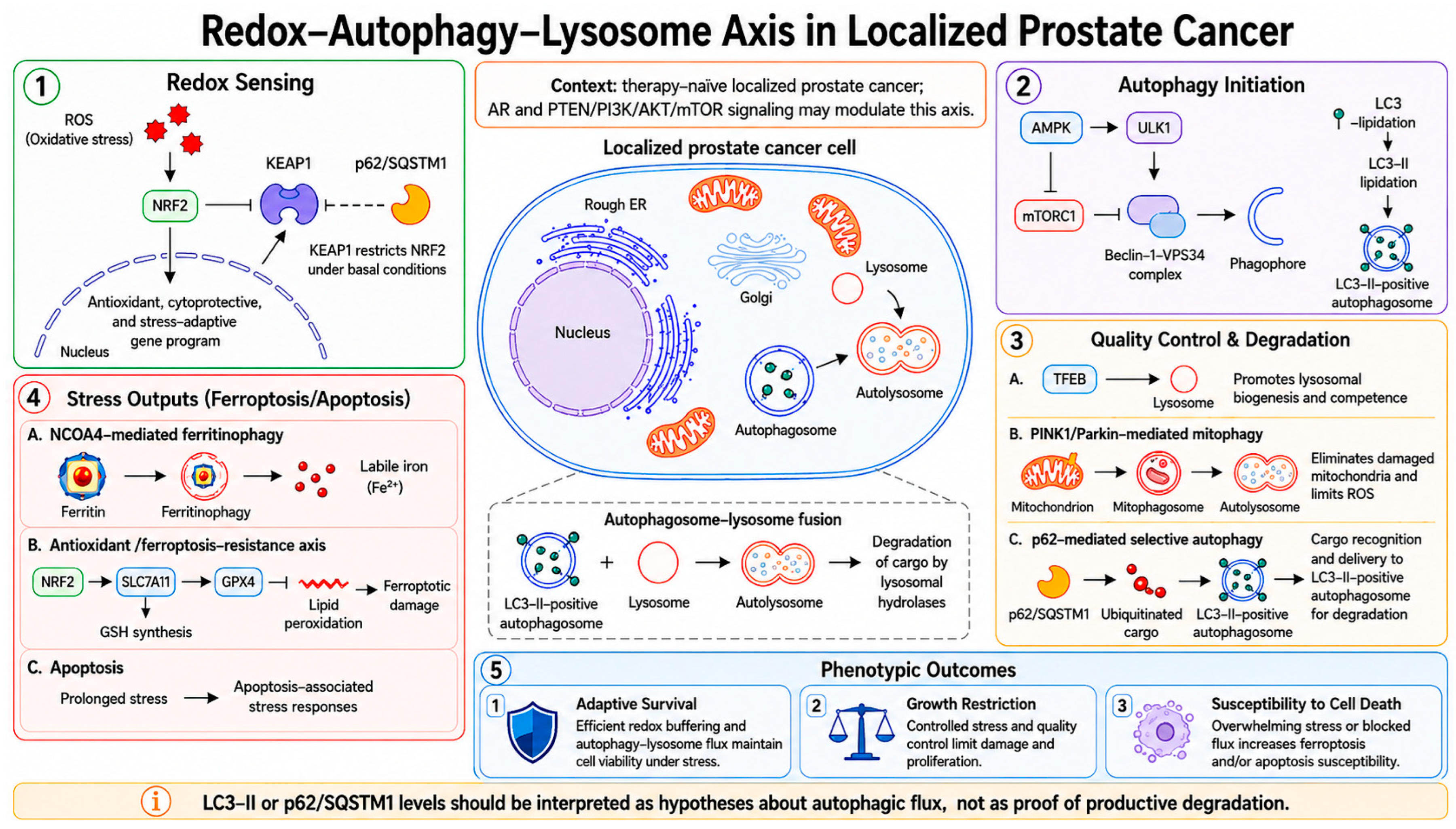
The meaning of autophagy in localized prostate cancer emerges from the integration of redox sensing, nutrient signaling, and degradative competence. AMPK-mTOR-ULK1 signaling, Beclin-1/VPS34-dependent nucleation, and prostate-cancer stress adaptation provide the initiation and disease-context layer [
4,
18,
19,
20,
21,
22,
23]. p62/SQSTM1-KEAP1-NRF2 signaling, TFEB-linked lysosomal biogenesis, and lysosomal damage responses define the redox-lysosome interpretive layer [
29,
30,
31,
32,
33,
59]. Mitophagy, ferritinophagy, and ferroptosis-related modules add organelle-specific and iron-dependent stress controls [
35,
42,
43]. The integrated architecture of these redox, autophagy initiation, quality control, and stress output modules is summarized in
Figure 1.
To clarify why isolated marker interpretation is insufficient,
Figure 1 organizes the main organelle-centered modules that shape the meaning of autophagy in localized prostate cancer, including redox sensing, AMPK-mTOR-ULK1 initiation, p62/SQSTM1 cargo handling, TFEB-linked lysosomal competence, mitophagy, ferritinophagy, lipid peroxidation, and downstream biological outputs.
The main implication of
Figure 1 is that autophagy-associated markers acquire biological meaning only when they are read within this organelle network, rather than as isolated upstream or downstream signals.
Among these axes, the p62/SQSTM1-KEAP1-NRF2 interface is particularly important, as it links autophagy status to antioxidant adaptation. In some prostate-cancer settings, p62/SQSTM1 accumulation and NRF2 activation may not indicate recovery, but rather a rewired cytoprotective state that helps malignant cells tolerate chronic stress. Recent EGCG work further supports caution, as autophagy-dependent regulation of the p62/SQSTM1-mediated antioxidant survival pathway can influence selective cancer cell death. p62/SQSTM1 behavior should therefore be interpreted alongside flux, pathway context, and redox-state readouts rather than as an isolated indicator of protective autophagy [
19,
20,
39,
40,
41,
59].
Autophagy in prostate cancer is also closely linked to lineage signaling and lysosomal regulation. Hypoxia-associated p62/SQSTM1 phosphorylation can promote androgen receptor degradation, while lysosomal status is coupled to TFEB translocation and mTORC1 signaling, two nodes that determine whether degradative capacity is preserved or exhausted [
21,
22,
29,
30,
31,
32,
33]. These links complicate any simple claim that changes in autophagy-related markers are inherently favorable or unfavorable. EGCG-related TFEB activation reported in renal cell carcinoma further supports the value of interpreting TFEB-linked lysosomal regulation as a mechanistic module, while still treating it as extra-prostatic evidence requiring prostate-specific validation [
65].
EGCG-related studies also illustrate why lysosomal competence should be treated as an interpretive determinant rather than as a secondary assay. Zhong et al. reported that EGCG counteracted HBV-associated incomplete autophagy by improving lysosomal acidification, thereby shifting the meaning of autophagy-associated readouts from stalled or incomplete processing toward a more degradative state. For localized prostate cancer, this finding should be interpreted as a mechanistic precedent rather than direct disease-specific evidence: when flavonoids are proposed to regulate autophagy, assays of lysosomal acidification, cargo turnover, and flux blockade are necessary to determine whether the observed response reflects productive degradation or unresolved marker accumulation [
63].
Methodology, therefore, matters as much as biology. Static accumulation of LC3 puncta or LC3-II does not, by itself, prove increased flux. The same signal may arise from enhanced autophagosome formation, impaired lysosomal degradation, altered membrane trafficking, CASM-related ATG8 lipidation, or broader stress-associated remodeling. The same caution applies to p62/SQSTM1, whose abundance reflects both synthesis and turnover [
15,
16,
23,
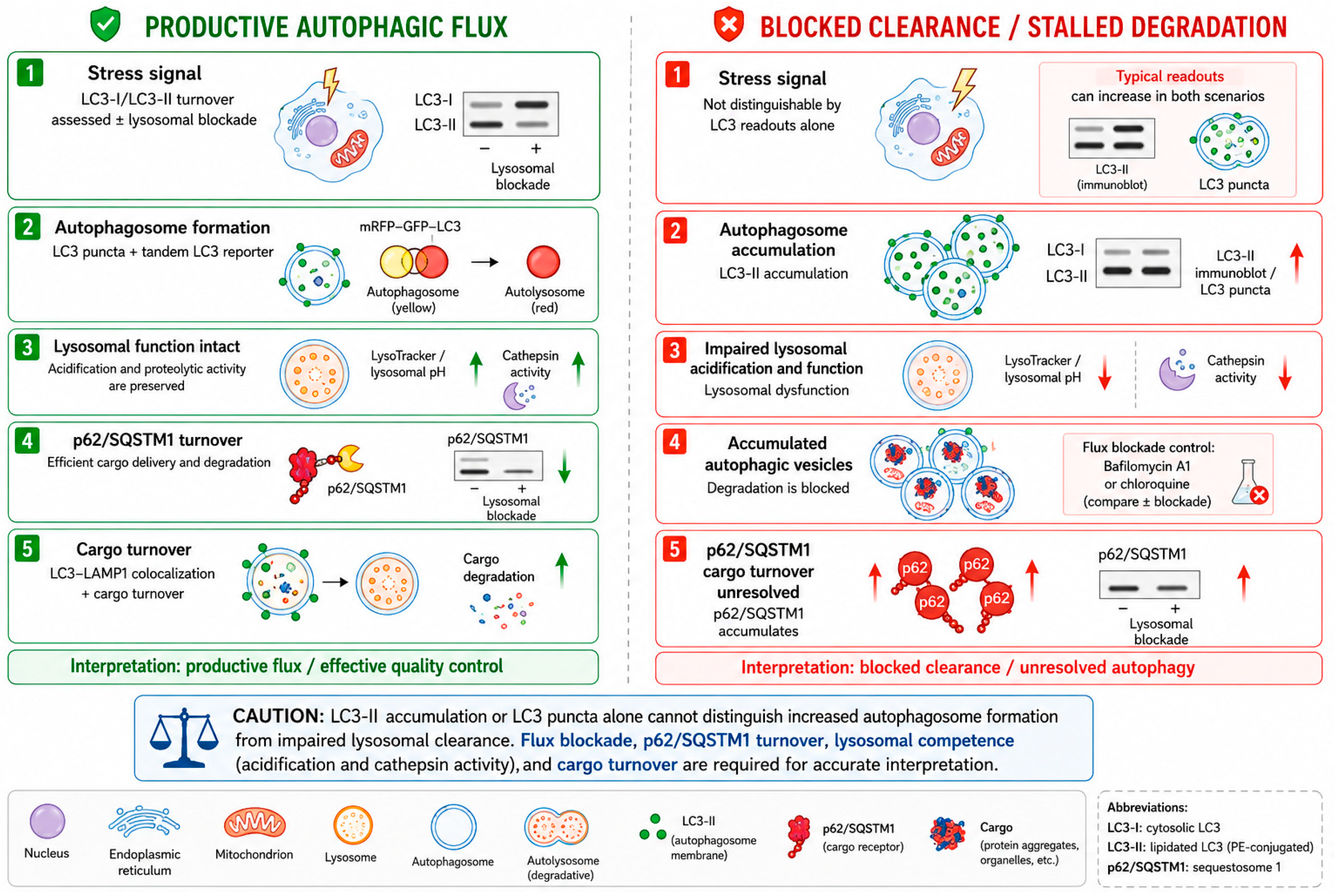
59]. This distinction between productive flux and blocked clearance is illustrated in
Figure 2.
Because LC3-II accumulation, LC3 puncta, and p62/SQSTM1 changes can arise from opposite biological situations,
Figure 2 separates productive autophagic flux from blocked clearance. This distinction is central to the proposed framework because both states may look similar when only static markers are measured.
Thus, flux-blockade and lysosomal-competence assays are essential, not optional.
Figure 2 shows that similar LC3/p62/SQSTM1 patterns may indicate either increased degradative flux or stalled clearance, making flux-blockade and lysosomal-competence assays essential rather than optional.
This is why the current autophagy-monitoring guidelines remain central to any serious interpretation. LC3 dynamics, p62/SQSTM1 behavior, pharmacological lysosomal blockades, and orthogonal imaging or colocalization approaches should be interpreted together rather than treated as interchangeable surrogates for flux [
15,
23].
In practical terms, flavonoid-associated autophagy claims are strongest when they show that lysosomal function remains intact, that cargo turnover is occurring, and that the resulting phenotype can be placed within a biologically coherent state rather than inferred from marker accumulation alone. Where lysosomal damage or repair is relevant, that state should also be separated from lysophagy and from endo-lysosomal damage responses more broadly [
29,
30,
31,
64,
65].
From a translational standpoint, a minimum autophagy package should therefore include at least one marker of autophagosome formation, one marker of cargo turnover, and one assay that directly addresses lysosomal competence or pharmacological flux blockade. In ferritinophagy-linked settings, NCOA4-aware readouts and ferroptosis-specific controls are also needed; in mitophagy-linked settings, mitochondrial quality-control measurements should be explicit rather than assumed [
42,
43]. These minimum criteria are summarized in
Table 2.
Not every pathway discussed in this review has been established directly in prostate-specific systems. Some components, particularly ferroptosis-linked responses and certain forms of flavonoid-induced redox stress, are informed in part by broader cancer literature. Those parallels are useful for interpretation, but they should not be treated as equivalent to prostate-specific confirmation [
35,
36,
42,
43,
62].
Accordingly, the mechanistic maps and case studies below apply this evidence-status logic to distinguish prostate-specific support from mechanisms that are extrapolated or hypothesis-generating. The tables should therefore be read as interpretive tools, not as claims that every pathway has equivalent validation in localized prostate cancer. The minimum evidentiary standard required to assign autophagic meaning in localized prostate cancer is summarized in
Table 3. This standard emphasizes that LC3-II accumulation, LC3 puncta, p62/SQSTM1 abundance, lysosomal status, and phenotype-level outcomes cannot be interpreted in isolation. Instead, each readout must be paired with complementary assays that clarify flux, lysosomal competence, cargo turnover, pathway specificity, and disease-stage relevance before any biological or translational meaning is assigned.
Operationalizing the decision framework requires a strict stepwise approach: from observed readout to required control, and finally to a provisional biological state. This sequence ensures that mere shifts in static markers are not mistakenly interpreted as therapeutic benefits. The stepwise decision logic is summarized in
Table 4.
The proposed framework also requires pathway-specific interpretation, as distinct stress-response modules may yield overlapping autophagy-associated readouts. Therefore, redox signaling, p62/SQSTM1-KEAP1-NRF2 adaptation, AMPK-mTOR-ULK1 initiation, PI3K/AKT/mTOR survival signaling, inflammatory stress, TFEB-linked lysosomal regulation, ferritinophagy, ferroptosis, and mitophagy should not be collapsed into a single directional label of autophagy induction or inhibition. The main pathway modules, core nodes, recommended readouts, and interpretive caveats are summarized in
Table 5.
5. A Flux-Aware Framework for Interpreting Autophagy Responses
The decision framework is applied through three linked questions: which disease window is being modeled, what level and type of redox stress is being imposed, and whether the lysosomal compartment remains sufficiently competent to complete cargo degradation. These questions convert isolated marker changes into a provisional biological state rather than a directional claim.
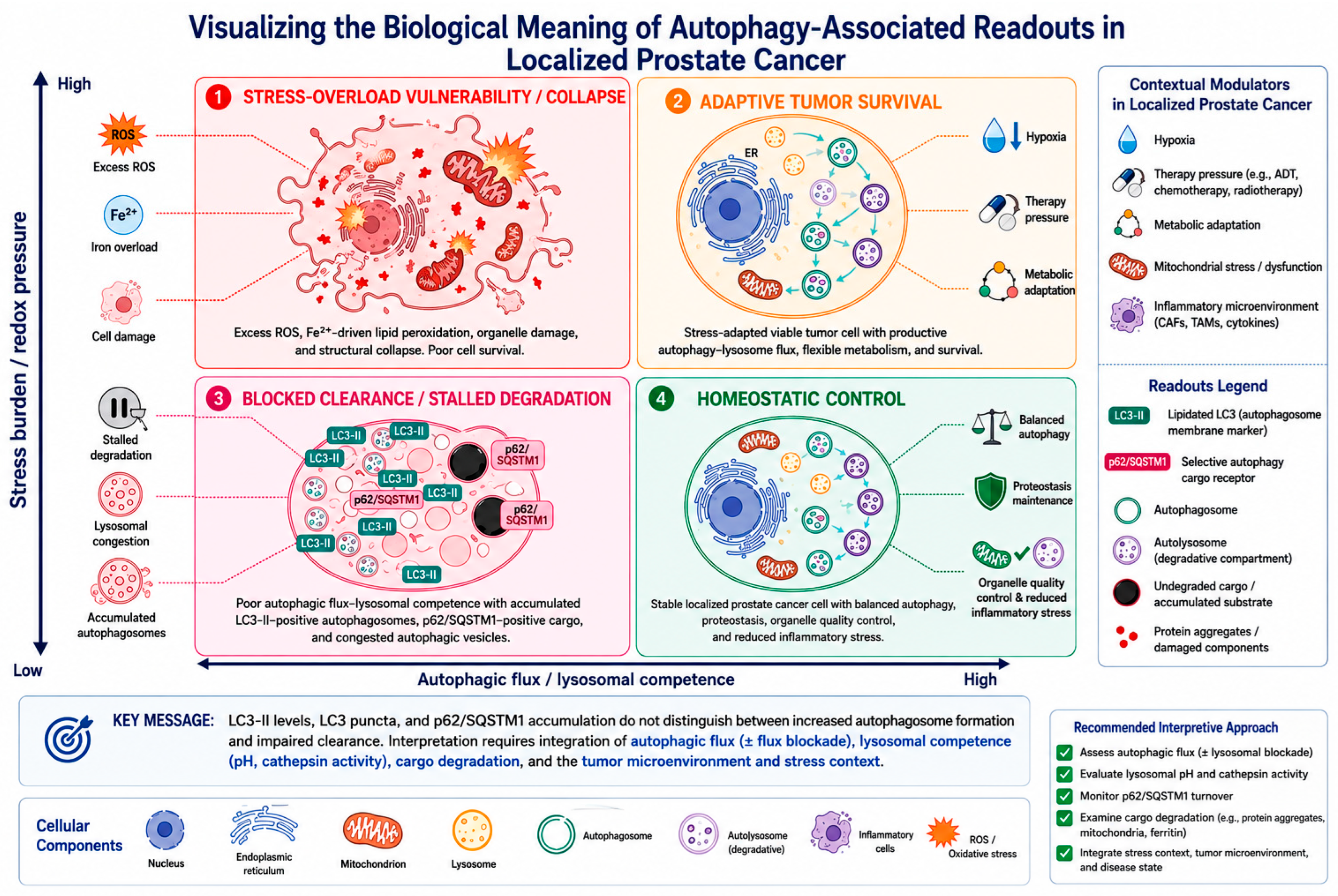
Disease stage defines the biological window, redox burden determines whether stress is buffered or amplified, and lysosomal competence determines whether the signal represents productive degradation or congestion within the degradative system. Together, stress burden/redox pressure and lysosomal competence generate the four-state model shown in
Figure 3.
The four-state model is not intended to replace established autophagy-monitoring guidelines. Rather, it translates flux-monitoring principles and lysosome-centric interpretation into disease-relevant categories for localized prostate cancer. In this respect, the model extends previous flux-based and prostate cancer autophagy frameworks by adding two contextual axes that are often underdeveloped in prostate cancer reviews: realistic tissue exposure and disease-window-specific biological meaning [
15,
16,
17,
24,
38,
57,
58].
This model is intended as a classification tool, not as a claim that all four states have equivalent levels of direct evidence in localized prostate cancer. Operationally, each state requires a different combination of flux-aware assays, lysosomal-competence readouts, pathway-specific controls, and phenotype-level endpoints. The evidence requirements, biological interpretation, and candidate translational vulnerabilities associated with each state are summarized in
Table 6.
This model is intended as a classification tool, not as a claim that all four states have equivalent levels of direct evidence in localized prostate cancer.
Operationally, homeostatic control is assigned when intact flux and lysosomal competence reduce damage without preserving malignant fitness; for example, low redox burden with cargo clearance in early lesions or surveillance tissue would fit this state. Adaptive tumor survival is conferred when the same intact flux supports viable tumor cells under hypoxia, androgen stress, therapeutic pressure, or metabolic strain. Thus, the boundary between the two states is not the presence of flux itself, but the phenotype and disease context coupled to that flux.
Redox burden matters because flavonoids are not fixed antioxidants. At modest stress intensity, they may dampen inflammatory or mitochondrial stress enough to support quality control and proteostasis. At higher intensity, or in cells already near the threshold for oxidative or iron-dependent injury, the same compounds may amplify vulnerability and drive the system toward apoptosis- or ferroptosis-linked collapse [
24,
25,
26,
27,
28,
35,
36]. This context dependence is also illustrated by EGCG studies in melanoma, NSCLC, and hepatoma models, where EGCG either decreased autophagy or leveraged autophagy inhibition to enhance drug-induced cell death, reinforcing the notion that autophagy modulation cannot be assigned a single direction across systems [
66,
67,
68].
Lysosomal competence is the third and often neglected axis. If degradation remains intact, increased autophagosome formation may indicate productive flux. If lysosomal function is compromised, however, similar marker changes may signify congestion within the degradative system. Without addressing this distinction, translational claims remain unstable [
4,
18,
22,
23].
A flavonoid-associated autophagy claim should be considered interpretable only when realistic exposure is linked to a defined readout package: markers of autophagosome formation, cargo turnover, lysosomal competence, and a phenotype consistent with homeostatic control, adaptive tumor survival, blocked clearance, or stress-overload vulnerability. At that level, flavonoid-associated biology is well-suited to mechanistic and translational inference [
15,
16,
23,
64,
69].
6. Stress-Test Case Studies for Organelle-Centered Autophagy Interpretation
The flavonoids most often discussed in localized prostate cancer should not be treated as a uniform therapeutic class. They are more informative when read as distinct stress perturbation case studies that expose where autophagy interpretation is relatively strong, where it remains chemically implausible, and where marker inflation continues to distort conclusions. These case studies are not intended to rank flavonoids therapeutically, but to test how the proposed framework handles different levels of exposure realism, mechanistic support, flux resolution, lysosomal competence, and disease relevance.
To avoid treating all compounds at the same evidence level, the case studies are stratified by the strength of human evidence, tissue pharmacology, and flux-aware validation, with human prostate tissue studies as the highest-exposure anchor and autophagy-monitoring guidance as the methodological anchor [
24,
45,
46,
54,
57,
58]. EGCG/green-tea catechins and soy isoflavones provide the most useful human or tissue-linked anchors, while quercetin, apigenin, luteolin, and carnosic acid are preclinical or mechanistic probes. Curcumin and resveratrol are included as non-flavonoid polyphenolic comparators to demonstrate that the framework is not limited to flavonoids [
52,
55,
56]. This evidence-stratification logic is summarized in
Table 7.
The representative flavonoid-associated perturbation case studies used to stress-test the framework are summarized in
Table 8. These examples are not intended to rank compounds therapeutically, but to show how different levels of exposure realism, redox modulation, lysosomal competence, pathway specificity, and disease relevance influence the interpretation of autophagy-associated readouts in localized prostate cancer.
6.1. When Tissue Exposure and Autophagy Readouts Begin to Converge
Green tea catechins provide one of the clearest examples of how tissue exposure and autophagy interpretation can be examined within the same disease window in localized prostate cancer, because they have been studied in HGPIN cohorts, pre-prostatectomy settings, and tissue-recovery designs in humans [
9,
10,
11,
12,
46,
47,
48]. This does not establish benefit on its own, but it makes catechins a useful interpretive benchmark for assessing whether exposure, sampling, and autophagy readouts are aligned.
Their value for the present review lies in the distinction between interpretable and weakly interpretable evidence. Stronger catechin studies link oral intervention to tissue recovery, controlled exposure, or biologically anchored sampling [
9,
10,
11,
12,
46,
47,
48]. By contrast, weaker mechanistic claims invoke high-dose in vitro LC3-II accumulation, EGCG-associated survival effects, or generic antioxidant responses without resolving metabolite realism, flux, or lysosomal function [
33,
34,
40,
64,
70,
71,
72,
73].
Pre-prostatectomy trials are particularly valuable because they do not need to establish long-term effectiveness to provide useful information. Their primary benefit is that they link drug intake, exposure to the target organ, and pharmacodynamic measurements within the same patient. This connection is exactly what the study of autophagy has been lacking in much of the field [
12].
Modernelli et al. provides a prostate-specific example of why EGCG cannot be treated as a simple anticancer autophagy inducer. In prostate cancer cells, EGCG antagonized bortezomib cytotoxicity via an autophagic mechanism, suggesting that EGCG-associated autophagy may support survival in some therapeutic contexts rather than uniformly promote tumor suppression. This finding is especially useful for the present framework because it links EGCG, prostate cancer, therapeutic stress, and context-dependent autophagy within a single experimental system [
31].
Beyond prostate-specific evidence, EGCG studies in other systems reinforce the need to resolve the degradative arm of the pathway. Zhong et al. showed that EGCG can oppose incomplete autophagy by enhancing lysosomal acidification, whereas Kim et al. reported that EGCG-induced autophagic flux can reduce TRAIL-induced apoptosis. These examples support the central point of this review: EGCG-related signals should be classified by flux, lysosomal competence, and phenotype rather than by LC3 or p62/SQSTM1 movement alone [
63,
68].
Catechins, therefore, serve as a benchmark for what an autophagy-oriented study looks like when exposure, tissue sampling, flux resolution, lysosomal readouts, and phenotype-level interpretation are aligned.
6.2. Soy Isoflavones, Daidzein, and Equol: Interpretation Requires Metabolic Stratification
Soy isoflavones remain compelling because they sit at the intersection of redox biology, endocrine signaling, microbiome-dependent metabolism, and tissue pharmacology. In localized prostate cancer, that combination makes them highly relevant but also unusually heterogeneous [
7,
13,
54].
The strongest interpretation arises when short-term intervention is linked to measurable exposure and to pathway-aware biological sampling, as in pre-prostatectomy work with genistein [
13]. The weakest interpretation appears when all participants are treated as chemically equivalent despite major differences in microbial conversion and metabolite production.
Equol status is especially important in this respect. Because only a subset of individuals efficiently converts daidzein to equol, similar interventions may generate substantially different exposures among patients. Ignoring that variability can blur real biological differences and make autophagy-related readouts appear inconsistent when the underlying chemistry is heterogeneous [
54,
74,
75].
Accordingly, soy interventions are most informative when metabolite identity, equol-producer phenotype, and tissue-level exposure are built into the design. Without that stratification, the interpretation of autophagy remains chemically underdetermined. This point expands the equol-producer phenotype into a broader host-metabolic stratification problem. Microbiome-dependent biotransformation can change the chemical species that reach the prostate, while obesity, diet, antibiotic exposure, and inflammatory tone may reshape redox state and lysosomal fitness. Future isoflavone studies should therefore predefine equol-producer status and, when feasible, profile microbial metabolites rather than treating all participants as chemically equivalent.
6.3. Apigenin and Luteolin: Mechanistic Probes of Stress-Program Interpretation
Apigenin and luteolin are often discussed together as broad anticancer flavones, but they are more useful as mechanistic probes than as independent translational candidates. Their main value lies in the autophagy-related questions they help clarify, rather than in any straightforward ranking of clinical potential [
36,
76].
Luteolin is particularly useful because it brings ferritinophagy, TFEB signaling, iron handling, and ferroptotic vulnerability more directly into the discussion than most other compounds in this review. That makes it a powerful example of why ROS alone is insufficient; iron flux, lipid peroxidation, GPX4/SLC7A11 status, and rescue controls are necessary to classify the induced state [
35,
36,
62]. A recent luteolin-focused review now provides background on the broader pharmacological context of luteolin. At the same time, the ferroptosis/ferritinophagy claim remains anchored in prostate-specific mechanistic evidence where available [
53].
Therefore, ferritinophagy- and ferroptosis-related modules are presented here as interpretive extensions that require prostate-specific validation, rather than as established mechanisms in localized prostate cancer.
Apigenin, by contrast, is frequently discussed in terms of apoptosis or autophagy-associated markers without equivalent attention to pharmacological realism or flux resolution. It therefore illustrates how quickly mechanistic richness can outpace interpretive discipline when autophagy is inferred rather than demonstrated [
8,
34,
66].
Taken together, these flavones are best seen as tools for stress-program interpretation: they can clarify when lysosomal engagement, ferritin turnover, or redox amplification is being mistaken for a generic anticancer autophagy signal.
6.4. Quercetin and Related Flavonols: The Problem of Static Marker Inflation
Quercetin illustrates one of the field’s most persistent interpretive problems: the tendency to convert any shift in LC3- or p62/SQSTM1-associated readouts into evidence of favorable autophagy. Because quercetin is widely available and mechanistically versatile, it is often used to support broad claims that exceed the actual strength of the data [
26,
27,
28].
That does not make quercetin unimportant. On the contrary, it may be one of the more useful system-level modulators in the group. The problem is that its literature is especially prone to static-marker inflation, exposure mismatch, and overgeneralized antioxidant language. In that sense, quercetin is a case study in how a plausible compound can still be interpreted poorly.
Human data remain informative mainly for feasibility and tolerability, not for strong claims about autophagy-driven efficacy. Quercetin is therefore more useful here as a cautionary example of interpretive excess than as a compound to be advanced based on static-marker changes alone.
Human studies that help classify, rather than merely claim, autophagy-associated responses in localized prostate cancer are summarized in
Table 9. These studies are most useful when they connect oral exposure, prostate tissue recovery, metabolite identity, pathology-linked endpoints, and biologically interpretable readouts. However, even in these clinically anchored settings, autophagy-related conclusions remain provisional unless tissue exposure is paired with flux-aware assays, lysosomal-competence measurements, and phenotype-level interpretation.
Human studies remain valuable in this field, but their value is interpretive rather than automatically confirmatory. They help define which classes have realistic exposure, which trial designs permit tissue sampling, and which biomarkers can be tied to clinically meaningful windows of disease [
7,
9,
10,
11,
12,
13,
14].
Taken together, the human studies literature should be synthesized by clinical window rather than by compound enthusiasm. HGPIN studies are useful for prevention-oriented biological signals; active-surveillance cohorts are useful for longitudinal risk stratification; and pre-prostatectomy window studies are the strongest design for pairing exposure with tissue pharmacodynamics and organelle-level readouts. The key limitation is that most human studies do not yet include a complete autophagy flux and lysosomal competence package.
The Pomi-T trial illustrates both the promise and the interpretive limit of complex polyphenol-rich interventions. Such mixtures may generate measurable biological signals, but once several bioactive components are combined, it becomes much harder to attribute an autophagy-related state to any one compound or mechanism [
14].
For that reason, the present evidence base supports neither broad dismissal nor generic optimism. Flavonoids remain useful only insofar as they help classify what kind of autophagy-related biology is produced under realistic conditions. Progress depends on clearly defined interpretive criteria rather than enthusiasm for any compound class.
The most informative evidence still comes from pre-prostatectomy studies and well-defined high-risk cohorts, where exposure can be linked to tissue pharmacodynamics. Studies focused mainly on PSA modulation or on heterogeneous mixtures are comparatively weaker for interpreting autophagy, even when they are biologically suggestive [
7,
9,
10,
11,
12,
13,
14].
7. Minimum Standards for Interpreting Autophagy-Associated Stress Responses in Localized Prostate Cancer
First, the chemical species under study should be pharmacologically plausible. For flavonoid-related stress perturbations, the parent aglycone tested in vitro should not be treated as a direct substitute for the conjugated or metabolized species recovered in plasma or prostate tissue. For non-dietary perturbations, the same principle applies: dose, duration, cell state, and tissue accessibility should be compatible with the disease window being modeled.
Second, autophagy must be assessed through flux-aware logic. LC3 and p62/SQSTM1 should be interpreted together, with lysosomal blockades or complementary assays to determine whether cargo turnover is occurring. Reviewers should be able to tell from the methods whether a reported state reflects productive flux, CASM-related ATG8 lipidation, stalled degradation, or altered lysosomal acidification [
15,
16,
23,
64,
69].
Constructive benchmarks are available. Stronger experimental designs do not merely report LC3-II or p62/SQSTM1; they pair these markers with lysosomal blockade, tandem LC3 reporters, lysosomal acidification, cargo turnover, viability, or rescue logic and pathway-specific markers such as NCOA4 or GPX4/SLC7A11 when ferritinophagy or ferroptosis is invoked. These examples convert a critique of static markers into a practical experimental standard.
Third, tissue and host context should be made visible. The equol-producing phenotype and microbial metabolite heterogeneity should be treated as exposure modifiers [
44,
74,
75]. Obesity, peri-prostatic adipose signaling, inflammatory tone, AR status, PTEN/PI3K/AKT context, and oxidative baseline can further reshape the meaning of the flavonoid-associated autophagy signal [
19,
20,
27,
28,
35,
39,
60,
61,
62]. Lysosomal competence should be treated as a distinct determinant rather than subsumed under generic stress biology [
15,
16,
23,
29,
30,
31,
32,
33]. Fourth, mechanistic movement should be paired with clinically interpretable anchors. Tissue pharmacodynamics, pathology-linked endpoints, repeat biopsy, imaging progression, or clearly staged surveillance metrics are more informative than biomarker drift in isolation [
7,
9,
10,
11,
12,
13,
14].
Common Failure Modes and Corrective Standards
A recurring failure mode is exposure mismatch: the biological conclusions are based on conditions unlikely to occur in the human prostate. The corrective standard is to anchor in vitro work to tissue-relevant concentrations or to explicitly justify departures from them.
A second failure mode is static-marker inflation: LC3-II accumulation, puncta formation, or changes in p62/SQSTM1 are reported as though they resolved flux on their own. The corrective standard is a minimal assay package that directly assesses cargo turnover and lysosomal competence and, where relevant, excludes CASM-related misclassification [
15,
16,
23].
A third failure mode is analytical distortion introduced during tissue handling. Ex vivo deconjugation can exaggerate apparent aglycone recovery, thereby inflating claims about which chemical species were truly present in vivo. The corrective standard is rigorous reporting of sample-processing and metabolite-recovery conditions [
49,
50].
A fourth failure mode is unmodeled biological heterogeneity. Equol status and microbial-metabolite production can alter exposure in ways that should be anticipated analytically [
54,
74,
75]. Inflammatory tone, obesity-associated adipose signaling, and tumor metabolic context can modify the interpretation of stress responses [
60,
61,
62], whereas baseline lysosomal fitness can shift the same marker pattern toward productive flux or blocked clearance [
29,
30,
31,
32,
33]. The corrective standard is a stratified design rather than a retrospective explanation.
Taken together, the main barriers are exposure mismatch, analytical distortion, static-marker inflation, unmodeled biological heterogeneity, and premature claims that conflate ferritinophagy- or mitophagy-associated signatures with resolved autophagic flux.
8. Study-Design Priorities for Organelle-Centered, Flux-Aware Autophagy Studies
Future studies should remain anchored in clinical settings where autophagy interpretation can be tested, including active surveillance cohorts, HGPIN or ASAP-associated studies, pre-prostatectomy intervention windows, and tissue-linked pharmacodynamic designs. In these settings, autophagy-associated readouts should be interpreted only when exposure realism, disease-stage context, flux resolution, lysosomal competence, and phenotype-level endpoints are evaluated together. The main interpretive barriers and corresponding design corrections are summarized in
Table 10.
Future studies should remain anchored in clinical settings where autophagy interpretation can actually be tested, particularly HGPIN, ASAP, active surveillance, and pre-prostatectomy cohorts. These settings allow the study of tissue biology, pharmacodynamic sampling, and short-term disease behavior together before late-stage selection pressures dominate the phenotype.
Pharmacology must move from a supporting role to a central one. Plasma measurements alone are insufficient; what matters is whether flavonoid-derived species reach the prostate, in what chemical forms they arrive, and whether those species engage the pathways later invoked to explain the phenotype.
The same principle applies to autophagy. Studies that rely solely on LC3-II accumulation do not tell us whether a compound promotes productive flux, stalls lysosomal clearance, corrects incomplete degradation, or triggers a broader stress program that happens to alter autophagy-related markers. Trial designs should therefore treat flux resolution and lysosomal function as requirements rather than optional mechanistic add-ons [
15,
16,
23,
64,
69]. Examples in EGCG-treated systems show that this package should include the degradative phase itself, because lysosomal acidification, lysosomal membrane integrity, and autophagic flux can change the biological interpretation of the same marker pattern [
17,
64,
69].
The host context deserves equal attention. Obesity, peri-prostatic adipose tissue, inflammatory tone, and tumor molecular background are not secondary variables; they can reshape redox stress, lipid handling, and the very meaning of autophagy-related readouts.
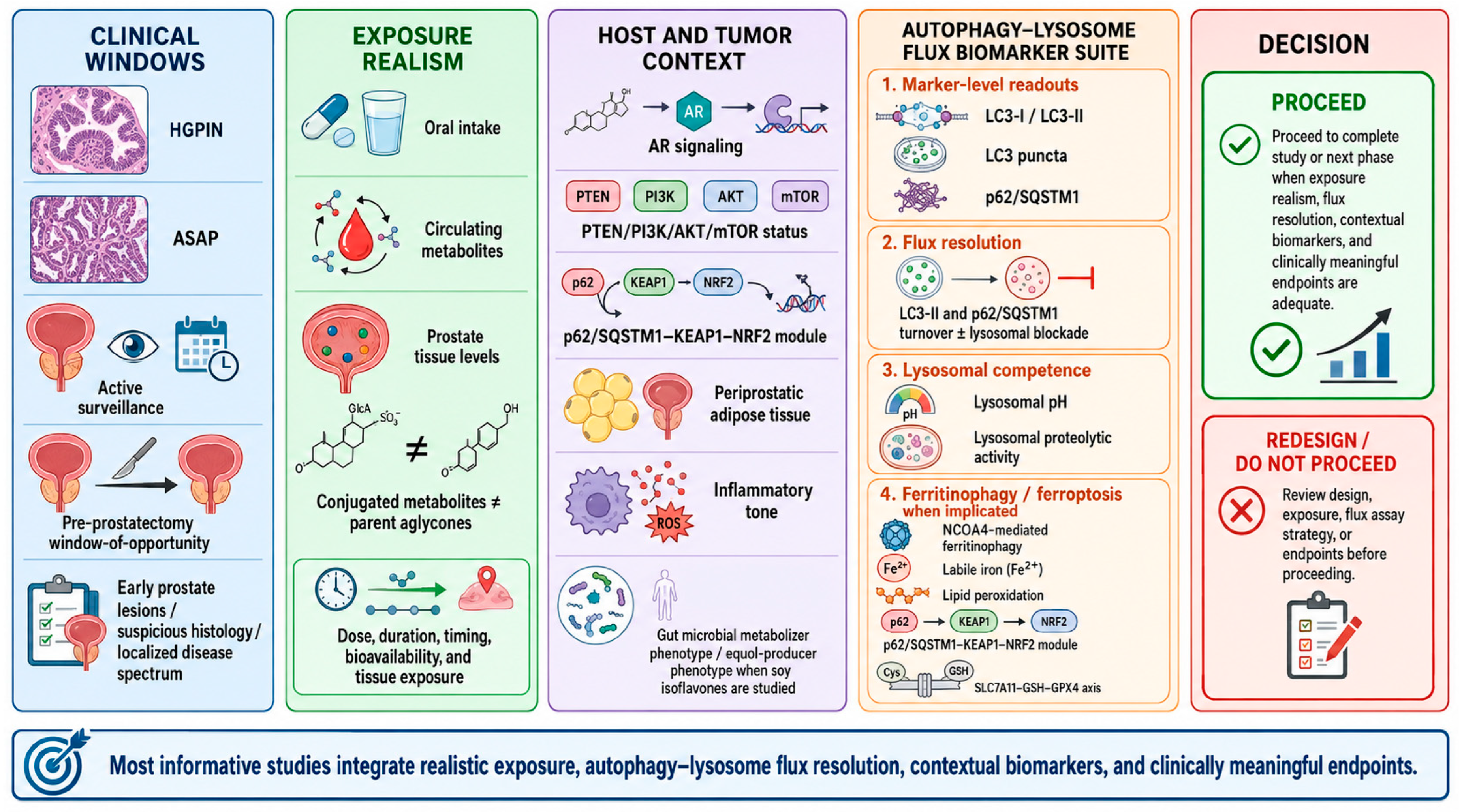
A stronger next-generation trial should therefore predefine a biomarker package aligned with the mechanism: circulating metabolite profiling, tissue compound quantification when feasible, LC3 plus p62/SQSTM1 plus lysosomal pH or proteolytic-activity assays, cargo-turnover measurements, and pathway-specific modules such as p62/SQSTM1-NRF2 signaling or iron and lipid peroxidation markers when ferritinophagy or ferroptosis is implicated [
40,
41,
44,
64,
69]. A study-design workflow based on this biomarker package is shown in
Figure 4.
For example, a pre-prostatectomy study could combine circulating metabolite profiling, quantification of prostate tissue compounds, LC3 and p62/SQSTM1 dynamics with lysosomal blockade or a tandem LC3 reporter, lysosomal pH or cathepsin activity, cargo-degradation assays, and paired tissue endpoints such as proliferation, oxidative damage, inflammatory tone, or pathology-linked features. Such a design would not simply ask whether autophagy increased; it would ask which autophagy-associated state was produced and whether that state created a testable organelle-centered vulnerability.
Figure 4 translates the conceptual framework into study-design priorities. It outlines how future studies should connect clinically relevant disease windows, realistic exposure conditions, host and tumor contexts, flux-aware biomarker packages, and decision points to advance or redesign translational studies.
Study advancement should depend on whether exposure, flux, organelle function, and phenotype point to the same biological state; discordant signals should trigger redesign rather than stronger mechanistic claims.
Future studies should also integrate prostate-specific metabolic rewiring, particularly lipid metabolism and androgen-linked bioenergetics, because these factors influence whether redox modulation is buffered, exploited, or pushed toward lethal stress [
60,
61,
62].
Different perturbations, including flavonoids, are best understood by testing them across localized disease rather than as entries in a single hierarchy of promise. In some study designs, the main question is tissue exposure; in others, it is metabolic stratification, lysosomal competence, ferritinophagy-aware classification, or the separation of adaptive buffering from stress overload.
Non-flavonoid examples illustrate the same logic. Carnosic acid, curcumin, and resveratrol can all generate autophagy-associated or organelle-stress readouts. However, those readouts should still be classified by exposure realism, flux, lysosomal competence, and phenotype rather than by the compound class itself [
52,
55,
56]. The inclusion of these examples, therefore, strengthens rather than broadens away from the central argument: prostate-specific or compound-specific activity is insufficient unless the observed LC3/p62/SQSTM1 pattern is resolved with flux and degradative controls [
24,
38,
57,
58]. This makes the framework a general interpretive tool for stress-response biology, not a flavonoid-specific argument.
In localized prostate cancer, flavonoid responses are used here as examples of a broader problem: autophagy-related claims become interpretable only when readouts are linked to exposure realism, flux, degradative capacity, and disease-stage context. Their value in this review lies as much in methodological as in biological grounds, because they serve as demanding test cases for determining whether observed readouts support restored homeostasis, adaptive tumor survival, blocked clearance, or stress-overload vulnerability.
The most credible subset of literature comprises studies that link realistic exposure to tissue pharmacology and biomarker packages capable of resolving the meaning of autophagy. This is where the field can move from descriptive claims to testable interpretive criteria.
8.1. Why This Organelle-Centered Framework Matters
Autophagy remains one of the most frequently invoked but often insufficiently resolved mechanisms in prostate cancer biology. In localized disease, this problem is especially important because early lesions, active surveillance cohorts, and pre-prostatectomy windows offer a unique opportunity to determine whether autophagy reflects tissue-level quality control, tumor-cell adaptation, blocked degradation, or a therapeutically exploitable stress state. If autophagy-associated readouts are interpreted without accounting for flux, lysosomal competence, exposure realism, and disease-stage context, the field risks overinterpreting marker changes as mechanistic evidence.
The practical contribution of this review is therefore not the proposal of a single compound class, biomarker, or therapeutic direction. Its main value lies in establishing an interpretive framework that helps distinguish biologically meaningful autophagy from static-marker inflation. By integrating lysosomal function, redox burden, p62/SQSTM1-NRF2 signaling, mitophagy, ferritinophagy, ferroptosis-aware controls, and clinically anchored prostate cancer settings, this review provides a more disciplined basis for deciding whether an autophagy-associated response is homeostatic, adaptive, blocked, or vulnerability-generating.
This distinction has direct translational relevance. A treatment or exposure that increases LC3-II or alters p62/SQSTM1 cannot be assumed to promote beneficial autophagy. It may instead preserve tumor-cell fitness, reflect defective lysosomal clearance, or indicate that cells are approaching a threshold of organelle stress that could be exploited therapeutically. For this reason, future studies should move beyond the language of autophagy induction or inhibition and toward state-based interpretation grounded in flux, lysosomal competence, and phenotype-level consequence.
In this sense, localized prostate cancer should be viewed not only as a disease context but also as an experimental window in which autophagy biology can be interpreted with greater clinical precision. The framework proposed here may help investigators design stronger mechanistic studies, reviewers evaluate autophagy-related claims more consistently, and translational researchers prioritize candidate organelle-centered vulnerabilities for future testing that are otherwise obscured by static readouts.
8.2. Limitations of the Framework
This review has limitations that should be acknowledged explicitly before translational conclusions are drawn. First, it is a structured narrative synthesis rather than a systematic review and therefore does not provide pooled effect estimates or formal risk-of-bias scoring. Second, several mechanistic modules discussed here, particularly TFEB-linked lysosomal regulation, ferritinophagy, ferroptosis-aware stress outputs, and selected flavonoid-autophagy interactions, rely partly on broader prostate cancer or extra-prostate evidence. Third, most available studies do not simultaneously measure realistic exposure, autophagic flux, lysosomal competence, cargo turnover, and phenotype-level endpoints. The proposed framework should therefore be used as a decision tool for assigning provisional biological meaning, not as definitive proof that any compound, pathway, or state has been fully validated in localized prostate cancer. Fourth, the framework is deliberately conservative: mechanisms derived from extra-prostate systems, mathematical modeling, or broader cancer biology are used to define testable hypotheses rather than to substitute for direct localized prostate cancer validation [
51].
8.3. Broader Applicability to Other Solid Tumors
Although developed for localized prostate cancer, the framework is transferable to other solid tumors in which stress biology, lysosomal function, and tissue pharmacology remain unresolved. Its direct transfer should be approached with caution: each tumor type will require its own disease-window anchors, exposure measurements, and organelle-specific readouts. The general principle, however, is broadly applicable: autophagy-associated marker movement becomes biologically informative only when flux, lysosomal competence, redox burden, tissue exposure, and phenotype converge.





 ,
,