


Organoids 2026, 5(2), 19; https://doi.org/10.3390/organoids5020019 - 12 Jun 2026
Abstract
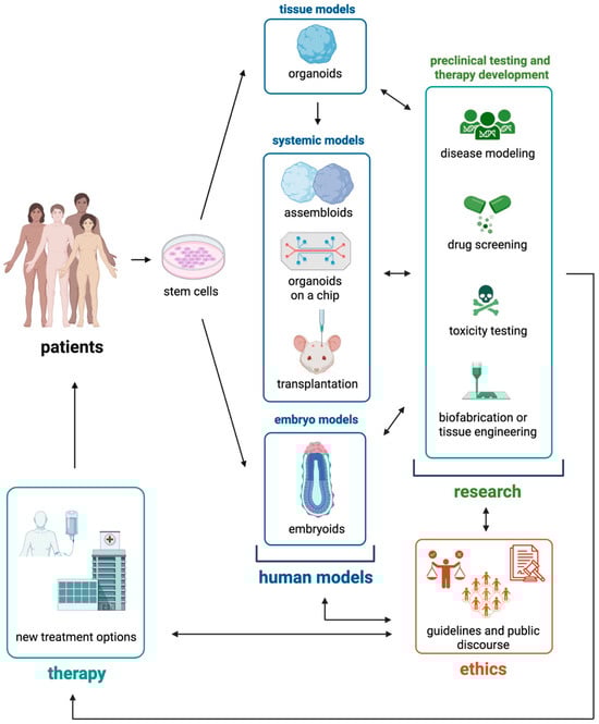
Patient-derived organoids (PDOs) provide ex vivo functional models that capture tumor drug-response patterns across multiple cancer types. Organoid drug sensitivity testing (ODST) has accumulated supportive evidence in single-tumor studies, yet it lacks a pan-cancer biostatistical framework that can support multi-cancer clinical decision-making. This
[...] Read more.
Patient-derived organoids (PDOs) provide ex vivo functional models that capture tumor drug-response patterns across multiple cancer types. Organoid drug sensitivity testing (ODST) has accumulated supportive evidence in single-tumor studies, yet it lacks a pan-cancer biostatistical framework that can support multi-cancer clinical decision-making. This article presents a pan-cancer ODST validation framework that integrates evidence synthesis, regulatory mapping, and adaptive trial design. The framework specifies analytical-performance standards, a three-stage validation architecture, and an explicit cross-tumor portability coefficient that quantifies the transferability of validated evidence among cancer types. Implementation barriers, including heterogeneous tissue-collection standards, variable establishment success, immunotherapy modeling limitations, and regulatory misalignment, are identified, and corresponding mitigation strategies are described. The framework supports a structured pathway from analytical validity to clinical utility for ODST across solid-tumor indications.
Full article
(This article belongs to the Special Issue Advances in Organoid Technology: Bridging the Gap between Research and Therapy)
►
Show Figures

Figure 1


