Next Issue
Volume 16, December
Previous Issue
Volume 16, October


J. Funct. Biomater., Volume 16, Issue 11 (November 2025) – 33 articles

Cover Story (view full-size image): 
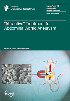
Abdominal aortic aneurysm (AAA) occurs when the distal aorta expands by 50% of its normal size (2 cm). A clinical threshold (5.5 cm for men or 5.0 cm for women) is used to determine when surgical intervention should occur. However, prior to this threshold, patients are solely monitored. Regenerative therapies during this “watchful waiting” period can be used to slow or possibly halt expansion. Previous studies have shown success with mesenchymal stem cell derived extracellular vesicles (EVs) for treatment of AAA. This study sought to create a magnetic delivery system of EVs for localized treatment of AAA. Magnetic particles were localized in vivo and encapsulated EVs were released for uptake into vascular smooth muscle cells, thus showing important proof of concept of this technology. View this paper
  • Issues are regarded as officially published after their release is announced to the table of contents alert mailing list.
  • You may sign up for e-mail alerts to receive table of contents of newly released issues.
  • PDF is the official format for papers published in both, html and pdf forms. To view the papers in pdf format, click on the "PDF Full-text" link, and use the free Adobe Reader to open them.
Order results
Result details
Section
Select all
Export citation of selected articles as:
15 pages, 1483 KB  
Article
Comparative Evaluation of Color Stability and Fracture Resistance of CAD/CAM and Chairside Provisional Restorations: An In Vitro Study
by Florina Titihazan, Ioana Veja, Cristian Zaharia, Tareq Hajaj, Cosmin Sinescu, George Dumitru Constantin and Mihai Rominu
J. Funct. Biomater. 2025, 16(11), 426; https://doi.org/10.3390/jfb16110426 - 20 Nov 2025
Viewed by 717
Abstract
Background and Objectives: Provisional restorations are essential in fixed prosthodontics, ensuring esthetics, function, and biological protection during treatment. Recent advances in CAD/CAM technologies have enabled the fabrication of provisional materials with enhanced color stability and fracture resistance compared to conventional chairside polymeric materials. [...] Read more.
Background and Objectives: Provisional restorations are essential in fixed prosthodontics, ensuring esthetics, function, and biological protection during treatment. Recent advances in CAD/CAM technologies have enabled the fabrication of provisional materials with enhanced color stability and fracture resistance compared to conventional chairside polymeric materials. This study aimed to compare the color stability and fracture strength of provisional crowns fabricated using CAD/CAM and a conventional direct chairside technique. Materials and Methods: A total of 40 provisional crowns (four materials, n = 10 each group) were fabricated for a mandibular molar 3.6 using two workflows: CAD/CAM-milled poly(methyl methacrylate) (PMMA), high-impact polymer composite (HIPC; Bredent), and Ambarino composite (Creamet), and directly fabricated 3M™ Protemp™ (Scutan technique), respectively. Color stability was evaluated after seven-day immersion in coffee and red wine at 37 °C using a spectrophotometer (CIE L*a*b* system). Fracture resistance (Fmax) was measured under axial load in a universal testing machine. Data were analyzed by one-way ANOVA and Tukey’s HSD (α = 0.05). Results: Significant differences were observed among materials (F(3,36) = 212.6, p < 0.001). HIPC showed the highest mean fracture resistance (2068.9 ± 104.0 N), followed by PMMA (1215.8 ± 61.4 N) and 3M™ Protemp™ (1183.4 ± 86.4 N), while Ambarino exhibited the lowest (555.4 ± 25.4 N). Regarding color stability, Ambarino demonstrated the smallest ΔE* (1.1 ± 0.2), followed by PMMA (2.0 ± 0.3), HIPC (2.8 ± 0.3), and Protemp™ (4.9 ± 0.4). Only Protemp™ exceeded the clinical perceptibility threshold (ΔE* > 3.3). Conclusions: Both manufacturing methods and material compositions significantly influence the optical and mechanical properties of provisional restorations. CAD/CAM-milled HIPC and PMMA provided superior fracture strength and clinically acceptable color stability, suggesting their suitability for long-term or high-load temporary crowns compared with chairside-fabricated composites, particularly in posterior regions. Full article
(This article belongs to the Special Issue Advances in Restorative Dentistry Materials)
Show Figures

Figure 1

19 pages, 7795 KB  
Article
Endothelial Cells Differentiated from Human Induced Pluripotent Stem Cells Form Aligned Network Structures in Engineered Neural Tissue
by Poppy O. Smith, Parmjit Jat and James B. Phillips
J. Funct. Biomater. 2025, 16(11), 425; https://doi.org/10.3390/jfb16110425 - 20 Nov 2025
Viewed by 670
Abstract
Background/Objectives: Endothelial cells play a key role in peripheral nerve regeneration, forming aligned vasculature which bridges the gap in the injured nerve tissue and guides the regrowing tissue. This work aimed to mimic key features of this aligned vasculature by differentiating endothelial cells [...] Read more.
Background/Objectives: Endothelial cells play a key role in peripheral nerve regeneration, forming aligned vasculature which bridges the gap in the injured nerve tissue and guides the regrowing tissue. This work aimed to mimic key features of this aligned vasculature by differentiating endothelial cells from human induced pluripotent stem cells (hiPSCs) and incorporating them into engineered neural tissue (EngNT). Methods: hiPSCs were differentiated into endothelial cells with the temporal addition of growth factors and biomolecules. These hiPSC-derived endothelial cells (hiPSC-ECs) were incorporated into EngNT fabricated from collagen hydrogels using the gel aspiration-ejection (GAE) technique and maintained in vitro to allow endothelial network formation. Results: At the mRNA and protein level, pluripotency marker expression decreased and endothelial cell marker expression increased over the course of hiPSC differentiation to endothelial cells. The derived endothelial cells expressed CD31, CD144, ENG, VEGFR2, and VWF, and formed network structures in the matrix tubulogenesis assay. hiPSC-ECs incorporated into EngNT were viable and aligned. They formed highly aligned tube-like structures containing lumens after four days in culture and the EngNT constructs supported neurite growth in vitro when co-cultured with rat dorsal root ganglion (DRG) neurons. Conclusions: This work rapidly generated engineered nerve tissue containing highly aligned endothelial tube-like structures, resembling key features of the early nerve regeneration bridge. Therefore, this 3D engineered tissue provides a platform to study the effects of endothelial cell structures in nerve repair treatment and translational development. Full article
Show Figures

Graphical abstract

17 pages, 1450 KB  
Article
In Vitro Evaluation of Biofilm Formation by Oral Microorganisms on Clear Aligner Materials: Influence of Mouthwash Exposure
by Vlad Tiberiu Alexa, Diana Obistioiu, Ramona Dumitrescu, Iuliana Cretescu, Anca Hulea, Vanessa Bolchis, Octavia Balean, Daniela Jumanca and Atena Galuscan
J. Funct. Biomater. 2025, 16(11), 424; https://doi.org/10.3390/jfb16110424 - 13 Nov 2025
Cited by 1 | Viewed by 797
Abstract
Clear aligners have gained popularity in orthodontics due to their aesthetics, comfort, and removability; however, their prolonged intraoral wear and frequent removal–reinsertion cycles create favorable conditions for microbial colonization. This in vitro study evaluated the efficacy of seven commercially available mouthwash formulations in [...] Read more.
Clear aligners have gained popularity in orthodontics due to their aesthetics, comfort, and removability; however, their prolonged intraoral wear and frequent removal–reinsertion cycles create favorable conditions for microbial colonization. This in vitro study evaluated the efficacy of seven commercially available mouthwash formulations in inhibiting biofilms of Streptococcus mutans, Streptococcus oralis, and Candida albicans formed on four different clear aligner materials. Standardized aligner fragments were incubated for 24 h with microbial suspensions to allow biofilm formation, treated for 1 min with one of the mouthwashes, and then assessed for residual viability through spectrophotometric optical density measurements after a further 24 h incubation. Biofilm inhibition varied according to both mouthwash composition and aligner material. The chlorhexidine-based rinse (MW-D) consistently showed the highest inhibition across microorganisms, while the fluoride–cetylpyridinium chloride rinse (MW-B) performed strongly for S. oralis and C. albicans. An essential oil-based formulation with xylitol (MW-G) showed notable antifungal activity against C. albicans. Monolayer polyurethane aligners generally achieved higher inhibition rates than multilayer or copolyester-based materials. These findings indicate that antimicrobial efficacy on aligners depends on both mouthwash type and material, supporting a tailored approach to biofilm management in clear aligner therapy to reduce the risk of caries, periodontal disease, and candidiasis. Full article
(This article belongs to the Special Issue Antimicrobial Biomaterials for Medical Applications)
Show Figures

Figure 1

32 pages, 19195 KB  
Article
Suitability of Mg-Nd and Mg-Zn Alloys to Obtain Biodegradable Structures for Bone Defects
by Veronica Manescu (Paltanea), Aurora Antoniac, Maria Cristina Moraru, Iulian Antoniac, Cosmin Mihai Cotrut, Sebastian Gradinaru, Alexandra Iulia Dreanca, Bogdan Sevastre, Romelia Pop, Flaviu Alexandru Tabaran, George Mihail Vlasceanu, Mariana Ionita and Marius Manole
J. Funct. Biomater. 2025, 16(11), 423; https://doi.org/10.3390/jfb16110423 - 12 Nov 2025
Viewed by 960
Abstract
Mg-based alloys are one of the most promising materials used in regenerative medicine for bone tissue engineering. Considering the increasing prevalence of a continuously aging population, as well as the high incidence of accidents and bone cancers, it is crucial to explore biomaterials [...] Read more.
Mg-based alloys are one of the most promising materials used in regenerative medicine for bone tissue engineering. Considering the increasing prevalence of a continuously aging population, as well as the high incidence of accidents and bone cancers, it is crucial to explore biomaterials that can serve as bone substitutes. After carefully analyzing the literature in the introduction section, we proposed two Mg-based alloys as suitable for obtaining biodegradable structures for bone defect treatment. To achieve trustworthy results, the alloys’ microstructure was investigated using microscopic techniques coupled with energy-dispersive spectroscopy and X-ray diffraction. The obtained results were comparable with those described in references on similar Mg alloys. Then, the mechanical compression properties were highlighted, and the in vitro corrosion behavior proved that Mg-Zn exhibited a reduced corrosion rate compared to the Mg-Nd alloy, as tested using electrochemical methods. However, the in vivo tests showed good biocompatibility for both magnesium alloys. In conclusion, both alloys are suitable for use as potential bone substitute applications, but it must be taken into consideration that Mg-Zn alloys present lower biodegradation and mechanical properties. For future investigations, we aim to develop bone substitutes made from these materials, specifically designed for small bone defect treatment and with patient-adapted geometry. Due to the differences mentioned above, various designs will be tested. Full article
Show Figures

Figure 1

23 pages, 5447 KB  
Article
3D-Printed Alginate–Chitosan Hydrogel Loaded with Cannabidiol as a Platform for Drug Delivery: Design and Mechanistic Characterization
by Hernan Santiago Garzon, Camilo Alfonso-Rodríguez, João G. S. Souza, Lina J. Suárez and Daniel R. Suárez
J. Funct. Biomater. 2025, 16(11), 422; https://doi.org/10.3390/jfb16110422 - 12 Nov 2025
Viewed by 882
Abstract
Alginate and chitosan (Ag/Cs) combined form an effective platform to develop biocompatible hydrogels with customizable properties for controlled drug release. Cannabidiol (CBD), a hydrophobic compound with anti-inflammatory and antibacterial effects, represents a powerful strategy to enhance their therapeutic performance. A/Cs hydrogels were produced [...] Read more.
Alginate and chitosan (Ag/Cs) combined form an effective platform to develop biocompatible hydrogels with customizable properties for controlled drug release. Cannabidiol (CBD), a hydrophobic compound with anti-inflammatory and antibacterial effects, represents a powerful strategy to enhance their therapeutic performance. A/Cs hydrogels were produced using the CELLINK® printer with 12 and 24 mg/mL of CBD. SEM and FTIR were assessed. Viscoelasticity was assessed using oscillatory rheology. Structural strength was evaluated via uniaxial compression. Swelling and absorption were measured gravimetrically under physiological conditions. CBD was successfully incorporated into the 3D-printed A/Cs hydrogel. Increasing the CBD concentration led to mechanical changes such as a dose-dependent decrease in G′ and a slight reduction in the linearity threshold (typically 10–30% from medium loads), while still maintaining G′ > G″. FTIR showed shifts in O–H/N–H and C=O, indicating hydrogen bonding without new reactive bands. Microscopic images revealed moderate pore compaction and increased tortuosity with dose. At higher CBD concentrations, the hydrogel resisted compression but could deform further before failure. Equilibrium swelling and absorption kinetics decreased with increasing dose, resulting in a reduced initial burst and lower water uptake capacity. The CBD-loaded hydrogel provides a mechanically suitable and molecularly stable platform for local drug release in the oral cavity. Full article
(This article belongs to the Special Issue Biomaterials and Bioengineering in Dentistry (2nd Edition))
Show Figures

Figure 1

15 pages, 6108 KB  
Article
Handheld Nonthermal Plasma Augmentation of Glass–Ceramic Spray Deposition on Zirconia Surface Characterization and MG-63/HGF-1 Cell Behavior: An In Vitro Study
by Sheng-Han Wu, Szu-Yu Lai, I-Ta Lee, Yuichi Mine, Huei-Yu Huang and Tzu-Yu Peng
J. Funct. Biomater. 2025, 16(11), 421; https://doi.org/10.3390/jfb16110421 - 11 Nov 2025
Viewed by 765
Abstract
Zirconia is widely used for customized implant abutments owing to its esthetics, strength, and biocompatibility; however, the optimal surface modification for soft-tissue sealing and bone metabolic remains uncertain. This study evaluated how glass–ceramic spray deposition (GCSD), with or without handheld nonthermal plasma (HNP), [...] Read more.
Zirconia is widely used for customized implant abutments owing to its esthetics, strength, and biocompatibility; however, the optimal surface modification for soft-tissue sealing and bone metabolic remains uncertain. This study evaluated how glass–ceramic spray deposition (GCSD), with or without handheld nonthermal plasma (HNP), alters zirconia surface physiochemistry and cellular responses. Field-emission scanning electron microscopy/energy-dispersive X-ray spectroscopy, surface roughness (Ra), wettability, and surface free energy (SFE) were measured. Human osteoblast-like cells (MG-63) and human gingival fibroblasts (HGF-1) were used to assess attachment and spreading, metabolic activity, cytotoxicity, and inflammatory response (tumor necrosis factor-α, TNF-α) (α = 0.05). GCSD produced an interlaced rod- and needle-like glass–ceramic layer, significantly increasing Ra and hydrophilicity. HNP further reduced surface contaminants, increased SFE, and enhanced wettability. The combination of GCSD and HNP yielded the greatest attachment and spreading for both cell types, without increases in cytotoxicity or TNF-α. GCSD with HNP creates a hydrophilic, micro-textured, chemically activated zirconia surface that maintains biocompatibility while promoting early attachment and bone metabolic activity, supporting its application for zirconia implant abutments. Full article
(This article belongs to the Special Issue Advanced Dental Restorative Composite Materials)
Show Figures

Figure 1

15 pages, 5141 KB  
Article
Biomechanical Influence of Different Cervical Micro-Thread Forms over Narrow-Diameter Implants (2.9 mm) Using Finite Element Analysis
by Qiannian Zhang, Waikit Lau, Nalini Cheong and Tonghan Zhang
J. Funct. Biomater. 2025, 16(11), 420; https://doi.org/10.3390/jfb16110420 - 11 Nov 2025
Viewed by 694
Abstract
Narrow-diameter implants (≤3.5 mm) have garnered significant attention due to their widespread application in areas with insufficient bone volume. However, their mechanical performance is limited. The cervical region, serving as a pivotal stress concentration zone, exhibits a thread form that directly modulates stress [...] Read more.
Narrow-diameter implants (≤3.5 mm) have garnered significant attention due to their widespread application in areas with insufficient bone volume. However, their mechanical performance is limited. The cervical region, serving as a pivotal stress concentration zone, exhibits a thread form that directly modulates stress distribution and determines the long-term stability of the implant–bone interface. This study was designed to investigate the influence of varying thread forms and face angles on microstrain and stress distribution patterns in narrow-diameter implants (NDIs) and their adjacent cortical bone structures. Through systematic modification of implant thread forms and face angle parameters, finite element analysis (FEA) was employed to develop nine distinct implant models featuring varied geometric characteristics. Each model was implanted into Type III bone tissue, followed by the application of a 100 N occlusal force, including a vertical load and an oblique load deviated 30 degrees lingually from the long axis of the implants. Subsequent biomechanical evaluation quantified peak von Mises stress concentrations at the bone–implant interface, maximum equivalent elastic strain distributions in peri-implant bone tissue, and abutment stress profile characteristics. The results indicated that in the RB thread group, the optimal thread face angle parameter was 60 degrees; in the B thread group, this optimal thread face angle parameter was 45 degrees, whereas in the V thread group, the optimal thread face angle parameter was 30 degrees. Full article
(This article belongs to the Special Issue Biomaterials and Biomechanics Modelling in Dental Implantology)
Show Figures

Figure 1

15 pages, 1670 KB  
Article
Optical and Mechanical Characteristics of One-Shade Composite Resins
by Jee Eun Shim, Hyun-Jung Kim, Soram Oh and Ji-Hyun Jang
J. Funct. Biomater. 2025, 16(11), 419; https://doi.org/10.3390/jfb16110419 - 8 Nov 2025
Viewed by 822
Abstract
This study evaluated the optical and mechanical properties of two single-shade composite resins compared with a conventional multi-shade composite. Omnichroma (OM), Metafil Bulk Fill ONE (BO), and Filtek Z350XT (Z350) were tested. Color adjustment was assessed using A3, B1, and C4 background cavities, [...] Read more.
This study evaluated the optical and mechanical properties of two single-shade composite resins compared with a conventional multi-shade composite. Omnichroma (OM), Metafil Bulk Fill ONE (BO), and Filtek Z350XT (Z350) were tested. Color adjustment was assessed using A3, B1, and C4 background cavities, and ΔE00 values were calculated. The translucency parameter (TP) was measured, and the flexural strength, flexural modulus, and depth of cure (B/T ratio) were determined. OM and BO showed better color adjustment performance on brighter (B1) backgrounds and decreased matching on darker (C4) ones. OM maintained stable color adjustment across cavity depths, while BO showed improved adjustment in shallower cavities. Both exhibited higher TP values than Z350. The control group (Z350) had the highest flexural strength and modulus, though BO’s flexural strength was comparable. OM and BO showed sufficient mechanical strength and a greater depth of cure compared to Z350. Our study indicated that the one-shade composite resins OM and BO exhibited better color adjustment performance compared to conventional composite resins due to the influence of the surrounding shades, with a better adjustment ability on brighter backgrounds. Additionally, OM and BO demonstrated sufficient strength and a higher depth of cure compared to the control group. Full article
(This article belongs to the Special Issue Biomechanical Studies and Biomaterials in Dentistry (2nd Edition))
Show Figures

Figure 1

25 pages, 913 KB  
Review
Advances in Bioactive Dental Adhesives for Caries Prevention: A State-of-the-Art Review
by Mohammed Zahedul Islam Nizami, Apissada Jindarojanakul, Qiang Ma, Sang J. Lee and Jirun Sun
J. Funct. Biomater. 2025, 16(11), 418; https://doi.org/10.3390/jfb16110418 - 7 Nov 2025
Viewed by 1608
Abstract
The long-term success of composite restorations largely depends on the performance of dental adhesives at the adhesive–tooth interface. Despite ongoing improvements, secondary caries remains the leading cause of restoration failure, primarily due to the adhesive layer’s susceptibility to hydrolytic degradation, bacterial invasion, and [...] Read more.
The long-term success of composite restorations largely depends on the performance of dental adhesives at the adhesive–tooth interface. Despite ongoing improvements, secondary caries remains the leading cause of restoration failure, primarily due to the adhesive layer’s susceptibility to hydrolytic degradation, bacterial invasion, and limited biological functionality. This review provides a comprehensive overview of recent advances in bioactive dental adhesives for preventing recurrent caries, focusing on their mechanisms of action, material performance, therapeutic functions, and clinical potential. Bioactive adhesives combine durable bonding with biofunctional benefits, including remineralization, antimicrobial activity, enzymatic inhibition, and support for tissue regeneration. By integrating these properties, they enhance both the durability of the adhesive interface and oral health. Recent strategies include the incorporation of ion-releasing fillers such as calcium phosphate and bioactive glass, antimicrobial monomers such as MDPB and quaternary ammonium methacrylates, enzymatic inhibitors, and hydrolytically stable resin matrices. Together, these components strengthen the adhesive interface and provide biologically active effects to prevent recurrent caries. Although in vitro findings are promising, challenges remain, including limited long-term clinical data, the absence of standardized evaluation protocols, and barriers to clinical translation. Addressing these gaps is essential to ensure predictable clinical outcomes. Bioactive dental adhesives represent a paradigm shift in restorative dentistry, evolving from passive bonding agents to multifunctional therapeutic materials. By combining structural durability with biological protection, they hold significant potential to prevent recurrent caries and improve the long-term success of composite restorations. Full article
(This article belongs to the Special Issue Biomaterials for Management of Dental Caries and Periodontal Disease)
Show Figures

Figure 1

25 pages, 1639 KB  
Review
The Role and Involvement of Functional Three-Dimensional Porcine-Derived Collagen Matrix Biomaterials in Periodontal Regeneration: A Comprehensive Review
by Cristian Cojocaru, Dana Gabriela Budala, Dragos Ioan Virvescu, Gabriel Rotundu, Florinel Cosmin Bida, Teona Tudorici, Zinovia Surlari, Mihaela Scurtu, Ancuta Goriuc and Ionut Luchian
J. Funct. Biomater. 2025, 16(11), 417; https://doi.org/10.3390/jfb16110417 - 7 Nov 2025
Viewed by 794
Abstract
Three-dimensional porcine matrix-derived biomaterials have emerged as valuable tools in periodontal regeneration, offering structural stability, biocompatibility, and favorable cellular responses. This review summarizes their physicochemical characteristics, biological mechanisms, and clinical performance in guided tissue and bone regeneration. Comparative analyses show superior handling, integration [...] Read more.
Three-dimensional porcine matrix-derived biomaterials have emerged as valuable tools in periodontal regeneration, offering structural stability, biocompatibility, and favorable cellular responses. This review summarizes their physicochemical characteristics, biological mechanisms, and clinical performance in guided tissue and bone regeneration. Comparative analyses show superior handling, integration potential, and regenerative predictability compared with collagen and synthetic scaffolds, especially in complex intrabony and furcation defects. Despite promising clinical outcomes, heterogeneity in processing techniques and limited long-term data still hinder standardization. Overall, porcine-derived scaffolds represent reliable and biologically active options for periodontal regeneration. Future innovation focusing on functionalization, cell integration, and patient-tailored design will define the next generation of predictable and biomimetic regenerative solutions. Full article
Show Figures

Graphical abstract

16 pages, 3306 KB  
Article
Optimisation of 3D Printing Parameters and Surface Modification for Porous Gyroid Structures in Beta Titanium Alloy Ti25Nb4Ta8Sn
by Zdeněk Tolde, Aleš Jíra, Jitřenka Jírů, Vojtěch Hybášek, Vojtěch Smola and Petr Vlčák
J. Funct. Biomater. 2025, 16(11), 416; https://doi.org/10.3390/jfb16110416 - 7 Nov 2025
Viewed by 831
Abstract
In recent years, 3D printing has become a key technology for producing intricate geometries with high precision. Beta titanium alloys (β-Ti), due to their excellent combination of strength, ductility, low elastic modulus, and biocompatibility, are widely used in the aerospace and medical industries. [...] Read more.
In recent years, 3D printing has become a key technology for producing intricate geometries with high precision. Beta titanium alloys (β-Ti), due to their excellent combination of strength, ductility, low elastic modulus, and biocompatibility, are widely used in the aerospace and medical industries. However, the unique microstructure formed during additive manufacturing characterised by porosity, residual stress, and anisotropy can significantly influence the mechanical performance and durability of these materials. This study examines how different printing parameters affect porosity, dimensional stability, and mechanical properties in the β-Ti alloy Ti25Nb4Ta8Sn. The investigation focuses on thin-walled samples and gyroid structures, which represent model geometries for porous biomedical components. These structures, defined by a periodic network of interconnected channels, provide a useful platform for studying the relationship between geometry and mechanical response. In addition, the effects of surface etching on the morphology and compressive behaviour of printed gyroid structures were evaluated. Compression testing was used to determine how etching alters load-bearing performance and to identify correlations between surface modification and mechanical response. The combined analysis enables optimisation of both printing and post-processing parameters for advanced biomedical applications. Full article
(This article belongs to the Special Issue Three-Dimensional-Printable Biomaterials for Bone Regeneration)
Show Figures

Figure 1

16 pages, 3701 KB  
Article
Early Osseointegration in a Sheep Tibia Model: Correlating Digital Periapical Radiograph Gray-Level and RGB-Derived Metrics with Histologic Tissue Composition
by Sergio Alexandre Gehrke, Jaime Aramburú Júnior, Tiago Luis Eilers Treichel, Germán Odella Colla, Gustavo Coura, Bruno Freitas Mello, Márcio de Carvalho Formiga, Fátima de Campos Buzzi, Sergio Rexhep Tari and Antonio Scarano
J. Funct. Biomater. 2025, 16(11), 415; https://doi.org/10.3390/jfb16110415 - 7 Nov 2025
Viewed by 1010
Abstract
Objective: This study aimed to evaluate peri-implant tissue changes during early osseointegration using a combined approach of digital radiographic analysis, RGB pseudocolorization, and histomorphometry in a sheep tibia model. Materials and Methods: Thirty titanium implants were placed in the tibiae of six adult [...] Read more.
Objective: This study aimed to evaluate peri-implant tissue changes during early osseointegration using a combined approach of digital radiographic analysis, RGB pseudocolorization, and histomorphometry in a sheep tibia model. Materials and Methods: Thirty titanium implants were placed in the tibiae of six adult sheep and evaluated at 14 and 28 days post-implantation. Digital periapical radiographs were acquired, grayscale values and RGB channel intensities were measured using Fiji/ImageJ, and compared with histological parameters (bone tissue, collagen, and medullary spaces) quantified from picrosirius–hematoxylin-stained sections. Manual overlay of radiographic and histological images was performed to ensure spatial correspondence of regions of interest. Statistical analyses assessed differences over time and correlations between image data and histological composition. Results: Radiographic grayscale values and histologically measured bone and collagen increased significantly from 14 to 28 days (p < 0.01), while medullary spaces decreased (p < 0.001), indicating progressive bone formation and matrix maturation. RGB analysis revealed significant increases in green channel intensity and decreases in red channel intensity (p < 0.05), while the blue channel remained stable. At 14 days, strong correlations were observed between blue channel intensity and bone tissue (r = 0.81; p = 0.015), and between green channel intensity and collagen (r = 0.98; p < 0.001). Visual overlays demonstrated alignment between radiographic high-density zones and histologically dense bone regions. Conclusions: RGB pseudocolorized radiographic analysis, correlated with histological findings, offers a non-invasive and reproducible method for early detection of peri-implant tissue maturation. This feasibility correlation study provides a foundation for future investigations integrating imaging, histology, and biomechanical testing. Full article
(This article belongs to the Special Issue Functional Biomaterial for Bone Regeneration (2nd Edition))
Show Figures

Figure 1

55 pages, 17120 KB  
Review
Magnetic Hydrogels as a Treatment for Oncological Pathologies
by Veronica Manescu (Paltanea), Adrian-Vasile Dumitru, Aurora Antoniac, Iulian Antoniac, Gheorghe Paltanea, Elena-Cristina Zeca (Berbecar), Mirela Gherghe, Iosif Vasile Nemoianu, Alexandru Streza, Costel Paun and Sebastian Gradinaru
J. Funct. Biomater. 2025, 16(11), 414; https://doi.org/10.3390/jfb16110414 - 5 Nov 2025
Viewed by 1207
Abstract
Cancer is considered today as a prevalent research direction due to the fact that, by 2050, more than 30 million cases will occur, followed by about 19 million deaths. It is expected that scholars will search for new, innovative, and localized therapies to [...] Read more.
Cancer is considered today as a prevalent research direction due to the fact that, by 2050, more than 30 million cases will occur, followed by about 19 million deaths. It is expected that scholars will search for new, innovative, and localized therapies to ensure a much more targeted treatment with reduced side effects. Magnetic hydrogels overcome the disadvantages of classical magnetic nanoparticles in various oncological domains, including magnetic hyperthermia, theragnostic, immunotherapy, and, notably, regenerative medicine and contrast substances. We will review the magnetic hydrogel topics that may be involved as a potential application for cancer. Firstly, we present the international context and subject importance in the framework of statistics estimated by some researchers. Then, the magnetic hydrogel synthesis method will be briefly described with examples extracted from the literature. Supplementary, we will emphasize the main attributes of an ideal magnetic hydrogel, and last but not least, we will review some of the latest in vitro and in vivo studies in a direct relationship with magnetic hyperthermia, chemotherapeutic drug release dynamics, and immunotherapy used as single strategies or in combination, by underling the magnetic properties of the hydrogels and importance of application of magnetic fields. We will conclude our review paper by discussing toxicity issues, future trends, limitations, and proposed new approaches to address them. Full article
Show Figures

Graphical abstract

17 pages, 10869 KB  
Article
The Effect of Titanium Dioxide Nanotubes and Graphene Compounds on the Proliferation and Osteogenic Differentiation of Rat BMSCs
by Chenyuan Zhu, Yuwei Deng, Jing Xu, Jin Wen, Qingfeng Huang and Weiqiang Yu
J. Funct. Biomater. 2025, 16(11), 413; https://doi.org/10.3390/jfb16110413 - 5 Nov 2025
Viewed by 720
Abstract
Graphene-based nanomaterials, including graphene oxide (GO) and graphene quantum dots (GQDs), exhibit exceptional properties, which might facilitate the functional modification of TiO2 nanotubes (NTs) for enhanced rapid osseointegration. This study investigated the effects of GO/GQD-deposited TiO2-NTs on cell proliferation, osteogenic [...] Read more.
Graphene-based nanomaterials, including graphene oxide (GO) and graphene quantum dots (GQDs), exhibit exceptional properties, which might facilitate the functional modification of TiO2 nanotubes (NTs) for enhanced rapid osseointegration. This study investigated the effects of GO/GQD-deposited TiO2-NTs on cell proliferation, osteogenic differentiation of rat bone marrow-derived mesenchymal stem cells (BMSCs), and early osseointegration in male 6-week-old Sprague Dawley (SD) rats. TiO2-NTs (control group) were fabricated on titanium substrates via anodic oxidation. GO and GQDs were electrochemically deposited onto the TiO2-NTs using cyclic voltammetry with 0.5 mg/mL GO and 0.1 mg/mL GQD dispersions to form NT-GO and NT-GQDs. In vitro assays evaluated cell adhesion, proliferation, and osteogenic differentiation. Implants were randomly inserted into one femoral epiphysis of nine rats (n = 3), and osseointegration was evaluated using micro-computed tomography and sequential fluorescence labeling at 2, 4, and 6 weeks post-implantation. Statistical analysis was conducted using ANOVA. Cyclic voltammetry successfully synthesized NT-GO and NT-GQDs, with Raman spectra confirming D and G bands. Both NT-GO and NT-GQDs exhibited superior cell adhesion, proliferation, and enhanced osteogenic differentiation compared with TiO2-NTs. Notably, the NT-GQDs significantly promoted new bone formation in vivo. The integration of graphene nanomaterials onto TiO2-NTs improves biocompatibility and accelerates osteogenesis, suggesting a promising strategy for enhancing osseointegration in orthopedic and dental implants. Full article
(This article belongs to the Special Issue Advanced Materials and Devices for Medical Interventions)
Show Figures

Graphical abstract

15 pages, 722 KB  
Article
Clinical and Radiographic Outcomes of Root Canal Obturation with Hydraulic Condensation and Tricalcium Silicate Bioceramic Sealer: A 12-Month Observational Study on Periapical Healing
by Kostadin Zhekov and Vesela Stefanova
J. Funct. Biomater. 2025, 16(11), 412; https://doi.org/10.3390/jfb16110412 - 5 Nov 2025
Viewed by 1616
Abstract
Successful endodontic treatment relies on effective shaping, disinfection and obturation. Calcium silicate sealers such as BioRoot™ RCS show promise due to their bioactivity and sealing properties, but more clinical evidence using standardized protocols is needed. This observational clinical study aimed to assess periapical [...] Read more.
Successful endodontic treatment relies on effective shaping, disinfection and obturation. Calcium silicate sealers such as BioRoot™ RCS show promise due to their bioactivity and sealing properties, but more clinical evidence using standardized protocols is needed. This observational clinical study aimed to assess periapical healing at 6 and 12 months following single-visit root canal treatment using BioRoot™ RCS with hydraulic condensation in teeth with irreversible pulpitis or apical periodontitis. Sixty-six teeth were treated using a standardized protocol: ProTaper Gold instrumentation, sonic-activated irrigation, and hydraulic condensation with gutta-percha cone and BioRoot™ RCS. Periapical healing was evaluated using the periapical index (PAI) at baseline, 6 months, and 12 months. Clinical success was defined as functional, asymptomatic teeth and a PAI ≤ 2. Statistical analysis included repeated measures of ANOVA and McNemar’s test. All 66 teeth remained asymptomatic and functional of 12 months, yielding a 100% survival rate. Clinical success was confirmed in 97% of cases. PAI scores decreased significantly over time (p < 0.001) in apical periodontitis cases. Single-visit endodontic treatment with BioRoot™ RCS and hydraulic condensation demonstrated excellent clinical and radiographic outcomes. This approach promotes resolution of apical periodontitis in non-vital cases and supports the preservation of periapical health in teeth initially diagnosed with irreversible pulpitis. Full article
Show Figures

Figure 1

28 pages, 1101 KB  
Review
Dental Implantology in Acromegaly: Pathophysiological Challenges, Biomaterial Interactions, and Future Directions—A Narrative Review
by Beata Wiśniewska, Sandra Spychała, Kosma Piekarski, Ewelina Golusińska-Kardach, Maria Stelmachowska-Banaś and Marzena Wyganowska
J. Funct. Biomater. 2025, 16(11), 411; https://doi.org/10.3390/jfb16110411 - 5 Nov 2025
Viewed by 838
Abstract
Introduction: Acromegaly is a chronic endocrine disorder caused by excessive secretion of growth hormone (GH) and insulin-like growth factor 1 (IGF-1). Acromegaly leads to a wide range of systemic alterations, including metabolic disturbances, abnormalities in bone microarchitecture, soft tissue overgrowth, and morphological changes [...] Read more.
Introduction: Acromegaly is a chronic endocrine disorder caused by excessive secretion of growth hormone (GH) and insulin-like growth factor 1 (IGF-1). Acromegaly leads to a wide range of systemic alterations, including metabolic disturbances, abnormalities in bone microarchitecture, soft tissue overgrowth, and morphological changes in the maxilla and mandible. All these factors may significantly complicate the planning and success of implant therapy. Study Aim: This narrative review aimed to critically analyze the impact of acromegaly on bone healing and osseointegration, with particular emphasis on the stability of implant biomaterials, and to assess whether the disease constitutes a contraindication to implant prosthetic treatment. Methods: A narrative literature review was conducted using the PubMed, Scopus, and Web of Science databases, covering publications from 2000 to August 2025. Manual screening of reference lists from key articles was also performed. Peer-reviewed publications in English, including experimental and preclinical studies, case reports, biomaterials research, and conceptual reviews, were included based on their relevance to acromegaly, bone metabolism, stomatognathic alterations, and implant therapy outcomes. No formal inclusion or exclusion criteria were applied, and methodological quality was not formally assessed, reflecting the exploratory and conceptual nature of this review. Results: Patients with acromegaly exhibit persistent structural bone deficits, such as reduced trabecular number, irregular trabecular distribution, and increased cortical porosity, despite normal or even elevated bone mineral density. In parallel, profound changes in soft tissues and dentition are observed, including macroglossia, diastemas, gingival overgrowth, and mandibular prognathism, which further complicate prosthetic rehabilitation. Animal studies suggest that GH and IGF-1 may support early osseointegration, although the long-term effects of their excess remain inconclusive. Clinical data, although limited, indicate that implant placement in patients with acromegaly is feasible when treatment is meticulously planned and carried out within an interdisciplinary setting. Standard biomaterials, such as titanium and its alloys, may undergo degradation under conditions of chronic inflammation and oxidative stress, underscoring the need for innovative solutions integrating bioactive and immunomodulatory materials, as well as patient-specific implants manufactured using 3D printing technologies. Conclusions: Acromegaly should not be regarded as an absolute contraindication to implant therapy; however, the current evidence is limited. Implant placement requires individualized planning, endocrine control, and interdisciplinary coordination. Further clinical and preclinical studies are needed to establish reliable treatment protocols for this population. Full article
(This article belongs to the Section Dental Biomaterials)
Show Figures

Figure 1

12 pages, 654 KB  
Article
Quantitative Suspension Test for the Evaluation of a Cold Sterilization System Based on Reducing Free Radicals Compared to Autoclave Sterilization Cycles
by Christian Cirillo, Daniele Botticelli and Stefano Benedicenti
J. Funct. Biomater. 2025, 16(11), 410; https://doi.org/10.3390/jfb16110410 - 4 Nov 2025
Viewed by 725
Abstract
Sterilization of medical devices is a critical process to ensure patient safety. However, traditional steam autoclaves may be unsuitable for heat-sensitive materials. In this study, we evaluated an innovative cold sterilization system based on the controlled generation of free radicals with reducing properties. [...] Read more.
Sterilization of medical devices is a critical process to ensure patient safety. However, traditional steam autoclaves may be unsuitable for heat-sensitive materials. In this study, we evaluated an innovative cold sterilization system based on the controlled generation of free radicals with reducing properties. The system has already been validated and marketed following the completion of numerous microbiological tests in compliance with UNI EN standards (13727, 13624, 17126, 14476, 14348). A quantitative suspension test was conducted under controlled conditions, comparing the microbial reduction achieved with the cold system to that obtained with a standard autoclave cycle. The system demonstrated bactericidal efficacy exceeding 6 log10, comparable to that of the autoclave cycle. The results suggest that the free radical system represents a safe, rapid, and effective alternative for the sterilization of heat-sensitive materials, with potential applications in both healthcare and industrial settings. Full article
(This article belongs to the Section Antibacterial Biomaterials)
Show Figures

Graphical abstract

16 pages, 10746 KB  
Article
Pre-Vascularized 3-Dimensional Skin Substitutes Promote Angiogenesis and Tissue Repair in a Murine Model of Refractory Skin Ulcers
by Shota Tojo, Hiromi Miyazaki, Takami Saiki, Yasuyuki Tsunoi, Shingo Nakamura and Ryuichi Azuma
J. Funct. Biomater. 2025, 16(11), 409; https://doi.org/10.3390/jfb16110409 - 3 Nov 2025
Viewed by 657
Abstract
Restoring blood flow is crucial for treating refractory ulcers. Despite advancements in various biomaterials, none incorporating pre-formed blood vessels have been commercialized. To address this, we developed a pre-vascularized three-dimensional (3D) skin substitute (PV-3D skin) designed to enhance healing when treating refractory ulcers. [...] Read more.
Restoring blood flow is crucial for treating refractory ulcers. Despite advancements in various biomaterials, none incorporating pre-formed blood vessels have been commercialized. To address this, we developed a pre-vascularized three-dimensional (3D) skin substitute (PV-3D skin) designed to enhance healing when treating refractory ulcers. This study aimed to evaluate the therapeutic role of PV-3D skin transplantation in refractory ulcer models, induced by applying mitomycin C to wounds in severe immunodeficient mice. The wounds were then treated with PV-3D skin, non-vascularized 3D skin, skin grafts, or wound dressings. The PV-3D skin group demonstrated healing dynamics comparable to those of the skin graft group, with similar tissue morphology and wound temperature changes. Furthermore, at day 7 post-transplantation, the PV-3D skin group demonstrated significantly higher hypoxia-inducible factor 1-alpha expression levels compared to the 3D skin group. By day 14, the PV-3D skin group exhibited a significantly larger vascular area compared to the 3D skin group. Notably, PV-3D skin treatment stimulated host-derived angiogenesis, thereby enhancing wound healing and reducing the recurrence of refractory ulcers. These results suggest that PV-3D skin transplantation offers a promising therapeutic approach for refractory ulcers, especially in terms of angiogenesis. Full article
Show Figures

Figure 1

12 pages, 3399 KB  
Article
Influence of Sintering Parameters on the Mechanical Behaviour of Lithium Disilicate Glass Ceramics: An In-Vitro Study
by Mai Soliman, Raghad Alotaibi, Abrar Almutairi, Asma Alzahrani, Reem Abunyan, Aseel Rozi, Dalia Alamri, Shahad Almakenzi, Elzahraa Eldwakhly and Alhanoof Aldegheishem
J. Funct. Biomater. 2025, 16(11), 408; https://doi.org/10.3390/jfb16110408 - 2 Nov 2025
Viewed by 585
Abstract
This study investigates the effect of different sintering parameters on the surface roughness and fracture resistance of different CAD/CAM lithium disilicate ceramics before and after thermocycling and simulated toothbrushing. Sixty lithium disilicate ceramic samples were categorized according to ceramic type (AMB: Amber Mill, [...] Read more.
This study investigates the effect of different sintering parameters on the surface roughness and fracture resistance of different CAD/CAM lithium disilicate ceramics before and after thermocycling and simulated toothbrushing. Sixty lithium disilicate ceramic samples were categorized according to ceramic type (AMB: Amber Mill, ECAD: IPS e.max CAD) and level of translucency (LT: low and HT: high) into four groups: AMB_LT, AMB_HT, ECAD_LT and ECAD_HT. Specimens were prepared to 2 mm thickness, crystallized, polished, and subjected to thermocycling to simulate five years of clinical aging. Simulated toothbrushing was performed using a soft-bristled mechanical brushing system under controlled force and strokes. Surface roughness was assessed using a profilometer before and after brushing, and fracture resistance was measured using a universal testing machine. Data were statistically evaluated using paired t-tests, one-way ANOVA with Bonferroni post hoc correction (p ≤ 0.05). LT lithium disilicate specimens exhibited significantly smoother surfaces than high-translucency counterparts. After thermocycling and brushing simulation, all groups showed a statistically significant increase in surface roughness, ranging between 0.239 ± 0.012 μm (AMB_LT) and 0.486 ± 0.014 μm (ECAD_HT). In terms of fracture resistance, the highest values were recorded for ECAD_HT (636 ± 8.29 N), and the lowest in the AMB_HT group (546.3 ± 21.9 N) with significant differences observed between materials and translucency levels. Sintering parameters and artificial aging significantly influenced the surface roughness and fracture resistance of lithium disilicate ceramics. Low-translucency variants demonstrated smoother surfaces and higher strength than high-translucency groups, while all materials exhibited increased roughness after aging. These findings provide guidance for the selection of lithium disilicate ceramics, balancing esthetic and mechanical requirements in anterior and posterior restorations. Full article
Show Figures

Figure 1

14 pages, 3772 KB  
Article
Phase-Pure Hydroxyapatite/β-Tricalcium Phosphate Scaffolds from Ultra-Pure Precursors: Composition Governs Porosity, Strength, and SBF Kinetics
by Panuwat Monviset, Kasama Srirussamee, Anak Khantachawana and Parichart Naruphontjirakul
J. Funct. Biomater. 2025, 16(11), 407; https://doi.org/10.3390/jfb16110407 - 31 Oct 2025
Viewed by 805
Abstract
Biphasic calcium phosphate (BCP)scaffolds comprising hydroxyapatite (HA) and β-tricalcium phosphate (β-TCP) were produced from ultra-pure precursors and processed under an α-TCP–avoiding schedule (1100 °C, 2 h). Quantitative X-ray diffraction (Rietveld/Profex) detected no α-TCP above the ~1 wt% limit of detection and quantified post-sintering [...] Read more.
Biphasic calcium phosphate (BCP)scaffolds comprising hydroxyapatite (HA) and β-tricalcium phosphate (β-TCP) were produced from ultra-pure precursors and processed under an α-TCP–avoiding schedule (1100 °C, 2 h). Quantitative X-ray diffraction (Rietveld/Profex) detected no α-TCP above the ~1 wt% limit of detection and quantified post-sintering phase fractions (wt% HA/β-TCP): 99.26/0.74, 68.51/31.49, and 27.57/72.43. Across compositions, SEM/ImageJ yielded similar mean macropore sizes (≈71–80 µm), while open porosity increased with the HA fraction (27.5 ± 1.8%, 39.1 ± 2.0%, 57.1 ± 2.4%). Compressive strength decreased accordingly (1.07 ± 0.25, 0.24 ± 0.01, 0.05 ± 0.02 MPa), consistent with non-load-bearing use. In ISO-compliant simulated body fluid (28 d), medium pH remained stable (7.33–7.43); mass loss and early Ca2+ depletion increased with β-TCP content, consistent with more extensive surface apatite formation in β-TCP-rich scaffolds. Collectively, these data are consistent with a composition-dependent sequence—β-TCP content → densification/porosity → strength → degradation/apatite kinetics—within the tested conditions and inform parameter-based tuning of BCP scaffolds for non-load-bearing indications (e.g., alveolar ridge preservation, craniofacial void filling). Full article
(This article belongs to the Special Issue Biomaterials for Bone Implant and Regeneration)
Show Figures

Figure 1

23 pages, 7319 KB  
Article
Corrosion-Modulating Effect of Pharmaceutical Agents in a Hybrid Coating System on Pure Magnesium
by Lara Moreno, Adrián Belarra-Rodriguez, Marta Mohedano, Laura Castro, Margarita Chevalier, Raul Arrabal and Endzhe Matykina
J. Funct. Biomater. 2025, 16(11), 406; https://doi.org/10.3390/jfb16110406 - 30 Oct 2025
Viewed by 875
Abstract
There is a knowledge gap about the effect of pharmaceutical agents on the biodegradation of Mg-based resorbable implants. The present work investigates how three common antibiotics and three anti-inflammatory drugs affect the corrosion of high-purity Mg, with and without ceramic and hybrid ceramic/polymeric [...] Read more.
There is a knowledge gap about the effect of pharmaceutical agents on the biodegradation of Mg-based resorbable implants. The present work investigates how three common antibiotics and three anti-inflammatory drugs affect the corrosion of high-purity Mg, with and without ceramic and hybrid ceramic/polymeric coatings, using electrochemical impedance spectroscopy and hydrogen evolution tests. A Ca-P-Si-based ceramic coating is developed using plasma electrolytic oxidation (PEO), after the AC voltage and frequency parameters are optimized. A hybrid coating included a PEO and a poly(ε-caprolactone) (PCL) top layer formed by dip coating. High-purity Mg exhibited an instantaneous onset of corrosion with a corrosion rate of 90 μm/year after 24 h of immersion in a modified α-MEM. A hybrid PEO/PCL coating prevents the onset of corrosion for at least 5 h and reduces the H2 evolution during the following 90 h by two times by the precipitation of 5–40 μm thick Ca-P surface deposits. Gentamicin, naproxen, streptomycin, ciprofloxacin and paracetamol were found to be corrosion accelerators with respect to bare h.p. Mg, whereas aspirin was found to be an inhibitor. Streptomycin-functionalized PEO/PCL system exhibited an active protection mechanism, triggered upon the release of the coating and substrate cations, associated with the coating defect-blocking action of the insoluble Me(II)-streptomycin chelates. Full article
(This article belongs to the Section Biomaterials for Drug Delivery)
Show Figures

Graphical abstract

25 pages, 7146 KB  
Article
Biopolymer Fibers of High Strength and Enhanced Orientation by the Synergy of High/Low Molecular Weight Chitosans in Hybrid Biomaterials Processed by Gel Spinning
by Tuan Anh Tran, Ingo Doench, Arnaud Kamdem Tamo, Shaghayegh Jahangir, Sofia Marquez-Bravo, Pamela Molina, Martin Helmstaedter, Aliuska Morales Helguera, Christian Gorzelanny and Anayancy Osorio-Madrazo
J. Funct. Biomater. 2025, 16(11), 405; https://doi.org/10.3390/jfb16110405 - 29 Oct 2025
Viewed by 795
Abstract
High-performance spun bionanocomposite fibers, composed of high-molecular-weight chitosan (HMW), low-molecular-weight chitosan “oligomers” (LMW), and cellulose nanofibers (CNFs), were successfully fabricated via gel spinning of viscous aqueous chitosan (CHI) based formulations into a NaOH coagulation bath. The X-ray diffraction (XRD) analysis revealed that the [...] Read more.
High-performance spun bionanocomposite fibers, composed of high-molecular-weight chitosan (HMW), low-molecular-weight chitosan “oligomers” (LMW), and cellulose nanofibers (CNFs), were successfully fabricated via gel spinning of viscous aqueous chitosan (CHI) based formulations into a NaOH coagulation bath. The X-ray diffraction (XRD) analysis revealed that the incorporation of cellulose nanofibers contributed to enhance crystallinity of chitosan in spun fibers. The spinning process, which comprised sequential acidic solubilization, basic neutralization, stretching, and drying steps, produced chitosan/CNF composite fibers with high crystallinity, further enhanced by the incorporation of low molecular weight chitosan. The cellulose nanofibers seem to promote CHI crystallization, by acting as nucleation sites for the nucleation and growth of chitosan crystals, with those latter of LMW further enhancing crystallization and orientation due to higher mobility of shorter polymer chains. Two-dimensional XRD patterns demonstrated the preferential alignment of both CNFs and chitosan crystals along the fiber axis. Increasing the proportion of short-chain chitosan led to a reduction of the viscosity of collodion, facilitating the spinning of solutions with higher polymer concentrations. The X-ray diffraction (XRD) analysis revealed that the addition of low-molecular-weight chitosan (LMW), with an intermediate molecular weight Mw of ~4.4 × 10⁴ g/mol, produced the most significant improvements in the crystallinity index (CrI) and orientation. This structural enhancement corresponded to superior mechanical properties like Young’s modulus, yield stress σy, and stress-at-break σb of the processed composite fibers. By incorporating that intermediate molecular weight chitosan, a Young’s modulus as high as 20 GPa was achieved for the spun composite fibers, which was twice higher than the modulus of around 10 GPa obtained by adding the lowest molecular weight chitosan of Mw ~ 2.9 × 10⁴ g/mol in the composite, and largely above the modulus of around 5 GPa obtained for fiber just spun with chitosan without incorporation of cellulose nanofibers. Full article
(This article belongs to the Section Synthesis of Biomaterials via Advanced Technologies)
Show Figures

Figure 1

16 pages, 2580 KB  
Article
Titanium Versus Bioabsorbable Magnesium Headless Compression Screw Fixation for Tibial Tubercle Osteotomy
by Mehmet Baris Ertan, Oguzhan Uslu, Firat Dogruoz, Omer Faruk Celik and Ozkan Kose
J. Funct. Biomater. 2025, 16(11), 404; https://doi.org/10.3390/jfb16110404 - 29 Oct 2025
Viewed by 713
Abstract
Purpose: This retrospective study aimed to compare the clinical and radiological outcomes of Fulkerson tibial tubercle osteotomy (TTO) fixed with either bioabsorbable magnesium (Mg) or titanium (Ti) headless compression screws, particularly focusing on the need for implant removal. Materials and Methods: 29 patients [...] Read more.
Purpose: This retrospective study aimed to compare the clinical and radiological outcomes of Fulkerson tibial tubercle osteotomy (TTO) fixed with either bioabsorbable magnesium (Mg) or titanium (Ti) headless compression screws, particularly focusing on the need for implant removal. Materials and Methods: 29 patients (19 Ti, 10 Mg screws) who underwent TTO between 2013 and 2023 were included. The primary outcome was implant removal; secondary outcomes included Kujala and Lysholm scores, complication rates, and radiographic union. Rehabilitation protocols were standardized, but early weight-bearing was delayed in the Mg group to mitigate potential implant degradation effects. Results: Both groups demonstrated significant improvements in Kujala and Lysholm Knee scores postoperatively, with no statistically significant differences between the groups. No cases of implant removal, fixation failure, nonunion, or significant range of motion loss were observed. Radiographically, persistent remnants of Mg screws were detected even after more than five years, raising concerns about incomplete bioabsorption. The Ti screws maintained mechanical stability without evidence of loss of fixation or the need for revision. Patient satisfaction and cosmetic outcomes were similar. Conclusion: The use of bioabsorbable Mg screws in TTO did not confer additional clinical or radiological benefits compared to headless Ti screws. Given the higher cost, the incomplete resorption observed at long-term follow-up, and the absence of an implant removal requirement in either group, the routine use of Mg screws cannot be recommended. Ti headless compression screws offer a cost-effective, reliable fixation method, achieving stable osteotomy healing without the need for secondary surgery. Full article
(This article belongs to the Section Bone Biomaterials)
Show Figures

Figure 1

31 pages, 3096 KB  
Review
Implications of Tissue Engineering for Tendon Repair and Regeneration
by Dana Ivanisova, Katarina Bevizova, Sara Vach Agocsova, Lubos Danisovic and Martina Culenova
J. Funct. Biomater. 2025, 16(11), 403; https://doi.org/10.3390/jfb16110403 - 28 Oct 2025
Viewed by 2239
Abstract
Tendon injuries affect millions of people globally and are among the most prevalent musculoskeletal conditions, frequently resulting in chronic pain, reduced mobility, and functional impairment. While conservative and surgical treatments are available, limitations such as low healing capacity, scar formation, and reduced biomechanics [...] Read more.
Tendon injuries affect millions of people globally and are among the most prevalent musculoskeletal conditions, frequently resulting in chronic pain, reduced mobility, and functional impairment. While conservative and surgical treatments are available, limitations such as low healing capacity, scar formation, and reduced biomechanics necessitate alternative approaches. Tissue engineering offers a promising solution by combining cells, scaffolds, and bioactive molecules to regenerate tendon tissue. This review presents key concepts and emerging trends, highlighting the cellular components, scaffold materials, and manufacturing processes. Tenocytes and mesenchymal stem cells are fundamental for tissue regeneration, as they synthesize extracellular matrix components and regulate inflammatory responses. Various natural and synthetic polymers have been fabricated into scaffolds that mimic the structure and biomechanics of natural tendons. Composite and hybrid scaffolds are utilized to improve the biocompatibility of natural materials with the mechanical stability of synthetic materials. Advanced technologies, such as electrospinning, freeze-drying, and 3D bioprinting, enable the creation of scaffolds with defined architecture and functional gradients, improving cell alignment, differentiation, and tendon–bone integration. Although promising preclinical data exists, major challenges remain in translating these strategies clinically, particularly vascularization, immune rejection, and mechanical stability. Continued interdisciplinary attempts in biomaterials science, cellular biology, and engineering are crucial to advancing clinically viable tendon tissue engineering. Full article
Show Figures

Figure 1

17 pages, 1803 KB  
Article
In Vitro and In Vivo Evaluation of a New Experimental Polydimethylsiloxane-Based Endodontic Sealer
by Fabiola Cardoso Maldonado, Cesar Gaitan Fonseca, Carlos Bermudez Jimenez, Luis Alejandro Aguilera Galaviz, Margarita L. Martinez-Fierro, Lorena Troncoso Vazquez and Martha Eugenia Reyes Ortiz
J. Funct. Biomater. 2025, 16(11), 402; https://doi.org/10.3390/jfb16110402 - 28 Oct 2025
Viewed by 926
Abstract
Successful root canal treatment depends on adequate obturation with biocompatible and non-cytotoxic materials. This study evaluated the in vitro and in vivo biological characteristics of an experimental polydimethylsiloxane (PDMS)-based endodontic sealer and compared it with Silco® and Sealapex® cement. Human dermal [...] Read more.
Successful root canal treatment depends on adequate obturation with biocompatible and non-cytotoxic materials. This study evaluated the in vitro and in vivo biological characteristics of an experimental polydimethylsiloxane (PDMS)-based endodontic sealer and compared it with Silco® and Sealapex® cement. Human dermal fibroblasts (HDFa) were exposed to polydimethylsiloxane-based sealer eluates, Silco® and Sealapex®, at concentrations of 1:200, 1:100, 1:50, 1:1, and undiluted eluate (1×) for 24, 48, and 72 h, and they were subcutaneously implanted in Wistar rats for 15, 30, and 45 days. Cell viability exceeded 90% at 24–48 h and remained at 85% at the highest concentration after 72 h. Sealapex® showed approximately 85% viability at 24 h, over 70% at 48 h, and remained below the cytotoxicity threshold at 72 h. Silco® showed a marked reduction, with values approaching 50% at 24 h. At 48 and 73 h, Silco® showed a significant reduction in cell viability. Histological analysis revealed only mild acute and chronic inflammation, with no statistically significant differences over time. These results indicate that the experimental sealant demonstrates favorable biological properties suitable for further clinical evaluation. Full article
(This article belongs to the Special Issue The 15th Anniversary of JFB—Endodontic Biomaterials)
Show Figures

Graphical abstract

31 pages, 12238 KB  
Article
Micropatterning and Nanodropletting of Titanium by Shifted Surface Laser Texturing Significantly Enhances In Vitro Osteogenesis of Healthy and Osteoporotic Mesenchymal Stromal Cells
by Theresia Stich, Francisca Alagboso, Girish Pattappa, Jin Chu, Denys Moskal, Michal Povolný, Maximilian Saller, Veronika Schönitzer, Konstantin J. Scholz, Fabian Cieplik, Volker Alt, Maximilian Rudert, Tomáš Kovářík, Tomáš Křenek and Denitsa Docheva
J. Funct. Biomater. 2025, 16(11), 401; https://doi.org/10.3390/jfb16110401 - 27 Oct 2025
Viewed by 837
Abstract
The key to proper implant integration in bone replacement is to orchestrate the complex interactions between materials and tissues. Moreover, due to the rapid demographic shift towards aging societies and the increase in elderly and osteoporotic patients, it is of great importance that [...] Read more.
The key to proper implant integration in bone replacement is to orchestrate the complex interactions between materials and tissues. Moreover, due to the rapid demographic shift towards aging societies and the increase in elderly and osteoporotic patients, it is of great importance that implant materials are osteointegrative in not only healthy but also compromised bone tissues. Here, titanium (Ti) scaffolds were subjected to shifted laser surface texturing (sLST) using a nanosecond pulsed laser to create an open pore macrotopography with micro-and nano-Ti droplets. In contrast to conventional laser texturing, which leads to high heat accumulation; in sLST, the frequency of laser pulses is low, allowing for resolidification, thereby creating a surface with abundant coverage micro-/nanodroplets. The main objective was to compare the cellular responses of human mesenchymal stromal cells (hMSCs) on sLST-textured Ti surfaces (LT-Ti) for the first time with standard sand-blasted, acid-etched surfaces (SLA-Ti). In-depth analyses of cell survival, proliferation, shape, mineralization, and gene expression were performed. Cell survival/proliferation was found to be similar on both surfaces; however, SEM imaging revealed differences in hMSC morphology. On LT-Ti, cells adopted well-rounded shapes, whereas on SLA-Ti they assumed more planar shapes. Bulk RNA sequencing performed after short-term culture on both surfaces disclosed expression changes in genes such as DUSP6, TNFSF12-TNFSF13 and SULT1A4. Remarkably, the osteogenic differentiation capacity of hMSCs was significantly enhanced on LT-Ti compared to SLA-Ti. Furthermore, aged/osteoporotic donor cohorts showed significantly enhanced matrix mineralization on LT-Ti. In conclusion, our novel results demonstrate that sLST-Ti surfaces are safe, highly biocompatible, can rescue patient-cohort-specific mineralization behavior, and therefore hold great potential for the development into next-generation implants, which are suitable for both the elderly and bone-compromised populations. Full article
(This article belongs to the Section Bone Biomaterials)
Show Figures

Graphical abstract

19 pages, 4496 KB  
Article
Influence of Machining, Polishing, and Glazing on Surface Properties and Biological Performance of Zirconia and Lithium Disilicate Dental Ceramics
by Youngchae Cho, Min-Gu Cho, Jeong-Hyun Ryu, Ji-Yeong Kim, Sung-Hwan Choi, Hyungjoon Shim, Min-Ho Hong and Deuk Yong Lee
J. Funct. Biomater. 2025, 16(11), 400; https://doi.org/10.3390/jfb16110400 - 27 Oct 2025
Viewed by 967
Abstract
Surface treatments play a crucial role in modifying the surface properties and biological performance of dental ceramics. This study investigated the effects of surface conditions on the wettability, cytocompatibility, and bacterial resistance of 4 mol% Y2O3-stabilized tetragonal zirconia polycrystal [...] Read more.
Surface treatments play a crucial role in modifying the surface properties and biological performance of dental ceramics. This study investigated the effects of surface conditions on the wettability, cytocompatibility, and bacterial resistance of 4 mol% Y2O3-stabilized tetragonal zirconia polycrystal (4Y–TZP) and two lithium disilicate (Li2Si2O5) glass ceramics (Amber® Mill (AM) and Amber® Mill Abut-Crown (AC)). Human gingival fibroblast (HGF-1) responses and biofilm formation on the machined, polished, and glazed samples were evaluated. The polished 4Y–TZP sample exhibited the highest water contact angle (WCA; 71.3°), while that of the AC samples decreased as the sample was machined (58.4°), polished (46.8°), and glazed (14.0°). The wettability, cytocompatibility, and bacterial resistance of the dental ceramics were significantly influenced by material type and surface condition. Among the surface-treated samples, the glazed specimens exhibited the lowest WCA and bulk density; thus, wettability is an important factor for cell proliferation and bacterial resistance. Among all samples, HGF-1 cells adhered well to the glazed ceramics and significantly proliferated over time. Particularly, the 4Y–TZP and AC glazed samples exhibited the lowest biomass and strong resistance to biofilm formation and bacterial adhesion. Thus, the glaze dramatically affected HGF-1 cell growth and antibiofilm formation. Full article
(This article belongs to the Special Issue Recent Advancements in Dental Restorative Materials)
Show Figures

Figure 1

21 pages, 3350 KB  
Article
Multifunctional Peptide-Based Biohybrid for Targeted Reduction of Metastatic Breast Carcinoma-Associated Osteolysis
by Nicole Stadler, Bingjie Gao, Maria Jose Silva, Joscha Borho, Eva Haunschild, Kaloian Koynov, Melanie Haffner-Luntzer, Anita Ignatius, Gilbert Weidinger, Seah Ling Kuan, Tanja Weil and Holger Barth
J. Funct. Biomater. 2025, 16(11), 399; https://doi.org/10.3390/jfb16110399 - 25 Oct 2025
Viewed by 1948
Abstract
Metastatic breast carcinoma (BC) cells are prone to spreading in the bone microenvironment, leading to a vicious cycle between local osteoclast-mediated osteolysis and tumor progression. Therefore, the targeted pharmacological down-modulation of BC cell proliferation as well as osteoclast differentiation and hyperactivity might represent [...] Read more.
Metastatic breast carcinoma (BC) cells are prone to spreading in the bone microenvironment, leading to a vicious cycle between local osteoclast-mediated osteolysis and tumor progression. Therefore, the targeted pharmacological down-modulation of BC cell proliferation as well as osteoclast differentiation and hyperactivity might represent a promising treatment option. We developed a multifunctional peptide nanocarrier combining bioactive EPI-X4 peptides and the Rho-inhibiting C3bot enzyme from Clostridium botulinum. C3bot is preferentially internalized into the cytosol of monocytic cells, including osteoclasts, where it inhibits Rho-mediated signal transduction. However, Rho-mediated cellular processes like migration and cell division can also be inhibited in non-monocytic cells if C3bot is delivered into their cytosol by a nanocarrier. To accomplish this, we designed a supramolecular transporter where one molecule of biotinylated C3bot and three biotinylated entities of the human EPI-X4 peptide-derived CXCR4 antagonist JM173 are assembled on avidin as a central platform. This modular transport system (JM173)3-Avi-C3 down-modulated osteoclast formation and hyperactivity and delivered the therapeutic cargo C3bot successfully into the cytosol of breast cancer cells, where it inhibited Rho. Full article
(This article belongs to the Special Issue Advanced Biomaterials in Cancer Therapeutics and Diagnosis)
Show Figures

Graphical abstract

14 pages, 1128 KB  
Article
Influence of Different Biomaterials Extracted from Autologous Blood on the Cell Migration of Stem Cells from Dental Pulp
by Janet N. Kirilova, Rositsa Z. Vladova, Viktoria P. Petrova, Sevda Yantcheva, Elitsa G. Deliverska and Nikolay D. Ishkitiev
J. Funct. Biomater. 2025, 16(11), 398; https://doi.org/10.3390/jfb16110398 - 24 Oct 2025
Viewed by 1728
Abstract
Background: This study aims to evaluate the effect of different types of platelet concentrates (autologous blood biomaterials) on the migration potential of human dental pulp stem (hDPSCs). Materials and Methods: Our team created a model of human dental pulp stem cells (hDPSCs). Various [...] Read more.
Background: This study aims to evaluate the effect of different types of platelet concentrates (autologous blood biomaterials) on the migration potential of human dental pulp stem (hDPSCs). Materials and Methods: Our team created a model of human dental pulp stem cells (hDPSCs). Various types of AB biomaterials were produced from blood samples from volunteers using the protocols presented: A-PRF+, Gel A-PRF+, and Solid PRF. The scratch wound healing assay was used to examine the closure of the experimental wounds on day 1 and day 14. The wound areas were quantified using Image J software. Statistical analysis was performed with the Kruskal–Wallis and Mann–Whitney U tests, as the data did not follow a normal distribution, which was confirmed by the Shapiro–Wilk test (p < 0.05). Results: The results demonstrate significantly faster closure of the experimental wounds on day 14 of the studied biomaterials AB: A-PRF+, Gel A-PRF+, and Solid PRF compared to the control group of cells. Gel A-PRF+ exhibited the most pronounced stimulatory effect on cell migration (p = 0.0036 vs. control), followed by Solid PRF and A-PRF+. Conclusions: The results indicate that autologous blood platelet concentrates stimulate the migration of hDPSCs in vitro. Gel A-PRF+ demonstrated the strongest effect, underscoring its potential clinical relevance for applications in tissue engineering. Full article
(This article belongs to the Special Issue Biomaterials Applied in Dental Sciences)
Show Figures

Figure 1

18 pages, 3261 KB  
Article
Biological Response of Osteoblasts to Zirconia Manufactured via FFF, DLP, and Milling
by Christoph J. Roser, Ralf Erber, Andreas Zenthöfer, Stefan Rues, Christopher J. Lux, Dorit Nötzel, Ralf Eickhoff and Thomas Hanemann
J. Funct. Biomater. 2025, 16(11), 397; https://doi.org/10.3390/jfb16110397 - 23 Oct 2025
Viewed by 900
Abstract
(1) Background: Zirconia (ZrO2) is increasingly used in dental implantology due to its biocompatibility and favorable mechanical and biological properties. While subtractive and stereolithographic additive manufacturing techniques are well established, the application of Fused Filament Fabrication (FFF) for zirconia-based dental implants [...] Read more.
(1) Background: Zirconia (ZrO2) is increasingly used in dental implantology due to its biocompatibility and favorable mechanical and biological properties. While subtractive and stereolithographic additive manufacturing techniques are well established, the application of Fused Filament Fabrication (FFF) for zirconia-based dental implants remains largely unexplored. (2) Methods: Cylindrical ZrO2 specimens were fabricated using three different manufacturing techniques: milling (MIL), Digital Light Processing (DLP), and FFF. Surface topography was analyzed via white-light interferometry. Human fetal osteoblasts (hFOBs 1.19) were cultured on the specimens to evaluate cell adhesion after 4 and 24 h, proliferation for 4 days, cell surface coverage after 4 and 24 h, and osteogenic gene expression (RUNX2, ALPL, and BGLAP) after 24 h, 48 h, 7 days, and 14 days. (3) Results: The FFF samples exhibited significantly higher surface roughness than the MIL and DLP specimens. After 24 h, enhanced cell adhesion and the highest proliferation rates were observed on FFF surfaces. At 14 days, gene expression analysis revealed elevated expression of BGLAP on FFF surfaces, suggesting advanced osteogenic differentiation compared to MIL and DLP. (4) Conclusions: The inherent surface roughness of FFF-printed zirconia appears to promote osteogenic activity without additional surface treatment. These findings suggest that FFF may constitute a viable manufacturing method for the fabrication of customized zirconia components in dental implantology, warranting further investigations, particularly regarding their mechanical performance. Full article
(This article belongs to the Special Issue Digital Design and Biomechanical Analysis of Dental Materials)
Show Figures

Figure 1

Previous Issue
Next Issue
Back to TopTop