Next Issue
Volume 9, March
Previous Issue
Volume 9, January


Cells, Volume 9, Issue 2 (February 2020) – 264 articles

Cover Story (view full-size image): 
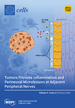
Tumors release chemokines that can recruit macrophages and dendritic cells to adjacent nerves. There, localization, morphology, and lipid content of epineural adipocytes change, coinciding with downregulation of perineural tight junction proteins and appearance of microlesions. View this paper.
  • Issues are regarded as officially published after their release is announced to the table of contents alert mailing list.
  • You may sign up for e-mail alerts to receive table of contents of newly released issues.
  • PDF is the official format for papers published in both, html and pdf forms. To view the papers in pdf format, click on the "PDF Full-text" link, and use the free Adobe Reader to open them.
Order results
Result details
Section
Select all
Export citation of selected articles as:
24 pages, 1105 KiB  
Review
MYC’s Fine Line Between B Cell Development and Malignancy
by Oriol de Barrios, Ainara Meler and Maribel Parra
Cells 2020, 9(2), 523; https://doi.org/10.3390/cells9020523 - 24 Feb 2020
Cited by 18 | Viewed by 9745
Abstract
The transcription factor MYC is transiently expressed during B lymphocyte development, and its correct modulation is essential in defined developmental transitions. Although temporary downregulation of MYC is essential at specific points, basal levels of expression are maintained, and its protein levels are not [...] Read more.
The transcription factor MYC is transiently expressed during B lymphocyte development, and its correct modulation is essential in defined developmental transitions. Although temporary downregulation of MYC is essential at specific points, basal levels of expression are maintained, and its protein levels are not completely silenced until the B cell becomes fully differentiated into a plasma cell or a memory B cell. MYC has been described as a proto-oncogene that is closely involved in many cancers, including leukemia and lymphoma. Aberrant expression of MYC protein in these hematological malignancies results in an uncontrolled rate of proliferation and, thereby, a blockade of the differentiation process. MYC is not activated by mutations in the coding sequence, and, as reviewed here, its overexpression in leukemia and lymphoma is mainly caused by gene amplification, chromosomal translocations, and aberrant regulation of its transcription. This review provides a thorough overview of the role of MYC in the developmental steps of B cells, and of how it performs its essential function in an oncogenic context, highlighting the importance of appropriate MYC regulation circuitry. Full article
(This article belongs to the Special Issue Regulation and Function of the Myc Oncogene)
Show Figures

Figure 1

18 pages, 4642 KiB  
Article
Detection of MET Alterations Using Cell Free DNA and Circulating Tumor Cells from Cancer Patients
by Patricia Mondelo-Macía, Carmela Rodríguez-López, Laura Valiña, Santiago Aguín, Luis León-Mateos, Jorge García-González, Alicia Abalo, Oscar Rapado-González, Mercedes Suárez-Cunqueiro, Angel Díaz-Lagares, Teresa Curiel, Silvia Calabuig-Fariñas, Aitor Azkárate, Antònia Obrador-Hevia, Ihab Abdulkader, Laura Muinelo-Romay, Roberto Diaz-Peña and Rafael López-López
Cells 2020, 9(2), 522; https://doi.org/10.3390/cells9020522 - 24 Feb 2020
Cited by 20 | Viewed by 4061
Abstract
MET alterations may provide a potential biomarker to evaluate patients who will benefit from treatment with MET inhibitors. Therefore, the purpose of the present study is to investigate the utility of a liquid biopsy-based strategy to assess MET alterations in cancer patients. We [...] Read more.
MET alterations may provide a potential biomarker to evaluate patients who will benefit from treatment with MET inhibitors. Therefore, the purpose of the present study is to investigate the utility of a liquid biopsy-based strategy to assess MET alterations in cancer patients. We analyzed MET amplification in circulating free DNA (cfDNA) from 174 patients with cancer and 49 healthy controls and demonstrated the accuracy of the analysis to detect its alteration in patients. Importantly, a significant correlation between cfDNA concentration and MET copy number (CN) in cancer patients (r = 0.57, p <10−10) was determined. Furthermore, we evaluated two approaches to detect the presence of MET on circulating tumor cells (CTCs), using the CellSearch® and Parsortix systems and monitored patients under anti-EGFR treatment (n = 30) combining both cfDNA and CTCs analyses. This follow-up provides evidence for the potential of MET CN assessment when patients develop resistance to anti-EGFR therapy and a significant association between the presence of CTCs MET+ and the Overall Survival (OS) in head and neck cancer patients (P = 0.05; HR = 6.66). In conclusion, we develop specific and noninvasive assays to monitor MET status in cfDNA/CTCs and demonstrate the utility of plasma MET CN determination as a biomarker for monitoring the appearance of resistance to anti-EGFR therapy. Full article
(This article belongs to the Special Issue Liquid Biopsy)
Show Figures

Figure 1

14 pages, 743 KiB  
Review
Nanoparticle-Based Delivery of Tumor Suppressor microRNA for Cancer Therapy
by Clodagh P. O’Neill and Róisín M. Dwyer
Cells 2020, 9(2), 521; https://doi.org/10.3390/cells9020521 - 24 Feb 2020
Cited by 58 | Viewed by 6781
Abstract
Improved understanding of microRNA expression and function in cancer has revealed a range of microRNAs that negatively regulate many oncogenic pathways, thus representing potent tumor suppressors. Therapeutic targeting of the expression of these microRNAs to the site of tumors and metastases provides a [...] Read more.
Improved understanding of microRNA expression and function in cancer has revealed a range of microRNAs that negatively regulate many oncogenic pathways, thus representing potent tumor suppressors. Therapeutic targeting of the expression of these microRNAs to the site of tumors and metastases provides a promising avenue for cancer therapy. To overcome challenges associated with microRNA degradation, transient expression and poor targeting, novel nanoparticles are being developed and employed to shield microRNAs for tumor-targeted delivery. This review focuses on studies describing a variety of both natural and synthetic nanoparticle delivery vehicles that have been engineered for tumor-targeted delivery of tumor suppressor microRNAs in vivo. Full article
(This article belongs to the Special Issue microRNA as Therapeutic Target)
Show Figures

Figure 1

16 pages, 9280 KiB  
Article
Computational Identification and Analysis of Ubiquinone-Binding Proteins
by Chang Lu, Wenjie Jiang, Hang Wang, Jinxiu Jiang, Zhiqiang Ma and Han Wang
Cells 2020, 9(2), 520; https://doi.org/10.3390/cells9020520 - 24 Feb 2020
Cited by 2 | Viewed by 2817
Abstract
Ubiquinone is an important cofactor that plays vital and diverse roles in many biological processes. Ubiquinone-binding proteins (UBPs) are receptor proteins that dock with ubiquinones. Analyzing and identifying UBPs via a computational approach will provide insights into the pathways associated with ubiquinones. In [...] Read more.
Ubiquinone is an important cofactor that plays vital and diverse roles in many biological processes. Ubiquinone-binding proteins (UBPs) are receptor proteins that dock with ubiquinones. Analyzing and identifying UBPs via a computational approach will provide insights into the pathways associated with ubiquinones. In this work, we were the first to propose a UBPs predictor (UBPs-Pred). The optimal feature subset selected from three categories of sequence-derived features was fed into the extreme gradient boosting (XGBoost) classifier, and the parameters of XGBoost were tuned by multi-objective particle swarm optimization (MOPSO). The experimental results over the independent validation demonstrated considerable prediction performance with a Matthews correlation coefficient (MCC) of 0.517. After that, we analyzed the UBPs using bioinformatics methods, including the statistics of the binding domain motifs and protein distribution, as well as an enrichment analysis of the gene ontology (GO) and the Kyoto Encyclopedia of Genes and Genomes (KEGG) pathway. Full article
(This article belongs to the Special Issue Biocomputing and Synthetic Biology in Cells)
Show Figures

Figure 1

25 pages, 3951 KiB  
Article
Microfluidic Device for On-Chip Immunophenotyping and Cytogenetic Analysis of Rare Biological Cells
by Kumuditha M. Weerakoon-Ratnayake, Swarnagowri Vaidyanathan, Nicholas Larkey, Kavya Dathathreya, Mengjia Hu, Jilsha Jose, Shalee Mog, Keith August, Andrew K. Godwin, Mateusz L. Hupert, Malgorzata A. Witek and Steven A. Soper
Cells 2020, 9(2), 519; https://doi.org/10.3390/cells9020519 - 24 Feb 2020
Cited by 6 | Viewed by 4856
Abstract
The role of circulating plasma cells (CPCs) and circulating leukemic cells (CLCs) as biomarkers for several blood cancers, such as multiple myeloma and leukemia, respectively, have recently been reported. These markers can be attractive due to the minimally invasive nature of their acquisition [...] Read more.
The role of circulating plasma cells (CPCs) and circulating leukemic cells (CLCs) as biomarkers for several blood cancers, such as multiple myeloma and leukemia, respectively, have recently been reported. These markers can be attractive due to the minimally invasive nature of their acquisition through a blood draw (i.e., liquid biopsy), negating the need for painful bone marrow biopsies. CPCs or CLCs can be used for cellular/molecular analyses as well, such as immunophenotyping or fluorescence in situ hybridization (FISH). FISH, which is typically carried out on slides involving complex workflows, becomes problematic when operating on CLCs or CPCs due to their relatively modest numbers. Here, we present a microfluidic device for characterizing CPCs and CLCs using immunofluorescence or FISH that have been enriched from peripheral blood using a different microfluidic device. The microfluidic possessed an array of cross-channels (2–4 µm in depth and width) that interconnected a series of input and output fluidic channels. Placing a cover plate over the device formed microtraps, the size of which was defined by the width and depth of the cross-channels. This microfluidic chip allowed for automation of immunofluorescence and FISH, requiring the use of small volumes of reagents, such as antibodies and probes, as compared to slide-based immunophenotyping and FISH. In addition, the device could secure FISH results in <4 h compared to 2–3 days for conventional FISH. Full article
Show Figures

Figure 1

15 pages, 2964 KiB  
Article
Protein Translocation Acquires Substrate Selectivity Through ER Stress-Induced Reassembly of Translocon Auxiliary Components
by Sohee Lee, Yejin Shin, Kyunggon Kim, Youngsup Song, Yongsub Kim and Sang-Wook Kang
Cells 2020, 9(2), 518; https://doi.org/10.3390/cells9020518 - 24 Feb 2020
Cited by 1 | Viewed by 3190
Abstract
Protein import across the endoplasmic reticulum membrane is physiologically regulated in a substrate-selective manner to ensure the protection of stressed ER from the overload of misfolded proteins. However, it is poorly understood how different types of substrates are accurately distinguished and disqualified during [...] Read more.
Protein import across the endoplasmic reticulum membrane is physiologically regulated in a substrate-selective manner to ensure the protection of stressed ER from the overload of misfolded proteins. However, it is poorly understood how different types of substrates are accurately distinguished and disqualified during translocational regulation. In this study, we found poorly assembled translocon-associated protein (TRAP) complexes in stressed ER. Immunoaffinity purification identified calnexin in the TRAP complex in which poor assembly inhibited membrane insertion of the prion protein (PrP) in a transmembrane sequence-selective manner, through translocational regulation. This reaction was induced selectively by redox perturbation, rather than calcium depletion, in the ER. The liberation of ERp57 from calnexin appeared to be the reason for the redox sensitivity. Stress-independent disruption of the TRAP complex prevented a pathogenic transmembrane form of PrP (ctmPrP) from accumulating in the ER. This study uncovered a previously unappreciated role for calnexin in assisting the redox-sensitive function of the TRAP complex and provided insights into the ER stress-induced reassembly of translocon auxiliary components as a key mechanism by which protein translocation acquires substrate selectivity. Full article
(This article belongs to the Section Intracellular and Plasma Membranes)
Show Figures

Graphical abstract

18 pages, 4128 KiB  
Article
Nrp1 is Activated by Konjac Ceramide Binding-Induced Structural Rigidification of the a1a2 Domain
by Seigo Usuki, Yoshiaki Yasutake, Noriko Tamura, Tomohiro Tamura, Kunikazu Tanji, Takashi Saitoh, Yuta Murai, Daisuke Mikami, Kohei Yuyama, Kenji Monde, Katsuyuki Mukai and Yasuyuki Igarashi
Cells 2020, 9(2), 517; https://doi.org/10.3390/cells9020517 - 24 Feb 2020
Cited by 3 | Viewed by 3961
Abstract
Konjac ceramide (kCer) is a plant-type ceramide composed of various long-chain bases and α-hydroxyl fatty acids. The presence of d4t,8t-sphingadienine is essential for semaphorin 3A (Sema3A)-like activity. Herein, we examined the three neuropilin 1 (Nrp1) domains (a1a2, b1b2, or c), and found that [...] Read more.
Konjac ceramide (kCer) is a plant-type ceramide composed of various long-chain bases and α-hydroxyl fatty acids. The presence of d4t,8t-sphingadienine is essential for semaphorin 3A (Sema3A)-like activity. Herein, we examined the three neuropilin 1 (Nrp1) domains (a1a2, b1b2, or c), and found that a1a2 binds to d4t,8t-kCer and possesses Sema3A-like activity. kCer binds to Nrp1 with a weak affinity of μM dissociation constant (Kd). We wondered whether bovine serum albumin could influence the ligand–receptor interaction that a1a2 has with a single high affinity binding site for kCer (Kd in nM range). In the present study we demonstrated the influence of bovine serum albumin. Thermal denaturation indicates that the a1a2 domain may include intrinsically disordered region (IDR)-like flexibility. A potential interaction site on the a1 module was explored by molecular docking, which revealed a possible Nrp1 activation mechanism, in which kCer binds to Site A close to the Sema3A-binding region of the a1a2 domain. The a1 module then accesses a2 as the IDR-like flexibility becomes ordered via kCer-induced protein rigidity of a1a2. This induces intramolecular interaction between a1 and a2 through a slight change in protein secondary structure. Full article
(This article belongs to the Special Issue Sphingolipids in Cancer Progression and Therapy)
Show Figures

Figure 1

12 pages, 3591 KiB  
Article
Transcriptional and Ultrastructural Analyses Suggest Novel Insights into Epithelial Barrier Impairment in Celiac Disease
by Agnieszka Sowińska, Yasser Morsy, Elżbieta Czarnowska, Beata Oralewska, Ewa Konopka, Marek Woynarowski, Sylwia Szymańska, Maria Ejmont, Michael Scharl, Joanna B. Bierła, Marcin Wawrzyniak and Bożena Cukrowska
Cells 2020, 9(2), 516; https://doi.org/10.3390/cells9020516 - 24 Feb 2020
Cited by 9 | Viewed by 3159
Abstract
Disruption of epithelial junctional complex (EJC), especially tight junctions (TJ), resulting in increased intestinal permeability, is supposed to activate the enhanced immune response to gluten and to induce the development of celiac disease (CD). This study is aimed to present the role of [...] Read more.
Disruption of epithelial junctional complex (EJC), especially tight junctions (TJ), resulting in increased intestinal permeability, is supposed to activate the enhanced immune response to gluten and to induce the development of celiac disease (CD). This study is aimed to present the role of EJC in CD pathogenesis. To analyze differentially expressed genes the next-generation mRNA sequencing data from CD326+ epithelial cells isolated from non-celiac and celiac patients were involved. Ultrastructural studies with morphometry of EJC were done in potential CD, newly recognized active CD, and non-celiac controls. The transcriptional analysis suggested disturbances of epithelium and the most significant gene ontology enriched terms in epithelial cells from CD patients related to the plasma membrane, extracellular exome, extracellular region, and extracellular space. Ultrastructural analyses showed significantly tighter TJ, anomalies in desmosomes, dilatations of intercellular space, and shorter microvilli in potential and active CD compared to controls. Enterocytes of fetal-like type and significantly wider adherence junctions were observed only in active CD. In conclusion, the results do not support the hypothesis that an increased passage of gluten peptides by unsealing TJ precedes CD development. However, increased intestinal permeability due to abnormality of epithelium might play a role in CD onset. Full article
(This article belongs to the Special Issue Epithelial Cell Mechanics: From Physiology to Pathology)
Show Figures

Figure 1

17 pages, 2345 KiB  
Article
Non-Phosphorylatable PEA-15 Sensitises SKOV-3 Ovarian Cancer Cells to Cisplatin
by Shahana Dilruba, Alessia Grondana, Anke C. Schiedel, Naoto T. Ueno, Chandra Bartholomeusz, Jindrich Cinatl Jr, Katie-May McLaughlin, Mark N. Wass, Martin Michaelis and Ganna V. Kalayda
Cells 2020, 9(2), 515; https://doi.org/10.3390/cells9020515 - 24 Feb 2020
Cited by 5 | Viewed by 3270
Abstract
The efficacy of cisplatin-based chemotherapy in ovarian cancer is often limited by the development of drug resistance. In most ovarian cancer cells, cisplatin activates extracellular signal-regulated kinase1/2 (ERK1/2) signalling. Phosphoprotein enriched in astrocytes (PEA-15) is a ubiquitously expressed protein, capable of sequestering ERK1/2 [...] Read more.
The efficacy of cisplatin-based chemotherapy in ovarian cancer is often limited by the development of drug resistance. In most ovarian cancer cells, cisplatin activates extracellular signal-regulated kinase1/2 (ERK1/2) signalling. Phosphoprotein enriched in astrocytes (PEA-15) is a ubiquitously expressed protein, capable of sequestering ERK1/2 in the cytoplasm and inhibiting cell proliferation. This and other functions of PEA-15 are regulated by its phosphorylation status. In this study, the relevance of PEA-15 phosphorylation state for cisplatin sensitivity of ovarian carcinoma cells was examined. The results of MTT-assays indicated that overexpression of PEA-15AA (a non-phosphorylatable variant) sensitised SKOV-3 cells to cisplatin. Phosphomimetic PEA-15DD did not affect cell sensitivity to the drug. While PEA-15DD facilitates nuclear translocation of activated ERK1/2, PEA-15AA acts to sequester the kinase in the cytoplasm as shown by Western blot. Microarray data indicated deregulation of thirteen genes in PEA-15AA-transfected cells compared to non-transfected or PEA-15DD-transfected variants. Data derived from The Cancer Genome Atlas (TCGA) showed that the expression of seven of these genes including EGR1 (early growth response protein 1) and FLNA (filamin A) significantly correlated with the therapy outcome in cisplatin-treated cancer patients. Further analysis indicated the relevance of nuclear factor erythroid 2-related factor 2/antioxidant response element (Nrf2/ARE) signalling for the favourable effect of PEA-15AA on cisplatin sensitivity. The results warrant further evaluation of the PEA-15 phosphorylation status as a potential candidate biomarker of response to cisplatin-based chemotherapy. Full article
(This article belongs to the Special Issue Molecular and Cellular Mechanisms of Cancers: Ovarian Cancer)
Show Figures

Figure 1

12 pages, 1367 KiB  
Article
Analysis of Killer Immunoglobulin-Like Receptor Genes in Colorectal Cancer
by Roberto Diaz-Peña, Patricia Mondelo-Macía, Antonio José Molina de la Torre, Rebeca Sanz-Pamplona, Víctor Moreno and Vicente Martín
Cells 2020, 9(2), 514; https://doi.org/10.3390/cells9020514 - 24 Feb 2020
Cited by 6 | Viewed by 2784
Abstract
Natural killer cells (NK cells) play a major role in the immune response to cancer. An important element of NK target recognition is the binding of human leucocyte antigen (HLA) class I molecules by killer immunoglobulin-like receptors (KIRs). Colorectal carcinoma (CRC) is one [...] Read more.
Natural killer cells (NK cells) play a major role in the immune response to cancer. An important element of NK target recognition is the binding of human leucocyte antigen (HLA) class I molecules by killer immunoglobulin-like receptors (KIRs). Colorectal carcinoma (CRC) is one of the most common types of inflammation-based cancer. The purpose of the present study was to investigate the presence of KIR genes and HLA class I and II alleles in 1074 CRC patients and 1272 controls. We imputed data from single-nucleotide polymorphism (SNP) Illumina OncoArray to identify associations at HLA (HLA–A, B, C, DPB1, DQA1, DQB1, and DRB1) and KIRs (HIBAG and KIR*IMP, respectively). For association analysis, we used PLINK (v1.9), the PyHLA software, and R version 3.4.0. Only three SNP markers showed suggestive associations (p < 10−3; rs16896742, rs28367832, and rs9277952). The frequency of KIR2DS3 was significantly increased in the CRC patients compared to healthy controls (p < 0.005). Our results suggest that the implication of NK cells in CRC may not act through allele combinations in KIR and HLA genes. Much larger studies in ethnically homogeneous populations are needed to rule out the possible role of allelic combinations in KIR and HLA genes in CRC risk. Full article
(This article belongs to the Special Issue Molecular and Cellular Mechanisms of Cancers: Colorectal Cancer)
Show Figures

Figure 1

22 pages, 5185 KiB  
Article
Helicobacter Pylori Targets the EPHA2 Receptor Tyrosine Kinase in Gastric Cells Modulating Key Cellular Functions
by Marina Leite, Miguel S. Marques, Joana Melo, Marta T. Pinto, Bruno Cavadas, Miguel Aroso, Maria Gomez-Lazaro, Raquel Seruca and Ceu Figueiredo
Cells 2020, 9(2), 513; https://doi.org/10.3390/cells9020513 - 24 Feb 2020
Cited by 18 | Viewed by 4556
Abstract
Helicobacter pylori, a stomach-colonizing Gram-negative bacterium, is the main etiological factor of various gastroduodenal diseases, including gastric adenocarcinoma. By establishing a life-long infection of the gastric mucosa, H. pylori continuously activates host-signaling pathways, in particular those associated with receptor tyrosine kinases. Using [...] Read more.
Helicobacter pylori, a stomach-colonizing Gram-negative bacterium, is the main etiological factor of various gastroduodenal diseases, including gastric adenocarcinoma. By establishing a life-long infection of the gastric mucosa, H. pylori continuously activates host-signaling pathways, in particular those associated with receptor tyrosine kinases. Using two different gastric epithelial cell lines, we show that H. pylori targets the receptor tyrosine kinase EPHA2. For long periods of time post-infection, H. pylori induces EPHA2 protein downregulation without affecting its mRNA levels, an effect preceded by receptor activation via phosphorylation. EPHA2 receptor downregulation occurs via the lysosomal degradation pathway and is independent of the H. pylori virulence factors CagA, VacA, and T4SS. Using small interfering RNA, we show that EPHA2 knockdown affects cell–cell and cell–matrix adhesion, invasion, and angiogenesis, which are critical cellular processes in early gastric lesions and carcinogenesis mediated by the bacteria. This work contributes to the unraveling of the underlying mechanisms of H. pylori–host interactions and associated diseases. Additionally, it raises awareness for potential interference between H. pylori infection and the efficacy of gastric cancer therapies targeting receptors tyrosine kinases, given that infection affects the steady-state levels and dynamics of some receptor tyrosine kinases (RTKs) and their signaling pathways. Full article
(This article belongs to the Special Issue Molecular and Cellular Mechanisms of Cancers: Gastric Cancer)
Show Figures

Graphical abstract

18 pages, 734 KiB  
Review
The Translocator Protein (TSPO) in Mitochondrial Bioenergetics and Immune Processes
by Calina Betlazar, Ryan J. Middleton, Richard Banati and Guo-Jun Liu
Cells 2020, 9(2), 512; https://doi.org/10.3390/cells9020512 - 24 Feb 2020
Cited by 66 | Viewed by 7365
Abstract
The translocator protein (TSPO) is an outer mitochondrial membrane protein that is widely used as a biomarker of neuroinflammation, being markedly upregulated in activated microglia in a range of brain pathologies. Despite its extensive use as a target in molecular imaging studies, the [...] Read more.
The translocator protein (TSPO) is an outer mitochondrial membrane protein that is widely used as a biomarker of neuroinflammation, being markedly upregulated in activated microglia in a range of brain pathologies. Despite its extensive use as a target in molecular imaging studies, the exact cellular functions of this protein remain in question. The long-held view that TSPO plays a fundamental role in the translocation of cholesterol through the mitochondrial membranes, and thus, steroidogenesis, has been disputed by several groups with the advent of TSPO knockout mouse models. Instead, much evidence is emerging that TSPO plays a fundamental role in cellular bioenergetics and associated mitochondrial functions, also part of a greater role in the innate immune processes of microglia. In this review, we examine the more direct experimental literature surrounding the immunomodulatory effects of TSPO. We also review studies which highlight a more central role for TSPO in mitochondrial processes, from energy metabolism, to the propagation of inflammatory responses through reactive oxygen species (ROS) modulation. In this way, we highlight a paradigm shift in approaches to TSPO functioning. Full article
(This article belongs to the Special Issue Study around Neuroinflammation)
Show Figures

Graphical abstract

17 pages, 4633 KiB  
Article
(R)-Salbutamol Improves Imiquimod-Induced Psoriasis-Like Skin Dermatitis by Regulating the Th17/Tregs Balance and Glycerophospholipid Metabolism
by Fei Liu, Shanping Wang, Bo Liu, Yukun Wang and Wen Tan
Cells 2020, 9(2), 511; https://doi.org/10.3390/cells9020511 - 24 Feb 2020
Cited by 20 | Viewed by 5935
Abstract
Psoriasis is a skin disease that is characterized by a high degree of inflammation caused by immune dysfunction. (R)-salbutamol is a bronchodilator for asthma and was reported to alleviate immune system reactions in several diseases. In this study, using imiquimod (IMQ)-induced [...] Read more.
Psoriasis is a skin disease that is characterized by a high degree of inflammation caused by immune dysfunction. (R)-salbutamol is a bronchodilator for asthma and was reported to alleviate immune system reactions in several diseases. In this study, using imiquimod (IMQ)-induced mouse psoriasis-like dermatitis model, we evaluated the therapeutic effects of (R)-salbutamol in psoriasis in vivo, and explored the metabolic pathway involved. The results showed that, compared with IMQ group, (R)-salbutamol treatment significantly ameliorated psoriasis, reversed the suppressive effects of IMQ on differentiation, excessive keratinocyte proliferation, and infiltration of inflammatory cells. Enzyme-linked immunosorbent assays (ELISA) showed that (R)-salbutamol markedly reduced the plasma levels of IL-17. Cell analysis using flow cytometry showed that (R)-salbutamol decreased the proportion of CD4+ Th17+ T cells (Th17), whereas it increased the percentage of CD25+ Foxp3+ regulatory T cells (Tregs) in the spleens. (R)-salbutamol also reduced the increased weight ratio of spleen to body. Furthermore, untargeted metabolomics showed that (R)-salbutamol affected three metabolic pathways, including (i) arachidonic acid metabolism, (ii) sphingolipid metabolism, and (iii) glycerophospholipid metabolism. These results demonstrated that (R)-salbutamol can alleviate IMQ-induced psoriasis through regulating Th17/Tregs cell response and glycerophospholipid metabolism. It may provide a new use of (R)-salbutamol in the management of psoriasis. Full article
(This article belongs to the Special Issue Molecular and Cellular Basis of Autoimmune Diseases)
Show Figures

Figure 1

23 pages, 1606 KiB  
Review
Lactate in Sarcoma Microenvironment: Much More than just a Waste Product
by Maria Letizia Taddei, Laura Pietrovito, Angela Leo and Paola Chiarugi
Cells 2020, 9(2), 510; https://doi.org/10.3390/cells9020510 - 24 Feb 2020
Cited by 23 | Viewed by 3944
Abstract
Sarcomas are rare and heterogeneous malignant tumors relatively resistant to radio- and chemotherapy. Sarcoma progression is deeply dependent on environmental conditions that sustain both cancer growth and invasive abilities. Sarcoma microenvironment is composed of different stromal cell types and extracellular proteins. In this [...] Read more.
Sarcomas are rare and heterogeneous malignant tumors relatively resistant to radio- and chemotherapy. Sarcoma progression is deeply dependent on environmental conditions that sustain both cancer growth and invasive abilities. Sarcoma microenvironment is composed of different stromal cell types and extracellular proteins. In this context, cancer cells may cooperate or compete with stromal cells for metabolic nutrients to sustain their survival and to adapt to environmental changes. The strict interplay between stromal and sarcoma cells deeply affects the extracellular metabolic milieu, thus altering the behavior of both cancer cells and other non-tumor cells, including immune cells. Cancer cells are typically dependent on glucose fermentation for growth and lactate is one of the most heavily increased metabolites in the tumor bulk. Currently, lactate is no longer considered a waste product of the Warburg metabolism, but novel signaling molecules able to regulate the behavior of tumor cells, tumor-stroma interactions and the immune response. In this review, we illustrate the role of lactate in the strong acidity microenvironment of sarcoma. Really, in the biological context of sarcoma, where novel targeted therapies are needed to improve patient outcomes in combination with current therapies or as an alternative treatment, lactate targeting could be a promising approach to future clinical trials. Full article
(This article belongs to the Special Issue Molecular and Cellular Mechanisms of Cancers: Bone Sarcomas)
Show Figures

Figure 1

24 pages, 6830 KiB  
Article
NOTO Transcription Factor Directs Human Induced Pluripotent Stem Cell-Derived Mesendoderm Progenitors to a Notochordal Fate
by Pauline Colombier, Boris Halgand, Claire Chédeville, Caroline Chariau, Valentin François-Campion, Stéphanie Kilens, Nicolas Vedrenne, Johann Clouet, Laurent David, Jérôme Guicheux and Anne Camus
Cells 2020, 9(2), 509; https://doi.org/10.3390/cells9020509 - 24 Feb 2020
Cited by 19 | Viewed by 4436
Abstract
The founder cells of the Nucleus pulposus, the centre of the intervertebral disc, originate in the embryonic notochord. After birth, mature notochordal cells (NC) are identified as key regulators of disc homeostasis. Better understanding of their biology has great potential in delaying the [...] Read more.
The founder cells of the Nucleus pulposus, the centre of the intervertebral disc, originate in the embryonic notochord. After birth, mature notochordal cells (NC) are identified as key regulators of disc homeostasis. Better understanding of their biology has great potential in delaying the onset of disc degeneration or as a regenerative-cell source for disc repair. Using human pluripotent stem cells, we developed a two-step method to generate a stable NC-like population with a distinct molecular signature. Time-course analysis of lineage-specific markers shows that WNT pathway activation and transfection of the notochord-related transcription factor NOTO are sufficient to induce high levels of mesendoderm progenitors and favour their commitment toward the notochordal lineage instead of paraxial and lateral mesodermal or endodermal lineages. This study results in the identification of NOTO-regulated genes including some that are found expressed in human healthy disc tissue and highlights NOTO function in coordinating the gene network to human notochord differentiation. Full article
Show Figures

Figure 1

15 pages, 2730 KiB  
Article
Convolutional Neural Networks–Based Image Analysis for the Detection and Quantification of Neutrophil Extracellular Traps
by Aneta Manda-Handzlik, Krzysztof Fiok, Adrianna Cieloch, Edyta Heropolitanska-Pliszka and Urszula Demkow
Cells 2020, 9(2), 508; https://doi.org/10.3390/cells9020508 - 24 Feb 2020
Cited by 6 | Viewed by 3512
Abstract
Over a decade ago, the formation of neutrophil extracellular traps (NETs) was described as a novel mechanism employed by neutrophils to tackle infections. Currently applied methods for NETs release quantification are often limited by the use of unspecific dyes and technical difficulties. Therefore, [...] Read more.
Over a decade ago, the formation of neutrophil extracellular traps (NETs) was described as a novel mechanism employed by neutrophils to tackle infections. Currently applied methods for NETs release quantification are often limited by the use of unspecific dyes and technical difficulties. Therefore, we aimed to develop a fully automatic image processing method for the detection and quantification of NETs based on live imaging with the use of DNA-staining dyes. For this purpose, we adopted a recently proposed Convolutional Neural Network (CNN) model called Mask R-CNN. The adopted model detected objects with quality comparable to manual counting—Over 90% of detected cells were classified in the same manner as in manual labelling. Furthermore, the inhibitory effect of GW 311616A (neutrophil elastase inhibitor) on NETs release, observed microscopically, was confirmed with the use of the CNN model but not by extracellular DNA release measurement. We have demonstrated that a modern CNN model outperforms a widely used quantification method based on the measurement of DNA release and can be a valuable tool to quantitate the formation process of NETs. Full article
(This article belongs to the Special Issue Bioinformatics and Computational Biology 2019)
Show Figures

Graphical abstract

16 pages, 900 KiB  
Review
Vasculogenic Mimicry: A Promising Prognosticator in Head and Neck Squamous Cell Carcinoma and Esophageal Cancer? A Systematic Review and Meta-Analysis
by Roosa Hujanen, Rabeia Almahmoudi, Sini Karinen, Bright I. Nwaru, Tuula Salo and Abdelhakim Salem
Cells 2020, 9(2), 507; https://doi.org/10.3390/cells9020507 - 24 Feb 2020
Cited by 19 | Viewed by 4053
Abstract
Vasculogenic mimicry (VM) is an intratumoral microcirculation pattern formed by aggressive cancer cells, which mediates tumor growth. In this study, we compiled the evidence from studies evaluating whether positive VM status can serve as a prognostic factor to patients with squamous cell carcinoma [...] Read more.
Vasculogenic mimicry (VM) is an intratumoral microcirculation pattern formed by aggressive cancer cells, which mediates tumor growth. In this study, we compiled the evidence from studies evaluating whether positive VM status can serve as a prognostic factor to patients with squamous cell carcinoma of the head and neck (HNSCC) or esophagus (ESCC). Comprehensive systematic searches were conducted using Cochrane Library, Ovid Medline, PubMed, and Scopus databases. We appraised the quality of studies and the potential for bias, and performed random-effect meta-analysis to assess the prognostic impact of VM on the overall survival (OS). Seven studies with 990 patients were eligible, where VM was detected in 34.24% of patients. Positive-VM was strongly associated with poor OS (hazard ratio = 0.50; 95% confidence interval: 0.38–0.64), which remained consistent following the subgroup analysis of the studies. Furthermore, VM was associated with more metastasis to local lymph nodes and more advanced stages of HNSCC and ESCC. In conclusion, this study provides clear evidence showing that VM could serve as a promising prognosticator for patients with either HNSCC or ESCC. Further studies are warranted to assess how VM can be implemented as a reliable staging element in clinical practice and whether it could provide a new target for therapeutic intervention. Full article
Show Figures

Figure 1

28 pages, 810 KiB  
Review
G-Protein-Coupled Receptors in CNS: A Potential Therapeutic Target for Intervention in Neurodegenerative Disorders and Associated Cognitive Deficits
by Shofiul Azam, Md. Ezazul Haque, Md. Jakaria, Song-Hee Jo, In-Su Kim and Dong-Kug Choi
Cells 2020, 9(2), 506; https://doi.org/10.3390/cells9020506 - 23 Feb 2020
Cited by 49 | Viewed by 11042
Abstract
Neurodegenerative diseases are a large group of neurological disorders with diverse etiological and pathological phenomena. However, current therapeutics rely mostly on symptomatic relief while failing to target the underlying disease pathobiology. G-protein-coupled receptors (GPCRs) are one of the most frequently targeted receptors for [...] Read more.
Neurodegenerative diseases are a large group of neurological disorders with diverse etiological and pathological phenomena. However, current therapeutics rely mostly on symptomatic relief while failing to target the underlying disease pathobiology. G-protein-coupled receptors (GPCRs) are one of the most frequently targeted receptors for developing novel therapeutics for central nervous system (CNS) disorders. Many currently available antipsychotic therapeutics also act as either antagonists or agonists of different GPCRs. Therefore, GPCR-based drug development is spreading widely to regulate neurodegeneration and associated cognitive deficits through the modulation of canonical and noncanonical signals. Here, GPCRs’ role in the pathophysiology of different neurodegenerative disease progressions and cognitive deficits has been highlighted, and an emphasis has been placed on the current pharmacological developments with GPCRs to provide an insight into a potential therapeutic target in the treatment of neurodegeneration. Full article
(This article belongs to the Special Issue Key Signalling Molecules in Aging and Neurodegeneration)
Show Figures

Figure 1

14 pages, 1670 KiB  
Review
Microtubule-Based Mechanisms of Pronuclear Positioning
by Johnathan L. Meaders and David R. Burgess
Cells 2020, 9(2), 505; https://doi.org/10.3390/cells9020505 - 23 Feb 2020
Cited by 15 | Viewed by 4685
Abstract
The zygote is defined as a diploid cell resulting from the fusion of two haploid gametes. Union of haploid male and female pronuclei in many animals occurs through rearrangements of the microtubule cytoskeleton into a radial array of microtubules known as the sperm [...] Read more.
The zygote is defined as a diploid cell resulting from the fusion of two haploid gametes. Union of haploid male and female pronuclei in many animals occurs through rearrangements of the microtubule cytoskeleton into a radial array of microtubules known as the sperm aster. The sperm aster nucleates from paternally-derived centrioles attached to the male pronucleus after fertilization. Nematode, echinoderm, and amphibian eggs have proven as invaluable models to investigate the biophysical principles for how the sperm aster unites male and female pronuclei with precise spatial and temporal regulation. In this review, we compare these model organisms, discussing the dynamics of sperm aster formation and the different force generating mechanism for sperm aster and pronuclear migration. Finally, we provide new mechanistic insights for how sperm aster growth may influence sperm aster positioning. Full article
(This article belongs to the Special Issue Manufacturing a Female Gamete: An Oocyte Story)
Show Figures

Figure 1

19 pages, 4264 KiB  
Article
RNA-Based Strategies for Cardiac Reprogramming of Human Mesenchymal Stromal Cells
by Paula Mueller, Markus Wolfien, Katharina Ekat, Cajetan Immanuel Lang, Dirk Koczan, Olaf Wolkenhauer, Olga Hahn, Kirsten Peters, Hermann Lang, Robert David and Heiko Lemcke
Cells 2020, 9(2), 504; https://doi.org/10.3390/cells9020504 - 22 Feb 2020
Cited by 8 | Viewed by 3815
Abstract
Multipotent adult mesenchymal stromal cells (MSCs) could represent an elegant source for the generation of patient-specific cardiomyocytes needed for regenerative medicine, cardiovascular research, and pharmacological studies. However, the differentiation of adult MSC into a cardiac lineage is challenging compared to embryonic stem cells [...] Read more.
Multipotent adult mesenchymal stromal cells (MSCs) could represent an elegant source for the generation of patient-specific cardiomyocytes needed for regenerative medicine, cardiovascular research, and pharmacological studies. However, the differentiation of adult MSC into a cardiac lineage is challenging compared to embryonic stem cells or induced pluripotent stem cells. Here we used non-integrative methods, including microRNA and mRNA, for cardiac reprogramming of adult MSC derived from bone marrow, dental follicle, and adipose tissue. We found that MSC derived from adipose tissue can partly be reprogrammed into the cardiac lineage by transient overexpression of GATA4, TBX5, MEF2C, and MESP1, while cells isolated from bone marrow, and dental follicle exhibit only weak reprogramming efficiency. qRT-PCR and transcriptomic analysis revealed activation of a cardiac-specific gene program and up-regulation of genes known to promote cardiac development. Although we did not observe the formation of fully mature cardiomyocytes, our data suggests that adult MSC have the capability to acquire a cardiac-like phenotype when treated with mRNA coding for transcription factors that regulate heart development. Yet, further optimization of the reprogramming process is mandatory to increase the reprogramming efficiency. Full article
(This article belongs to the Special Issue Stem Cell Research on Cardiology)
Show Figures

Figure 1

12 pages, 508 KiB  
Review
Telomeres and Telomerase in Heart Ontogenesis, Aging and Regeneration
by Denis Nalobin, Svetlana Alipkina, Anna Gaidamaka, Alexander Glukhov and Zaza Khuchua
Cells 2020, 9(2), 503; https://doi.org/10.3390/cells9020503 - 22 Feb 2020
Cited by 8 | Viewed by 4223
Abstract
The main purpose of the review article is to assess the contributions of telomere length and telomerase activity to the cardiac function at different stages of development and clarify their role in cardiac disorders. It has been shown that the telomerase complex and [...] Read more.
The main purpose of the review article is to assess the contributions of telomere length and telomerase activity to the cardiac function at different stages of development and clarify their role in cardiac disorders. It has been shown that the telomerase complex and telomeres are of great importance in many periods of ontogenesis due to the regulation of the proliferative capacity of heart cells. The review article also discusses the problems of heart regeneration and the identification of possible causes of dysfunction of telomeres and telomerase. Full article
(This article belongs to the Section Mitochondria)
Show Figures

Figure 1

14 pages, 5020 KiB  
Communication
The Timing and Extent of Motor Neuron Vulnerability in ALS Correlates with Accumulation of Misfolded SOD1 Protein in the Cortex and in the Spinal Cord
by Baris Genc, Oge Gozutok, Nuran Kocak and P. Hande Ozdinler
Cells 2020, 9(2), 502; https://doi.org/10.3390/cells9020502 - 22 Feb 2020
Cited by 7 | Viewed by 4138
Abstract
Understanding the cellular and molecular basis of selective vulnerability has been challenging, especially for motor neuron diseases. Developing drugs that improve the health of neurons that display selective vulnerability relies on in vivo cell-based models and quantitative readout measures that translate to patient [...] Read more.
Understanding the cellular and molecular basis of selective vulnerability has been challenging, especially for motor neuron diseases. Developing drugs that improve the health of neurons that display selective vulnerability relies on in vivo cell-based models and quantitative readout measures that translate to patient outcome. We initially developed and characterized UCHL1-eGFP mice, in which motor neurons are labeled with eGFP that is stable and long-lasting. By crossing UCHL1-eGFP to amyotrophic lateral sclerosis (ALS) disease models, we generated ALS mouse models with fluorescently labeled motor neurons. Their examination over time began to reveal the cellular basis of selective vulnerability even within the related motor neuron pools. Accumulation of misfolded SOD1 protein both in the corticospinal and spinal motor neurons over time correlated with the timing and extent of degeneration. This further proved simultaneous degeneration of both upper and lower motor neurons, and the requirement to consider both upper and lower motor neuron populations in drug discovery efforts. Demonstration of the direct correlation between misfolded SOD1 accumulation and motor neuron degeneration in both cortex and spinal cord is important for building cell-based assays in vivo. Our report sets the stage for shifting focus from mice to diseased neurons for drug discovery efforts, especially for motor neuron diseases. Full article
(This article belongs to the Section Cellular Pathology)
Show Figures

Figure 1

15 pages, 567 KiB  
Review
Arteriogenesis of the Spinal Cord—The Network Challenge
by Florian Simon, Markus Udo Wagenhäuser, Albert Busch, Hubert Schelzig and Alexander Gombert
Cells 2020, 9(2), 501; https://doi.org/10.3390/cells9020501 - 22 Feb 2020
Cited by 13 | Viewed by 6215
Abstract
Spinal cord ischemia (SCI) is a clinical complication following aortic repair that significantly impairs the quality and expectancy of life. Despite some strategies, like cerebrospinal fluid drainage, the occurrence of neurological symptoms, such as paraplegia and paraparesis, remains unpredictable. Beside the major blood [...] Read more.
Spinal cord ischemia (SCI) is a clinical complication following aortic repair that significantly impairs the quality and expectancy of life. Despite some strategies, like cerebrospinal fluid drainage, the occurrence of neurological symptoms, such as paraplegia and paraparesis, remains unpredictable. Beside the major blood supply through conduit arteries, a huge collateral network protects the central nervous system from ischemia—the paraspinous and the intraspinal compartment. The intraspinal arcades maintain perfusion pressure following a sudden inflow interruption, whereas the paraspinal system first needs to undergo arteriogenesis to ensure sufficient blood supply after an acute ischemic insult. The so-called steal phenomenon can even worsen the postoperative situation by causing the hypoperfusion of the spine when, shortly after thoracoabdominal aortic aneurysm (TAAA) surgery, muscles connected with the network divert blood and cause additional stress. Vessels are a conglomeration of different cell types involved in adapting to stress, like endothelial cells, smooth muscle cells, and pericytes. This adaption to stress is subdivided in three phases—initiation, growth, and the maturation phase. In fields of endovascular aortic aneurysm repair, pre-operative selective segmental artery occlusion may enable the development of a sufficient collateral network by stimulating collateral vessel growth, which, again, may prevent spinal cord ischemia. Among others, the major signaling pathways include the phosphoinositide 3 kinase (PI3K) pathway/the antiapoptotic kinase (AKT) pathway/the endothelial nitric oxide synthase (eNOS) pathway, the Erk1, the delta-like ligand (DII), the jagged (Jag)/NOTCH pathway, and the midkine regulatory cytokine signaling pathways. Full article
(This article belongs to the Special Issue Arteriogenesis and Therapeutic Neovascularization)
Show Figures

Figure 1

14 pages, 563 KiB  
Review
Molecular Diagnostics in Human Papillomavirus-Related Head and Neck Squamous Cell Carcinoma
by Katherine C. Wai, Madeleine P. Strohl, Annemieke van Zante and Patrick K. Ha
Cells 2020, 9(2), 500; https://doi.org/10.3390/cells9020500 - 22 Feb 2020
Cited by 18 | Viewed by 5077
Abstract
The incidence of human papillomavirus (HPV)-related head and neck squamous cell carcinoma continues to increase. Accurate diagnosis of the HPV status of a tumor is vital, as HPV+ versus HPV– tumors represent two unique biological and clinical entities with different treatment strategies. High-risk [...] Read more.
The incidence of human papillomavirus (HPV)-related head and neck squamous cell carcinoma continues to increase. Accurate diagnosis of the HPV status of a tumor is vital, as HPV+ versus HPV– tumors represent two unique biological and clinical entities with different treatment strategies. High-risk HPV subtypes encode oncoproteins E6 and E7 that disrupt cellular senescence and ultimately drive tumorigenesis. Current methods for detection of HPV take advantage of this established oncogenic pathway and detect HPV at various biological stages. This review article provides an overview of the existing technologies employed for the detection of HPV and their current or potential future role in management and prognostication. Full article
(This article belongs to the Special Issue HPV-Associated Malignancies: Screening, Prevention and Treatment)
Show Figures

Figure 1

18 pages, 5527 KiB  
Article
High Levels of Class I Major Histocompatibility Complex mRNA Are Present in Epstein–Barr Virus-Associated Gastric Adenocarcinomas
by Farhad Ghasemi, Steven F. Gameiro, Tanner M. Tessier, Allison H. Maciver and Joe S. Mymryk
Cells 2020, 9(2), 499; https://doi.org/10.3390/cells9020499 - 21 Feb 2020
Cited by 17 | Viewed by 2766
Abstract
Epstein–Barr virus (EBV) is responsible for approximately 9% of stomach adenocarcinomas. EBV-encoded microRNAs have been reported as reducing the function of the class I major histocompatibility complex (MHC-I) antigen presentation apparatus, which could allow infected cells to evade adaptive immune responses. Using data [...] Read more.
Epstein–Barr virus (EBV) is responsible for approximately 9% of stomach adenocarcinomas. EBV-encoded microRNAs have been reported as reducing the function of the class I major histocompatibility complex (MHC-I) antigen presentation apparatus, which could allow infected cells to evade adaptive immune responses. Using data from nearly 400 human gastric carcinomas (GCs), we assessed the impact of EBV on MHC-I heavy and light chain mRNA levels, as well as multiple other components essential for antigen processing and presentation. Unexpectedly, mRNA levels of these genes were as high, or higher, in EBV-associated gastric carcinomas (EBVaGCs) compared to normal control tissues or other GC subtypes. This coordinated upregulation could have been a consequence of the higher intratumoral levels of interferon γ in EBVaGCs, which correlated with signatures of increased infiltration by T and natural killer (NK) cells. These results indicate that EBV-encoded products do not effectively reduce mRNA levels of the MHC-I antigen presentation apparatus in human GCs. Full article
(This article belongs to the Special Issue Molecular and Cellular Mechanisms of Cancers: Gastric Cancer)
Show Figures

Figure 1

12 pages, 562 KiB  
Review
Plasma Membrane Transporters as Biomarkers and Molecular Targets in Cholangiocarcinoma
by Jose J.G. Marin, Rocio I.R. Macias, Candela Cives-Losada, Ana Peleteiro-Vigil, Elisa Herraez and Elisa Lozano
Cells 2020, 9(2), 498; https://doi.org/10.3390/cells9020498 - 21 Feb 2020
Cited by 6 | Viewed by 3568
Abstract
The dismal prognosis of patients with advanced cholangiocarcinoma (CCA) is due, in part, to the extreme resistance of this type of liver cancer to available chemotherapeutic agents. Among the complex mechanisms accounting for CCA chemoresistance are those involving the impairment of drug uptake, [...] Read more.
The dismal prognosis of patients with advanced cholangiocarcinoma (CCA) is due, in part, to the extreme resistance of this type of liver cancer to available chemotherapeutic agents. Among the complex mechanisms accounting for CCA chemoresistance are those involving the impairment of drug uptake, which mainly occurs through transporters of the superfamily of solute carrier (SLC) proteins, and the active export of drugs from cancer cells, mainly through members of families B, C and G of ATP-binding cassette (ABC) proteins. Both mechanisms result in decreased amounts of active drugs able to reach their intracellular targets. Therefore, the “cancer transportome”, defined as the set of transporters expressed at a given moment in the tumor, is an essential element for defining the multidrug resistance (MDR) phenotype of cancer cells. For this reason, during the last two decades, plasma membrane transporters have been envisaged as targets for the development of strategies aimed at sensitizing cancer cells to chemotherapy, either by increasing the uptake or reducing the export of antitumor agents by modulating the expression/function of SLC and ABC proteins, respectively. Moreover, since some elements of the transportome are differentially expressed in CCA, their usefulness as biomarkers with diagnostic and prognostic purposes in CCA patients has been evaluated. Full article
Show Figures

Graphical abstract

31 pages, 3670 KiB  
Review
Ribosome and Translational Control in Stem Cells
by Mathieu Gabut, Fleur Bourdelais and Sébastien Durand
Cells 2020, 9(2), 497; https://doi.org/10.3390/cells9020497 - 21 Feb 2020
Cited by 46 | Viewed by 6550
Abstract
Embryonic stem cells (ESCs) and adult stem cells (ASCs) possess the remarkable capacity to self-renew while remaining poised to differentiate into multiple progenies in the context of a rapidly developing embryo or in steady-state tissues, respectively. This ability is controlled by complex genetic [...] Read more.
Embryonic stem cells (ESCs) and adult stem cells (ASCs) possess the remarkable capacity to self-renew while remaining poised to differentiate into multiple progenies in the context of a rapidly developing embryo or in steady-state tissues, respectively. This ability is controlled by complex genetic programs, which are dynamically orchestrated at different steps of gene expression, including chromatin remodeling, mRNA transcription, processing, and stability. In addition to maintaining stem cell homeostasis, these molecular processes need to be rapidly rewired to coordinate complex physiological modifications required to redirect cell fate in response to environmental clues, such as differentiation signals or tissue injuries. Although chromatin remodeling and mRNA expression have been extensively studied in stem cells, accumulating evidence suggests that stem cell transcriptomes and proteomes are poorly correlated and that stem cell properties require finely tuned protein synthesis. In addition, many studies have shown that the biogenesis of the translation machinery, the ribosome, is decisive for sustaining ESC and ASC properties. Therefore, these observations emphasize the importance of translational control in stem cell homeostasis and fate decisions. In this review, we will provide the most recent literature describing how ribosome biogenesis and translational control regulate stem cell functions and are crucial for accommodating proteome remodeling in response to changes in stem cell fate. Full article
(This article belongs to the Special Issue Translational Machinery to Understand and Fight Cancer)
Show Figures

Figure 1

13 pages, 3433 KiB  
Article
The Cytokine Nicotinamide Phosphoribosyltransferase (eNAMPT; PBEF; Visfatin) Acts as a Natural Antagonist of C-C Chemokine Receptor Type 5 (CCR5)
by Simone Torretta, Giorgia Colombo, Cristina Travelli, Sara Boumya, Dmitry Lim, Armando A. Genazzani and Ambra A. Grolla
Cells 2020, 9(2), 496; https://doi.org/10.3390/cells9020496 - 21 Feb 2020
Cited by 18 | Viewed by 3072
Abstract
(1) Background: Extracellular nicotinamide phosphoribosyltrasferase (eNAMPT) is released by various cell types with pro-tumoral and pro-inflammatory properties. In cancer, eNAMPT regulates tumor growth through the activation of intracellular pathways, suggesting that it acts through a putative receptor, although its nature is still elusive. [...] Read more.
(1) Background: Extracellular nicotinamide phosphoribosyltrasferase (eNAMPT) is released by various cell types with pro-tumoral and pro-inflammatory properties. In cancer, eNAMPT regulates tumor growth through the activation of intracellular pathways, suggesting that it acts through a putative receptor, although its nature is still elusive. It has been shown, using surface plasma resonance, that eNAMPT binds to the C-C chemokine receptor type 5 (CCR5), although the physiological meaning of this finding is unknown. The aim of the present work was to characterize the pharmacodynamics of eNAMPT on CCR5. (2) Methods: HeLa CCR5-overexpressing stable cell line and B16 melanoma cells were used. We focused on some phenotypic effects of CCR5 activation, such as calcium release and migration, to evaluate eNAMPT actions on this receptor. (3) Results: eNAMPT did not induce ERK activation or cytosolic Ca2+-rises alone. Furthermore, eNAMPT prevents CCR5 internalization mediated by Rantes. eNAMPT pretreatment inhibits CCR5-mediated PKC activation and Rantes-dependent calcium signaling. The effect of eNAMPT on CCR5 was specific, as the responses to ATP and carbachol were unaffected. This was strengthened by the observation that eNAMPT inhibited Rantes-induced Ca2+-rises and Rantes-induced migration in a melanoma cell line. (4) Conclusions: Our work shows that eNAMPT binds to CCR5 and acts as a natural antagonist of this receptor. Full article
Show Figures

Graphical abstract

3 pages, 199 KiB  
Editorial
Novel Therapeutic Approach to Induce Autophagy in a Drosophila Model for Huntington’s Disease
by Marta Martinez-Vicente
Cells 2020, 9(2), 495; https://doi.org/10.3390/cells9020495 - 21 Feb 2020
Cited by 1 | Viewed by 2334
Abstract
Autophagy induction is an attractive therapeutic approach to ameliorate aggregate accumulation in many neurodegenerative diseases [...] Full article
(This article belongs to the Special Issue Autophagy in Neurodegenerative Diseases)
18 pages, 5613 KiB  
Article
TLR2 Signaling Pathway Combats Streptococcus uberis Infection by Inducing Mitochondrial Reactive Oxygen Species Production
by Bin Li, Zhixin Wan, Zhenglei Wang, Jiakun Zuo, Yuanyuan Xu, Xiangan Han, Vanhnaseng Phouthapane and Jinfeng Miao
Cells 2020, 9(2), 494; https://doi.org/10.3390/cells9020494 - 21 Feb 2020
Cited by 22 | Viewed by 3130
Abstract
Mastitis caused by Streptococcus uberis (S. uberis) is a common and difficult-to-cure clinical disease in dairy cows. In this study, the role of Toll-like receptors (TLRs) and TLR-mediated signaling pathways in mastitis caused by S. uberis was investigated using mouse models [...] Read more.
Mastitis caused by Streptococcus uberis (S. uberis) is a common and difficult-to-cure clinical disease in dairy cows. In this study, the role of Toll-like receptors (TLRs) and TLR-mediated signaling pathways in mastitis caused by S. uberis was investigated using mouse models and mammary epithelial cells (MECs). We used S. uberis to infect mammary glands of wild type, TLR2−/− and TLR4−/− mice and quantified the adaptor molecules in TLR signaling pathways, proinflammatory cytokines, tissue damage, and bacterial count. When compared with TLR4 deficiency, TLR2 deficiency induced more severe pathological changes through myeloid differentiation primary response 88 (MyD88)-mediated signaling pathways during S. uberis infection. In MECs, TLR2 detected S. uberis infection and induced mitochondrial reactive oxygen species (mROS) to assist host in controlling the secretion of inflammatory factors and the elimination of intracellular S. uberis. Our results demonstrated that TLR2-mediated mROS has a significant effect on S. uberis-induced host defense responses in mammary glands as well as in MECs. Full article
(This article belongs to the Special Issue Innate-Acquired Linkage in Immunotherapy)
Show Figures

Figure 1

Previous Issue
Next Issue
Back to TopTop