Next Issue
Volume 14, September-2
Previous Issue
Volume 14, August-2


Cells, Volume 14, Issue 17 (September-1 2025) – 106 articles

Cover Story (view full-size image): 
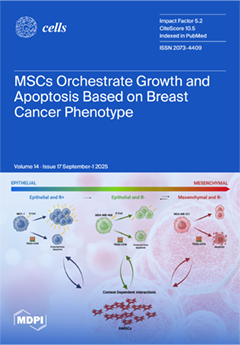
The leading cause of death in breast cancer (BrCa) is the conversion of dormant micro-metastases into outgrowth. Factors that enable these cells to persist in dormancy or progress into rapid proliferation are not fully defined; however, studies suggest a significant role of mesenchymal stem cells (MSCs) in modulating the metastatic progression of BrCa. Using various cultures of immortalized human MSCs with different breast cancer cell lines—MCF-7, MDA-MB-468 and MDA-MB-231—we illustrate that the effects of ihMSC on these cell lines are context-dependent, showing differential proliferation effects and apoptotic phenotypes and protection from induced cell death based on the phenotype of the target BrCa cell. This suggests that the MSCs regulate the metastatic behavior of breast cancer cells through complex Juxtacrine–Paracrine signaling. View this paper
  • Issues are regarded as officially published after their release is announced to the table of contents alert mailing list.
  • You may sign up for e-mail alerts to receive table of contents of newly released issues.
  • PDF is the official format for papers published in both, html and pdf forms. To view the papers in pdf format, click on the "PDF Full-text" link, and use the free Adobe Reader to open them.
Order results
Result details
Section
Select all
Export citation of selected articles as:
18 pages, 1172 KB  
Review
The Zebrafish as a Model for Ocular Translational Research: From Retinal Repair to Regeneration
by Bijorn Omar Balzamino, Mariagrazia Severino, Concetta Cafiero, Marco Coassin, Antonio Di Zazzo and Alessandra Micera
Cells 2025, 14(17), 1405; https://doi.org/10.3390/cells14171405 - 8 Sep 2025
Viewed by 231
Abstract
In the last years, the zebrafish model has become a primary model system for vertebrate tissue regeneration, particularly for neurodegeneration and metabolic disease. Zebrafish (Danio rerio) are small freshwater teleosts valued for disease modelling, which are widely used in genetic laboratories, [...] Read more.
In the last years, the zebrafish model has become a primary model system for vertebrate tissue regeneration, particularly for neurodegeneration and metabolic disease. Zebrafish (Danio rerio) are small freshwater teleosts valued for disease modelling, which are widely used in genetic laboratories, as a key model for studying neurodegenerative, metabolic, cardiac and dystrophic diseases, supporting the goal of identifying new therapeutic targets and approaches. Zebrafish can proliferate and produce/regenerate neurons. In response to retinal injury, zebrafish can regenerate multiple classes of retinal neurons and particularly, Müller glia-derived progenitor cells (MGPCs) can regenerate all types of neurons and restore visual function upon injury. The Jak/Stat-pathway of zebrafish retina represents one of the cell-signalling pathways involved in reprogramming Müller glia into MGPCs. In this era characterized by a revolution in experimental models and the future of omics, zebrafish might represent a suitable animal model for studying retinal degeneration and regeneration. In this context, the review is not meant to be entirely comprehensive of the zebrafish field, but it will highlight the usefulness of this model in discovering some mechanisms underlying retinal repair and regeneration. Full article
(This article belongs to the Special Issue Advances in Zebrafish Disease Models)
Show Figures

Figure 1

27 pages, 1202 KB  
Review
Integrating Multi-Omics in Endometrial Cancer: From Molecular Insights to Clinical Applications
by Hye Kyeong Kim and Taejin Kim
Cells 2025, 14(17), 1404; https://doi.org/10.3390/cells14171404 - 8 Sep 2025
Viewed by 294
Abstract
Endometrial cancer (EC) is the most common gynecologic malignancy in developed countries, and its incidence is increasing globally. While early-stage ECs generally show good prognosis, advanced or recurrent cases and those with aggressive histologic subtypes exhibit poor outcomes. Traditional histopathologic classification, however, fails [...] Read more.
Endometrial cancer (EC) is the most common gynecologic malignancy in developed countries, and its incidence is increasing globally. While early-stage ECs generally show good prognosis, advanced or recurrent cases and those with aggressive histologic subtypes exhibit poor outcomes. Traditional histopathologic classification, however, fails to reflect the molecular heterogeneity of EC, limiting its role in guiding treatment. Recent developments in multi-omics have enhanced our understanding of EC biology, which supports more personalized treatment strategies. The Cancer Genome Atlas (TCGA) classification has provided a more systematic molecular framework for stratifying risk and identifying prognostic and therapeutic biomarkers. This review discusses the latest developments in multi-omics-based classification of EC, highlights emerging diagnostic and therapeutic strategies, and summarizes ongoing clinical trials that aim to translate molecular discoveries into improved outcomes. Full article
(This article belongs to the Special Issue Signaling Pathways in Endometrial Cancer Cells)
Show Figures

Figure 1

17 pages, 3271 KB  
Article
Ectopic HLA-II Expression in ESCC: Exploration of Its Relationship with Neoantigen Burden and Patient Survival
by Yupei Ji, Zhizhong Wang, Zhenguo Cheng, Shuangshuang Lu, Nick R. Lemoine, Renato Baleeiro, Louisa S. Chard Dunmall and Yaohe Wang
Cells 2025, 14(17), 1403; https://doi.org/10.3390/cells14171403 - 8 Sep 2025
Viewed by 188
Abstract
Ectopic expression of human leukocyte antigen class II (HLA-II) on tumor cells correlates with anti-tumor immunity and prognosis in various cancers, but its role in esophageal squamous cell carcinoma (ESCC) remains unclear. Methods: HLA-II expression was evaluated in 34 ESCC tissue sections and [...] Read more.
Ectopic expression of human leukocyte antigen class II (HLA-II) on tumor cells correlates with anti-tumor immunity and prognosis in various cancers, but its role in esophageal squamous cell carcinoma (ESCC) remains unclear. Methods: HLA-II expression was evaluated in 34 ESCC tissue sections and a 102-sample tissue microarray (TMA) using immunohistochemistry (IHC) and in 10 ESCC cell lines via flow cytometry. Transcriptome sequencing of KYSE270, KYSE180, KYSE450, and KYSE510 was performed to investigate HLA-II regulatory mechanisms, while tumor samples from 104 ESCC patients were analyzed for neoantigen load. The prognostic significance of neoantigen burden was assessed using Cox regression. Results: HLA-II was ectopically expressed in ESCC, with positivity rates of 20.59% (34 tissues) and 25.49% (TMA). Among 10 ESCC cell lines, only KYSE270 exhibited spontaneous HLA-II expression. Transcriptome analysis revealed 1278 KYSE270-specific genes enriched in immune-related pathways (e.g., “Cytokine–cytokine receptor interaction”), suggesting immune-mediated HLA-II regulation. IFN-γ stimulation induced HLA-II expression in KYSE180, KYSE450, and KYSE510, indicating broader inducible HLA-II potential. In 104 patients, MHC-II-restricted neoantigen burden varied widely (0–75) and lacked direct correlation with HLA-II expression. Additionally, MHC-II-restricted neoantigen load was not significantly associated with overall survival (p > 0.05). Conclusion: Ectopic HLA-II expression in ESCC may influence the tumor immune microenvironment, while the prognostic value of MHC-II-restricted neoantigen burden in ESCC remains unclear, providing potential implications for immunotherapy strategies. Full article
(This article belongs to the Section Cell Microenvironment)
Show Figures

Figure 1

21 pages, 3708 KB  
Article
Autism Spectrum Disorder Induced Pluripotent Stem Cells Display Dysregulated Calcium Signaling During Neural Differentiation
by Abdullah J. AlShawaf, Sarah A. AlNassar, Norah AlGhamdi, Cristiana Mattei, Shiang Y. Lim, Mirella Dottori and Futwan A. Al-Mohanna
Cells 2025, 14(17), 1402; https://doi.org/10.3390/cells14171402 - 8 Sep 2025
Viewed by 304
Abstract
Autism Spectrum Disorder (ASD) is a neurodevelopmental condition that affects communication, social interaction, and behavior. Calcium (Ca2+) signaling dysregulation has been frequently highlighted in genetic studies as a contributing factor to aberrant developmental processes in ASD. Herein, we used ASD and [...] Read more.
Autism Spectrum Disorder (ASD) is a neurodevelopmental condition that affects communication, social interaction, and behavior. Calcium (Ca2+) signaling dysregulation has been frequently highlighted in genetic studies as a contributing factor to aberrant developmental processes in ASD. Herein, we used ASD and control induced pluripotent stem cells (iPSCs) to investigate transcriptomic and functional Ca2+ dynamics at various stages of differentiation to cortical neurons. Idiopathic ASD and control iPSC lines underwent the dual SMAD inhibition differentiation protocol to direct their fate toward cortical neurons. Samples from multiple time points along the course of differentiation were processed for bulk RNA sequencing, spanning the following sequential stages: the iPSC stage, neural induction (NI) stage, neurosphere (NSP) stage, and differentiated cortical neuron (Diff) stage. Our transcriptomic analyses suggested that the numbers of Ca2+ signaling-relevant differentially expressed genes between ASD and control samples were higher in the iPSC and Diff stages. Accordingly, samples from the iPSC and Diff stages were processed for Ca2+ imaging studies. Results revealed that iPSC-stage ASD samples displayed elevated maximum Ca2+ levels in response to ATP compared to controls. By contrast, in the Diff stage, ASD neurons showed reduced maximum Ca2+ levels in response to ATP but increased maximum Ca2+ levels in response to KCl and DHPG relative to controls. Considering the distinct functional signaling contexts of these stimuli, this differential profile of receptor- and ionophore-mediated Ca2+ response suggests that aberrant calcium homeostasis underlies the pathophysiology of ASD neurons. Our data provides functional evidence for Ca2+ signaling dysregulation during neurogenesis in idiopathic ASD. Full article
(This article belongs to the Special Issue Molecular Mechanisms of Autism Spectrum Disorder)
Show Figures

Figure 1

15 pages, 2661 KB  
Article
miR-451 Is a Driver of Lipotoxic Injury in Patients with Diabetic Cardiomyopathy
by Sarah Costantino, Shafeeq A. Mohammed, Federico Ranocchi, Francesco Zito, Valentina Delfine, Nazha Hamdani, Maria Cristina Vinci, Giovanni Melina and Francesco Paneni
Cells 2025, 14(17), 1401; https://doi.org/10.3390/cells14171401 - 8 Sep 2025
Viewed by 207
Abstract
MicroRNA 451 (miR-451) is emerging as a pivotal mediator of cardiac damage in experimental models of diabetic cardiomyopathy. Whether miR-451 plays a detrimental role in the human diabetic myocardium is unknown. The present study investigates miR-451’s role in patients with type 2 diabetes [...] Read more.
MicroRNA 451 (miR-451) is emerging as a pivotal mediator of cardiac damage in experimental models of diabetic cardiomyopathy. Whether miR-451 plays a detrimental role in the human diabetic myocardium is unknown. The present study investigates miR-451’s role in patients with type 2 diabetes (T2D). We show that miR-451 is upregulated in myocardial specimens from T2D patients compared to controls without diabetes and correlates with cardiometabolic parameters, the myocardial triglyceride content and cardiac expression of lipotoxic genes as well as echocardiographic indices of left ventricular dysfunction. Calcium-binding protein 39 (Cab39)—a known target of miR-451 in mouse hearts—was downregulated in T2D patients vs. controls, and its expression negatively correlated with that of miR-451. In cultured human cardiomyocytes (CMs), Ago2 immunoprecipitation confirmed Cab39 to be a direct target of miR-451. Treatment with a high amount of glucose (25mM) and palmitic acid (PA) mimicked miR-451 upregulation and Cab39 downregulation in human CMs. These changes were associated with increased TGs and markers of lipotoxic injury, such as elevated oxidative stress levels, mitochondrial dysfunction and apoptosis. Targeting miR-451 led to restoration of Cab39 levels while rescuing diabetes-induced lipotoxic injury and metabolic dysfunction. By contrast, miR-451 overexpression recapitulated features of lipotoxic damage. Our findings indicate miR-451 to be a potential target for the prevention of myocardial lipotoxic injury in diabetes. Full article
Show Figures

Figure 1

21 pages, 1901 KB  
Review
Targeting the NO–sGC–cGMP Pathway: Mechanisms of Action of Vericiguat in Chronic Heart Failure
by Tine Bajec and Gregor Poglajen
Cells 2025, 14(17), 1400; https://doi.org/10.3390/cells14171400 - 8 Sep 2025
Viewed by 434
Abstract
The recent advancements in the medical management of patients with chronic heart failure with reduced ejection fraction (HFrEF) is the soluble guanylate cyclase (sGC) stimulator, vericiguat. Clinical trials have demonstrated that vericiguat effectively lowers plasma levels of NT-proBNP and reduces the risk of [...] Read more.
The recent advancements in the medical management of patients with chronic heart failure with reduced ejection fraction (HFrEF) is the soluble guanylate cyclase (sGC) stimulator, vericiguat. Clinical trials have demonstrated that vericiguat effectively lowers plasma levels of NT-proBNP and reduces the risk of cardiovascular death or hospitalization in HFrEF patients, making it a class IIb recommendation for patients with worsening heart failure despite receiving guideline-directed medical therapy. However, the precise pathophysiological mechanisms underlying these clinical benefits remain unexplored. This review aims to present the signalling pathways associated with maladaptive remodeling and heart failure progression that can be modulated by sGC stimulators, focusing on the antihypertrophic, antifibrotic, and anti-inflammatory effects of NO–sGC–cGMP signalling observed in preclinical studies. A better understanding of the mechanisms of action of sGC stimulators could optimize heart failure treatment strategies and enable tailoring of therapies to individual patient profiles. Full article
(This article belongs to the Special Issue New Research on Immunity and Inflammation in Cardiovascular Disease)
Show Figures

Figure 1

11 pages, 215 KB  
Editorial
Uncovering the Cellular and Molecular Landscape of Gynecological Disorders
by Qiwei Yang
Cells 2025, 14(17), 1399; https://doi.org/10.3390/cells14171399 - 8 Sep 2025
Viewed by 353
Abstract
Gynecological disorders encompass a diverse array of conditions that affect the female reproductive system, including the uterus, ovaries, fallopian tubes, cervix, vagina, and external genitalia [...] Full article
(This article belongs to the Special Issue Cellular and Molecular Mechanisms in Gynecological Disorders)
34 pages, 2282 KB  
Review
Cancer-Associated Fibroblasts in Solid Tumors and Sarcomas: Heterogeneity, Function, and Therapeutic Implications
by Omar Badran, Idan Cohen and Gil Bar-Sela
Cells 2025, 14(17), 1398; https://doi.org/10.3390/cells14171398 - 7 Sep 2025
Viewed by 524
Abstract
Cancer-associated fibroblasts (CAFs) are crucial regulators of the tumor microenvironment (TME), promoting cancer progression, immune suppression, and therapy resistance. Single-cell transcriptomics has identified at least five distinct CAF subtypes: myofibroblastic (myCAFs), inflammatory (iCAFs), antigen-presenting (apCAFs), metabolic (meCAFs), and vascular/developmental (vCAFs/dCAFs), each with unique [...] Read more.
Cancer-associated fibroblasts (CAFs) are crucial regulators of the tumor microenvironment (TME), promoting cancer progression, immune suppression, and therapy resistance. Single-cell transcriptomics has identified at least five distinct CAF subtypes: myofibroblastic (myCAFs), inflammatory (iCAFs), antigen-presenting (apCAFs), metabolic (meCAFs), and vascular/developmental (vCAFs/dCAFs), each with unique localization, signaling, and functions. While CAFs are well studied in epithelial cancers, their roles in sarcomas are less understood despite the shared mesenchymal origin of tumor and stromal cells. This overlap blurs the line between malignant and non-malignant fibroblasts, raising fundamental questions about the identity of CAFs in mesenchymal tumors. In this narrative review, we explore the heterogeneity and plasticity of CAFs across solid tumors, focusing on their role in immune evasion, epithelial-to-mesenchymal transition (EMT), and resistance to chemotherapy, targeted therapy, and immunotherapy. We highlight emerging evidence on CAF-like cells in sarcomas and their contribution to tumor invasion, immune exclusion, and metastatic niche formation. We also assess new strategies to target or reprogram CAFs and suggest that CAF profiling may serve as a potential biomarker for patient stratification. Understanding CAF biology across various tumor types, including those with dense stroma and immunologically cold sarcomas, is crucial for developing more effective, personalized cancer treatments. Full article
Show Figures

Figure 1

24 pages, 4260 KB  
Article
Distinct Inflammatory Responses of hiPSC-Derived Endothelial Cells and Cardiomyocytes to Cytokines Involved in Immune Checkpoint Inhibitor-Associated Myocarditis
by Samantha Conte, Isaure Firoaguer, Simon Lledo, Thi Thom Tran, Claire El Yazidi, Stéphanie Simoncini, Zohra Rebaoui, Claire Guiol, Christophe Chevillard, Régis Guieu, Denis Puthier, Franck Thuny, Jennifer Cautela and Nathalie Lalevée
Cells 2025, 14(17), 1397; https://doi.org/10.3390/cells14171397 - 7 Sep 2025
Viewed by 412
Abstract
Inflammatory cytokines, particularly interferon-γ (IFN-γ), are markedly elevated in the peripheral blood of patients with immune checkpoint inhibitor-induced myocarditis (ICI-M). Endomyocardial biopsies from these patients also show GBP-associated inflammasome overexpression. While both factors are implicated in ICI-M pathophysiology, their interplay and cellular targets [...] Read more.
Inflammatory cytokines, particularly interferon-γ (IFN-γ), are markedly elevated in the peripheral blood of patients with immune checkpoint inhibitor-induced myocarditis (ICI-M). Endomyocardial biopsies from these patients also show GBP-associated inflammasome overexpression. While both factors are implicated in ICI-M pathophysiology, their interplay and cellular targets remain poorly characterized. Our aim was to elucidate how ICI-M-associated cytokines affect the viability and inflammatory responses of endothelial cells (ECs) and cardiomyocytes (CMs) using human induced pluripotent stem cell (hiPSC)-derived models. ECs and CMs were differentiated from the same hiPSC line derived from a healthy donor. Cells were exposed either to IFN-γ alone or to an inflammatory cytokine cocktail (CCL5, GZMB, IL-1β, IL-2, IL-6, IFN-γ, TNF-α). We assessed large-scale transcriptomic changes via microarray and evaluated inflammatory, apoptotic, and cell death pathways at cellular and molecular levels. hiPSC-ECs were highly sensitive to cytokine exposure, displaying significant mortality and marked transcriptomic changes in immunity- and inflammation-related pathways. In contrast, hiPSC-CM showed limited transcriptional changes and reduced susceptibility to cytokine-induced death. In both cell types, cytokine treatment upregulated key components of the inflammasome pathway, including regulators (GBP5, GBP6, P2X7, NLRC5), a core component (AIM2), and the effector GSDMD. Increased GBP5 expression and CASP-1 cleavage mirrored the findings found elsewhere in endomyocardial biopsies from ICI-M patients. This hiPSC-based model reveals a distinct cellular sensitivity to ICI-M-related inflammation, with endothelial cells showing heightened vulnerability. These results reposition endothelial dysfunction, rather than cardiomyocyte injury alone, as a central mechanism in ICI-induced myocarditis. Modulating endothelial inflammasome activation, particularly via AIM2 inhibition, could offer a novel strategy to mitigate cardiac toxicity while preserving antitumor efficacy. Full article
(This article belongs to the Special Issue New Research on Immunity and Inflammation in Cardiovascular Disease)
Show Figures

Figure 1

43 pages, 4033 KB  
Review
Updated Applications of Stem Cells in Hypoplastic Left Heart Syndrome
by Rui Xiao, Haleema Darr, Zarif Khan and Qingzhong Xiao
Cells 2025, 14(17), 1396; https://doi.org/10.3390/cells14171396 - 6 Sep 2025
Viewed by 1528
Abstract
Hypoplastic left heart syndrome (HLHS) is a severe congenital heart disease affecting 2–3 neonates every 10,000 live births. While prior research has highlighted associations of HLHS with specific chromosomal abnormalities and genetic mutations, the precise pathophysiology remains elusive. Despite early surgical intervention potentially [...] Read more.
Hypoplastic left heart syndrome (HLHS) is a severe congenital heart disease affecting 2–3 neonates every 10,000 live births. While prior research has highlighted associations of HLHS with specific chromosomal abnormalities and genetic mutations, the precise pathophysiology remains elusive. Despite early surgical intervention potentially allowing most HLHS patients to survive their critical heart disease with a single-ventricle physiology, patients frequently experience complications of arrhythmias and right ventricular heart failure, culminating in the need for an eventual heart transplant. Scarcity of suitable donors combined with limited understanding of mechanisms of development highlights the need for furthering our understanding of HLHS and alternative treatment options. Over the past decades, stem cell research has significantly advanced our understanding of cardiac conditions, repair, development, and therapy, opening the door for a new exciting field of regenerative medicine in cardiology with significant implications for HLHS. This review serves to provide a comprehensive overview of a much focused-on area related to HLHS. Specifically, we will first discuss the key pathophysiological basis and signalling molecules of HLHS. We then outline the emerging role of stem cell-based therapy, with a focus on adult stem cells and pluripotent stem cells (PSCs) in uncovering the pathophysiology of HLHS and optimising future treatment directions. Finally, we will also explore the latest and possible future directions of stem cell-derived techniques such as cardiac organoids and bioengineering cardiac tissues and their utility for investigating disease mechanisms, drug screening, and novel therapy for HLHF. Full article
Show Figures

Figure 1

19 pages, 6972 KB  
Article
Development and Characterization of a Novel Lineage of Renal Progenitor Cells for Potential Use in Feline Chronic Kidney Disease: A Preliminary Study
by Lara Carolina Mario, Juliana de Paula Nhanharelli, Jéssica Borghesi, Rafaela Rodrigues Ribeiro, Hianka Jasmyne Costa de Carvalho, Thamires Santos da Silva, Mariano del Sol, Rodrigo da Silva Nunes Barreto, Sandra Maria Barbalho and Maria Angelica Miglino
Cells 2025, 14(17), 1395; https://doi.org/10.3390/cells14171395 - 6 Sep 2025
Viewed by 438
Abstract
Chronic kidney disease (CKD) is a common and serious condition in felines. Accordingly, several cell therapies have been studied over the past decades for effective treatments. This study aimed to develop a new lineage of renal progenitor cells for use in cats with [...] Read more.
Chronic kidney disease (CKD) is a common and serious condition in felines. Accordingly, several cell therapies have been studied over the past decades for effective treatments. This study aimed to develop a new lineage of renal progenitor cells for use in cats with CKD. Metanephric and mesonephric progenitor cells were obtained from mesonephros and metanephros tissues of feline conceptuses at four distinct gestational stages. The cultured cells were characterized by their morphology, tumorigenic potential, immunophenotype determined by flow cytometry, and differentiation potential. We then conducted a pilot study in CKD-affected cats, comparing intraperitoneal injections of cultured metanephric progenitor cells (n = 4) to a placebo solution (n = 3). All four cell types exhibited adhesion and colony formation, but showed no tumorigenic potential. Cells tested positive for renal progenitor markers (CD117, Nephron, and WT1), confirming their identity. Treated cats showed no statistically significant differences (p ≤ 0.05) in any of the data analyzed. However, caregivers reported a voluntary increase in appetite after cell administration. Veterinarians confirmed this information during double-blind evaluations conducted after treatment. Although this data are qualitative, no clinical deterioration was observed in cats. Our results suggest that this new lineage of renal progenitor cells did not induce immediate adverse effects, thus supporting its potential for use in cell-based therapies. However, further studies are needed to evaluate its efficacy in treating renal diseases. Full article
(This article belongs to the Special Issue New Advances in Tissue Engineering and Regeneration)
Show Figures

Figure 1

24 pages, 2696 KB  
Article
Proteomics of Patient-Derived Striatal Medium Spiny Neurons in Multiple System Atrophy
by Nadine J. Smandzich, Andreas Pich, Thomas Gschwendtberger, Stephan Greten, Lan Ye, Martin Klietz, Alessio Di Fonzo, Lisa M. Henkel and Florian Wegner
Cells 2025, 14(17), 1394; https://doi.org/10.3390/cells14171394 - 6 Sep 2025
Viewed by 403
Abstract
The rare and rapidly progressive neurodegenerative disease multiple system atrophy (MSA) mainly affects the striatum and other subcortical brain regions. In this atypical Parkinsonian syndrome, the protein alpha-synuclein aggregates and misfolds in neurons as well as glial cells and is released in elevated [...] Read more.
The rare and rapidly progressive neurodegenerative disease multiple system atrophy (MSA) mainly affects the striatum and other subcortical brain regions. In this atypical Parkinsonian syndrome, the protein alpha-synuclein aggregates and misfolds in neurons as well as glial cells and is released in elevated amounts by hypoexcitable neurons. Mitochondrial dysregulation affects the biosynthesis of coenzyme Q10 and the activity of the respiratory chain, as shown in an induced pluripotent stem cell (iPSC) model. Proteome studies of cerebrospinal fluid and brain tissue from MSA patients yielded inconsistent results regarding possible protein changes due to small and combined groups of atypical Parkinsonian syndromes. In this study, we analysed the cellular proteome of MSA patient-derived striatal GABAergic medium spiny neurons. We observed 25 significantly upregulated and 16 significantly downregulated proteins in MSA cell lines compared to matched healthy controls. Various protein types involved in diverse molecular functions and cellular processes emphasise the multifaceted pathomechanisms of MSA. These data could contribute to the development of novel disease-modifying treatment strategies for MSA patients. Full article
(This article belongs to the Special Issue Role of Alpha-Synuclein in Neurodegenerative Diseases)
Show Figures

Figure 1

22 pages, 1566 KB  
Review
Integrating Macrophages into Human-Engineered Cardiac Tissue
by Yi Peng Zhao and Barry M. Fine
Cells 2025, 14(17), 1393; https://doi.org/10.3390/cells14171393 - 6 Sep 2025
Viewed by 417
Abstract
Heart disease remains a leading cause of morbidity and mortality worldwide, necessitating the development of in vivo models for therapeutic development. Advances in biomedical engineering in the past decade have led to the promising rise of human-based engineered cardiac tissues (hECTs) using novel [...] Read more.
Heart disease remains a leading cause of morbidity and mortality worldwide, necessitating the development of in vivo models for therapeutic development. Advances in biomedical engineering in the past decade have led to the promising rise of human-based engineered cardiac tissues (hECTs) using novel scaffolds and pluripotent stem cell derivatives. This has led to a new frontier of human-based models for improved preclinical development. At the same time, there has been significant progress in elucidating the importance of the immune system and, in particular, macrophages, particularly during myocardial injury. This review summarizes new methods and findings for deriving macrophages from human pluripotent stem cells (hPSCs) and advances in integrating these cells into cardiac tissue. Key challenges include immune cell infiltration in 3D constructs, maintenance of tissue architecture, and modeling aged or diseased cardiac microenvironments. By integrating immune components, hECTs can serve as powerful tools to unravel the complexities of cardiac pathology and develop targeted therapeutic strategies. Full article
(This article belongs to the Special Issue Immune Cells from Pluripotent Stem Cells)
Show Figures

Figure 1

17 pages, 2593 KB  
Article
Immunophenotypic Profile of Normal Hematopoietic Populations in Human Bone Marrow: Influence of Gender and Aging as a Basis for Reference Value Establishment
by Flavia Arandas de Sousa, Rodolfo Patussi Correa, Laiz Cameirão Bento, Luiz Fabiano Presente Taniguchi, Nydia Strachman Bacal and Luciana Cavalheiro Marti
Cells 2025, 14(17), 1392; https://doi.org/10.3390/cells14171392 - 6 Sep 2025
Viewed by 558
Abstract
The purpose of this study was to evaluate normal values of healthy human bone marrow (n = 56) and identify gender- and age-related variations using cell lineage markers and maturational curves. Using 10-color quantitative flow cytometry, various cell types were identified, including [...] Read more.
The purpose of this study was to evaluate normal values of healthy human bone marrow (n = 56) and identify gender- and age-related variations using cell lineage markers and maturational curves. Using 10-color quantitative flow cytometry, various cell types were identified, including B cells, T cells, NK cells, granulocytes, monocytes, erythroblasts, plasma cells, basophils, mast cells, and dendritic cells. Results revealed significant age-related declines in the absolute counts of nucleated cells (p = 0.001), including CD34+ immature B cells (p = 0.006) and CD34- immature B cells (p = 0.004). Declines were also observed for T cells (p = 0.002), cytotoxic T cells (p < 0.001), double-negative T cells (p = 0.0001), NK cells (p = 0.007), CD16- NK cells (p < 0.001), metamyelocytes (p = 0.002), neutrophils (p = 0.001), basophils (p = 0.009), promonocytes (p = 0.001), mature monocytes (p = 0.007), and plasmacytoid dendritic cells (p = 0.001). Gender differences showed males had more intermediate monocytes (p = 0.009) compared to females. In summary, this study provides normal values for hematopoietic cells, highlighting age- and gender-related disparities critical for understanding hematopoietic dynamics. Full article
Show Figures

Figure 1

17 pages, 3273 KB  
Review
LRP5: A Multifaceted Co-Receptor in Development, Disease, and Therapeutic Target
by Abdulmajeed F. Alrefaei
Cells 2025, 14(17), 1391; https://doi.org/10.3390/cells14171391 - 5 Sep 2025
Viewed by 413
Abstract
Low-density lipoprotein receptor-related protein 5 (LRP5) is a multifunctional transmembrane coreceptor that plays a pivotal role in development and disease. Wnt/β-catenin signaling is the primary downstream signaling pathway activated by LRP5. Furthermore, some LRP5 functions are mediated by noncanonical pathways, such as AKT/P21 [...] Read more.
Low-density lipoprotein receptor-related protein 5 (LRP5) is a multifunctional transmembrane coreceptor that plays a pivotal role in development and disease. Wnt/β-catenin signaling is the primary downstream signaling pathway activated by LRP5. Furthermore, some LRP5 functions are mediated by noncanonical pathways, such as AKT/P21 and TGF-β/Smad signaling. Pathologically, both loss-of-function and gain-of-function mutations in LRP5 produce distinct phenotypes, ranging from osteoporosis-pseudoglioma syndrome to high bone mass disorders. Beyond the skeletal system, LRP5 has emerged as a key regulator of retinal angiogenesis, vascular integrity, renal tubular function, neurodevelopment, and lipid metabolism. Its physiological functions are highlighted by its ability to influence adipocyte differentiation, insulin sensitivity, and neuronal synaptic plasticity. Moreover, LRP5 displays a dual role in development and disease progression. Although it plays a protective role in acute injuries such as myocardial infarction and acute kidney injury, LRP5 also contributes to chronic pathologies such as tubulointerstitial fibrosis, polycystic kidney disease, and atherosclerosis through fibrotic and inflammatory pathways. Recent therapeutic interest has focused on modulating LRP5 activity using agents such as anti-Dickkopf-related protein 1 antibody, sclerostin inhibitors, polyclonal antibodies, CRISPR/Cas9 knockout, and some natural products. This review discusses the current understanding of LRP5's physiological and pathological roles across organ systems and highlights its therapeutic potential, emphasizing the need for targeted approaches considering its context-dependent effects. Full article
(This article belongs to the Section Tissues and Organs)
Show Figures

Figure 1

32 pages, 7407 KB  
Review
Cardiac Development, Cellular Composition and Function: From Regulatory Mechanisms to Applications
by Huan-Yu Zhao, Jie-Bing Jiang, Shu-Na Wang and Chao-Yu Miao
Cells 2025, 14(17), 1390; https://doi.org/10.3390/cells14171390 - 5 Sep 2025
Viewed by 548
Abstract
Cardiogenesis and heart cell composition and function constitute fundamental areas of cardiovascular medicine research, and exploring their underlying mechanisms is closely tied to the goals of precision medicine. This review comprehensively examines the composition and functions of the heart from embryonic organogenesis to [...] Read more.
Cardiogenesis and heart cell composition and function constitute fundamental areas of cardiovascular medicine research, and exploring their underlying mechanisms is closely tied to the goals of precision medicine. This review comprehensively examines the composition and functions of the heart from embryonic organogenesis to maturity, and highlights the main breakthroughs of treatment strategies associated with these processes. By elaborating on the spatiotemporally specific signaling pathways and transcriptional networks that drive heart organogenesis and progenitor cell fate determination during the pivotal stages of cardiac development, and by systematically presenting the molecular biomarkers and functional characteristics of the principal cell types in mature heart, the latest advancements in related applications are summarized, with a particular emphasis on breakthroughs in gene/cell therapy, organoid development, and tissue engineering and regenerative medicine. This paper provides a theoretical foundation for precision interventions and regenerative medicine in cardiovascular disease using an axis that integrates cardiogenesis, cellular architecture, and therapeutic translation. Full article
Show Figures

Figure 1

21 pages, 1055 KB  
Article
Prognostic Associations and Functional Implications of Angiogenesis-Related miRNA Variants in Ischemic Stroke
by Chang Soo Ryu, Kee-Ook Lee, Eun Ju Ko, Hyeon Woo Park, Jae Hyun Lee, Ok Joon Kim and Nam Keun Kim
Cells 2025, 14(17), 1389; https://doi.org/10.3390/cells14171389 - 5 Sep 2025
Viewed by 359
Abstract
Ischemic stroke is a multifactorial cerebrovascular disease that remains a leading cause of long-term disability and mortality worldwide. Despite advances in acute treatment, recurrence rates remain high, and nearly half of survivors experience persistent neurological deficits. Therefore, identifying genetic biomarkers that contribute to [...] Read more.
Ischemic stroke is a multifactorial cerebrovascular disease that remains a leading cause of long-term disability and mortality worldwide. Despite advances in acute treatment, recurrence rates remain high, and nearly half of survivors experience persistent neurological deficits. Therefore, identifying genetic biomarkers that contribute to early diagnosis, risk prediction, and therapeutic improvement is increasingly important. MicroRNAs, small non-coding RNAs involved in gene regulation, have been recognized for their critical roles in vascular development and angiogenesis. This study investigated the association between angiogenesis-related miRNA gene polymorphisms and ischemic stroke risk using a population-based case–control design. Genotyping and statistical analysis revealed that miR-21 rs13137 A > T and miR-126 rs4636297 G > A were significantly associated with stroke susceptibility. The TT genotype of miR-21 rs13137 demonstrated a protective effect (p = 0.019); the AA genotype of miR-126 rs4636297 was associated with increased risk (p = 0.006), along with its dominant model (p = 0.007). Additionally, deep learning models were utilized to evaluate gene–gene and gene–environment interactions, enhancing predictive accuracy and identifying synergistic effects between miRNA polymorphisms and clinical risk factors. In summary, specific miRNA variants may serve as novel biomarkers for ischemic stroke, providing valuable insight into genetic susceptibility and supporting the advancement of precision medicine strategies. Full article
Show Figures

Figure 1

21 pages, 987 KB  
Review
m6A mRNA Methylation in Hematopoiesis: The Importance of Writing, Erasing, and Reading
by Antonia-Gerasimina Vasilopoulou, Eleni Kalafati, Ekati Drakopoulou and Nicholas P. Anagnou
Cells 2025, 14(17), 1388; https://doi.org/10.3390/cells14171388 - 5 Sep 2025
Viewed by 472
Abstract
Over recent years, epitranscriptomic research has provided a new layer of gene regulation during hematopoietic development and aberrant hematopoiesis. Among the 170 identified RNA chemical marks, N6-methyladenosine (m6A) is the most abundant in eukaryotic cells and plays a critical [...] Read more.
Over recent years, epitranscriptomic research has provided a new layer of gene regulation during hematopoietic development and aberrant hematopoiesis. Among the 170 identified RNA chemical marks, N6-methyladenosine (m6A) is the most abundant in eukaryotic cells and plays a critical role in various biological processes. This dynamic modification is regulated by a series of methyltransferases, demethylases, and m6A binding proteins, known as writers, erasers, and readers, respectively. Emerging evidence suggests that m6A modification and its regulators are involved in every aspect of normal hematopoietic development, from the emergence of hematopoietic stem cells to the generation of mature blood cells. Also, it has been established that abnormal expression of m6A regulators is implicated in the initiation of blood diseases. In this review, we summarize the latest findings regarding the role of m6A in erythropoiesis and highlight its implications in the pathophysiology of hemoglobin disorders. Full article
(This article belongs to the Section Stem Cells)
Show Figures

Graphical abstract

31 pages, 1475 KB  
Review
TREM2 in Neurodegenerative Diseases: Mechanisms and Therapeutic Potential
by Ling Li, Xiaoxiao Zheng, Hongyue Ma, Mingxia Zhu, Xiuli Li, Xiaodan Sun and Xinhong Feng
Cells 2025, 14(17), 1387; https://doi.org/10.3390/cells14171387 - 5 Sep 2025
Viewed by 562
Abstract
Neurodegenerative diseases, including Alzheimer’s disease (AD), Parkinson’s disease (PD), and amyotrophic lateral sclerosis (ALS), represent significant global health challenges, affecting millions and straining healthcare systems. These disorders involve progressive neuronal loss and cognitive decline, with incompletely elucidated underlying mechanisms. Chronic neuroinflammation is increasingly [...] Read more.
Neurodegenerative diseases, including Alzheimer’s disease (AD), Parkinson’s disease (PD), and amyotrophic lateral sclerosis (ALS), represent significant global health challenges, affecting millions and straining healthcare systems. These disorders involve progressive neuronal loss and cognitive decline, with incompletely elucidated underlying mechanisms. Chronic neuroinflammation is increasingly recognized as a critical contributor to disease progression. The brain’s resident immune cells, microglia, are central to this inflammatory response. When overactivated, microglia and other immune cells, such as peripheral macrophages, can exacerbate inflammation and accelerate disease development. Triggering Receptor Expressed on Myeloid Cells 2 (TREM2) is a transmembrane receptor of the immunoglobulin superfamily that demonstrates high expression on microglia in the central nervous system. TREM2 serves a vital role in regulating phagocytosis, synaptic pruning, and energy metabolism. This review examines the functions of TREM2 in neurodegenerative diseases and its potential as a therapeutic target, aiming to inform future treatment strategies. Full article
Show Figures

Figure 1

14 pages, 3199 KB  
Article
Efficacy of Conventional and Novel Tyrosine Kinase Inhibitors for Uncommon EGFR Mutations—An In Vitro Study
by Hana Oiki, Kenichi Suda, Akira Hamada, Toshio Fujino, Keiko Obata, Yoshihisa Kobayashi, Kazuko Sakai, Shota Fukuda, Shuta Ohara, Masaoki Ito, Junichi Soh, Kazuto Nishio, Tetsuya Mitsudomi and Yasuhiro Tsutani
Cells 2025, 14(17), 1386; https://doi.org/10.3390/cells14171386 - 4 Sep 2025
Viewed by 548
Abstract
Afatinib and osimertinib are current treatment options for non-small cell lung cancer (NSCLC) patients with uncommon epidermal growth factor receptor (EGFR) mutations, although their efficacy is limited. To explore potentially effective drugs for these patients, we evaluated the efficacy of conventional [...] Read more.
Afatinib and osimertinib are current treatment options for non-small cell lung cancer (NSCLC) patients with uncommon epidermal growth factor receptor (EGFR) mutations, although their efficacy is limited. To explore potentially effective drugs for these patients, we evaluated the efficacy of conventional EGFR tyrosine kinase inhibitors (TKIs) and novel third-generation (3G) TKIs using in vitro models. Ba/F3 cells transformed with each of the five most frequent uncommon EGFR mutations, Del18 (delE709_T710insD), E709K, G719A, S768I, and L861Q, were used. The growth inhibitory effects of five novel 3G-TKIs, almonertinib, lazertinib, furmonertinib, rezivertinib, and befotertinib, in addition to currently available TKIs, were evaluated. We also explored for secondary resistant mutations to afatinib or osimertinib and TKIs that can overcome these resistances. Afatinib was active against all uncommon EGFR mutations tested. The 3G-TKIs were all active against the L861Q mutation and were inactive against the S768I mutation. Furmonertinib and befotertinib showed efficacy against exon 18 mutations (Del18, E709K, and G719A). In the acquired resistance models to afatinib or osimertinib, we found T790M or a novel T725M secondary mutation, respectively, both of which could be overcome by lazertinib. However, some afatinib-resistant cells acquired V769L/M secondary mutations that were refractory to all EGFR-TKIs tested. In conclusion, afatinib exhibited broad activity and some 3G-TKIs showed promising efficacy in the front-line setting. Lazertinib is a potential second-line option after acquisition of resistance to afatinib or osimertinib. Full article
(This article belongs to the Section Cellular Pathology)
Show Figures

Graphical abstract

28 pages, 636 KB  
Systematic Review
Machine Learning for Multi-Omics Characterization of Blood Cancers: A Systematic Review
by Sultan Qalit Alhamrani, Graham Roy Ball, Ahmed A. El-Sherif, Shaza Ahmed, Nahla O. Mousa, Shahad Ali Alghorayed, Nader Atallah Alatawi, Albalawi Mohammed Ali, Fahad Abdullah Alqahtani and Refaat M. Gabre
Cells 2025, 14(17), 1385; https://doi.org/10.3390/cells14171385 - 4 Sep 2025
Viewed by 593
Abstract
Artificial Intelligence and machine learning are increasingly used to interrogate complex biological data. This systematic review evaluates their application to multi-omics for the molecular characterization of hematological malignancies, an area with unmet clinical need. We searched PubMed, Embase, Institute of Electrical and Electronics [...] Read more.
Artificial Intelligence and machine learning are increasingly used to interrogate complex biological data. This systematic review evaluates their application to multi-omics for the molecular characterization of hematological malignancies, an area with unmet clinical need. We searched PubMed, Embase, Institute of Electrical and Electronics Engineers Xplore, and Web of Science from January 2015 to December 2024. Two reviewers screened records, extracted data, and used a modified appraisal emphasizing explainability, performance, reproducibility, and ethics. From 2847 records, 89 studies met inclusion criteria. Studies focused on acute myeloid leukemia (34), acute lymphoblastic leukemia (23), and multiple myeloma (18). Other hematological diseases were less frequently studied. Methods included Support Vector Machines, Random Forests, and deep learning (28, 25, and 24 studies). Multi-omics integration was reported in 23 studies. External validation occurred in 31 studies, and explainability in 19. The median diagnostic area under the curve was 0.87 (interquartile range 0.81 to 0.94); deep learning reached 0.91 but offered the least explainability. Artificial Intelligence and machine learning show promise for molecular characterization, yet gaps in validation, interpretability, and standardization remain. Priorities include external validation, interpretable modeling, harmonized evaluation, and standardized reporting with shared benchmarks to enable safe, reproducible clinical translation. Full article
(This article belongs to the Section Cell Methods)
Show Figures

Figure 1

19 pages, 2817 KB  
Article
A Synthetic Sponge System Against miRNAs of the miR-17/92 Cluster Targets Transcriptional MYC Dosage Compensation in Aneuploid Cancer
by Diana M. Bravo-Estupiñan, Carsten Geiß, Jorge L. Arias-Arias, Mariela Montaño-Samaniego, Ricardo Chinchilla-Monge, Christian Marín-Müller, Steve Quirós-Barrantes, Anne Régnier-Vigouroux, Miguel Ibáñez-Hernández and Rodrigo A Mora-Rodríguez
Cells 2025, 14(17), 1384; https://doi.org/10.3390/cells14171384 - 4 Sep 2025
Viewed by 418
Abstract
Background: Genomic instability, a hallmark of cancer, leads to copy number variations disrupting gene dosage balance and contributing to tumor progression. One of the most affected oncogenes is MYC, whose overexpression is tightly regulated to avoid cytotoxicity. In aneuploid cancer cells, gene dosage [...] Read more.
Background: Genomic instability, a hallmark of cancer, leads to copy number variations disrupting gene dosage balance and contributing to tumor progression. One of the most affected oncogenes is MYC, whose overexpression is tightly regulated to avoid cytotoxicity. In aneuploid cancer cells, gene dosage compensation mechanisms involving microRNAs (miRNAs) from the miR-17/92 cluster contribute in regulating MYC expression. Targeting this miRNA-mediated compensation system represents a promising therapeutic strategy leading to an uncontrolled and lethal MYC overexpression. Results: Synthetic miRNA sponges targeting miR-17, miR-19a, and miR-20a, key regulators of MYC dosage compensation, were designed and validated. Breast cancer cells (MCF7) with stable exogenous MYC overexpression were used to assess the impact of sponge constructs on MYC regulation. Quantitative RT-PCR revealed a significant reduction in miRNA expression and a corresponding increase in endogenous MYC levels upon sponge treatment. Functional assays in multiple colorectal cancer cell lines with varying MYC copy numbers demonstrated a time-dependent increase in cell death following sponge transfection. Cytotoxic effects increased with MYC copy number, confirming a correlation between gene dosage sensitivity and therapeutic response. Conclusions: Our findings demonstrate that miRNA sponges targeting the miR-17/92 cluster can effectively disrupt MYC dosage compensation, leading to selective cytotoxicity in MYC-amplified cancer cells. Full article
(This article belongs to the Special Issue MicroRNAs: Regulators of Cellular Fate)
Show Figures

Figure 1

26 pages, 2348 KB  
Article
Voluntary Wheel Running Mitigates Disease in an Orai1 Gain-of-Function Mouse Model of Tubular Aggregate Myopathy
by Thomas N. O’Connor, Nan Zhao, Haley M. Orciuoli, Sundeep Malik, Alice Brasile, Laura Pietrangelo, Miao He, Linda Groom, Jennifer Leigh, Zahra Mahamed, Chen Liang, Feliciano Protasi and Robert T. Dirksen
Cells 2025, 14(17), 1383; https://doi.org/10.3390/cells14171383 - 4 Sep 2025
Viewed by 529
Abstract
Tubular aggregate myopathy (TAM) is an inherited skeletal muscle disease associated with progressive muscle weakness, cramps, and myalgia. Tubular aggregates (TAs) are regular arrays of highly ordered and densely packed straight-tubules observed in muscle biopsies; the extensive presence of TAs represent a key [...] Read more.
Tubular aggregate myopathy (TAM) is an inherited skeletal muscle disease associated with progressive muscle weakness, cramps, and myalgia. Tubular aggregates (TAs) are regular arrays of highly ordered and densely packed straight-tubules observed in muscle biopsies; the extensive presence of TAs represent a key histopathological hallmark of this disease in TAM patients. TAM is caused by gain-of-function mutations in proteins that coordinate store-operated Ca2+ entry (SOCE): STIM1 Ca2+ sensor proteins in the sarcoplasmic reticulum (SR) and Ca2+-permeable ORAI1 channels in the surface membrane. Here, we assessed the therapeutic potential of endurance exercise in the form of voluntary wheel running (VWR) in mitigating TAs and muscle weakness in Orai1G100S/+ (GS) mice harboring a gain-of-function mutation in the ORAI1 pore. Six months of VWR exercise significantly increased specific force production, upregulated biosynthetic and protein translation pathways, and normalized both mitochondrial protein expression and morphology in the soleus of GS mice. VWR also restored Ca2+ store content, reduced the incidence of TAs, and normalized pathways involving the formation of supramolecular complexes in fast twitch muscles of GS mice. In summary, sustained voluntary endurance exercise improved multiple skeletal muscle phenotypes observed in the GS mouse model of TAM. Full article
Show Figures

Figure 1

46 pages, 8337 KB  
Review
Numerical Modelling of Keratinocyte Behaviour: A Comprehensive Review of Biochemical and Mechanical Frameworks
by Sarjeel Rashid, Raman Maiti and Anish Roy
Cells 2025, 14(17), 1382; https://doi.org/10.3390/cells14171382 - 4 Sep 2025
Viewed by 657
Abstract
Keratinocytes are the primary cells of the epidermis layer in our skin. They play a crucial role in maintaining skin health, responding to injuries, and counteracting disease progression. Understanding their behaviour is essential for advancing wound healing therapies, improving outcomes in regenerative medicine, [...] Read more.
Keratinocytes are the primary cells of the epidermis layer in our skin. They play a crucial role in maintaining skin health, responding to injuries, and counteracting disease progression. Understanding their behaviour is essential for advancing wound healing therapies, improving outcomes in regenerative medicine, and developing numerical models that accurately mimic skin deformation. To create physically representative models, it is essential to evaluate the nuanced ways in which keratinocytes deform, interact, and respond to mechanical and biochemical signals. This has prompted researchers to investigate various computational methods that capture these dynamics effectively. This review summarises the main mathematical and biomechanical modelling techniques (with particular focus on the literature published since 2010). It includes reaction–diffusion frameworks, finite element analysis, viscoelastic models, stochastic simulations, and agent-based approaches. We also highlight how machine learning is being integrated to accelerate model calibration, improve image-based analyses, and enhance predictive simulations. While these models have significantly improved our understanding of keratinocyte function, many approaches rely on idealised assumptions. These may be two-dimensional unicellular analysis, simplistic material properties, or uncoupled analyses between mechanical and biochemical factors. We discuss the need for multiscale, integrative modelling frameworks that bridge these computational and experimental approaches. A more holistic representation of keratinocyte behaviour could enhance the development of personalised therapies, improve disease modelling, and refine bioengineered skin substitutes for clinical applications. Full article
(This article belongs to the Section Cellular Biophysics)
Show Figures

Figure 1

13 pages, 1180 KB  
Review
Polo-like Kinase 4: A Molecular Culprit in Skin Cancer Pathogenesis
by Tanya Jaiswal, Durdana Muntaqua and Nihal Ahmad
Cells 2025, 14(17), 1381; https://doi.org/10.3390/cells14171381 - 4 Sep 2025
Viewed by 416
Abstract
Skin cancer remains a significant global health challenge, with rising incidence and associated mortality in late-stage and drug-resistant cases. This underscores a continuing need for more effective novel therapeutic options that can be utilized for efficient management of skin cancers. A promising approach [...] Read more.
Skin cancer remains a significant global health challenge, with rising incidence and associated mortality in late-stage and drug-resistant cases. This underscores a continuing need for more effective novel therapeutic options that can be utilized for efficient management of skin cancers. A promising approach involves exploiting novel targets, which are dysregulated in skin cancer, either alone or in combination with existing therapeutics. Among these, polo-like kinases (PLKs), a family of serine/threonine kinases, has emerged as promising candidates due to their essential role in cell cycle and maintaining genomic stability, key hallmarks of cancer. Within this family, polo-like kinase 4 (PLK4) stands out as a structurally distinct member and the master regulator of centriole duplication, ensuring this process occurs only once per cell division. Dysregulation of PLK4 can disrupt genomic integrity, contributing to tumorigenesis, thus making it a promising target for cancer management. Notably, PLK4 is frequently overexpressed in several cancers, including skin cancer, and its precise role in skin cancer is an area of current investigation. Further, several small-molecule PLK4 inhibitors such as centrinone, YLZ-F5, CFI-400945, and RP-1664 have demonstrated efficacy in targeting PLK4. Among these, CFI-400945 has advanced to clinical trials, where it has shown modest anti-cancer activity. In this review, we provide a comprehensive overview of the known functions of PLK4 in skin cancer. Additionally, we discuss potential mechanistic insights into PLK4′s involvement in skin cancer progression by extrapolating evidence from studies in other cancer types including colorectal cancer, thyroid cancer, lymphomas, leukemia, etc., while identifying gaps for future research. Full article
Show Figures

Figure 1

22 pages, 1081 KB  
Review
Pseudouridine Synthase 7 in Cancer: Functions, Mechanisms, and Therapeutic Potential
by Qiwei Yang, Thomas G. Boyer and Ayman Al-Hendy
Cells 2025, 14(17), 1380; https://doi.org/10.3390/cells14171380 - 4 Sep 2025
Viewed by 518
Abstract
Pseudouridylation, the most abundant RNA modification, plays a critical role in modulating RNA structure, stability, and function. Among the family of pseudouridine synthases, Pseudouridine Synthase 7 (PUS7) has recently gained attention for its emerging roles in human health and disease. Originally characterized for [...] Read more.
Pseudouridylation, the most abundant RNA modification, plays a critical role in modulating RNA structure, stability, and function. Among the family of pseudouridine synthases, Pseudouridine Synthase 7 (PUS7) has recently gained attention for its emerging roles in human health and disease. Originally characterized for its function in modifying tRNA and small non-coding RNAs, PUS7 is now recognized as a dynamic regulator of mRNA pseudouridylation, influencing gene expression at the post-transcriptional level. Aberrant expressions or activity of PUS7 have been linked to a variety of pathological conditions, including cancers such as colon cancer, glioblastoma, pancreatic cancer, and neuroblastoma, as well as potential roles in neurodevelopmental disorders and immune regulation. Through mechanisms involving translational reprogramming, stress adaptation, and epitranscriptomic remodeling, PUS7 contributes to disease progression and cellular plasticity. This review summarizes the current understanding of PUS7 biology, its functional relevance in the contexts of cancer progression, and the growing interest in targeting RNA-modifying enzymes for therapeutic intervention. Uncovering the full spectrum of PUS7-mediated pseudouridylation and its downstream effects holds promise for advancing our understanding of RNA-based regulation in human diseases, including gynecological disorders. Full article
Show Figures

Figure 1

17 pages, 3777 KB  
Article
Heparanase-Neutralizing Monoclonal Antibody (mAb A54) Attenuates Tumor Growth and Metastasis
by Uri Barash, Malik Farhoud, Maali Odeh, Eliezer Huberman, Liang Wu and Israel Vlodavsky
Cells 2025, 14(17), 1379; https://doi.org/10.3390/cells14171379 - 4 Sep 2025
Viewed by 467
Abstract
Heparanase is the only human enzyme responsible for heparan sulfate (HS) breakdown, an activity that remodels the extracellular matrix (ECM) and strongly drives cancer metastasis and angiogenesis. Compelling evidence implies that heparanase promotes essentially all aspects of the tumorigenic process, namely, tumor initiation, [...] Read more.
Heparanase is the only human enzyme responsible for heparan sulfate (HS) breakdown, an activity that remodels the extracellular matrix (ECM) and strongly drives cancer metastasis and angiogenesis. Compelling evidence implies that heparanase promotes essentially all aspects of the tumorigenic process, namely, tumor initiation, vascularization, growth, metastasis, and chemoresistance. A key mechanism by which heparanase accelerates cancer progression is by enabling the release and bioavailability of HS-bound growth factors, chemokines, and cytokines, residing in the tumor microenvironment and supporting tumor growth and metastasis. The currently available heparanase inhibitors are mostly HS/heparin-like compounds that lack specificity and exert multiple off-target side effects. To date, only four such compounds have progressed to clinical trials, and none have been approved for clinical use. We have generated and characterized an anti-heparanase monoclonal antibody (A54 mAb) that specifically inhibits heparanase enzymatic activity (ECM degradation assay) and cellular uptake. Importantly, A54 mAb attenuates xenograft tumor growth and metastasis (myeloma, glioma, pancreatic, and breast carcinomas) primarily when administered (syngeneic or immunocompromised mice) in combination with conventional anti-cancer drugs. Co-crystallization of the A54 Fab fragment and the heparanase enzyme revealed that the interaction between the two proteins takes place adjacent to the enzyme HS/heparin binding domain II (HBDII; Pro271-Ala276), likely hindering heparanase from interacting with HS substrates via steric occlusion of the active site cleft. Collectively, we have generated and characterized a novel mAb that specifically neutralizes heparanase enzymatic activity and attenuates its pro-tumorigenic effects in preclinical models, paving the way for its clinical examination against cancer, inflammation, and other diseases. Full article
Show Figures

Figure 1

13 pages, 2353 KB  
Review
Tertiary Lymphoid Structures in Human Melanoma: Molecular Mechanisms and Therapeutic Opportunities
by Gelare Ghajar-Rahimi, Ishika Patel and Nabiha Yusuf
Cells 2025, 14(17), 1378; https://doi.org/10.3390/cells14171378 - 4 Sep 2025
Viewed by 465
Abstract
Tertiary lymphoid structures (TLSs) are ectopic lymphoid aggregates often found in chronic inflammatory conditions, including cancer. These structures, which share many cellular and functional features with secondary lymphoid organs, can profoundly influence the tumor microenvironment by promoting local anti-tumor immune activation. TLSs have [...] Read more.
Tertiary lymphoid structures (TLSs) are ectopic lymphoid aggregates often found in chronic inflammatory conditions, including cancer. These structures, which share many cellular and functional features with secondary lymphoid organs, can profoundly influence the tumor microenvironment by promoting local anti-tumor immune activation. TLSs have been observed in various cancers, including melanoma, and are associated with improved responses to immunotherapy and clinical outcomes. However, our understanding of the molecular mechanisms underlying TLS formation and function remains incomplete. This review summarizes the current findings on TLSs in human melanoma, drawing from multiple studies to provide an updated overview. We discuss the cellular composition, spatial distribution, and genetic signatures of TLSs at different stages of melanoma pathogenesis and in subtypes including acral, uveal, and desmoplastic melanoma. Additionally, we examine the influence of tumor mutational burden (TMB) and complement activation on TLS formation, as well as the role of TLSs in immune checkpoint inhibitor therapy. We also highlight the potential of TLSs as indicators for disease progression and treatment response, and review preclinical strategies aimed at inducing TLSs to improve therapeutic outcomes. This synthesis aims to support ongoing research into the role of TLSs in melanoma immunobiology. Full article
Show Figures

Figure 1

17 pages, 32794 KB  
Article
Histopathological Characteristics and Multi-Omics Analysis of Ocular Pigmentation Defects in Albino Percocypris pingi
by Senyue Liu, Xiaoyun Wu, Qiaolin Zou, Jiansheng Lai, Yongqiang Deng, Yang Feng, Chengyan Mou, Mingjiang Song, Pengcheng Li, Jun Du, Yan Liu, Qiang Li and Ya Liu
Cells 2025, 14(17), 1377; https://doi.org/10.3390/cells14171377 - 4 Sep 2025
Viewed by 416
Abstract
Percocypris pingi was listed in the China Vertebrate Red List in 2015, and albino P. pingi exhibits remarkable ocular phenotypes due to melanin synthesis defects, including the deficiency of melanin granules in the iris and retinal pigment epithelium (RPE). However, the regulatory mechanism [...] Read more.
Percocypris pingi was listed in the China Vertebrate Red List in 2015, and albino P. pingi exhibits remarkable ocular phenotypes due to melanin synthesis defects, including the deficiency of melanin granules in the iris and retinal pigment epithelium (RPE). However, the regulatory mechanism of pigment loss in the eyes of albino P. pingi has not yet been clarified. This study systematically revealed the potential mechanisms underlying the obstruction of ocular melanin synthesis in albino P. pingi through histopathological analysis, transcriptomics, and proteomics techniques. The results showed that the synergistic effects of abnormal H+ transport mediated by SLC45A2, excessive activation of retinol metabolism, and cytoskeletal transport disorders led to the inhibition of tyrosinase activity and retention of pigment granules, ultimately causing melanin deficiency in the eyes. This study first elucidates the molecular network of ocular albinism in fish from a multi-omics perspective, providing a new perspective for the mechanistic research of pigmentation disorders in vertebrates. Full article
(This article belongs to the Special Issue Retinal Disorders: Cellular Mechanisms and Targeted Therapies)
Show Figures

Figure 1

21 pages, 6817 KB  
Article
Prepubertal Diabetes Stagnates Testicular Development by Skewing Autophagy Homeostasis in Leydig Cells
by Zonghao Tang and Youkun Zheng
Cells 2025, 14(17), 1376; https://doi.org/10.3390/cells14171376 - 4 Sep 2025
Viewed by 411
Abstract
The maturation of testicular Leydig cells during the prepubertal stage is crucial for establishing male fertility. While diabetes is recognized as a significant detrimental factor affecting male testicular function, its impact specifically during the prepubertal period remains largely unknown. We hypothesized that prepubertal [...] Read more.
The maturation of testicular Leydig cells during the prepubertal stage is crucial for establishing male fertility. While diabetes is recognized as a significant detrimental factor affecting male testicular function, its impact specifically during the prepubertal period remains largely unknown. We hypothesized that prepubertal diabetes may impair testicular development by disrupting Leydig cell maturation. Using streptozotocin (STZ) administration, we established a prepubertal diabetic rat model and investigated the effects of diabetes on testicular development 2 and 4 weeks post-STZ treatment. Diabetes significantly hampered testicular development, manifesting as a decreased testicular weight, structural abnormalities, reduced testosterone levels, and increased inflammatory responses. As anticipated, prepubertal diabetes stagnated Leydig cell maturation and increased Leydig cell apoptosis. Mechanistic studies revealed that autophagy is essential for maintaining homeostasis and facilitating differentiation in immature Leydig cells but is significantly inhibited by hyperglycemia. Dysregulation of autophagy impaired the mitochondrial network, triggering inflammatory responses, suppressing steroidogenic capacity, and accumulating reactive oxygen species (ROS). Elevated ROS levels exacerbated the inflammatory response in the Leydig cells in an NLRP3-dependent manner. Inhibition of NLRP3 ameliorated the hyperglycemia-induced inflammation and decline in steroidogenic ability. Collectively, these findings demonstrate that hyperglycemia suppresses autophagy induction and enhances ROS accumulation in Leydig cells. This cascade promotes inflammation and inhibits steroidogenesis, thereby impeding testicular development in prepubertal diabetic rats. Full article
Show Figures

Figure 1

Previous Issue
Back to TopTop