Next Issue
Volume 14, June-1
Previous Issue
Volume 14, May-1


Cells, Volume 14, Issue 10 (May-2 2025) – 79 articles

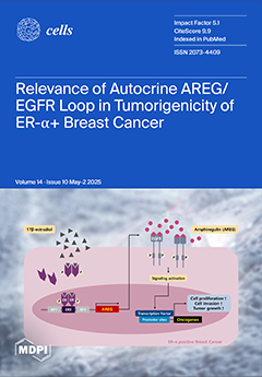
Cover Story (view full-size image): Despite the development of therapeutics targeting hormone receptors, recurrence remains a challenge. In previous studies, we found that EGFR+/ER+ breast cancer had a significantly lower survival rate. Therefore, we hypothesized that identifying an intermediary between EGFR and ER will play an important role in increasing the survival rate. In this study, we identify amphiregulin (AREG) as a key intermediary between EGFR and ER. E2-induced AREG plays an important role in promoting tumorigenicity. In addition, oncogenic effects of E2 were prevented by AREG knockdown. Our findings reveal that abnormal AREG expression is associated with a poor prognosis in EGFR+/ER+ breast cancer patients. Thus, a change in therapeutic strategy is required to increase the efficiency of EGFR+/ER+ breast cancer treatment. View this paper
  • Issues are regarded as officially published after their release is announced to the table of contents alert mailing list.
  • You may sign up for e-mail alerts to receive table of contents of newly released issues.
  • PDF is the official format for papers published in both, html and pdf forms. To view the papers in pdf format, click on the "PDF Full-text" link, and use the free Adobe Reader to open them.
Order results
Result details
Section
Select all
Export citation of selected articles as:
16 pages, 2271 KiB  
Article
Inhibition of GPX4 by Toxoplasma gondii Promotes Ferroptosis and Enhances Its Proliferation in Acute and Chronic Infection
by Yanlong Gu, Zhipeng Niu, Hui-Hong Lu, Si-Ang Li and Dong-Hui Zhou
Cells 2025, 14(10), 756; https://doi.org/10.3390/cells14100756 - 21 May 2025
Viewed by 127
Abstract
Toxoplasma gondii (T. gondii) is an intracellular parasite that extensively infects warm-blooded animals, causing toxoplasmosis and posing a significant threat to global public health. In this study, we investigated the association between T. gondii infection and ferroptosis in host cells, as [...] Read more.
Toxoplasma gondii (T. gondii) is an intracellular parasite that extensively infects warm-blooded animals, causing toxoplasmosis and posing a significant threat to global public health. In this study, we investigated the association between T. gondii infection and ferroptosis in host cells, as well as the regulatory role of glutathione peroxidase 4 (GPX4). Our findings revealed that mice infected with RH and PRU strains of T. gondii exhibited significantly elevated levels of reactive oxygen species and malondialdehyde in brain and liver tissues. Concurrently, the expression of GPX4, a critical negative regulator of ferroptosis, was downregulated, which correlated with the elevated parasite burden. In Vero cells, T. gondii infection similarly inhibited GPX4 expression, whereas GPX4 overexpression suppressed T. gondii proliferation. These results indicate that T. gondii infection can promote ferroptosis in host cells and that GPX4 plays a pivotal role in regulating infection and proliferation. This study provides novel insights into the pathogenic mechanisms of T. gondii and identifies GPX4 as a regulatory factor that constrains parasite proliferation, offering new approaches for toxoplasmosis prevention and control. Full article
Show Figures

Figure 1

17 pages, 625 KiB  
Review
Survivin Interference and SurVaxM as an Adjunct Therapy for Glioblastoma Multiforme
by Willie James Elliott, Nandini Gurramkonda, Maheedhara R. Guda, Andrew J. Tsung and Kiran K. Velpula
Cells 2025, 14(10), 755; https://doi.org/10.3390/cells14100755 - 21 May 2025
Viewed by 107
Abstract
Glioblastoma, IDH wild-type WHO Grade IV, is a devastating diagnosis in pediatric and adult populations with a poor prognosis and median overall survival of less than two years. Despite the advent of the Stupp protocol and advances in neurosurgical tumor resection techniques, there [...] Read more.
Glioblastoma, IDH wild-type WHO Grade IV, is a devastating diagnosis in pediatric and adult populations with a poor prognosis and median overall survival of less than two years. Despite the advent of the Stupp protocol and advances in neurosurgical tumor resection techniques, there has been minimal change to both the quantity and quality of life in individuals diagnosed. Provided the extensive research on survivin’s association with glioblastoma tumor microenvironment, this review suggests that priming the individual’s immune systems to the tumor-promoting protein may reduce tumor burden through multiple mechanisms, including the arrest of the G2/M phase, microtubule dysfunction, induction of autophagy, and ultimately activation of apoptosis in glioblastoma cells. SurVaxM, a multiple peptide, survivin-specific vaccine, may assist in tumor cell destruction by eliciting the production of cytotoxic T-cells specific to survivin-expression glioblastoma tumors. Although phase I and II clinical trials suggest relatively safe adverse effects and potential efficacy, additional research is necessary to evaluate further how this vaccine may compare to standard treatment. Full article
Show Figures

Figure 1

32 pages, 21562 KiB  
Article
Major Traumatic Injury and Exposure to Mitochondrial-Derived Damage-Associated Molecular Patterns Promotes Neutrophil Survival Accompanied by Stabilisation of the Anti-Apoptotic Protein Mcl-1
by Thomas Nicholson, Michael Macleod, Antonio Belli, Janet M. Lord and Jon Hazeldine
Cells 2025, 14(10), 754; https://doi.org/10.3390/cells14100754 - 21 May 2025
Viewed by 94
Abstract
Traumatic injury leads to an extension of the half-life of circulating neutrophils. However, how quickly neutrophil apoptosis is delayed post-injury is currently unknown, as are the underlying mechanisms and factors that promote this extension of lifespan. During the ultra-early (≤1 h) and acute [...] Read more.
Traumatic injury leads to an extension of the half-life of circulating neutrophils. However, how quickly neutrophil apoptosis is delayed post-injury is currently unknown, as are the underlying mechanisms and factors that promote this extension of lifespan. During the ultra-early (≤1 h) and acute (4–12 and 48–72 h) post-injury phases, we collected blood samples from 73 adult trauma patients. Following ex vivo culture, neutrophil apoptosis was measured, alongside caspase-3 activation and expression of the anti-apoptotic protein Mcl-1. To identify factors that may promote neutrophil survival post-trauma, neutrophils from healthy controls (HCs) were cultured with mitochondrial-derived damage-associated molecular patterns (mtDAMPs) or mitochondrial DNA (mtDNA). Accompanied by reduced mitochondrial membrane depolarisation, delayed Mcl-1 turnover, and reduced caspase-3 activation, the ex vivo lifespan of neutrophils from trauma patients was significantly enhanced in a protein synthesis-independent manner within minutes to hours after injury. Neutrophils from HCs exhibited delayed apoptosis when cultured in media supplemented with trauma patient serum, which occurred alongside stabilisation of Mcl-1. Culturing HCs neutrophils with mtDAMPs or mtDNA significantly delayed apoptosis rates, promoted stabilisation of Mcl-1, and reduced caspase-3 activation. The release of mtDAMPs from damaged tissue may drive post-trauma immune dysregulation by promoting the survival of dysfunctional neutrophils. Full article
(This article belongs to the Collection Feature Papers in ‘Cellular Immunology’)
Show Figures

Graphical abstract

24 pages, 754 KiB  
Review
Chimeric Autoantibody Receptor- and/or Peptide-MHC-Based CAR Therapies for Targeted Elimination of Antigen-Specific B or T Cells in Hypersensitivity Disorders Such as Allergies and Autoimmune Diseases
by Isidora Protić-Rosić, Al Nasar Ahmed Sehgal, Sebastian Wrighton, Birgit Heller and Winfried F. Pickl
Cells 2025, 14(10), 753; https://doi.org/10.3390/cells14100753 - 21 May 2025
Viewed by 230
Abstract
Hypersensitivity reactions are dysregulated and potentially devastating immune responses, characterized by a tendency to become chronic. They target either self-proteins or harmless foreign proteins and are driven by both T and B cells. Although numerous symptomatic treatment options for hypersensitivity reactions have been [...] Read more.
Hypersensitivity reactions are dysregulated and potentially devastating immune responses, characterized by a tendency to become chronic. They target either self-proteins or harmless foreign proteins and are driven by both T and B cells. Although numerous symptomatic treatment options for hypersensitivity reactions have been established over recent decades, only a few antigen-specific, causal approaches capable of specifically targeting the pathogenic autoreactive T and/or B cells have been developed. Among these are cell-based treatment modalities involving chimeric antigen receptor (CAR)- or chimeric autoantibody-receptor (CAAR)-expressing cells. These therapies utilize B- or T-cell antigens, presented as B-cell epitopes or peptide-major histocompatibility complexes (pMHCs) to serve as bait. The latter are coupled to potent activation domains derived from the TCR/CD3 complex itself, such as the zeta or CD3 chains, as well as domains from bona fide co-stimulatory molecules (e.g., CD28, 4-1BB). Recent in vitro and in vivo studies have demonstrated the therapeutic potential of these ATMP-based strategies in eliminating autoreactive lymphocytes and alleviating hypersensitivity reactions. This systematic review provides a comprehensive overview of the current status of antigen-specific CAR and CAAR T-cell therapies, highlighting novel directions as well as the ongoing challenges within this promising research field. Full article
Show Figures

Figure 1

18 pages, 1949 KiB  
Article
Utilization of Flow Cytometry, Metabolomic Analyses and a Feline Infectious Peritonitis Case Study to Evaluate the Physiological Impact of Polyprenyl Immunostimulant
by Irene Lee, Amar Desai, Akshay Patil, Yan Xu, Kelley Pozza-Adams and Anthony J Berdis
Cells 2025, 14(10), 752; https://doi.org/10.3390/cells14100752 - 21 May 2025
Viewed by 135
Abstract
Measles, hepatitis C, and COVID-19 are significant human diseases caused by RNA viruses. While vaccines exist to prevent infections, there are a small number of currently available therapeutic agents that can effectively treat these diseases after infection occurs. This study explores a new [...] Read more.
Measles, hepatitis C, and COVID-19 are significant human diseases caused by RNA viruses. While vaccines exist to prevent infections, there are a small number of currently available therapeutic agents that can effectively treat these diseases after infection occurs. This study explores a new therapeutic strategy using a small molecule designated polyprenyl immunostimulant (PI) to increase innate immune responses and combat viral infections. Using a multi-disciplinary approach, this study quantifies the effects of PI in mice and THP-1 cells using flow cytometry to identify immune phenotypic markers and mass spectroscopy to monitor the metabolomic profiles of immune cells perturbed by PI treatment. The metabolomic studies identified that sphinganine and ceramide, which are precursors of sphingosine-1-phosphate (S1P), were the common metabolites upregulated in THP-1 and mice blood. Sphingosine-1-phosphate can mediate the trafficking of T cells, whereas ceramide can signal the activation and proliferation of T cells, thereby modulating the mammalian host’s immunity. To demonstrate proof-of-principle, a case study was conducted to examine the benefit of administering PI to improve the outcomes of a feline co-infected with two distinct RNA viruses—feline leukemia virus and feline infectious peritonitis virus. Both viruses produce deadly symptoms that closely resemble RNA viruses that infect humans. The results identify quantifiable cellular and metabolic markers arising from PI treatment that can be used to establish a platform measuring the efficacy of PI in modulating the innate immune system. Full article
Show Figures

Figure 1

13 pages, 2729 KiB  
Article
Influence of Unidirectional Vacuum Application on Bone Healing in Maxillofacial Surgery
by Tom Alexander Schröder, Athanasios Karasavvas, Maximilian Bauckloh, Matthias C. Schulz, Günter Lauer and Lysann Michaela Kroschwald
Cells 2025, 14(10), 751; https://doi.org/10.3390/cells14100751 - 21 May 2025
Viewed by 143
Abstract
Negative-pressure wound therapy (NPWT) using vacuum-assisted closure (VAC) is a well known tissue defect bridging method that applies a vacuum pump to sterile, open-cell foam dressings via suction tubes. Although it has mostly been described for soft tissue use, there are also a [...] Read more.
Negative-pressure wound therapy (NPWT) using vacuum-assisted closure (VAC) is a well known tissue defect bridging method that applies a vacuum pump to sterile, open-cell foam dressings via suction tubes. Although it has mostly been described for soft tissue use, there are also a few studies concerning its use on hard tissue. However, as oral and maxillofacial surgery has to deal with both soft and hard tissue, which lie next to each other in these regions, there is a particular need to assess the influence of negative pressure on bone. Therefore, the effects of different negative pressure levels (530 mbar and 725 mbar) and atmospheric pressure (1013 mbar) on bone tissue cultures and osteoblast cell cultures were investigated over periods of 1, 3, and 6 weeks. During the culture period, osteoblast growth and the tissue regeneration of bone defects were studied in vitro using tissue cultures that were histologically supplemented by cytological investigations and quantitative RNA expression studies. In the bone defect model, there was a faster defect reduction using NPWT; the effect was especially strong for 530 mbar. Compared to the control group, up to 30% more newly generated bone tissue was detected. This effect on the mineralization capacity was assessed by the mRNA expression of osteogenic marker genes, as well as the receptor activator of nuclear factor κB ligand (RANKL) and osteoprotegerin (OPG), two multifaceted cytokines that regulate bone metabolism. The influence of negative pressure consequently resulted in a decreased RANKL/OPG ratio in osteoblasts. Associated with the upregulation of marker genes to up to 400%, including Col1, BMP4, OCN, and RUNX2, the decrease in the RANKL/OPG ratio to 41% indicates the stimulation of osteogenesis. Since VAC has been shown to be a safe and effective method to close wounds in general, these data suggest that patients suffering from compound bone and soft tissue defects in the maxillofacial area may benefit from an adapted therapy approach accelerating both soft and hard tissue regeneration. Full article
Show Figures

Figure 1

19 pages, 33190 KiB  
Article
Jun N-Terminal Kinase Inhibitor Suppresses CASK Deficiency-Induced Cerebellar Granular Cell Death in MICPCH Syndrome Model Mice
by Qi Guo, Emi Kouyama-Suzuki, Yoshinori Shirai and Katsuhiko Tabuchi
Cells 2025, 14(10), 750; https://doi.org/10.3390/cells14100750 - 20 May 2025
Viewed by 286
Abstract
Microcephaly with pontine and cerebellar hypoplasia (MICPCH) syndrome is a severe neurodevelopmental disorder caused by a deficiency in the X-linked gene calcium/calmodulin-dependent serine protein kinase (CASK). A better understanding of the role of CASK in the pathophysiology of neurodevelopmental disorders may provide insights [...] Read more.
Microcephaly with pontine and cerebellar hypoplasia (MICPCH) syndrome is a severe neurodevelopmental disorder caused by a deficiency in the X-linked gene calcium/calmodulin-dependent serine protein kinase (CASK). A better understanding of the role of CASK in the pathophysiology of neurodevelopmental disorders may provide insights into novel therapeutic and diagnostic strategies for MICPCH syndrome and other neurodegenerative diseases. To investigate this, we generated CASK knockout (KO) cerebellar granule (CG) cell culture from CASK floxed (CASKflox/flox) mice by infecting lentiviruses expressing codon-improved Cre recombinase (iCre). We performed RNA-sequencing (RNA-seq) on these cells and found that CASK-KO CG cells underwent apoptosis by activating intracellular Jun N-terminal kinase (JNK) signaling and upregulating reactive oxygen species (ROS)-related gene expression. We also performed mouse gait analysis and limb clasping behavior experiments on trans-heterozygous CASK-KO and Hprt-eGFP (CASK+/- HprteGFP/+) mice. The CASK+/- HprteGFP/+ mice exhibited cerebellar ataxic phenotypes as judged by the scores of these experiments compared to the CASK wild-type control (CASK+/+ HprteGFP/+) mice. Interestingly, the administration of the JNK inhibitor, JNK-IN-8, in CASK-KO CG cell cultures increased CG cell survival by reducing ROS generation. Moreover, injection of JNK-IN-8 into the cerebellum of CASK+/- HprteGFP/+ mice suppressed CG cell death and alleviated cerebellar ataxic phenotypes in vivo. In conclusion, JNK-IN-8 suppresses the cell death and activation of the ROS pathway in CASK-KO CG cells in both in vitro and in vivo models, suggesting its potential as a therapeutic strategy for cerebellar neurodegeneration in MICPCH syndrome. Full article
(This article belongs to the Section Cells of the Nervous System)
Show Figures

Graphical abstract

27 pages, 2016 KiB  
Review
Extracellular Vesicles in the Crosstalk of Autophagy and Apoptosis: A Role for Lipid Rafts
by Agostina Longo, Valeria Manganelli, Roberta Misasi, Gloria Riitano, Tuba Rana Caglar, Elena Fasciolo, Serena Recalchi, Maurizio Sorice and Tina Garofalo
Cells 2025, 14(10), 749; https://doi.org/10.3390/cells14100749 - 20 May 2025
Viewed by 137
Abstract
Autophagy and apoptosis are two essential mechanisms regulating cell fate. Although distinct, their signaling pathways are closely interconnected through various crosstalk mechanisms. Lipid rafts are described to act as both physical and functional platforms during the early stages of autophagic and apoptotic processes. [...] Read more.
Autophagy and apoptosis are two essential mechanisms regulating cell fate. Although distinct, their signaling pathways are closely interconnected through various crosstalk mechanisms. Lipid rafts are described to act as both physical and functional platforms during the early stages of autophagic and apoptotic processes. Only recently has a role for lipid raft-associated molecules in regulating EV biogenesis and release begun to emerge. In particular, lipids of EV membranes are essential components in conferring stability to these vesicles in different extracellular environments and/or to facilitate binding or uptake into recipient cells. In this review we highlight these aspects, focusing on the role of lipid molecules during apoptosis and secretory autophagy pathways. We describe the molecular machinery that connects autophagy and apoptosis with vesicular trafficking and lipid metabolism during the release of EVs, and how their alterations contribute to the development of various diseases, including autoimmune disorders and cancer. Overall, these findings emphasize the complexity of autophagy/apoptosis crosstalk and its key role in cellular dynamics, supporting the role of lipid rafts as new therapeutic targets. Full article
(This article belongs to the Special Issue Crosstalk of Autophagy and Apoptosis: Recent Advances)
Show Figures

Figure 1

25 pages, 1419 KiB  
Review
Cancer Vulnerabilities Through Targeting the ATR/Chk1 and ATM/Chk2 Axes in the Context of DNA Damage
by Anell Fernandez, Maider Artola, Sergio Leon, Nerea Otegui, Aroa Jimeno, Diego Serrano and Alfonso Calvo
Cells 2025, 14(10), 748; https://doi.org/10.3390/cells14100748 - 20 May 2025
Viewed by 336
Abstract
Eliciting DNA damage in tumor cells continues to be one of the most successful strategies against cancer. This is the case for classical chemotherapy drugs and radiotherapy. In the modern era of personalized medicine, this strategy tries to identify specific vulnerabilities found in [...] Read more.
Eliciting DNA damage in tumor cells continues to be one of the most successful strategies against cancer. This is the case for classical chemotherapy drugs and radiotherapy. In the modern era of personalized medicine, this strategy tries to identify specific vulnerabilities found in each patient’s tumor, to inflict DNA damage in certain cell contexts that end up in massive cancer cell death. Cells rely on multiple DNA repair pathways to fix DNA damage, but cancer cells frequently exhibit defects in these pathways, many times being tolerant to the damage. Key vulnerabilities, such as BRCA1/BRCA2 mutations, have been exploited with PARP inhibitors, leveraging synthetic lethality to selectively kill tumor cells and improving patients’ survival. In the DNA damage response (DDR) network, kinases ATM, ATR, Chk1, and Chk2 coordinate DNA repair, cell cycle arrest, and apoptosis. Inhibiting these proteins enhances tumor sensitivity to DNA-damaging therapies, especially in DDR-deficient cancers. Several small-molecule inhibitors targeting ATM/Chk2 or ATR/Chk1 are currently being tested in preclinical and/or clinical settings, showing promise in cancer models and patients. Additionally, pharmacological blockade of ATM/Chk2 and ATR/Chk1 axes enhances the effects of immunotherapy by increasing tumor immunogenicity, promoting T-cell infiltration and activating immune responses. Combining ATM/Chk2- or ATR/Chk1-targeting drugs with conventional chemotherapy, radiotherapy or immune checkpoint inhibitors offers a compelling strategy to improve treatment efficacy, overcome resistance, and enhance patients’ survival in modern oncology. Full article
(This article belongs to the Special Issue Unlocking the Secrets Behind Drug Resistance at the Cellular Level)
Show Figures

Graphical abstract

28 pages, 1535 KiB  
Review
The Sebaceous Gland: A Key Player in the Balance Between Homeostasis and Inflammatory Skin Diseases
by Sarah Mosca, Monica Ottaviani, Stefania Briganti, Anna Di Nardo and Enrica Flori
Cells 2025, 14(10), 747; https://doi.org/10.3390/cells14100747 - 20 May 2025
Viewed by 156
Abstract
The sebaceous gland (SG) is an integral part of the pilosebaceous unit and is a very active and dynamic organ that contributes significantly to the maintenance of skin homeostasis. In addition to its primary role in sebum production, the SG is involved in [...] Read more.
The sebaceous gland (SG) is an integral part of the pilosebaceous unit and is a very active and dynamic organ that contributes significantly to the maintenance of skin homeostasis. In addition to its primary role in sebum production, the SG is involved in the maintenance of skin barrier function, local endocrine/neuroendocrine function, the innate immune response, and the regulation of skin bacterial colonization. Structural and functional alterations of SGs leading to the dysregulation of sebum production/composition and immune response may contribute to the pathogenesis of inflammatory dermatoses. This review summarises the current knowledge on the contribution of SGs to the pathogenesis of common inflammatory skin diseases. These findings are crucial for the development of more effective therapeutic strategies for the treatment of inflammatory dermatoses. Full article
(This article belongs to the Special Issue Sebaceous Gland in Skin Health and Disease)
Show Figures

Figure 1

13 pages, 1045 KiB  
Article
All-Trans Retinoic Acid Induces Differentiation and Downregulates Stemness Markers and MGMT Expression in Glioblastoma Stem Cells
by Justin Tang and Raymond Yang
Cells 2025, 14(10), 746; https://doi.org/10.3390/cells14100746 - 20 May 2025
Viewed by 171
Abstract
Background: Glioblastoma (GBM) remains almost uniformly fatal, owing in part to therapy-resistant cancer stem-like cells (CSCs) and to temozolomide (TMZ) resistance driven by O6-methylguanine-DNA methyltransferase (MGMT). Differentiation therapy with all-trans retinoic acid (ATRA) has the potential to attenuate stemness and sensitize [...] Read more.
Background: Glioblastoma (GBM) remains almost uniformly fatal, owing in part to therapy-resistant cancer stem-like cells (CSCs) and to temozolomide (TMZ) resistance driven by O6-methylguanine-DNA methyltransferase (MGMT). Differentiation therapy with all-trans retinoic acid (ATRA) has the potential to attenuate stemness and sensitize GBM to TMZ. We therefore asked whether ATRA reduces expression of key CSC markers and MGMT in established GBM lines. Methods: Two established human GBM cell lines, U87-MG and A172, were cultured under neurosphere-promoting conditions to enrich for potential stem-like subpopulations. Cells were treated with either 1 µM ATRA or vehicle control (DMSO) for 5 days. Total RNA was extracted, and cDNA was synthesized. Quantitative Real-Time PCR (qPCR) assessed relative mRNA expression levels of key stemness transcription factors (SOX2, NES) and the DNA repair gene MGMT and corresponding protein levels were measured by an Enzyme-Linked Immunosorbent Assay (ELISA). Gene expression was normalized to the geometric mean of two validated housekeeping genes (GAPDH, ACTB). Relative quantification was calculated using the ΔΔCt method, and statistical significance was determined using Student’s t-tests. Results: ATRA markedly suppressed stemness and MGMT in both lines. In U87-MG, SOX2 mRNA fell 3.7-fold (p = 0.0008) and protein 2.99-fold (148.3 ± 6.0 → 49.7 ± 2.7 pg µg−1; p = 0.0002); Nestin dropped 4.1-fold (p = 0.0005) and 3.51-fold (450.0 ± 17.3 → 128.3 ± 4.4 pg µg−1; p = 0.00008). MGMT decreased 2.6-fold at transcript level (p = 0.0065) and 2.11-fold at protein level (81.7 ± 4.4 → 38.7 ± 1.8 pg µg−1; p = 0.0005). In A172, SOX2 was reduced 2.9-fold (p = 0.0041) and 2.31-fold (p = 0.0007); Nestin 3.3-fold (p = 0.0028) and 2.79-fold (p = 0.00009). MGMT declined 2.2-fold (p = 0.0132) and 1.82-fold (p = 0.0015), respectively. Conclusions: Five-day exposure to ATRA diminishes SOX2, Nestin, and MGMT at both mRNA and protein levels in stem-enriched GBM cultures, supporting the premise that ATRA-induced differentiation can concurrently blunt CSC traits and TMZ-resistance mechanisms. These data provide a molecular rationale for testing ATRA in combination regimens aimed at improving GBM therapy. Full article
(This article belongs to the Special Issue The Pivotal Role of Tumor Stem Cells in Glioblastoma)
Show Figures

Figure 1

27 pages, 1400 KiB  
Review
Perspectives of Targeting Autophagy as an Adjuvant to Anti-PD-1/PD-L1 Therapy for Colorectal Cancer Treatment
by Nasrah ALKhemeiri, Sahar Eljack and Maha Mohamed Saber-Ayad
Cells 2025, 14(10), 745; https://doi.org/10.3390/cells14100745 - 20 May 2025
Viewed by 303
Abstract
Colorectal cancer (CRC) is the third most common cancer in the world, with increasing incidence and mortality rates. Standard conventional treatments for CRC are surgery, chemotherapy, and radiotherapy. Recently, immunotherapy has been introduced as a promising alternative to CRC treatment that utilizes patients’ [...] Read more.
Colorectal cancer (CRC) is the third most common cancer in the world, with increasing incidence and mortality rates. Standard conventional treatments for CRC are surgery, chemotherapy, and radiotherapy. Recently, immunotherapy has been introduced as a promising alternative to CRC treatment that utilizes patients’ immune system to combat cancer cells. The beneficial effect of immune checkpoint inhibitors, specifically anti-PD-1/ PD-L1, has been ascribed to the abundance of DNA replication errors that result in the formation of neoantigens. Such neoantigens serve as distinct flags that amplify the immune response when checkpoint inhibitors (ICIs) are administered. DNA replication errors in CRC patients are expressed as two statuses: the first is the deficient mismatch repair (MSI-H/dMMR) with a higher overall immune response and survival rate than the second status of patients with proficient mismatch repair (MSS/pMMR). There is a limitation to using anti-PD-1/PD-L1 as it is only confined to MSI-H/dMMR, where there is an abundance of T-cell inhibitory ligands (PD-L1). This calls for investigating new therapeutic interventions to widen the scope of ICIs’ role in the treatment of CRC. Autophagy modulation provides a good example. Autophagy is a cellular process that plays a crucial role in maintaining cellular homeostasis and has been studied for its impact on tumor development, progression, and response to treatment. In this review, we aim to highlight autophagy as a potential determinant in tumor immune response and to study the impact of autophagy on the tumor immune microenvironment. Moreover, we aim to investigate the value of a combination of anti-PD-1/PD-L1 agents with autophagy modulators as an adjuvant therapeutic approach for CRC treatment. Full article
(This article belongs to the Special Issue Autophagy and Tumor Microenvironment)
Show Figures

Graphical abstract

21 pages, 1766 KiB  
Article
MvfR Shapes Pseudomonas aeruginosa Interactions in Polymicrobial Contexts: Implications for Targeted Quorum-Sensing Inhibition
by Kelsey M. Wheeler, Myung Whan Oh, Julianna Fusco, Aishlinn Mershon, Erin Kim, Antonia De Oliveira and Laurence G. Rahme
Cells 2025, 14(10), 744; https://doi.org/10.3390/cells14100744 - 20 May 2025
Viewed by 230
Abstract
Infections often occur in complex niches consisting of multiple bacteria. Despite the increasing awareness, there is a fundamental gap in understanding which interactions govern microbial community composition. Pseudomonas aeruginosa is frequently isolated from monomicrobial and polymicrobial human infections. This pathogen forms polymicrobial infections [...] Read more.
Infections often occur in complex niches consisting of multiple bacteria. Despite the increasing awareness, there is a fundamental gap in understanding which interactions govern microbial community composition. Pseudomonas aeruginosa is frequently isolated from monomicrobial and polymicrobial human infections. This pathogen forms polymicrobial infections with other ESKAPEE pathogens and defies eradication by conventional therapies. By analyzing the competition within co-cultures of P. aeruginosa and representative secondary pathogens that commonly co-infect patients, we demonstrate the antagonism of P. aeruginosa against other ESKAPEE pathogens and the contribution of this pathogen’s multiple quorum-sensing (QS) systems in these interactions. QS is a highly conserved bacterial cell-to-cell communication mechanism that coordinates collective gene expressions at the population level, and it is also involved in P. aeruginosa virulence. Using a collection of P. aeruginosa QS mutants of the three major systems, LasR/LasI, MvfR/PqsABCDE, and RhlR/RhlI, and mutants of several QS-regulated functions, we reveal that MvfR and, to a lesser extent, LasR and RhlR, control competition between P. aeruginosa and other microbes, possibly through their positive impact on pyoverdine, pyochelin, and phenazine genes. We show that MvfR inhibition alters competitive interspecies interactions and preserves the coexistence of P. aeruginosa with the ESKAPEE pathogens tested while disarming the pathogens’ ability to form biofilm and adhere to lung epithelial cells. Our results highlight the role of MvfR inhibition in modulating microbial competitive interactions across multiple species, while simultaneously attenuating virulence traits. These findings reveal the complexity and importance of QS in interspecies interactions and underscore the impact of the anti-virulence approach in microbial ecology and its importance for treating polymicrobial infections. Full article
Show Figures

Figure 1

18 pages, 3604 KiB  
Article
The Effects of Neuronal Fyn Knockdown in the Hippocampus in the Rat Kainate Model of Temporal Lobe Epilepsy
by Nikhil S. Rao, Marson Putra, Christina Meyer, Sirisha Parameswaran and Thimmasettappa Thippeswamy
Cells 2025, 14(10), 743; https://doi.org/10.3390/cells14100743 - 19 May 2025
Viewed by 190
Abstract
Previous studies have demonstrated neuronal and microglial Fyn, a Src family kinase (SFK), and how its interactions with tau contribute to epileptogenesis. Saracatinib, a Fyn/SFK inhibitor, modifies disease progression in rat kainate (KA) epilepsy models. In this study, we investigated neuronal-specific fyn knockdown [...] Read more.
Previous studies have demonstrated neuronal and microglial Fyn, a Src family kinase (SFK), and how its interactions with tau contribute to epileptogenesis. Saracatinib, a Fyn/SFK inhibitor, modifies disease progression in rat kainate (KA) epilepsy models. In this study, we investigated neuronal-specific fyn knockdown effects on Fyn–tau signaling, neurodegeneration, and gliosis using a calcium/calmodulin-dependent protein kinase II (CaMKII)-promoter-driven adeno-associated viral vector (AAV9)-mediated fyn-shRNA injection in the rat hippocampus. Eight days following AAV administration, rats received repeated low-dose KA injections intraperitoneally to induce status epilepticus (SE). Both fyn-shRNA and control groups showed comparable SE severity, indicating inadequate neuronal fyn knockdown at this timepoint. Two weeks post fyn-shRNA injection, hippocampal Fyn significantly decreased, alongside reductions in NR2B, pNR2BY1472, PSD95, and total tau. There was also a compensatory activation of SFK (pSFKY416:Fyn) and tau hyperphosphorylation (AT8:total tau), negatively correlating with NeuN expression. Proximity ligation assay indicated unchanged Fyn–tau interactions, suggesting tau interactions with alternative SH3 domain proteins. Persistent neuronal loss, astrogliosis, and microgliosis suggested limited effectiveness of neuronal-specific fyn knockdown at this timepoint. An extended-duration fyn knockdown study, or using broad SFK inhibitors such as saracatinib or tau-SH3 blocking peptides, may effectively prevent SE-induced epileptogenesis. Full article
Show Figures

Graphical abstract

16 pages, 2135 KiB  
Article
Endometriosis Cell Spheroids Undergo Mesothelial Clearance in a Similar Manner to Ovarian Cancer Cell Spheroids
by Allison A. Kloeckner and Sarah R. Walker
Cells 2025, 14(10), 742; https://doi.org/10.3390/cells14100742 - 19 May 2025
Viewed by 275
Abstract
Endometriosis is a gynecological disease characterized by the presence of endometrium-like cells located outside the uterus. The most widely accepted theory for endometriosis development, retrograde menstruation, does not account for extra-pelvic lesions or ones found on other organs in the peritoneal cavity. Similar [...] Read more.
Endometriosis is a gynecological disease characterized by the presence of endometrium-like cells located outside the uterus. The most widely accepted theory for endometriosis development, retrograde menstruation, does not account for extra-pelvic lesions or ones found on other organs in the peritoneal cavity. Similar to ovarian cancer, endometriosis cells can interact with the mesothelial cells of the peritoneal cavity. In ovarian cancer metastasis, ovarian cancer cell spheroids attach and push away the mesothelial cells lining the peritoneal cavity, clearing the mesothelial layer. Since endometriosis cells are known to interact with the mesothelium, we hypothesized that endometriosis cells would be able to form spheroids capable of undergoing mesothelial clearance. To test this, we designed an in vitro mesothelial clearance assay using endometriosis spheroids and a mesothelial cell monolayer. Our results demonstrate that normal and endometriotic epithelial cell spheroids can perform mesothelial clearance similar to ovarian cancer spheroids, though normal endometrial cells do not clear as well as endometriosis cells. Additionally, we demonstrated that our mesothelial clearance assay can test potential pharmacological therapies for endometriosis prior to clinical trials. These results give insight into the development of endometriosis lesions, but further research is needed to determine the mechanisms behind mesothelial clearance in endometriosis. Full article
(This article belongs to the Special Issue Cellular and Molecular Mechanisms in Gynecological Disorders)
Show Figures

Graphical abstract

48 pages, 2181 KiB  
Review
Tumor-Associated Macrophages: Polarization, Immunoregulation, and Immunotherapy
by Abdullah Farhan Saeed
Cells 2025, 14(10), 741; https://doi.org/10.3390/cells14100741 - 19 May 2025
Viewed by 544
Abstract
Tumor-associated macrophages’ (TAMs) origin, polarization, and dynamic interaction in the tumor microenvironment (TME) influence cancer development. They are essential for homeostasis, monitoring, and immune protection. Cells from bone marrow or embryonic progenitors dynamically polarize into pro- or anti-tumor M2 or M1 phenotypes based [...] Read more.
Tumor-associated macrophages’ (TAMs) origin, polarization, and dynamic interaction in the tumor microenvironment (TME) influence cancer development. They are essential for homeostasis, monitoring, and immune protection. Cells from bone marrow or embryonic progenitors dynamically polarize into pro- or anti-tumor M2 or M1 phenotypes based on cytokines and metabolic signals. Recent advances in TAM heterogeneity, polarization, characterization, immunological responses, and therapy are described here. The manuscript details TAM functions and their role in resistance to PD-1/PD-L1 blockade. Similarly, TAM-targeted approaches, such as CSF-1R inhibition or PI3Kγ-driven reprogramming, are discussed to address anti-tumor immunity suppression. Furthermore, innovative biomarkers and combination therapy may enhance TAM-centric cancer therapies. It also stresses the relevance of this distinct immune cell in human health and disease, which could impact future research and therapies. Full article
Show Figures

Graphical abstract

21 pages, 7234 KiB  
Article
Activated Immune and Complement C3 Are Potential Contributors in MASH via Stimulating Neutrophil Extracellular Traps
by Ao Liu, Xiaoling Deng, Shuhui Hou, Yuwen Xi and Keshu Xu
Cells 2025, 14(10), 740; https://doi.org/10.3390/cells14100740 - 19 May 2025
Viewed by 265
Abstract
The number of metabolic dysfunction-associated steatotic liver disease (MASLD) patients is increasing rapidly. More attention has been paid to the relationship between immunity and MASLD. This study explored the roles of serum autoantibodies, immunoglobulins, and complements in MASLD. A total of 182 MASLD [...] Read more.
The number of metabolic dysfunction-associated steatotic liver disease (MASLD) patients is increasing rapidly. More attention has been paid to the relationship between immunity and MASLD. This study explored the roles of serum autoantibodies, immunoglobulins, and complements in MASLD. A total of 182 MASLD patients were investigated and grouped by autoantibody or NAS scores. Correlation between immunology and clinical features was assessed. In addition, metabolic dysfunction-associated steatohepatitis (MASH) models were constructed to verify the findings. Neutrophils were isolated from mice and treated with complement C3 to investigate the association between C3 and neutrophil extracellular traps (NETs). IgG, IgM, and NAS scores in the autoantibody positive group were significantly higher than those in the autoantibody negative group. Antinuclear antibodies (ANA), IgA, IgE, IgG, C3, C4, ALT, and AST were related to MASH. Meanwhile, IgA and C3 correlated with the severity of MASLD. The ROC curve showed that IgA > 2.990 g/L or C3 > 1.115 g/L predicted the presence of MASH. More importantly, IgG, activated C3, and NETs were increased in MASH. C3 stimulation directly induced NET formation in the neutrophils. Immunity systems were activated in MASH and elevated IgG activated C3 to stimulate the production of NETs, thus exacerbating MASH. Full article
Show Figures

Figure 1

15 pages, 885 KiB  
Review
The Role of Complement in the Pathogenesis and Treatment of Myasthenia Gravis
by Armando Martinez Salazar, Sepideh Mokhtari, Edwin Peguero and Muhammad Jaffer
Cells 2025, 14(10), 739; https://doi.org/10.3390/cells14100739 - 19 May 2025
Viewed by 276
Abstract
Myasthenia gravis is an antibody-mediated autoimmune condition characterized by defects in cholinergic transmission at the neuromuscular junction. In AchR antibody-positive patients, complement activation plays a prominent role in the disease process, which appears to be mediated by the activation of the membrane attack [...] Read more.
Myasthenia gravis is an antibody-mediated autoimmune condition characterized by defects in cholinergic transmission at the neuromuscular junction. In AchR antibody-positive patients, complement activation plays a prominent role in the disease process, which appears to be mediated by the activation of the membrane attack complex. Since IgG4 is not a good complement activator, the role of complement in MuSK antibody-positive myasthenia gravis patients is negligible. Experimental animal models of myasthenia gravis have shown promise with the antagonism of different elements of the complement cascade, with positive clinical outcomes. This has led to the development of the first C5 inhibitors approved for myasthenia gravis with AchR antibodies: eculizumab, ravulizumab, and zilucoplan. Other clinical trials are currently in progress, investigating the potential therapeutic role of other targets, including the Factor B inhibition or hepatic synthesis of the C5 protein. Other proposed potential targets that have not yet been clinically tested are also discussed in this review article. Full article
Show Figures

Figure 1

14 pages, 3409 KiB  
Review
Neutrophils at the Crossroads of Inflammatory Bowel Disease and Atherosclerosis: A State-of-the-Art Review
by Vadim Genkel, Yana Zaripova, Alla Kuznetsova, Alena Sluchanko, Anna Minasova, Maria Zotova, Anna Saenko, Albina Savochkina and Anastasiya Dolgushina
Cells 2025, 14(10), 738; https://doi.org/10.3390/cells14100738 - 18 May 2025
Viewed by 242
Abstract
Inflammatory bowel disease (IBD) is a growing global problem, particularly in regions with low sociodemographic indices and growing populations. IBD incidence is increasing among children and adolescents, leading to a growing economic burden. The prevalence of atherosclerotic cardiovascular diseases among patients with IBD [...] Read more.
Inflammatory bowel disease (IBD) is a growing global problem, particularly in regions with low sociodemographic indices and growing populations. IBD incidence is increasing among children and adolescents, leading to a growing economic burden. The prevalence of atherosclerotic cardiovascular diseases among patients with IBD is also higher than in the general population. While mortality rates have decreased, cardiovascular disease (CVD) remains a significant contributor to mortality and disability in IBD patients. According to the current understanding, neutrophils play an important role in both the atherogenesis and pathogenesis of IBD. This review addresses the state of the art of neutrophil involvement in the development of atherosclerosis and IBD. In the present review, we summarize the currently available evidence regarding neutrophils as a possible key driver of extraintestinal manifestations of IBD and cardiovascular complications. We provide a discussion on the potential role of neutrophil-derived markers in the development of new approaches for the precise diagnosis of atherosclerosis in patients with IBD, as well as new therapeutic targets. Full article
(This article belongs to the Section Cells of the Cardiovascular System)
Show Figures

Figure 1

23 pages, 19280 KiB  
Article
Recognizing Epithelial Cells in Prostatic Glands Using Deep Learning
by Liton Devnath, Puneet Arora, Anita Carraro, Jagoda Korbelik, Mira Keyes, Gang Wang, Martial Guillaud and Calum MacAulay
Cells 2025, 14(10), 737; https://doi.org/10.3390/cells14100737 - 18 May 2025
Viewed by 200
Abstract
Artificial intelligence (AI) is becoming an integral part of pathological assessment and diagnostic procedures in modern pathology. As most prostate cancers (PCa) arise from glandular epithelial tissue, an AI-based methodology has been developed to recognize glandular epithelial nuclei in prostate biopsy tissue. An [...] Read more.
Artificial intelligence (AI) is becoming an integral part of pathological assessment and diagnostic procedures in modern pathology. As most prostate cancers (PCa) arise from glandular epithelial tissue, an AI-based methodology has been developed to recognize glandular epithelial nuclei in prostate biopsy tissue. An integrated machine-learning network, named GlandNet, was developed to correctly recognize the epithelial cells within prostate glands using cell-centric patches selected from the core biopsy specimens. Feulgen-Thionin (a DNA stoichiometric label) was used to stain biopsy sections (4–7 µm in thickness) from 82 active surveillance patients diagnosed with PCa. Images of these sections were human-annotated, and the resultant dataset consisted of 1,264,772 segmented, cell-centric nuclei patches, of which 449,879 were centered on epithelial gland nuclei from 110 needle biopsies (training set: n = 66; validation set: n = 22; and test set: n = 22). The training of GlandNet used semi-supervised machine-learning knowledge of the training and validation cohorts and integrated both human and AI predictions to enhance its performance on the test cohort. The performance was evaluated against a consensus deliberation from three observers. The GlandNet demonstrated an average accuracy, sensitivity, specificity, and F1-score of 94.1%, 95.7%, 87.8%, and 95.2%, respectively, when tested on the 20,735 glandular cells found in the three needle biopsies with the visually best consensus predictions. Conversely, the average accuracy, sensitivity, specificity, and F1-score were 90.9%, 86.4%, 94.0%, and 89.7% when assessed on 57,217 cells found in the three needle biopsies with the visually worst consensus predictions. GlandNet is a first-generation AI with an excellent ability to differentiate between epithelial and stromal nuclei in core biopsies from patients with early prostate cancer. Full article
(This article belongs to the Special Issue The Artificial Intelligence to the Rescue of Cancer Research)
Show Figures

Figure 1

21 pages, 1497 KiB  
Article
Physiological Oxygen Levels in the Microenvironment Program Ex Vivo-Generated Conventional Dendritic Cells Toward a Tolerogenic Phenotype
by Antonia Peter, Morgane Vermeulen, Mats Van Delen, Amber Dams, Stefanie Peeters, Hans De Reu, Waleed F. A. Marei, Zwi N. Berneman and Nathalie Cools
Cells 2025, 14(10), 736; https://doi.org/10.3390/cells14100736 - 18 May 2025
Viewed by 291
Abstract
Dendritic cells (DCs) are critical regulators of immune homeostasis, balancing tolerance and immunity through antigen presentation and T cell modulation. While the influence of hypoxia (<2% O2) on DC function in pathological settings is well-documented, the impact of physiological O2 [...] Read more.
Dendritic cells (DCs) are critical regulators of immune homeostasis, balancing tolerance and immunity through antigen presentation and T cell modulation. While the influence of hypoxia (<2% O2) on DC function in pathological settings is well-documented, the impact of physiological O2 levels remains underexplored. This study investigates the role of physioxia (4% O2) in programming mature DCs toward a tolerogenic phenotype compared to atmospheric conditions (21% O2) typically present in in vitro assays. DC cultures generated under 4% O2 exhibited a reduced monocyte-to-DC transformation rate, increased lactate production, a semi-mature surface marker profile, and increased surface expression of the tolerance-associated marker ILT4. T cell priming was altered only when atmospheric DCs were co-cultured under physioxia, suggesting an O2-dependent threshold for immunostimulatory capacity. These findings highlight the complexity of O2-dependent mechanisms in DC-T cell interactions, revealing a delicate balance between tolerance and immunogenicity. Our results underscore the need for physiologically relevant O2 conditions in DC research to better reflect in vivo behavior and inform immunotherapy design. Overall, this study advances understanding of how microenvironmental cues shape DC biology, with implications for immune tolerance, autoimmunity, and cancer immunotherapy. Full article
(This article belongs to the Special Issue Cellular and Molecular Mechanisms in Immune Regulation)
Show Figures

Figure 1

23 pages, 5127 KiB  
Article
The Competitive Loss of Cerebellar Granule and Purkinje Cells Driven by X-Linked Mosaicism in a Female Mouse Model of CASK-Related Disorders
by Takuma Mori, Mengyun Zhou, Ken Kunugitani, Taichi Akatsuka, Yukina Yoshida, Emi Kouyama-Suzuki, Shin Kobayashi, Yoshinori Shirai and Katsuhiko Tabuchi
Cells 2025, 14(10), 735; https://doi.org/10.3390/cells14100735 - 17 May 2025
Viewed by 593
Abstract
CASK-related disorders are a form of female-restricted intellectual disabilities associated with cerebellar and pontine hypoplasia. The CASK gene is regulated by X-chromosome inactivation, which results in a mosaic distribution of CASK-expressing and CASK-deficient neurons in the female brain. This mosaic distribution is believed [...] Read more.
CASK-related disorders are a form of female-restricted intellectual disabilities associated with cerebellar and pontine hypoplasia. The CASK gene is regulated by X-chromosome inactivation, which results in a mosaic distribution of CASK-expressing and CASK-deficient neurons in the female brain. This mosaic distribution is believed to play a key role in the pathophysiology of X-linked neurological disorders; however, the detailed brain structure has not been extensively characterized. In this study, we used CASK heterozygous knockout (CASK-hKO) mice combined with X-linked GFP reporter mice to investigate motor abilities and the distribution of CASK-expressing cells in the brains of female CASK-hKO mice. The CASK-hKO mice exhibited motor deficits and cerebellar hypoplasia similar to those observed in patients with CASK-related disorders. Interestingly, although half of the cerebellar granule cells were CASK-negative during early postnatal development, almost all Purkinje cells and cerebellar granule cells were CASK-positive in adulthood, suggesting that CASK expression may determine the survival of cerebellar granule cells during postnatal development. We also analyzed CASK-hypomorphic mice, which express 50% less CASK than wild-type mice, and compared hemizygous males and heterozygous females. The CASK-hypomorphic heterozygous females displayed a thinner cerebellar cortex and a higher probability of CASK-positive granule cells in CASK-hKO females, suggesting that the survival of cerebellar granule cells is regulated by a combination of cell-autonomous and cell-competitive mechanisms between CASK-expressing and CASK-deficient cells, which are generated by X-chromosome inactivation. These findings provide new insights into the relationship between the mosaic distribution of cells established by X-chromosome inactivation and the pathophysiology of CASK-related disorders. Full article
(This article belongs to the Collection Molecular Insights into Neurodegenerative Diseases)
Show Figures

Graphical abstract

15 pages, 17805 KiB  
Article
Accumulation of Small-Size, Highly Dispersive Mesoporous Silica Nanoparticles in a Tumor in Both Chorioallantoic Membrane and Mouse Models
by Aoi Komatsu, Yuya Higashi, Cong-Kai Lin, Yi-Ping Chen, Si-Han Wu, Minoru Suzuki, Kotaro Matsumoto and Fuyuhiko Tamanoi
Cells 2025, 14(10), 734; https://doi.org/10.3390/cells14100734 - 17 May 2025
Viewed by 218
Abstract
(1) Background: The chorioallantoic membrane (CAM) model has the potential to contribute to the development of personalized medicine based on individual cancer patients. We previously established the CAM model using patient-derived CIC-DUX4 sarcoma cells. We also used the CAM model for characterization and [...] Read more.
(1) Background: The chorioallantoic membrane (CAM) model has the potential to contribute to the development of personalized medicine based on individual cancer patients. We previously established the CAM model using patient-derived CIC-DUX4 sarcoma cells. We also used the CAM model for characterization and a comparison with the mouse model by examining the tumor accumulation of small-size, highly dispersive mesoporous silica nanoparticles (MSNs). (2) Method: In this study, we transplanted a variety of cancer cell lines, including patient-derived osteosarcoma (OS) and extraskeletal osteosarcoma (ESOS) cells. Patient-derived OS, ESOS and other cell lines were transplanted onto CAMs. The proliferation of cancer cells within CAM tumors was confirmed using H&E staining. For the comparison of the CAM and mouse models, rhodamine B-labeled MSNs were administered intravenously to CAMs and to xenograft mice. Tumor accumulation was evaluated by examining fluorescence and by confocal microscopy. The biodistribution of MSNs was examined by measuring the Si content by ICP. (3) Results: H&E staining demonstrated the proliferation of cancer cells of OS, ESOS and others on CAMs. While growth patterns and morphologies varied among different cancer types, H&E staining confirmed the establishment of tumors. As for the tumor accumulation, both the CAM and mouse models showed that MSNs were selectively accumulated in the tumors in both the CAM and mouse models. (4) Conclusions: We have expanded the range of CAM models by using a variety of cancer cells, including patient-derived cell lines. We also report that the small-size, highly dispersive MSNs exhibit excellent tumor accumulation in both the CAM and mouse models. These results point to the usefulness of the CAM model for patient-derived cancer cells as well as for evaluating drug carriers for tumor targeting. Full article
Show Figures

Figure 1

23 pages, 9402 KiB  
Review
Coupling of Intracellular Calcium Homeostasis and Formation and Secretion of Matrix Vesicles: Their Role in the Mechanism of Biomineralization
by Azzurra Margiotta
Cells 2025, 14(10), 733; https://doi.org/10.3390/cells14100733 - 17 May 2025
Viewed by 183
Abstract
The human bone is a dynamic, highly vascularized tissue composed of 60–70% minerals, which include mainly calcium phosphate (CaP) in the form of hydroxyapatite (HA) crystals, 30% organic matrix composed of type I collagen fibers, and less than 5% water and lipids. The [...] Read more.
The human bone is a dynamic, highly vascularized tissue composed of 60–70% minerals, which include mainly calcium phosphate (CaP) in the form of hydroxyapatite (HA) crystals, 30% organic matrix composed of type I collagen fibers, and less than 5% water and lipids. The crystals are formed inside the matrix vesicles (MVs) and are then released in the organic collagen-based fibrous matrix. Extracellular matrix (ECM) formation and mineralization processes, named osteogenesis, are associated with human mesenchymal stem cells (hMSCs) undergoing differentiation into osteoblasts (osteoblastogenesis). Osteogenesis is regulated by multiple intracellular signaling and genetic pathways and by environmental factors. Calcium flow is finely regulated and plays a key role in both osteoblastogenesis and osteogenesis. The formation and accumulation of CaP, the biogenesis of MVs, their secretion, and the deposition of HA crystals to fill the organic bone matrix are the fundamental events in the biomineralization process. In this paper, I will describe and discuss the recent findings and hypothesis on the molecular mechanism regulating this process. Full article
(This article belongs to the Special Issue Signaling Regulation of Bone and Tooth Development)
Show Figures

Figure 1

35 pages, 2316 KiB  
Review
Modeling Tumor Microenvironment Complexity In Vitro: Spheroids as Physiologically Relevant Tumor Models and Strategies for Their Analysis
by Shrey Shah and Gerard G. M. D’Souza
Cells 2025, 14(10), 732; https://doi.org/10.3390/cells14100732 - 17 May 2025
Viewed by 389
Abstract
Drug delivery to solid tumors is challenged by multiple physiological barriers arising from the tumor microenvironment, including dense extracellular matrix, cellular heterogeneity, hypoxic gradients, and elevated interstitial fluid pressure. These features hinder the uniform distribution and accumulation of therapeutics, reducing treatment efficacy. Despite [...] Read more.
Drug delivery to solid tumors is challenged by multiple physiological barriers arising from the tumor microenvironment, including dense extracellular matrix, cellular heterogeneity, hypoxic gradients, and elevated interstitial fluid pressure. These features hinder the uniform distribution and accumulation of therapeutics, reducing treatment efficacy. Despite their widespread use, conventional two-dimensional monolayer cultures fail to reproduce these complexities, contributing to the poor translational predictability of many preclinical candidates. Three-dimensional multicellular tumor spheroids have emerged as more representative in vitro models that capture essential features of tumor architecture, stromal interactions, and microenvironmental resistance mechanisms. Spheroids exhibit spatially organized regions of proliferation, quiescence, and hypoxia, and can incorporate non-tumor cells to mimic tumor–stroma crosstalk. Advances in spheroid analysis now enable detailed evaluation of drug penetration, cellular migration, cytotoxic response, and molecular gradients using techniques such as optical and confocal imaging, large-particle flow cytometry, biochemical viability assays, and microfluidic integration. By combining physiological relevance with analytical accessibility, spheroid models support mechanistic studies of drug transport and efficacy under tumor-like conditions. Their adoption into routine preclinical workflows has the potential to improve translational accuracy while reducing reliance on animal models. Full article
(This article belongs to the Special Issue 3D Cultures and Organ-on-a-Chip in Cell and Tissue Cultures)
Show Figures

Graphical abstract

36 pages, 2520 KiB  
Review
Revisiting Pathogen Exploitation of Clathrin-Independent Endocytosis: Mechanisms and Implications
by Oliver Goldmann and Eva Medina
Cells 2025, 14(10), 731; https://doi.org/10.3390/cells14100731 - 16 May 2025
Viewed by 142
Abstract
Endocytosis is a specialized transport mechanism in which the cell membrane folds inward to enclose large molecules, fluids, or particles, forming vesicles that are transported within the cell. It plays a crucial role in nutrient uptake, immune responses, and cellular communication. However, many [...] Read more.
Endocytosis is a specialized transport mechanism in which the cell membrane folds inward to enclose large molecules, fluids, or particles, forming vesicles that are transported within the cell. It plays a crucial role in nutrient uptake, immune responses, and cellular communication. However, many pathogens exploit the endocytic pathway to invade and survive within host cells, allowing them to evade the immune system and establish infection. Endocytosis can be classified as clathrin-mediated (CME) or clathrin-independent (CIE), based on the mechanism of vesicle formation. Unlike CME, which involves the formation of clathrin-coated vesicles that bud from the plasma membrane, CIE does not rely on clathrin-coated vesicles. Instead, other mechanisms facilitate membrane invagination and vesicle formation. CIE encompasses a variety of pathways, including caveolin-mediated, Arf6-dependent, and flotillin-dependent pathways. In this review, we discuss key features of CIE pathways, including cargo selection, vesicle formation, routes taken by internalized cargo, and the regulatory mechanisms governing CIE. Many viruses and bacteria hijack host cell CIE mechanisms to facilitate intracellular trafficking and persistence. We also revisit the exploitation of CIE by bacterial and viral pathogens, highlighting recent discoveries in entry mechanisms, intracellular fate, and host-pathogen interactions. Understanding how pathogens manipulate CIE in host cells can inform the development of novel antimicrobial and immunomodulatory interventions, offering new avenues for disease prevention and treatment. Full article
Show Figures

Figure 1

25 pages, 1823 KiB  
Review
PANoptosis as a Two-Edged Sword in Colorectal Cancer: A Pathogenic Mechanism and Therapeutic Opportunity
by Györgyi Műzes and Ferenc Sipos
Cells 2025, 14(10), 730; https://doi.org/10.3390/cells14100730 - 16 May 2025
Viewed by 150
Abstract
The examination of PANoptosis in colorectal cancer is particularly important, as many tumor cells can evade apoptotic cell death while continuing to proliferate through inflammatory mediators and creating an immunosuppressive environment. The PANoptosome functions as a regulatory complex that unites proteins governing pyroptotic, [...] Read more.
The examination of PANoptosis in colorectal cancer is particularly important, as many tumor cells can evade apoptotic cell death while continuing to proliferate through inflammatory mediators and creating an immunosuppressive environment. The PANoptosome functions as a regulatory complex that unites proteins governing pyroptotic, apoptotic, and necroptotic pathways, rather than allowing distinct death pathways to compete. The expression and functional status of key molecules within the PANoptosome, such as ZBP1, RIPK1, RIPK3, CASP8, and ASC, may influence tumor viability and immune detection. The tumorigenic impact of PANoptosis is complex and predominantly manifests through chronic inflammation, immune response modulation, and changes in the tumor microenvironment. PANoptosis also aids in the defense against colon cancer by directly eradicating tumor cells and modifying the cellular environment. The expression profile of PANoptosis components may possess prognostic and predictive significance. The therapeutic ramifications of PANoptosis in colorectal cancer are now being investigated through many avenues. It provides an opportunity to develop targeted therapeutic techniques. In contrast, it may also be pertinent in conjunction with immunotherapy, as PANoptosis signifies an immunogenic type of cell death and may consequently enhance the anti-tumor immune response. A thorough comprehension of how these parameters influence PANoptosis is crucial for practical implementation. Full article
(This article belongs to the Collection Molecular and Cellular Mechanisms of Cancers: Colorectal Cancer)
Show Figures

Figure 1

22 pages, 805 KiB  
Review
G-Protein-Coupled Receptors in Chronic Kidney Disease Induced by Hypertension and Diabetes
by Huidi Tang, Kang Li, Zhan Shi and Jichao Wu
Cells 2025, 14(10), 729; https://doi.org/10.3390/cells14100729 - 16 May 2025
Viewed by 145
Abstract
Hypertension and diabetes are two common causes of chronic kidney disease. Hypertension can induce renal vascular injury, glomerular damage, podocyte loss, and tubular injury, leading to tubulointerstitial fibrosis. A number of factors influence the regulation of hypertension, among which G-protein-coupled receptors (GPCRs) have [...] Read more.
Hypertension and diabetes are two common causes of chronic kidney disease. Hypertension can induce renal vascular injury, glomerular damage, podocyte loss, and tubular injury, leading to tubulointerstitial fibrosis. A number of factors influence the regulation of hypertension, among which G-protein-coupled receptors (GPCRs) have been studied extensively because they are desirable targets for drug development. Compared to hypertension, the regulatory effects of GPCRs on hypertensive kidney disease (HKD) are less generalized. In this review, we discussed the GPCRs involved in hypertensive kidney disease, such as angiotensin II receptors (AT1R and AT2R), Mas receptor (MasR), Mas-related G-protein-coupled receptor member D (MrgD), relaxin family receptor 1 (RXFP1), adenosine receptors (A1, A2A, A2B, and A3), purinergic P2Y receptors, and endothelin receptors (ETA and ETB). The progression of HKD is rarely reversed but can be retarded by ameliorating the hypertensive microenvironment in the kidneys. However, simply reducing blood pressure cannot stop the progression of HKD. Diabetic nephropathy (DN) is the most common cause of end-stage renal disease (ESRD), which is a major cause of morbidity and mortality in diabetes. Many GPCRs are involved in DN. Here, we select some well-studied GPCRs that are directly associated with the pathogenesis of DN to illustrate their mechanisms. The main purpose of this review is to provide an overview of the GPCRs involved in the occurrence and progression of HKD and DN and their probable pathophysiological mechanisms, which we hope will help in developing new therapeutic strategies. Full article
(This article belongs to the Section Cell Signaling)
Show Figures

Figure 1

19 pages, 4954 KiB  
Article
Genetic Predisposition and Mitochondrial Dysfunction in Sudden Cardiac Death: Role of MCU Complex Genetic Variations
by Haoliang Meng, Yan He, Yukun Rui, Mengqi Cai, Dongke Fu, Wanli Bi, Bin Luo and Yuzhen Gao
Cells 2025, 14(10), 728; https://doi.org/10.3390/cells14100728 - 16 May 2025
Viewed by 174
Abstract
Sudden cardiac death (SCD) is a major cause of cardiovascular mortality, with coronary artery disease-related SCD (SCD-CAD) being the most prevalent form. Genetic factors and mitochondrial dysfunction, particularly in calcium homeostasis, are critical in SCD-CAD. However, the specific genetic factors linked to mitochondrial [...] Read more.
Sudden cardiac death (SCD) is a major cause of cardiovascular mortality, with coronary artery disease-related SCD (SCD-CAD) being the most prevalent form. Genetic factors and mitochondrial dysfunction, particularly in calcium homeostasis, are critical in SCD-CAD. However, the specific genetic factors linked to mitochondrial dysfunction in SCD-CAD remain poorly understood. In this case-control study, we analyzed 229 SCD-CAD cases and 598 controls from a Southern Han Chinese population, focusing on 12 insertion-deletion (indel) variants across six mitochondrial calcium uniporter (MCU) complex genes. We used capillary electrophoresis-based multiplex genotyping and performed logistic regression and haplotype analyses to assess the association of these variants with SCD-CAD susceptibility. Four significant indel variants and three risk-associated haplotypes were identified. Two of these indels were previously validated in the GWAS catalog as strongly linked to cardiac disorders. Additionally, Mendelian randomization (MR) analysis revealed a causal relationship between elevated levels of the SMDT1-encoded MCU regulator and increased risks of cardiovascular diseases, including coronary atherosclerosis, myocardial infarction, and cardiomyopathy. These findings highlight the role of MCU complex variants in SCD-CAD susceptibility and suggest their potential as biomarkers for cardiovascular risk stratification. Further research with larger cohorts is needed to confirm these results and explore underlying mechanisms. Full article
(This article belongs to the Special Issue Molecular Pathogenesis of Cardiovascular Diseases)
Show Figures

Graphical abstract

25 pages, 13199 KiB  
Article
Taurine Prevents Impairments in Skin Barrier Function and Dermal Collagen Synthesis Triggered by Sleep Deprivation-Induced Estrogen Circadian Rhythm Disruption
by Qi Shao, Zhaoyang Wang, Yifang Li, Xun Tang, Ziyi Li, Huan Xia, Qihong Wu, Ruxue Chang, Chunna Wu, Tao Meng, Yufei Fan, Yadong Huang and Yan Yang
Cells 2025, 14(10), 727; https://doi.org/10.3390/cells14100727 - 16 May 2025
Viewed by 253
Abstract
Sleep deprivation is a prevalent issue that disrupts the circadian rhythm of estrogen, particularly estradiol, thereby significantly affecting women’s skin health and appearance. These disruptions can impair skin barrier functionality and decrease dermal collagen synthesis. In this study, our results demonstrate that topical [...] Read more.
Sleep deprivation is a prevalent issue that disrupts the circadian rhythm of estrogen, particularly estradiol, thereby significantly affecting women’s skin health and appearance. These disruptions can impair skin barrier functionality and decrease dermal collagen synthesis. In this study, our results demonstrate that topical taurine supplementation promotes the expression of tight junction (TJ)-related proteins and enhances collagen production, effectively restoring skin homeostasis in sleep-deprived female mice. Mechanistically, taurine upregulates the expression of TMEM38B, a gene encoding the TRIC-B trimeric cation channel, resulting in increased intracellular calcium ion levels. This, in turn, promotes the upregulation of TJ-related proteins, such as ZO-1, occludin, and claudin-11 in epidermal cells, while also enhancing the expression of type III collagen in fibroblasts, thus restoring skin homeostasis. These findings suggest that taurine may serve as an alternative to estradiol, effectively improving skin homeostasis disrupted by sleep deprivation while mitigating the potential risks associated with exogenous estrogen supplementation. Collectively, these results provide preliminary insights into the protective mechanisms of taurine against sleep deprivation-induced skin impairments and establish a foundation for its potential application in treating skin conditions related to estrogen imbalances, such as skin aging in menopausal women. Full article
Show Figures

Figure 1

Previous Issue
Next Issue
Back to TopTop