Next Issue
Volume 14, May-2
Previous Issue
Volume 14, April-2


Cells, Volume 14, Issue 9 (May-1 2025) – 56 articles

Cover Story (view full-size image): 
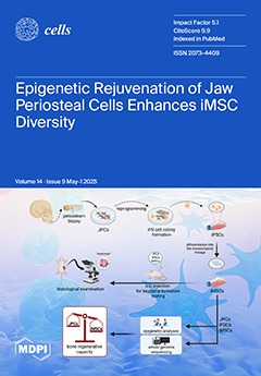
By reprogramming jaw periosteal cells (JPCs), we aim to create iPSC-derived mesenchymal stem cells (iMSCs) with superior regenerative capabilities. DNA methylation and gene expression analyses revealed many parallels with their JPC predecessors, as well as unique epigenetic patterns in iMSCs, including rejuvenation. DNA methylation-based biological age clocks showed that iPSCs reset to a near-zero age, a youthful state preserved even in the derived iMSCs. This confirmed safety, indicated through the absence of teratoma formation, positions iMSCs as a promising tool for enhancing bone tissue regeneration, suggesting significant therapeutic potential awaiting validation by future studies. View this paper
  • Issues are regarded as officially published after their release is announced to the table of contents alert mailing list.
  • You may sign up for e-mail alerts to receive table of contents of newly released issues.
  • PDF is the official format for papers published in both, html and pdf forms. To view the papers in pdf format, click on the "PDF Full-text" link, and use the free Adobe Reader to open them.
Order results
Result details
Section
Select all
Export citation of selected articles as:
23 pages, 4185 KiB  
Article
Experimental and Mathematical Model of Platelet Hemostasis Kinetics
by Bogdan Gerda, Anastasiya Volkova, Irina Dobrylko, Aleksandra Yu. Andreyeva, Thomas Dandekar, Mikhail A. Panteleev, Stepan Gambaryan and Igor Mindukshev
Cells 2025, 14(9), 677; https://doi.org/10.3390/cells14090677 - 7 May 2025
Viewed by 229
Abstract
Upon activation, platelets undergo rapid phenotypic transitions to maintain hemostasis, yet the kinetics governing these transitions remain poorly quantified. We present an integrated experimental and mathematical model describing platelet transitions between resting, activated, aggregating, inhibited, and exhausted phenotypes, determined by experiment rate constants [...] Read more.
Upon activation, platelets undergo rapid phenotypic transitions to maintain hemostasis, yet the kinetics governing these transitions remain poorly quantified. We present an integrated experimental and mathematical model describing platelet transitions between resting, activated, aggregating, inhibited, and exhausted phenotypes, determined by experiment rate constants for these reactions. Theoretical simulations of platelet transitions accurately describe the independently determined experimental read-out. Platelet aggregation under the conditions used directly correlates with the activation of αIIbβ3 integrins, demonstrating that the parameters of platelet aggregation achieved by the laser diffraction technique can be used for the evaluation of the rapid activation and deactivation kinetics of αIIbβ3 integrins. We demonstrate that platelet desensitization occurs at multiple activation stages, with distinct kinetic profiles for shape change and integrin deactivation. We also show that even 5 s of receptor-mediated PKA activation (iloprost) is sufficient for a complete inhibition of ADP-induced platelet aggregation. However, when iloprost was added after platelet stimulation by ADP, platelet activation was not fully inhibited, and after 180 s, aggregation became irreversible. The presented data help to understand the mechanisms of platelet transition between different phenotypes. The model effectively characterizes key physiological phenotypes and can serve as a modular framework for integration into more comprehensive models. Full article
(This article belongs to the Special Issue Molecular and Cellular Insights into Platelet Function)
Show Figures

Figure 1

25 pages, 5432 KiB  
Article
Optimization of In-Situ Exosome Enrichment Methodology On-a-Chip to Mimic Tumor Microenvironment Induces Cancer Stemness in Glioblastoma Tumor Model
by Saleheh Saffar, Ali Ghiaseddin, Shiva Irani and Amir Ali Hamidieh
Cells 2025, 14(9), 676; https://doi.org/10.3390/cells14090676 - 6 May 2025
Viewed by 341
Abstract
Understanding cancer etiology requires replicating the tumor microenvironment (TME), which significantly differs from standard in vitro cultures due to nutrient limitations, acidic pH, and oxidative stress. To address this, a microfluidic bioreactor (µBR) with an expanded culture surface was designed to optimize exosome [...] Read more.
Understanding cancer etiology requires replicating the tumor microenvironment (TME), which significantly differs from standard in vitro cultures due to nutrient limitations, acidic pH, and oxidative stress. To address this, a microfluidic bioreactor (µBR) with an expanded culture surface was designed to optimize exosome enrichment and glioblastoma cell behavior. Using response surface methodology (RSM), key parameters—including medium exchange volume and interval time—were optimized, leading to about a six-fold increase in exosome concentration without artificial inducers. Characterization techniques (SEM, AFM, DLS, RT-qPCR, and ELISA) confirmed significant alterations in exosome profiles, cancer stemness, and epithelial-mesenchymal transition (EMT)-related markers. Notably, EMT was induced in the µBR system, with a six-fold increase in HIF-1α protein despite normoxic conditions, suggesting activation of compensatory signaling pathways. Molecular analysis showed upregulation of SOX2, OCT4, and Notch1, with SOX2 protein reaching 28 ng/mL, while it was undetectable in traditional culture. Notch1 concentration tripled in the µBR system, correlating with enhanced stemness and phenotypic heterogeneity. Immunofluorescent microscopy confirmed nuclear SOX2 accumulation and co-expression of SOX2 and HIF-1α in dedifferentiated CSC-like cells, demonstrating tumor heterogeneity. These findings highlight the µBR’s ability to enhance stemness and mimic glioblastoma’s aggressive phenotype, establishing it as a valuable platform for tumor modeling and therapeutic development. Full article
(This article belongs to the Section Cell Microenvironment)
Show Figures

Figure 1

29 pages, 759 KiB  
Review
Targeting Glioma Stem Cells: Therapeutic Opportunities and Challenges
by Asma Mahdi, Mohamed Aittaleb and Fadel Tissir
Cells 2025, 14(9), 675; https://doi.org/10.3390/cells14090675 - 6 May 2025
Viewed by 360
Abstract
Glioblastoma (GBM), or grade 4 glioma, is the most common and aggressive primary brain tumor in adults with a median survival of 15 months. Increasing evidence suggests that GBM’s aggressiveness, invasiveness, and therapy resistance are driven by glioma stem cells (GSCs), a subpopulation [...] Read more.
Glioblastoma (GBM), or grade 4 glioma, is the most common and aggressive primary brain tumor in adults with a median survival of 15 months. Increasing evidence suggests that GBM’s aggressiveness, invasiveness, and therapy resistance are driven by glioma stem cells (GSCs), a subpopulation of tumor cells that share molecular and functional characteristics with neural stem cells (NSCs). GSCs are heterogeneous and highly plastic. They evade conventional treatments by shifting their state and entering in quiescence, where they become metabolically inactive and resistant to radiotherapy and chemotherapy. GSCs can exit quiescence and be reactivated to divide into highly proliferative tumor cells which contributes to recurrence. Understanding the molecular mechanisms regulating the biology of GSCs, their plasticity, and the switch between quiescence and mitotic activity is essential to shape new therapeutic strategies. This review examines the latest evidence on GSC biology, their role in glioblastoma progression and recurrence, emerging therapeutic approaches aimed at disrupting their proliferation and survival, and the mechanisms underlying their resistance to therapy. Full article
(This article belongs to the Special Issue Therapeutic Targets in Glioblastoma)
Show Figures

Figure 1

12 pages, 4686 KiB  
Communication
From Quiescence to Activation: The Reciprocal Regulation of Ras and Rho Signaling in Hepatic Stellate Cells
by Saeideh Nakhaei-Rad, Silke Pudewell, Amin Mirzaiebadizi, Kazem Nouri, Doreen Reichert, Claus Kordes, Dieter Häussinger and Mohammad Reza Ahmadian
Cells 2025, 14(9), 674; https://doi.org/10.3390/cells14090674 - 5 May 2025
Viewed by 375
Abstract
Chronic liver diseases are marked by persistent inflammation and can evolve into liver fibrosis, cirrhosis, and hepatocellular carcinoma. In an affected liver, hepatic stellate cells (HSCs) transition from a quiescent to an activated state and adopt a myofibroblast-like cell phenotype. While these activated [...] Read more.
Chronic liver diseases are marked by persistent inflammation and can evolve into liver fibrosis, cirrhosis, and hepatocellular carcinoma. In an affected liver, hepatic stellate cells (HSCs) transition from a quiescent to an activated state and adopt a myofibroblast-like cell phenotype. While these activated cells play a role in supporting liver regeneration, they can also have detrimental effects on liver function as the disease progresses to fibrosis and cirrhosis. These findings highlight the dynamic switching between different signaling pathways involving Ras, Rho GTPases, and Notch signaling. Notably, two specific members of the Ras and Rho GTPases, Eras and Rnd3, are predominantly expressed in quiescent HSCs, while Mras and Rhoc are more abundant in their activated forms. In addition, this study highlights the critical role of cytosolic Notch1 in quiescent HSCs and Rock in activated HSCs. We hypothesize that distinct yet interdependent intracellular signaling networks regulate HSC fate decisions in two key ways: by maintaining HSC quiescence and homeostasis and by facilitating HSC activation, thereby influencing processes such as proliferation, transdifferentiation, and mesenchymal transition. The proposed signaling model, combined with specific methodological tools for maintaining HSCs in a quiescent state, will deepen our understanding of the mechanisms underlying chronic liver disease and may also pave the way for innovative therapies. These therapies could include small molecule drugs targeting Ras- and Rho-dependent pathways. Full article
(This article belongs to the Topic Signaling Pathways in Liver Disease)
Show Figures

Figure 1

22 pages, 2441 KiB  
Review
The Distinct Role of HIF-1α and HIF-2α in Hypoxia and Angiogenesis
by Mouayad Zuheir Bakleh and Ayman Al Haj Zen
Cells 2025, 14(9), 673; https://doi.org/10.3390/cells14090673 - 4 May 2025
Viewed by 674
Abstract
Hypoxia results in a wide range of adaptive physiological responses, including metabolic reprogramming, erythropoiesis, and angiogenesis. The response to hypoxia at the cellular level is mainly regulated by hypoxia-inducible factors (HIFs): HIF1α and HIF2α isoforms. Although structurally similar and overlapping gene targets, both [...] Read more.
Hypoxia results in a wide range of adaptive physiological responses, including metabolic reprogramming, erythropoiesis, and angiogenesis. The response to hypoxia at the cellular level is mainly regulated by hypoxia-inducible factors (HIFs): HIF1α and HIF2α isoforms. Although structurally similar and overlapping gene targets, both isoforms can exhibit distinct expression patterns and functions in some conditions of hypoxia. The interaction between these isoforms, known as the “HIF switch”, determines their coordinated function under varying oxygen levels and exposure time. In angiogenesis, HIF-1α is rapidly stabilized under acute hypoxia, prompting a metabolic shift from oxidative phosphorylation to glycolysis and initiating angiogenesis by activating endothelial cells and extracellular matrix remodeling. Conversely, HIF-2α regulates cell responses to chronic hypoxia by sustaining genes critical for vascular remodeling and maturation. The current review highlights the different roles and regulatory mechanisms of HIF-1α and HIF-2α isoforms, focusing on their involvement in cell metabolism and the multi-step process of angiogenesis. Tuning the specific targeting of HIF isoforms and finding the right therapeutic window is essential to obtaining the best therapeutic effect in diseases such as cancer and vascular ischemic diseases. Full article
(This article belongs to the Special Issue The Role of Hypoxia-Inducible Factors (HIFs) in Human Diseases)
Show Figures

Figure 1

22 pages, 1371 KiB  
Review
Iron Metabolism and Muscle Aging: Where Ferritinophagy Meets Mitochondrial Quality Control
by Rosa Di Lorenzo, Emanuele Marzetti, Helio José Coelho-Junior, Riccardo Calvani, Vito Pesce, Francesco Landi, Christiaan Leeuwenburgh and Anna Picca
Cells 2025, 14(9), 672; https://doi.org/10.3390/cells14090672 - 3 May 2025
Viewed by 441
Abstract
In older adults with reduced physical performance, an increase in the labile iron pool within skeletal muscle is observed. This accumulation is associated with an altered expression of mitochondrial quality control (MQC) markers and increased mitochondrial DNA damage, supporting the hypothesis that impaired [...] Read more.
In older adults with reduced physical performance, an increase in the labile iron pool within skeletal muscle is observed. This accumulation is associated with an altered expression of mitochondrial quality control (MQC) markers and increased mitochondrial DNA damage, supporting the hypothesis that impaired MQC contributes to muscle dysfunction during aging. The autophagy–lysosome system plays a critical role in MQC by tagging and engulfing proteins and organelles for degradation in lysosomes. The endolysosomal system is also instrumental in transferrin recycling, which, in turn, regulates cellular iron uptake. In the neuromuscular system, the autophagy–lysosome system supports the structural integrity of neuromuscular junctions, and its dysfunction contributes to muscle atrophy. While MQC was thought to protect against iron-induced cell death, the discovery of ferroptosis, a form of iron-dependent cell death, has highlighted a complex interplay between MQC and iron-inflicted damage. Ferritinophagy, the autophagic degradation of ferritin, if overactivated, can induce ferroptosis. Alternatively, aging may impair ferritinophagy, leading to ferritin accumulation and the release of toxic labile iron under stress, exacerbating oxidative damage and cellular senescence. Physical activity supports muscle health also by preserving mitochondrial quantity and quality and enhancing bioenergetics. However, therapeutic strategies for preventing or reversing physical function decline in aging are still lacking due to the insufficient understanding of the underlying mechanisms. Unveiling how disruptions in iron homeostasis impact muscle quality in older adults may allow for the development of therapeutic strategies targeting iron handling to alleviate age-associated muscle decline. Full article
(This article belongs to the Special Issue Autophagy Meets Aging 2025)
Show Figures

Figure 1

15 pages, 4838 KiB  
Article
Hydrogen Peroxide Modulates the Timely Activation of Jun and Erk in Schwann Cells at the Injury Site and Is Required for Motor Axon Regeneration
by Samuele Negro, Chiara Baggio, Marika Tonellato, Marco Stazi, Giorgia D’Este, Aram Megighian, Cesare Montecucco and Michela Rigoni
Cells 2025, 14(9), 671; https://doi.org/10.3390/cells14090671 - 3 May 2025
Viewed by 513
Abstract
Peripheral nervous system (PNS) neurons, including motor neurons (MNs), possess a remarkable ability to regenerate and reinnervate target muscles following nerve injury. This process is orchestrated by a combination of intrinsic neuronal properties and extrinsic factors, with Schwann cells (SCs) playing a central [...] Read more.
Peripheral nervous system (PNS) neurons, including motor neurons (MNs), possess a remarkable ability to regenerate and reinnervate target muscles following nerve injury. This process is orchestrated by a combination of intrinsic neuronal properties and extrinsic factors, with Schwann cells (SCs) playing a central role. Upon injury, SCs transition into a repair phenotype that allows axonal regeneration through molecular signaling and structural guidance. However, the identity of the SCs’ reprogramming factors is only partially known. We previously identified hydrogen peroxide (H2O2) as an early and key driver of nerve repair, inducing gene expression rewiring in SCs to support nerve re-growth. In this study, we quantitatively assessed the role of H2O2 in the activation of key pro-regenerative signaling pathways in SCs following sciatic nerve compression, specifically the extracellular signal-regulated kinase 1/2 (ERK1/2) and c-Jun, which are essential for functional nerve recovery. Notably, we found that H2O2 neutralization does not impact degeneration, but it significantly affects the regenerative response. Collectively, our findings establish H2O2 as a promising regulator of the Schwann cell injury response at the injury site, linking oxidative signaling to the molecular mechanisms governing nerve regeneration. Full article
(This article belongs to the Special Issue Unveiling Axon-Glia Communication in Health and Disease)
Show Figures

Figure 1

20 pages, 8006 KiB  
Article
Early Development of an Innovative Nanoparticle-Based Multimodal Tool for Targeted Drug Delivery: A Step-by-Step Approach
by Chiara Barattini, Angela Volpe, Daniele Gori, Daniele Lopez, Alfredo Ventola, Stefano Papa, Mariele Montanari and Barbara Canonico
Cells 2025, 14(9), 670; https://doi.org/10.3390/cells14090670 - 3 May 2025
Viewed by 325
Abstract
Prostate cancer is the most common tumor in men in developed countries and it often responds poorly to conventional treatments. Monoclonal antibody (MoAb) therapy, for this pathology, has grown tremendously in the past decades, exploiting naked and conjugated antibodies to cytotoxic payloads to [...] Read more.
Prostate cancer is the most common tumor in men in developed countries and it often responds poorly to conventional treatments. Monoclonal antibody (MoAb) therapy, for this pathology, has grown tremendously in the past decades, exploiting naked and conjugated antibodies to cytotoxic payloads to form antibody drug conjugates (ADCs). Several studies have been carried out conjugating biomolecules against prostate-specific membrane antigen (PSMA), highly expressed in this tumor, to cytotoxic drugs. Nano-based formulations show high potential in targeted drug delivery to enhance the bioavailability of drugs. Our research aimed to evaluate the feasibility of setting up a nanoparticle-based multimodal tool for targeted drug delivery, describing the step-by-step approach and to perform a first screening of these fluorescent PEGylated silica nanoparticles employed in selective cancer cell targeting and killing. These nanoparticles featured a core–shell structure to contemporarily conjugate the antibody and the cytotoxic payload monomethyl auristatin E (MMAE) using a step-by-step approach. We compared the cytotoxic effect of this multimodal nanotool near the antibody-MMAE and free MMAE. We found a lower cytotoxicity effect of the nanoparticle-based construct compared to free drugs, likely because of the preservation of the previously observed receptor-mediated endocytosis. Nanomedicine is confirmed as a powerful alternative to organic drug delivery systems, even if some aspects, such as drug loading efficacy, release, scalable manufacturing and long-term stability, need to be deepened. Full article
Show Figures

Figure 1

29 pages, 2144 KiB  
Review
Adipose Tissue-Derived Therapies for Osteoarthritis: Multifaceted Mechanisms and Clinical Prospects
by Hanwen Zhang, Oliver Felthaus and Lukas Prantl
Cells 2025, 14(9), 669; https://doi.org/10.3390/cells14090669 - 2 May 2025
Viewed by 519
Abstract
Osteoarthritis (OA) is a degenerative joint disease that significantly impacts quality of life and poses a growing economic burden. Adipose tissue-derived therapies, including both cell-based and cell-free products, have shown promising potential in promoting cartilage repair, modulating inflammation, and improving joint function. Recent [...] Read more.
Osteoarthritis (OA) is a degenerative joint disease that significantly impacts quality of life and poses a growing economic burden. Adipose tissue-derived therapies, including both cell-based and cell-free products, have shown promising potential in promoting cartilage repair, modulating inflammation, and improving joint function. Recent studies and clinical trials have demonstrated their regenerative effects, highlighting their feasibility as a novel treatment approach for OA. This review summarises the therapeutic mechanisms and latest advancements in adipose tissue-derived therapies, providing insights into their clinical applications and future prospects. Full article
(This article belongs to the Collection Research on Adipose Stem Cells)
Show Figures

Figure 1

26 pages, 1303 KiB  
Review
Expanding the Use of SGLT2 Inhibitors in T2D Patients Across Clinical Settings
by Alessandro Cuttone, Vittorio Cannavò, Raouf Mastan Sheik Abdullah, Pierluigi Fugazzotto, Giada Arena, Simona Brancati, Andrea Muscarà, Carmela Morace, Cristina Quartarone, Domenica Ruggeri, Giovanni Squadrito and Giuseppina Tiziana Russo
Cells 2025, 14(9), 668; https://doi.org/10.3390/cells14090668 - 2 May 2025
Viewed by 907
Abstract
Sodium-glucose cotransporter-2 inhibitors (SGLT2i) are currently recommended in patients with type 2 diabetes (T2D) to reduce serum glucose levels. Moreover, robust evidence has clearly demonstrated their beneficial cardiovascular and renal effects, making this class of drugs pivotal for the treatment of T2D, especially [...] Read more.
Sodium-glucose cotransporter-2 inhibitors (SGLT2i) are currently recommended in patients with type 2 diabetes (T2D) to reduce serum glucose levels. Moreover, robust evidence has clearly demonstrated their beneficial cardiovascular and renal effects, making this class of drugs pivotal for the treatment of T2D, especially when complicated by diabetic kidney disease or heart failure. However, several other comorbidities are frequently encountered in T2D patients beyond these long-term diabetes complications, especially in the internal medicine setting. For some of these comorbidities, such as MAFLD and cognitive impairment, the association with diabetes is increasingly recognized, with the hypothesis of a common pathophysiologic background, whereas, for others, a coincident epidemiology linked to the ageing of populations, including that of T2D subjects, may be advocated. In the effort of personalizing T2D treatment, evidence on the potential effects of SGLT2i in these different clinical conditions is accumulating. The purpose of this narrative review is to update current literature on the effects of SGLT2i for the treatment of T2D in different clinical settings beyond glycaemic control, and to elucidate potential molecular mechanisms by which they exert these effects. Full article
Show Figures

Figure 1

18 pages, 3150 KiB  
Article
Targeting ATF5, CEBPB, and CEBPD with Cell-Penetrating Dpep Sensitizes Tumor Cells to NK-92MI Cell Cytotoxicity
by Qing Zhou, Markus D. Siegelin and Lloyd A. Greene
Cells 2025, 14(9), 667; https://doi.org/10.3390/cells14090667 - 2 May 2025
Viewed by 254
Abstract
Natural killer (NK) cells are an important innate defense against malignancies, and exogenous sources of NK cells have been developed as anti-cancer agents. Nevertheless, the apparent limitations of NK cells in clearing cancers have suggested that their efficacy might be augmented by combination [...] Read more.
Natural killer (NK) cells are an important innate defense against malignancies, and exogenous sources of NK cells have been developed as anti-cancer agents. Nevertheless, the apparent limitations of NK cells in clearing cancers have suggested that their efficacy might be augmented by combination with other treatments. We have developed cell-penetrating peptides that target the transcription factors ATF5, CEBPB, and CEBPD and that promote apoptotic cancer cell death both in vitro and in vivo without apparent toxicity to non-transformed cells. We report here that one such peptide, Dpep, significantly sensitizes a variety of tumor cell types to the cytotoxic activity of the NK cell line, NK-92MI. Such sensitization requires pre-exposure of tumor cells to Dpep and does not appear due to effects of Dpep on NK cells themselves. Our findings suggest that Dpep acts in this context to lower the apoptotic threshold of tumor cells to NK cell toxicity. Additionally, while Dpep pre-treatment does not prevent tumor cells from causing NK cell “inactivation”, it sensitizes cancer cells to repeated rounds of exposure to fresh NK cells. These findings thus indicate that Dpep pre-treatment is an effective strategy to sensitize cancer cells to the cytotoxic actions of NK cells. Full article
Show Figures

Figure 1

13 pages, 4773 KiB  
Article
Microgliosis in the Spinal Dorsal Horn Early After Peripheral Nerve Injury Is Associated with Damage to Primary Afferent Aβ-Fibers
by Yuto Shibata, Yuki Matsumoto, Keita Kohno, Yasuharu Nakashima and Makoto Tsuda
Cells 2025, 14(9), 666; https://doi.org/10.3390/cells14090666 - 2 May 2025
Viewed by 317
Abstract
Neuropathic pain results from a lesion or disease affecting the somatosensory nervous system. Injury to primary afferent nerves leads to microgliosis in the spinal dorsal horn (SDH), which plays a crucial role in developing neuropathic pain. Within the SDH, primary afferent fibers broadly [...] Read more.
Neuropathic pain results from a lesion or disease affecting the somatosensory nervous system. Injury to primary afferent nerves leads to microgliosis in the spinal dorsal horn (SDH), which plays a crucial role in developing neuropathic pain. Within the SDH, primary afferent fibers broadly project, and microglia are nearly ubiquitously distributed under normal conditions. However, not all microglia react to injuries affecting primary afferent fibers, resulting in spatially heterogeneous microgliosis within the SDH. The mechanisms underlying this phenomenon remain elusive. In this study, the spatial relationship between microgliosis and the projections of injured nerves was investigated by generating mice that had expressed tdTomato in the fourth lumbar dorsal root ganglion (L4-DRG) neurons via intra-L4-spinal nerve (SpN) injection of adeno-associated viral vectors. After transection of the L4-SpN, we found that microgliosis in the SDH selectively occurred in the innervation territories of the injured primary afferent fibers. However, denervating transient receptor potential vanilloid 1 (TRPV1)-expressing primary afferent fibers in the SDH through intrathecal injection of capsaicin did not trigger microgliosis, nor did it influence the microgliosis induced by L4-SpN injury. Conversely, pharmacological damage to myelinated DRG neurons, including Aβ-fibers, was sufficient to induce microgliosis. Furthermore, L4-SpN injury also induced microgliosis in the gracile nucleus, which primarily receives innervation from Aβ-fibers. These findings suggest that microgliosis in the SDH shortly after peripheral nerve injury is predominantly associated with damage to primary afferent Aβ-fibers. Full article
(This article belongs to the Special Issue Molecular Mechanisms of Neuropathic Pain)
Show Figures

Figure 1

23 pages, 2869 KiB  
Review
Getting Blood out of a Stone: Vascularization via Spheroids and Organoids in 3D Bioprinting
by Daria Revokatova, Polina Bikmulina, Zahra Heydari, Anna Solovieva, Massoud Vosough, Anastasia Shpichka and Peter Timashev
Cells 2025, 14(9), 665; https://doi.org/10.3390/cells14090665 - 1 May 2025
Viewed by 263
Abstract
Current developments in bioequivalent technology have led to the creation of excellent models that mimic the structure and function of human organs. These models are based on the original tissues and organs of the human body, but they lack the complex interaction with [...] Read more.
Current developments in bioequivalent technology have led to the creation of excellent models that mimic the structure and function of human organs. These models are based on the original tissues and organs of the human body, but they lack the complex interaction with the extensive network of vasculature, and this is a major challenge for these models. A functional vasculature is essential for oxygen, nutrient, and waste exchange. It is also responsible for inductive biochemical exchange, and provides a structural pattern for organ growth. In vitro systems, containing no perfusable vessels, suffer from the quick formation of a necrotic core of organoids, and further development does not occur due to increased metabolic demands. Another key limitation of 3D-based techniques is the absence of accurate architectural structures and large-scale tissue sizes. Recently, new 3D bioprinting methods have been developed for organoids and spheroids as living building blocks. These methods aim to address some of the challenges associated with 3D technologies. In this review, we discuss recent strategies for vascularization via organoids and spheroids, which are used as structural units in bioprinting to recreate natural organs and tissues with ever-increasing accuracy in structure and function. Full article
Show Figures

Figure 1

18 pages, 11391 KiB  
Article
RBFOX1 Regulates Calcium Signaling and Enhances SERCA2 Translation
by Sadiq Umar, Wuqiang Zhu, Fernando Souza-Neto, Ingrid Bender, Steven C. Wu, Chastity L. Healy, Timothy D. O’Connell and Jop H. van Berlo
Cells 2025, 14(9), 664; https://doi.org/10.3390/cells14090664 - 1 May 2025
Viewed by 273
Abstract
RBFOX1 is an RNA-binding protein that regulates alternative splicing and RNA processing in the neurons, skeletal muscle, and heart. We intended to define the role of RBFOX1 in regulating calcium homeostasis to maintain normal cardiac function. We generated cardiomyocyte-specific Rbfox1 gene-deletion mice (cKO). [...] Read more.
RBFOX1 is an RNA-binding protein that regulates alternative splicing and RNA processing in the neurons, skeletal muscle, and heart. We intended to define the role of RBFOX1 in regulating calcium homeostasis to maintain normal cardiac function. We generated cardiomyocyte-specific Rbfox1 gene-deletion mice (cKO). The cardiomyocyte-specific deletion of RBFOX1 was confirmed by Western blotting and immunohistochemistry. The cKO mice showed mild hypertrophy and depressed cardiac function under homeostatic conditions, which did not deteriorate with age. Pressure overload by trans-aortic constriction (TAC) caused exaggerated cardiac hypertrophy and accelerated heart failure in cKO compared with wild-type mice. We performed Western blotting to assess the expression of important Ca2+-handling proteins, which showed alterations in the phosphorylation of PLN and CAMKII and decreased expression of SERCA2. We measured the Ca2+ dynamics and noted significantly delayed Ca2+ reuptake into the sarcoplasmic reticulum. Importantly, the decrease in SERCA2 expression was not due to reduced mRNA expression or altered splicing. To assess the possibility of the post-transcriptional regulation of SERCA2 expression by RBFOX1, we performed RNA immunoprecipitation (RIP), which showed the binding of RBFOX1 protein to Serca2 mRNA, which was confirmed in luciferase assays with the Serca2a 3′-untranslated region fused to luciferase. Finally, we performed a puromycin incorporation experiment, which showed that RBFOX1 enhances SERCA2 protein translation. Our results show that RBFOX1 plays a crucial role in regulating the expression of Ca2+-handling genes to maintain normal cardiac function. We show an important post-transcriptional role of RBFOX1 in regulating SERCA2 expression. Full article
(This article belongs to the Section Cells of the Cardiovascular System)
Show Figures

Figure 1

28 pages, 7160 KiB  
Review
Inhibitors of Cyclic Dinucleotide Phosphodiesterases and Cyclic Oligonucleotide Ring Nucleases as Potential Drugs for Various Diseases
by Christopher S. Vennard, Samson Marvellous Oladeji and Herman O. Sintim
Cells 2025, 14(9), 663; https://doi.org/10.3390/cells14090663 - 30 Apr 2025
Viewed by 272
Abstract
The phosphodiester linkage is found in DNA, RNA and many signaling molecules, such as cyclic mononucleotide, cyclic dinucleotides (CDNs) and cyclic oligonucleotides (cONs). Enzymes that cleave the phosphodiester linkage (nucleases and phosphodiesterases) play important roles in cell persistence and fitness and have therefore [...] Read more.
The phosphodiester linkage is found in DNA, RNA and many signaling molecules, such as cyclic mononucleotide, cyclic dinucleotides (CDNs) and cyclic oligonucleotides (cONs). Enzymes that cleave the phosphodiester linkage (nucleases and phosphodiesterases) play important roles in cell persistence and fitness and have therefore become targets for various diseased states. While various inhibitors have been developed for nucleases and cyclic mononucleotide phosphodiesterases, and some have become clinical successes, there is a paucity of inhibitors of the recently discovered phosphodiesterases or ring nucleases that cleave CDNs and cONs. Inhibitors of bacterial c-di-GMP or c-di-AMP phosphodiesterases have the potential to be used as anti-virulence compounds, while compounds that inhibit the degradation of 3′,3′-cGAMP, cA3, cA4, cA6 could serve as antibiotic adjuvants as the accumulation of these second messengers leads to bacterial abortive infection. In humans, 2′3′-cGAMP plays critical roles in antiviral and antitumor responses. ENPP1 (the 2′3′-cGAMP phosphodiesterase) or virally encoded cyclic dinucleotide phosphodiesterases, such as poxin, however, blunt this response. Inhibitors of ENPP1 or poxin-like enzymes have the potential to be used as anticancer and antiviral agents, respectively. This review summarizes efforts made towards the discovery and development of compounds that inhibit CDN phosphodiesterases and cON ring nucleases. Full article
Show Figures

Graphical abstract

25 pages, 951 KiB  
Review
mTORopathies in Epilepsy and Neurodevelopmental Disorders: The Future of Therapeutics and the Role of Gene Editing
by Marina Ottmann Boff, Fernando Antônio Costa Xavier, Fernando Mendonça Diz, Júlia Budelon Gonçalves, Laura Meireles Ferreira, Jean Zambeli, Douglas Bottega Pazzin, Thales Thor Ramos Previato, Helena Scartassini Erwig, João Ismael Budelon Gonçalves, Fernanda Thays Konat Bruzzo, Daniel Marinowic, Jaderson Costa da Costa and Gabriele Zanirati
Cells 2025, 14(9), 662; https://doi.org/10.3390/cells14090662 - 30 Apr 2025
Viewed by 448
Abstract
mTORopathies represent a group of neurodevelopmental disorders linked to dysregulated mTOR signaling, resulting in conditions such as tuberous sclerosis complex, focal cortical dysplasia, hemimegalencephaly, and Smith–Kingsmore Syndrome. These disorders often manifest with epilepsy, cognitive impairments, and, in some cases, structural brain anomalies. The [...] Read more.
mTORopathies represent a group of neurodevelopmental disorders linked to dysregulated mTOR signaling, resulting in conditions such as tuberous sclerosis complex, focal cortical dysplasia, hemimegalencephaly, and Smith–Kingsmore Syndrome. These disorders often manifest with epilepsy, cognitive impairments, and, in some cases, structural brain anomalies. The mTOR pathway, a central regulator of cell growth and metabolism, plays a crucial role in brain development, where its hyperactivation leads to abnormal neuroplasticity, tumor formation, and heightened neuronal excitability. Current treatments primarily rely on mTOR inhibitors, such as rapamycin, which reduce seizure frequency and tumor size but fail to address underlying genetic causes. Advances in gene editing, particularly via CRISPR/Cas9, offer promising avenues for precision therapies targeting the genetic mutations driving mTORopathies. New delivery systems, including viral and non-viral vectors, aim to enhance the specificity and efficacy of these therapies, potentially transforming the management of these disorders. While gene editing holds curative potential, challenges remain concerning delivery, long-term safety, and ethical considerations. Continued research into mTOR mechanisms and innovative gene therapies may pave the way for transformative, personalized treatments for patients affected by these complex neurodevelopmental conditions. Full article
Show Figures

Figure 1

29 pages, 12311 KiB  
Article
Expanded Phenotype of the Cln6nclf Mouse Model
by Victoria Chaoul, Sara Saab, Omar Shmoury, Ramy Alam, Lynn Al Aridi, Nadine J. Makhoul, Jihane Soueid and Rose-Mary Boustany
Cells 2025, 14(9), 661; https://doi.org/10.3390/cells14090661 - 30 Apr 2025
Viewed by 426
Abstract
Neuronal ceroid lipofuscinoses (NCLs) are a group of autosomal recessive neurogenetic disorders caused by mutations in 14 different genes. CLN6 disease manifests as variant late-infantile NCL (vLINCL) or as an adult variant. In childhood, symptoms include speech delay, vision loss, cognitive and motor [...] Read more.
Neuronal ceroid lipofuscinoses (NCLs) are a group of autosomal recessive neurogenetic disorders caused by mutations in 14 different genes. CLN6 disease manifests as variant late-infantile NCL (vLINCL) or as an adult variant. In childhood, symptoms include speech delay, vision loss, cognitive and motor decline, seizures, and early death. An in-depth characterization of a naturally occurring Cln6 mutant mouse (Cln6nclf) is presented, with implications for translational research. The expanded phenotype provides data showing early death, vision loss, and motor deficits in male and female Cln6nclf mice. Diminished visual acuity in Cln6nclf mice was noted at 28 weeks of age, but the pathological loss of retinal layers began as early as 2 weeks or postnatal day 14 (P14). Apoptosis was confirmed by TUNEL staining in the Cln6nclf mouse brain at P8 and in the retina at P12. A peak in glial fibrillary acidic protein (GFAP) expression was established as a normal developmental phenomenon in the wild-type and Cln6nclf mouse brain cerebellum and the CA2–CA3 regions of the hippocampus at P8. In Cln6nclf mice, GFAP levels were elevated at P12 in the cerebellum and hippocampus. In the retina, a developmental peak in gliosis was absent, with increased astrogliosis noted at P6 and P8 in female and male Cln6nclf mice, respectively. This highlights the lack of a sex-dependent response in wild-type mice. These novel data position the Cln6nclf mouse model as a useful tool for screening potential therapeutics for human CLN6 disease. Full article
Show Figures

Graphical abstract

22 pages, 6834 KiB  
Article
Immunosuppression of Tumor-Derived Factors Modulated Neutrophils in Upper Tract Urothelial Carcinoma Through Upregulation of Arginase-1 via ApoA1-STAT3 Axis
by Chih-Chia Chang, Chia-Bin Chang, Cheng-Huang Shen, Ming-Yang Lee, Yeong-Chin Jou, Chun-Liang Tung, Wei-Hong Lai, Chi-Feng Hung, Meilin Wang, Ya-Yan Lai, Pi-Che Chen and Shu-Fen Wu
Cells 2025, 14(9), 660; https://doi.org/10.3390/cells14090660 - 30 Apr 2025
Viewed by 285
Abstract
Upper tract urothelial carcinoma (UTUC) presents aggressive features and a tumor microenvironment with T cell depletion. However, the role of tumor-associated neutrophils in UTUC remains unclear. This study aimed to investigate how UTUC tumor-derived factors modulate neutrophils and their impact on T cell [...] Read more.
Upper tract urothelial carcinoma (UTUC) presents aggressive features and a tumor microenvironment with T cell depletion. However, the role of tumor-associated neutrophils in UTUC remains unclear. This study aimed to investigate how UTUC tumor-derived factors modulate neutrophils and their impact on T cell immune responses. Our findings demonstrate that UTUC secreted tumor-derived factors, with apolipoprotein A1 (Apo-A1) being the predominant factor, which upregulated arginase-1 expression in neutrophils. STAT3 activation was responsible for the upregulation of arginase-1 in neutrophils. Blocking the interactions between Apo-A1 and its receptors reduced arginase-1 expression in neutrophils treated with tumor tissue culture supernatant (TTCS). Moreover, both CD4+ T and CD8+ T cell proliferation were inhibited by neutrophils treated with Apo-A1 or TTCS. Importantly, blocking Apo-A1 signaling in neutrophils reversed the inhibitory effects on T cells. In UTUC patients, the neutrophil-to-lymphocyte ratio was higher than that in healthy subjects. The expression of arginase-1 in neutrophils and the level of Apo-A1 within UTUC tumors were negatively correlated with tumor-infiltrating CD4+ T cells. Additionally, neutrophils from UTUC patients showed increased expression of arginase-1 and exhibited inhibitory effects of T cell functions. These findings suggest that UTUC orchestrates an immune-suppressive microenvironment through Apo-A1-mediated upregulation of arginase-1 in neutrophils, ultimately leading to the inhibition of T cell proliferation. Full article
Show Figures

Figure 1

17 pages, 514 KiB  
Review
Advancing Obstructive Airway Disease Treatment: Dual PDE3/4 Inhibition as a Therapeutic Strategy
by Rinzhin T. Sherpa, Cynthia J. Koziol-White and Reynold A. Panettieri, Jr.
Cells 2025, 14(9), 659; https://doi.org/10.3390/cells14090659 - 30 Apr 2025
Viewed by 461
Abstract
Obstructive airway diseases, including asthma and chronic obstructive pulmonary disease (COPD), evoke significant global health concerns manifested by airway inflammation and obstruction. Despite their differing origins, shared pathophysiological features and responses to therapeutic interventions highlight common molecular mechanisms. Standard treatments include inhaled bronchodilators, [...] Read more.
Obstructive airway diseases, including asthma and chronic obstructive pulmonary disease (COPD), evoke significant global health concerns manifested by airway inflammation and obstruction. Despite their differing origins, shared pathophysiological features and responses to therapeutic interventions highlight common molecular mechanisms. Standard treatments include inhaled bronchodilators, with combination therapies offering enhanced symptom control. Cyclic AMP (cAMP) plays a crucial role in airway relaxation. Phosphodiesterase (PDE) decreases cAMP levels, thereby attenuating the relaxation of airway smooth muscle, making it a promising therapeutic target. The balance between cAMP production and degradation is essential for regulating airway tone and function. PDE inhibitors for the treatment of obstructive airway diseases have suffered challenges, with adverse side effects of prospective inhibitors causing clinical failures. Efforts to develop PDE inhibitors with an improved safety profile could prove to be beneficial as an add-on treatment for severe asthma and COPD. The recent FDA approval of Ensifentrine, a dual PDE3/4 inhibitor, can significantly advance COPD management by improving bronchodilation, reducing inflammation, and lowering exacerbation rates with favorable safety outcomes. Full article
Show Figures

Figure 1

16 pages, 1313 KiB  
Article
Bilateral Germ Cell Tumor of the Testis: Biological and Clinical Implications for a Stem Versus Genetic Origin of Cancers
by Jamaal C. Jackson, Darren Sanchez, Aron Y. Joon, Marcos R. Estecio, Andrew C. Johns, Amishi Y. Shah, Matthew Campbell, John F. Ward, Louis L. Pisters, Charles C. Guo, Miao Zhang, Niki M. Zacharias and Shi-Ming Tu
Cells 2025, 14(9), 658; https://doi.org/10.3390/cells14090658 - 30 Apr 2025
Viewed by 397
Abstract
Germ cell tumors of the testis (GCTs) provide an ideal tumor model to investigate the cellular versus genetic origin of cancers. In this single institutional study, we evaluated 38 patients with bilateral GCT, including tumors that occurred simultaneously (synchronous) and those occurring at [...] Read more.
Germ cell tumors of the testis (GCTs) provide an ideal tumor model to investigate the cellular versus genetic origin of cancers. In this single institutional study, we evaluated 38 patients with bilateral GCT, including tumors that occurred simultaneously (synchronous) and those occurring at different times (metachronous). For nine of these patients, DNA was isolated from the right and left GCT to determine the genomic and epigenetic differences between tissues using whole-exome sequencing (WES) and reduced representation bisulfite sequencing (RRBS). We found that seminomas and non-seminomas are molecularly distinct based on DNA methylation and not due to synchronous or metachronous disease. In addition, we did not observe conservation of genetic mutations in right and left GCT in either synchronous or metachronous disease. Our data suggest a cellular origin for bilateral GCT. Full article
Show Figures

Figure 1

18 pages, 2187 KiB  
Article
Effect of Recombinant Human Amelogenin on the Osteogenic Differentiation Potential of SHED
by Akira Hirabae, Ryo Kunimatsu, Yuki Yoshimi, Kodai Rikitake, Shintaro Ogashira, Ayaka Nakatani, Shuzo Sakata and Kotaro Tanimoto
Cells 2025, 14(9), 657; https://doi.org/10.3390/cells14090657 - 30 Apr 2025
Viewed by 351
Abstract
This study aimed to explore how amelogenin can improve stem cells from human exfoliated deciduous teeth (SHED)–based bone regeneration and promote tissue healing as a treatment for critical-sized bone defects. SHED was induced into bone differentiation by using osteogenic differentiation medium. Real-time polymerase [...] Read more.
This study aimed to explore how amelogenin can improve stem cells from human exfoliated deciduous teeth (SHED)–based bone regeneration and promote tissue healing as a treatment for critical-sized bone defects. SHED was induced into bone differentiation by using osteogenic differentiation medium. Real-time polymerase chain reaction, alkaline phosphatase (ALP) staining and quantification, and Alizarin Red S staining, as well as calcium and osteocalcin quantification were performed to assess differentiation. On day 18, a significant increase was observed in the expression of RUNX2, CBFB, BGLAP, COL1, BMP2, BMP4, NOTCH1, NOTCH2, and NES. Osteocalcin gene expression continued to increase significantly. ALP activity was significantly higher in the amelogenin-treated group than in the control group on days 7, 10, and 14. On day 14, enhanced ALP staining was observed in the amelogenin-treated group. Calcium and osteocalcin levels were significantly higher in the amelogenin-treated group than in the control group on day 21. This study suggests that combining SHED and amelogenin may be effective for bone regeneration, offering a potential new approach in regenerative medicine. Full article
(This article belongs to the Section Tissues and Organs)
Show Figures

Figure 1

25 pages, 14263 KiB  
Article
The Six-Transmembrane Epithelial Antigen of the Prostate (STEAP) 3 Regulates the Myogenic Differentiation of Yunan Black Pig Muscle Satellite Cells (MuSCs) In Vitro via Iron Homeostasis and the PI3K/AKT Pathway
by Wei Zhang, Minying Zhang, Jiaqing Zhang, Sujuan Chen, Keke Zhang, Xuejing Xie, Chaofan Guo, Jiyuan Shen, Xiaojian Zhang, Huarun Sun, Liya Guo, Yuliang Wen, Lei Wang and Jianhe Hu
Cells 2025, 14(9), 656; https://doi.org/10.3390/cells14090656 - 29 Apr 2025
Viewed by 280
Abstract
The myogenic differentiation of muscle satellite cells (MuSCs) is an important biological process that plays a key role in the regeneration and repair of skeletal muscles. However, the mechanisms regulating myoblast myogenesis require further investigation. In this study, we found that STEAP3 is [...] Read more.
The myogenic differentiation of muscle satellite cells (MuSCs) is an important biological process that plays a key role in the regeneration and repair of skeletal muscles. However, the mechanisms regulating myoblast myogenesis require further investigation. In this study, we found that STEAP3 is involved in myogenic differentiation based on the Yunan black pig MuSCs model in vitro using cell transfection and other methods. Furthermore, the expression of myogenic differentiation marker genes MyoG and MyoD and the number of myotubes formed by the differentiation of cells from the si-STEAP3 treated group were significantly decreased but increased in the STEAP3 overexpression group compared to that in the control group. STEAP3 played a role in iron ion metabolism, affecting myogenic differentiation via the uptake of iron ions and enhancing IRP-IRE homeostasis. STEAP3 also activated the PI3K/AKT pathway, thus promoting myoblast differentiation of Yunan black pig MuSCs. The results of this study showed that STEAP3 overexpression increased intracellular iron ion content and activated the homeostatic IRP-IRE system to regulate intracellular iron ion metabolism. Full article
(This article belongs to the Section Cell Signaling)
Show Figures

Figure 1

32 pages, 15039 KiB  
Article
Enhanced Expression of Mitochondrial Magmas Protein in Ovarian Carcinomas: Magmas Inhibition Facilitates Antitumour Effects, Signifying a Novel Approach for Ovarian Cancer Treatment
by Ali Raza, Ashfaqul Hoque, Rodney Luwor, Ruth M. Escalona, Jason Kelly, Revati Sharma, Fadi Charchar, Simon Chu, Mary K. Short, Paul T. Jubinsky, George Kannourakis and Nuzhat Ahmed
Cells 2025, 14(9), 655; https://doi.org/10.3390/cells14090655 - 29 Apr 2025
Viewed by 398
Abstract
Mitochondrial-associated granulocyte macrophage colony-stimulating factor (Magmas) is a unique protein located in the inner membrane of mitochondria, with an active role in scavenging reactive oxygen species (ROS) in cellular systems. Ovarian cancer (OC), one of the deadliest gynaecological cancers, is characterised by genomic [...] Read more.
Mitochondrial-associated granulocyte macrophage colony-stimulating factor (Magmas) is a unique protein located in the inner membrane of mitochondria, with an active role in scavenging reactive oxygen species (ROS) in cellular systems. Ovarian cancer (OC), one of the deadliest gynaecological cancers, is characterised by genomic instability, affected by ROS production in the tumour microenvironment. This manuscript discusses the role of Magmas and efficacy of its novel small molecule inhibitor BT#9 in OC progression, metastasis, and chemoresistance. Magmas expression levels were significantly elevated in high-grade human OC compared to benign tumours by immunohistochemistry. The inhibition of Magmas by BT#9 enhanced ROS production and reduced mitochondrial membrane permeability, basal respiration, mitochondrial ATP production, and cellular functions, such as the proliferation and migration of OC cell lines in vitro. Oral administration of BT#9 in vivo significantly reduced tumour growth and spread and enhanced the survival of mice without having any effect on the peritoneal organs. These data suggest that Magmas is functionally important for OC growth and spread by affecting ROS levels and that the inhibition of Magmas activity by BT#9 may provide novel clinical benefits for patients with this malignancy. Full article
(This article belongs to the Special Issue Mitochondria and Metabolism in Cancer Stem Cells (CSCs))
Show Figures

Graphical abstract

20 pages, 1436 KiB  
Article
Gene Expression of Extracellular Matrix Proteins, MMPs, and TIMPs in Post-Operative Tissues of Chronic Rhinosinusitis Patients
by Zygimantas Vaitkus, Astra Vitkauskiene, Liutauras Labanauskas, Justinas Vaitkus, Povilas Lozovskis, Saulius Vaitkus and Ieva Janulaityte
Cells 2025, 14(9), 654; https://doi.org/10.3390/cells14090654 - 29 Apr 2025
Viewed by 310
Abstract
Chronic rhinosinusitis (CRS) is a persistent inflammatory condition of the sinus mucosa characterized by significant tissue remodeling. This study aimed to evaluate the gene expression of extracellular matrix (ECM) proteins, matrix metalloproteinases (MMPs), and tissue inhibitors of metalloproteinases (TIMPs) in post-operative tissues of [...] Read more.
Chronic rhinosinusitis (CRS) is a persistent inflammatory condition of the sinus mucosa characterized by significant tissue remodeling. This study aimed to evaluate the gene expression of extracellular matrix (ECM) proteins, matrix metalloproteinases (MMPs), and tissue inhibitors of metalloproteinases (TIMPs) in post-operative tissues of CRS patients. A total of 30 patients diagnosed with CRS, divided into CRSwNP (with nasal polyps) and CRSsNP (without nasal polyps) groups, were compared with a control group of 10 individuals undergoing nasal surgeries for non-CRS conditions. Gene expression analysis was conducted using quantitative real-time PCR, and plasma cytokine levels were measured via ELISA. Results indicated significantly higher expression of collagen I, collagen III, fibronectin, vimentin, periostin, and tenascin C in CRS tissues, especially in CRSsNP patients. Conversely, elastin expression was markedly lower. MMP-2, MMP-9, TIMP-1, and TIMP-2 expression was significantly altered, with CRSsNP showing lower levels compared to CRSwNP and controls. TGF-β1 expression was elevated in both CRS groups, particularly in CRSsNP, highlighting its role in fibrosis and ECM remodeling. Additionally, increased plasma concentrations of TSLP and TGF-β1 suggest epithelial activation and immune dysregulation in CRS. These findings underscore distinct remodeling profiles in CRS endotypes, emphasizing the need for targeted therapeutic strategies based on molecular phenotyping. Understanding ECM dysregulation and inflammatory pathways in CRS may lead to improved, individualized treatment approaches. Full article
(This article belongs to the Special Issue Molecular and Cellular Mechanisms of Treating Fibrosis)
Show Figures

Figure 1

17 pages, 1346 KiB  
Review
Targeted Redox Regulation α-Ketoglutarate Dehydrogenase Complex for the Treatment of Human Diseases
by Ryan J. Mailloux
Cells 2025, 14(9), 653; https://doi.org/10.3390/cells14090653 - 29 Apr 2025
Viewed by 361
Abstract
α-ketoglutarate dehydrogenase complex (KGDHc) is a crucial enzyme in the tricarboxylic acid (TCA) cycle that intersects monosaccharides, amino acids, and fatty acid catabolism with oxidative phosphorylation (OxPhos). A key feature of KGDHc is its ability to sense changes in the redox environment through [...] Read more.
α-ketoglutarate dehydrogenase complex (KGDHc) is a crucial enzyme in the tricarboxylic acid (TCA) cycle that intersects monosaccharides, amino acids, and fatty acid catabolism with oxidative phosphorylation (OxPhos). A key feature of KGDHc is its ability to sense changes in the redox environment through the reversible oxidation of the vicinal lipoic acid thiols of its dihydrolipoamide succinyltransferase (DLST; E2) subunit, which controls its activity and, by extension, OxPhos. This characteristic inculcates KGDHc with redox regulatory properties for the modulation of metabolism and mediating of intra- and intercellular signals. The innate capacity of KGDHc to participate in the regulation of cell redox homeodynamics also occurs through the production of mitochondrial hydrogen peroxide (mtH2O2), which is generated by the dihydrolipoamide dehydrogenase (DLD; E3) downstream from the E2 subunit. Reversible covalent redox modification of the E2 subunit controls this mtH2O2 production by KGDHc, which not only protects from oxidative distress but also modulates oxidative eustress pathways. The importance of KGDHc in modulating redox homeodynamics is underscored by the pathogenesis of neurological and metabolic disorders that occur due to the hyper-generation of mtH2O2 by this enzyme complex. This also implies that the targeted redox modification of the E2 subunit could be a potential therapeutic strategy for limiting the oxidative distress triggered by KGDHc mtH2O2 hyper-generation. In this short article, I will discuss recent findings demonstrating KGDHc is a potent mtH2O2 source that can trigger the manifestation of several neurological and metabolic diseases, including non-alcoholic fatty liver disease (NAFLD), inflammation, and cancer, and the targeted redox modification of the E2 subunit could alleviate these syndromes. Full article
(This article belongs to the Special Issue Charming Micro-Insights into Health and Diseases)
Show Figures

Graphical abstract

25 pages, 6758 KiB  
Article
Dopamine Receptor D3 Induces Transient, mTORC1-Dependent Autophagy That Becomes Persistent, AMPK-Mediated, and Neuroprotective in Experimental Models of Huntington’s Disease
by Diego Luis-Ravelo, Felipe Fumagallo-Reading, Alejandro Febles-Casquero, Jonathan Lopez-Fernandez, Daniel J. Marcellino and Tomas Gonzalez-Hernandez
Cells 2025, 14(9), 652; https://doi.org/10.3390/cells14090652 - 29 Apr 2025
Viewed by 378
Abstract
Huntington disease’s (HD) is a neurodegenerative disorder caused by the expansion of a polyglutamine region (PolyQ) within the huntingtin protein (HTT). Mutated huntingtin (mHTT) is cytotoxic, particularly for striatal medium spiny neurons (MSNs), whose degeneration is the hallmark of HD. Autophagy inducers currently [...] Read more.
Huntington disease’s (HD) is a neurodegenerative disorder caused by the expansion of a polyglutamine region (PolyQ) within the huntingtin protein (HTT). Mutated huntingtin (mHTT) is cytotoxic, particularly for striatal medium spiny neurons (MSNs), whose degeneration is the hallmark of HD. Autophagy inducers currently available promote the clearance of toxic proteins. However, due to their low selectivity and the possibility that prolonged autophagy hampers essential processes in unaffected cells, researchers have questioned their benefits in neurodegenerative diseases. Since MSNs express dopamine receptors D2 (DRD2) and D3 (DRD3) and DRD2/DRD3 agonists may activate autophagy, here, we explored how healthy and mHTT-challenged cells respond to prolonged DRD2/DRD3 agonist treatment. Autophagy activation and its effects on mHTT/polyQ clearance were studied in R6/1 mice (a genetic model of HD), their wild-type littermates, and DRD2- and DRD3-HEK cells expressing a pathogenic (Q74) and a non-pathogenic (Q23) polyQ fragment of mHTT treated with the DRD2/DRD3 agonist pramipexole. Two forms of DRD3-mediated autophagy were found: a transient mTORC1-dependent in WT mice and Q23-DRD3-HEK cells and a persistent AMPK-ULK1-activated in R6/1 mice and Q74-DRD3-HEK cells. This also promoted a robust clearance of soluble mHTT/polyQ and neuroprotection in striatal neurons and DRD3-HEK cells. The findings indicate that DRD3-induced autophagy may be a safe, disease-modifying intervention in HD patients. Full article
(This article belongs to the Special Issue Molecular Therapeutic Advances for Neurodegenerative Diseases)
Show Figures

Figure 1

20 pages, 38507 KiB  
Article
Multipotent Mesenchymal Stem Cell Therapy for Vascular Dementia
by Eun-Young Kim, Ki-Sung Hong, Dong-Hun Lee, Eun Chae Lee, Hyung-Min Chung, Se-Pill Park, Man Ryul Lee and Jae Sang Oh
Cells 2025, 14(9), 651; https://doi.org/10.3390/cells14090651 - 29 Apr 2025
Viewed by 429
Abstract
Vascular dementia (VD), characterized by cognitive decline and behavioral disorders, has seen a rapid increase in prevalence in recent years. However, effective treatments for VD remain unavailable. Due to its regenerative potential, stem cell therapy has garnered attention as a promising approach for [...] Read more.
Vascular dementia (VD), characterized by cognitive decline and behavioral disorders, has seen a rapid increase in prevalence in recent years. However, effective treatments for VD remain unavailable. Due to its regenerative potential, stem cell therapy has garnered attention as a promising approach for VD treatment, yet it has shown limited effects on cognitive and behavioral impairments caused by the disease. To address this limitation, this study aimed to develop a novel treatment using human embryonic stem cell-derived multipotent mesenchymal stem cells (MMSCs). The therapeutic efficacy of MMSCs was evaluated using a vascular dementia mouse model induced by bilateral carotid artery stenosis (BCAS). The effects of MMSCs were assessed through behavioral tests and postmortem brain tissue analysis, including mRNA expression analysis and hematoxylin and eosin (H&E) staining. MMSCs treatment significantly improved both working memory and long-term memory. Histological analysis revealed enhanced angiogenesis, preservation of blood–brain barrier integrity, and improved hippocampal organization. Furthermore, MMSCs treatment reduced the expression of Rock1/2, indicating suppression of neuroinflammatory and apoptotic pathways. These findings suggest that MMSCs offer a sustainable and effective therapeutic approach for vascular dementia. Full article
Show Figures

Figure 1

21 pages, 1275 KiB  
Review
Inflammation in Schizophrenia: The Role of Disordered Oscillatory Mechanisms
by Lucinda J. Speers and David K. Bilkey
Cells 2025, 14(9), 650; https://doi.org/10.3390/cells14090650 - 29 Apr 2025
Viewed by 495
Abstract
Schizophrenia is a chronic, debilitating disorder with diverse symptomatology, including disorganised cognition and behaviour. Despite considerable research effort, we have only a limited understanding of the underlying brain dysfunction. A significant proportion of individuals with schizophrenia exhibit high levels of inflammation, and inflammation [...] Read more.
Schizophrenia is a chronic, debilitating disorder with diverse symptomatology, including disorganised cognition and behaviour. Despite considerable research effort, we have only a limited understanding of the underlying brain dysfunction. A significant proportion of individuals with schizophrenia exhibit high levels of inflammation, and inflammation associated with maternal immune system activation is a risk factor for the disorder. In this review, we outline the potential role of inflammation in the disorder, with a particular focus on how cytokine release might affect the development and function of GABAergic interneurons. One consequence of this change in inhibitory control is a disruption in oscillatory processes in the brain. These changes disrupt the spatial and temporal synchrony of neural activity in the brain, which, by disturbing representations of time and space, may underlie some of the disorganisation symptoms observed in the disorder. Full article
(This article belongs to the Special Issue Inflammatory Pathways in Psychiatric Disorders)
Show Figures

Figure 1

18 pages, 8189 KiB  
Article
Study on γδT-Cell Degranulation at Maternal–Fetal Interface via iKIR–HLA-C Axis
by Diana Manchorova, Marina Alexandrova, Antonia Terzieva, Ivaylo Vangelov, Ljubomir Djerov, Iana Hristova, Gil Mor and Tanya Dimova
Cells 2025, 14(9), 649; https://doi.org/10.3390/cells14090649 - 29 Apr 2025
Viewed by 391
Abstract
Maternal–fetal tolerance mechanisms are crucial during human pregnancy to prevent the immune rejection of the embryo. A well-known mechanism blocking NK-cell cytotoxicity is the interaction of their inhibitory killer-cell immunoglobulin-like receptors (iKIR) with HLA-C molecules on the target cells. In this study, we [...] Read more.
Maternal–fetal tolerance mechanisms are crucial during human pregnancy to prevent the immune rejection of the embryo. A well-known mechanism blocking NK-cell cytotoxicity is the interaction of their inhibitory killer-cell immunoglobulin-like receptors (iKIR) with HLA-C molecules on the target cells. In this study, we aimed to investigate the expression of iKIRs (KIR2DL1 and KIR2DL2/3) on the matched decidual and peripheral γδT cells and the localization of HLA-C ligands throughout human pregnancy. The degranulation of γδT cells of pregnant and non-pregnant women in the presence of trophoblast cells was evaluated as well. Our results showed a higher proportion of iKIR-positive γδT cells at the maternal–fetal interface early in human pregnancy compared to the paired blood of pregnant women and full-term pregnancy decidua. In accordance, HLA-C was intensively expressed by the intermediate cytotrophoblasts and decidua-invading extravillous trophoblasts (EVTs) in early but not late pregnancy. Decidual γδT cells during early pregnancy showed higher spontaneous degranulation compared to their blood pairs, but neither decidual nor peripheral γδ T cells increased their degranulation in the presence of Sw71 EVT-like cells. The latter were unable to suppress the higher cytotoxicity of γδT cells, suggesting a complex regulatory landscape beyond NK-like activity inhibition. Full article
(This article belongs to the Section Cellular Immunology)
Show Figures

Graphical abstract

21 pages, 1147 KiB  
Review
B Cell Lineage in the Human Endometrium: Physiological and Pathological Implications
by Kotaro Kitaya
Cells 2025, 14(9), 648; https://doi.org/10.3390/cells14090648 - 29 Apr 2025
Viewed by 409
Abstract
Immunocompetent cells of B lineage function in the humoral immunity system in the adaptive immune responses. B cells differentiate into plasmacytes upon antigen-induced activation and produce different subclasses of immunoglobulins/antibodies. Secreted immunoglobulins not only interact with pathogens to inactivate and neutralize them, but [...] Read more.
Immunocompetent cells of B lineage function in the humoral immunity system in the adaptive immune responses. B cells differentiate into plasmacytes upon antigen-induced activation and produce different subclasses of immunoglobulins/antibodies. Secreted immunoglobulins not only interact with pathogens to inactivate and neutralize them, but also involve the complement system to exert antibacterial activities and trigger opsonization. Endometrium is a mucosal tissue that lines the mammalian uterus and is indispensable for the establishment of a successful pregnancy. The lymphocytes of B cell lineage are a minority in the human cycling endometrium. Human endometrial B cells have therefore been understudied so far. However, the disorders of the female reproductive tract, including chronic endometritis and endometriosis, have highlighted the importance of further research on the endometrial B cell lineage. This review aims to revisit lymphopoiesis, maturation, commitment, and survival of B cells, shedding light on their physiological and pathological implications in the human endometrium. Full article
Show Figures

Figure 1

Previous Issue
Next Issue
Back to TopTop