Next Issue
Volume 13, July-2
Previous Issue
Volume 13, June-2


Cells, Volume 13, Issue 13 (July-1 2024) – 92 articles

Cover Story (view full-size image): 
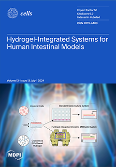
The luminal surface of the intestinal epithelium is protected by a vital mucus layer essential for lubrication, hydration, and symbiotic bacterial relationships. Replicating this structure in vitro is challenging. We developed a hydrogel-integrated millifluidic tissue chamber to apply defined shear stress to intestinal cells cultured on flat or 3D-structured hydrogel scaffolds. The chamber holds nine hydrogel scaffolds made from bioactive decellularized and methacrylated small intestinal submucosa with adjustable stiffness. Validated with HT29-MTX cells, the system provides a biomimetic cell culture substrate, inducing multi-lineage differentiation, increasing mucus production, and enhancing the 3D organization of organoid-derived human intestinal stem cells from the terminal ileum under physiological shear stress. View this paper
  • Issues are regarded as officially published after their release is announced to the table of contents alert mailing list.
  • You may sign up for e-mail alerts to receive table of contents of newly released issues.
  • PDF is the official format for papers published in both, html and pdf forms. To view the papers in pdf format, click on the "PDF Full-text" link, and use the free Adobe Reader to open them.
Order results
Result details
Section
Select all
Export citation of selected articles as:
18 pages, 7658 KiB  
Article
Uncovering the Interaction between TRAF1 and MAVS in the RIG-I Pathway to Enhance the Upregulation of IRF1/ISG15 during Classical Swine Fever Virus Infection
by Liyuan Zhang, Rongze Tang, Dongli Liang, Wenfeng Wang, Kaijun Min, Tingrong Luo and Xiaoning Li
Cells 2024, 13(13), 1165; https://doi.org/10.3390/cells13131165 - 8 Jul 2024
Cited by 3 | Viewed by 2061
Abstract
Classical swine fever (CSF) is caused by the classical swine fever virus (CSFV), which poses a threat to swine production. The activation of host innate immunity through linker proteins such as tumor necrosis factor receptor (TNF-R)-associated factor (TRAF) is crucial for the induction [...] Read more.
Classical swine fever (CSF) is caused by the classical swine fever virus (CSFV), which poses a threat to swine production. The activation of host innate immunity through linker proteins such as tumor necrosis factor receptor (TNF-R)-associated factor (TRAF) is crucial for the induction of the NF-κB pathway. Recent research has revealed the involvement of mitochondrial antiviral-signaling protein (MAVS) in the interaction with TRAF2, 3, 5, and 6 to activate both the NF-κB and IRF3 pathways. This study revealed that CSFV infection led to the upregulation of TRAF1 mRNA and protein levels; moreover, TRAF1 overexpression inhibited CSFV replication, while TRAF1 knockdown promoted replication, highlighting its importance in the host response to CSFV infection. Additionally, the expression of RIG-I, MAVS, TRAF1, IRF1, and ISG15 were detected in PK-15 cells infected with CSFV, revealing that TRAF1 plays a role in regulating IRF1 and ISG15 within the RIG-I pathway. Furthermore, Co-IP, GST pull-down, and IFA analyses demonstrated that TRAF1 interacted with MAVS and co-localized in the cytoplasm during CSFV infection. Ultimately, TRAF1 acted as a novel member of the TRAF family, bound to MAVS as a linker molecule, and functioned as a mediator downstream of MAVS in the RIG-I/MAVS pathway against CSFV replication. Full article
Show Figures

Figure 1

29 pages, 840 KiB  
Review
Unraveling the Potential Underlying Mechanisms of Mild Behavioral Impairment: Focusing on Amyloid and Tau Pathology
by Efthalia Angelopoulou, Anastasia Bougea, Alexandros Hatzimanolis, Nikolaos Scarmeas and Sokratis G. Papageorgiou
Cells 2024, 13(13), 1164; https://doi.org/10.3390/cells13131164 - 8 Jul 2024
Cited by 3 | Viewed by 2803
Abstract
The emergence of sustained neuropsychiatric symptoms (NPS) among non-demented individuals in later life, defined as mild behavioral impairment (MBI), is linked to a higher risk of cognitive decline. However, the underlying pathophysiological mechanisms remain largely unexplored. A growing body of evidence has shown [...] Read more.
The emergence of sustained neuropsychiatric symptoms (NPS) among non-demented individuals in later life, defined as mild behavioral impairment (MBI), is linked to a higher risk of cognitive decline. However, the underlying pathophysiological mechanisms remain largely unexplored. A growing body of evidence has shown that MBI is associated with alterations in structural and functional neuroimaging studies, higher genetic predisposition to clinical diagnosis of Alzheimer’s disease (AD), as well as amyloid and tau pathology assessed in the blood, cerebrospinal fluid, positron-emission tomography (PET) imaging and neuropathological examination. These findings shed more light on the MBI-related potential neurobiological mechanisms, paving the way for the development of targeted pharmacological approaches. In this review, we aim to discuss the available clinical evidence on the role of amyloid and tau pathology in MBI and the potential underlying pathophysiological mechanisms. Dysregulation of the hypothalamic–pituitary–adrenal (HPA) axis, disruption of neurotrophic factors, such as the brain-derived neurotrophic factor (BDNF), abnormal neuroinflammatory responses including the kynurenine pathway, dysregulation of transforming growth factor beta (TGF-β1), epigenetic alterations including micro-RNA (miR)-451a and miR-455-3p, synaptic dysfunction, imbalance in neurotransmitters including acetylcholine, dopamine, serotonin, gamma-aminobutyric acid (GABA) and norepinephrine, as well as altered locus coeruleus (LC) integrity are some of the potential mechanisms connecting MBI with amyloid and tau pathology. The elucidation of the underlying neurobiology of MBI would facilitate the design and efficacy of relative clinical trials, especially towards amyloid- or tau-related pathways. In addition, we provide insights for future research into our deeper understanding of its underlying pathophysiology of MBI, and discuss relative therapeutic implications. Full article
(This article belongs to the Collection Molecular Insights into Neurodegenerative Diseases)
Show Figures

Figure 1

18 pages, 4068 KiB  
Article
Type I Interferon Activates PD-1 Expression through Activation of the STAT1-IRF2 Pathway in Myeloid Cells
by Liyan Liang, Yingcui Yang, Kaidi Deng, Yanmin Wu, Yan Li, Liya Bai, Yinsong Wang and Chunwan Lu
Cells 2024, 13(13), 1163; https://doi.org/10.3390/cells13131163 - 8 Jul 2024
Cited by 1 | Viewed by 2136
Abstract
PD-1 (Programmed cell death protein 1) regulates the metabolic reprogramming of myeloid-derived suppressor cells and myeloid cell differentiation, as well as the type I interferon (IFN-I) signaling pathway in myeloid cells in the tumor microenvironment. PD-1, therefore, is a key inhibitory receptor in [...] Read more.
PD-1 (Programmed cell death protein 1) regulates the metabolic reprogramming of myeloid-derived suppressor cells and myeloid cell differentiation, as well as the type I interferon (IFN-I) signaling pathway in myeloid cells in the tumor microenvironment. PD-1, therefore, is a key inhibitory receptor in myeloid cells. However, the regulation of PD-1 expression in myeloid cells is unknown. We report that the expression level of PDCD1, the gene that encodes the PD-1 protein, is positively correlated with the levels of IFNB1 and IFNAR1 in myeloid cells in human colorectal cancer. Treatment of mouse myeloid cell lines with recombinant IFNβ protein elevated PD-1 expression in myeloid cells in vitro. Knocking out IFNAR1, the gene that encodes the IFN-I-specific receptor, diminished the inductive effect of IFNβ on PD-1 expression in myeloid cells in vitro. Treatment of tumor-bearing mice with a lipid nanoparticle-encapsulated IFNβ-encoding plasmid (IFNBCOL01) increased IFNβ expression, resulting in elevated PD-1 expression in tumor-infiltrating myeloid cells. At the molecular level, we determined that IFNβ activates STAT1 (signal transducer and activator of transcription 1) and IRFs (interferon regulatory factors) in myeloid cells. Analysis of the cd279 promoter identified IRF2-binding consensus sequence elements. ChIP (chromatin immunoprecipitation) analysis determined that the pSTAT1 directly binds to the irf2 promoter and that IRF2 directly binds to the cd279 promoter in myeloid cells in vitro and in vivo. In colon cancer patients, the expression levels of STAT1, IRF2 and PDCD1 are positively correlated in tumor-infiltrating myeloid cells. Our findings determine that IFNβ activates PD-1 expression at least in part by an autocrine mechanism via the stimulation of the pSTAT1-IRF2 axis in myeloid cells. Full article
Show Figures

Figure 1

11 pages, 4595 KiB  
Communication
The Use of Hexokinase 2-Displacing Peptides as an Anti-Neoplastic Approach for Malignant Peripheral Nerve Sheath Tumors
by Francesco Ciscato, Ionica Masgras, Alessandro Gori, Marco Fantuz, Greta Bergamaschi, Denis Komarov, Martina La Spina, Shiva Ghasemi-Firouzabadi, Marco Pizzi, Angelo Paolo Dei Tos, Federica Chiara, Alessandro Carrer and Andrea Rasola
Cells 2024, 13(13), 1162; https://doi.org/10.3390/cells13131162 - 8 Jul 2024
Viewed by 1843
Abstract
Malignant Peripheral Nerve Sheath Tumors (MPNSTs) are aggressive sarcomas that can arise both sporadically and in patients with the genetic syndrome Neurofibromatosis type 1 (NF1). Prognosis is dismal, as large dimensions, risk of relapse, and anatomical localization make surgery poorly effective, and no [...] Read more.
Malignant Peripheral Nerve Sheath Tumors (MPNSTs) are aggressive sarcomas that can arise both sporadically and in patients with the genetic syndrome Neurofibromatosis type 1 (NF1). Prognosis is dismal, as large dimensions, risk of relapse, and anatomical localization make surgery poorly effective, and no therapy is known. Hence, the identification of MPNST molecular features that could be hit in an efficient and selective way is mandatory to envision treatment options. Here, we find that MPNSTs express high levels of the glycolytic enzyme Hexokinase 2 (HK2), which is known to shield cancer cells from noxious stimuli when it localizes at MAMs (mitochondria-associated membranes), contact sites between mitochondria and endoplasmic reticulum. A HK2-targeting peptide that dislodges HK2 from MAMs rapidly induces a massive death of MPNST cells. After identifying different matrix metalloproteases (MMPs) expressed in the MPNST microenvironment, we have designed HK2-targeting peptide variants that harbor cleavage sites for these MMPs, making such peptides activatable in the proximity of cancer cells. We find that the peptide carrying the MMP2/9 cleavage site is the most effective, both in inhibiting the in vitro tumorigenicity of MPNST cells and in hampering their growth in mice. Our data indicate that detaching HK2 from MAMs could pave the way for a novel anti-MPNST therapeutic strategy, which could be flexibly adapted to the protease expression features of the tumor microenvironment. Full article
Show Figures

Figure 1

19 pages, 3552 KiB  
Article
Tumor Necrosis Factor-Alpha Modulates Expression of Genes Involved in Cytokines and Chemokine Pathways in Proliferative Myoblast Cells
by Angela María Alvarez, Carlos Eduardo Madureira Trufen, Marcus Vinicius Buri, Marcela Bego Nering de Sousa, Francisco Ivanio Arruda-Alves, Flavio Lichtenstein, Ursula Castro de Oliveira, Inácio de Loiola Meirelles Junqueira-de-Azevedo, Catarina Teixeira and Vanessa Moreira
Cells 2024, 13(13), 1161; https://doi.org/10.3390/cells13131161 - 8 Jul 2024
Cited by 3 | Viewed by 2006
Abstract
Skeletal muscle regeneration after injury is a complex process involving inflammatory signaling and myoblast activation. Pro-inflammatory cytokines like tumor necrosis factor-alpha (TNF-α) are key mediators, but their effects on gene expression in proliferating myoblasts are unclear. We performed the RNA sequencing of TNF-α [...] Read more.
Skeletal muscle regeneration after injury is a complex process involving inflammatory signaling and myoblast activation. Pro-inflammatory cytokines like tumor necrosis factor-alpha (TNF-α) are key mediators, but their effects on gene expression in proliferating myoblasts are unclear. We performed the RNA sequencing of TNF-α treated C2C12 myoblasts to elucidate the signaling pathways and gene networks regulated by TNF-α during myoblast proliferation. The TNF-α (10 ng/mL) treatment of C2C12 cells led to 958 differentially expressed genes compared to the controls. Pathway analysis revealed significant regulation of TNF-α signaling, along with the chemokine and IL-17 pathways. Key upregulated genes included cytokines (e.g., IL-6), chemokines (e.g., CCL7), and matrix metalloproteinases (MMPs). TNF-α increased myogenic factor 5 (Myf5) but decreased MyoD protein levels and stimulated the release of MMP-9, MMP-10, and MMP-13. TNF-α also upregulates versican and myostatin mRNA. Overall, our study demonstrates the TNF-α modulation of distinct gene expression patterns and signaling pathways that likely contribute to enhanced myoblast proliferation while suppressing premature differentiation after muscle injury. Elucidating the mechanisms involved in skeletal muscle regeneration can aid in the development of regeneration-enhancing therapeutics. Full article
(This article belongs to the Special Issue Recent Research on Muscle Homeostasis and Regeneration)
Show Figures

Figure 1

19 pages, 18184 KiB  
Article
Regeneration of Propriospinal Axons in Rat Transected Spinal Cord Injury through a Growth-Promoting Pathway Constructed by Schwann Cells Overexpressing GDNF
by Xiaolong Du, Shengqi Zhang, Aytak Khabbaz, Kristen Lynn Cohen, Yihong Zhang, Samhita Chakraborty, George M. Smith, Hongxing Wang, Amol P. Yadav, Naikui Liu and Lingxiao Deng
Cells 2024, 13(13), 1160; https://doi.org/10.3390/cells13131160 - 8 Jul 2024
Cited by 2 | Viewed by 1940
Abstract
Unsuccessful axonal regeneration in transected spinal cord injury (SCI) is mainly attributed to shortage of growth factors, inhibitory glial scar, and low intrinsic regenerating capacity of severely injured neurons. Previously, we constructed an axonal growth permissive pathway in a thoracic hemisected injury by [...] Read more.
Unsuccessful axonal regeneration in transected spinal cord injury (SCI) is mainly attributed to shortage of growth factors, inhibitory glial scar, and low intrinsic regenerating capacity of severely injured neurons. Previously, we constructed an axonal growth permissive pathway in a thoracic hemisected injury by transplantation of Schwann cells overexpressing glial-cell-derived neurotrophic factor (SCs-GDNF) into the lesion gap as well as the caudal cord and proved that this novel permissive bridge promoted the regeneration of descending propriospinal tract (dPST) axons across and beyond the lesion. In the current study, we subjected rats to complete thoracic (T11) spinal cord transections and examined whether these combinatorial treatments can support dPST axons’ regeneration beyond the transected injury. The results indicated that GDNF significantly improved graft–host interface by promoting integration between SCs and astrocytes, especially the migration of reactive astrocyte into SCs-GDNF territory. The glial response in the caudal graft area has been significantly attenuated. The astrocytes inside the grafted area were morphologically characterized by elongated and slim process and bipolar orientation accompanied by dramatically reduced expression of glial fibrillary acidic protein. Tremendous dPST axons have been found to regenerate across the lesion and back to the caudal spinal cord which were otherwise difficult to see in control groups. The caudal synaptic connections were formed, and regenerated axons were remyelinated. The hindlimb locomotor function has been improved. Full article
(This article belongs to the Collection Cell Biology of Spinal Cord Injury and Repair)
Show Figures

Figure 1

14 pages, 4037 KiB  
Article
Voltage-Gated Ion Channels Are Transcriptional Targets of Sox10 during Oligodendrocyte Development
by Christian Peters, Tim Aberle, Elisabeth Sock, Jessica Brunner, Melanie Küspert, Simone Hillgärtner, Hannah M. Wüst and Michael Wegner
Cells 2024, 13(13), 1159; https://doi.org/10.3390/cells13131159 - 7 Jul 2024
Cited by 1 | Viewed by 1711
Abstract
The transcription factor Sox10 is an important determinant of oligodendroglial identity and influences oligodendroglial development and characteristics at various stages. Starting from RNA-seq data, we here show that the expression of several voltage-gated ion channels with known expression and important function in oligodendroglial [...] Read more.
The transcription factor Sox10 is an important determinant of oligodendroglial identity and influences oligodendroglial development and characteristics at various stages. Starting from RNA-seq data, we here show that the expression of several voltage-gated ion channels with known expression and important function in oligodendroglial cells depends upon Sox10. These include the Nav1.1, Cav2.2, Kv1.1, and Kir4.1 channels. For each of the four encoding genes, we found at least one regulatory region that is activated by Sox10 in vitro and at the same time bound by Sox10 in vivo. Cell-specific deletion of Sox10 in oligodendroglial cells furthermore led to a strong downregulation of all four ion channels in a mouse model and thus in vivo. Our study provides a clear functional link between voltage-gated ion channels and the transcriptional regulatory network in oligodendroglial cells. Furthermore, our study argues that Sox10 exerts at least some of its functions in oligodendrocyte progenitor cells, in myelinating oligodendrocytes, or throughout lineage development via these ion channels. By doing so, we present one way in which oligodendroglial development and properties can be linked to neuronal activity to ensure crosstalk between cell types during the development and function of the central nervous system. Full article
(This article belongs to the Collection Oligodendrocyte Physiology and Pathology Function)
Show Figures

Graphical abstract

12 pages, 1818 KiB  
Article
Precise Serial Microregistration Enables Quantitative Microscopy Imaging Tracking of Human Skin Cells In Vivo
by Yunxian Tian, Zhenguo Wu, Harvey Lui, Jianhua Zhao, Sunil Kalia, InSeok Seo, Hao Ou-Yang and Haishan Zeng
Cells 2024, 13(13), 1158; https://doi.org/10.3390/cells13131158 - 7 Jul 2024
Viewed by 1497
Abstract
We developed an automated microregistration method that enables repeated in vivo skin microscopy imaging of the same tissue microlocation and specific cells over a long period of days and weeks with unprecedented precision. Applying this method in conjunction with an in vivo multimodality [...] Read more.
We developed an automated microregistration method that enables repeated in vivo skin microscopy imaging of the same tissue microlocation and specific cells over a long period of days and weeks with unprecedented precision. Applying this method in conjunction with an in vivo multimodality multiphoton microscope, the behavior of human skin cells such as cell proliferation, melanin upward migration, blood flow dynamics, and epidermal thickness adaptation can be recorded over time, facilitating quantitative cellular dynamics analysis. We demonstrated the usefulness of this method in a skin biology study by successfully monitoring skin cellular responses for a period of two weeks following an acute exposure to ultraviolet light. Full article
(This article belongs to the Special Issue Advanced Technology for Cellular Imaging)
Show Figures

Graphical abstract

19 pages, 864 KiB  
Review
Role of the Innate Immune Response in Glomerular Disease Pathogenesis: Focus on Podocytes
by Wadih Issa, Rachel Njeim, Arianna Carrazco, George W. Burke and Alla Mitrofanova
Cells 2024, 13(13), 1157; https://doi.org/10.3390/cells13131157 - 6 Jul 2024
Cited by 5 | Viewed by 3527
Abstract
Accumulating evidence indicates that inflammatory and immunologic processes play a significant role in the development and progression of glomerular diseases. Podocytes, the terminally differentiated epithelial cells, are crucial for maintaining the integrity of the glomerular filtration barrier. Once injured, podocytes cannot regenerate, leading [...] Read more.
Accumulating evidence indicates that inflammatory and immunologic processes play a significant role in the development and progression of glomerular diseases. Podocytes, the terminally differentiated epithelial cells, are crucial for maintaining the integrity of the glomerular filtration barrier. Once injured, podocytes cannot regenerate, leading to progressive proteinuric glomerular diseases. However, emerging evidence suggests that podocytes not only maintain the glomerular filtration barrier and are important targets of immune responses but also exhibit many features of immune-like cells, where they are involved in the modulation of the activity of innate and adaptive immunity. This dual role of podocytes may lead to the discovery and development of new therapeutic targets for treating glomerular diseases. This review aims to provide an overview of the innate immunity mechanisms involved in podocyte injury and the progression of proteinuric glomerular diseases. Full article
(This article belongs to the Special Issue Podocyte Pathophysiology)
Show Figures

Figure 1

20 pages, 7681 KiB  
Article
A Short Sequence Targets Transmembrane Proteins to Primary Cilia
by Viviana Macarelli, Edward C. Harding, David C. Gershlick and Florian T. Merkle
Cells 2024, 13(13), 1156; https://doi.org/10.3390/cells13131156 - 6 Jul 2024
Viewed by 2567
Abstract
Primary cilia are finger-like sensory organelles that extend from the bodies of most cell types and have a distinct lipid and protein composition from the plasma membrane. This partitioning is maintained by a diffusion barrier that restricts the entry of non-ciliary proteins, and [...] Read more.
Primary cilia are finger-like sensory organelles that extend from the bodies of most cell types and have a distinct lipid and protein composition from the plasma membrane. This partitioning is maintained by a diffusion barrier that restricts the entry of non-ciliary proteins, and allows the selective entry of proteins harboring a ciliary targeting sequence (CTS). However, CTSs are not stereotyped and previously reported sequences are insufficient to drive efficient ciliary localisation across diverse cell types. Here, we describe a short peptide sequence that efficiently targets transmembrane proteins to primary cilia in all tested cell types, including human neurons. We generate human-induced pluripotent stem cell (hiPSC) lines stably expressing a transmembrane construct bearing an extracellular HaloTag and intracellular fluorescent protein, which enables the bright, specific labeling of primary cilia in neurons and other cell types to facilitate studies of cilia in health and disease. We demonstrate the utility of this resource by developing an image analysis pipeline for the automated measurement of primary cilia to detect changes in their length associated with altered signaling or disease state. Full article
(This article belongs to the Special Issue The Role of Cilia in Health and Diseases)
Show Figures

Graphical abstract

17 pages, 11970 KiB  
Article
Suppressing PD-L1 Expression via AURKA Kinase Inhibition Enhances Natural Killer Cell-Mediated Cytotoxicity against Glioblastoma
by Trang T. T. Nguyen, Qiuqiang Gao, Jeong-Yeon Mun, Zhe Zhu, Chang Shu, Aaron Naim, Meri Rogava, Benjamin Izar, Mike-Andrew Westhoff, Georg Karpel-Massler and Markus D. Siegelin
Cells 2024, 13(13), 1155; https://doi.org/10.3390/cells13131155 - 6 Jul 2024
Cited by 1 | Viewed by 2743
Abstract
Immunotherapies have shown significant promise as an impactful strategy in cancer treatment. However, in glioblastoma multiforme (GBM), the most prevalent primary brain tumor in adults, these therapies have demonstrated lower efficacy than initially anticipated. Consequently, there is an urgent need for strategies to [...] Read more.
Immunotherapies have shown significant promise as an impactful strategy in cancer treatment. However, in glioblastoma multiforme (GBM), the most prevalent primary brain tumor in adults, these therapies have demonstrated lower efficacy than initially anticipated. Consequently, there is an urgent need for strategies to enhance the effectiveness of immune treatments. AURKA has been identified as a potential drug target for GBM treatment. An analysis of the GBM cell transcriptome following AURKA inhibition revealed a potential influence on the immune system. Our research revealed that AURKA influenced PD-L1 levels in various GBM model systems in vitro and in vivo. Disrupting AURKA function genetically led to reduced PD-L1 levels and increased MHC-I expression in both established and patient-derived xenograft GBM cultures. This process involved both transcriptional and non-transcriptional pathways, partly implicating GSK3β. Interfering with AURKA also enhanced NK-cell-mediated elimination of GBM by reducing PD-L1 expression, as evidenced in rescue experiments. Furthermore, using a mouse model that mimics GBM with patient-derived cells demonstrated that Alisertib decreased PD-L1 expression in living organisms. Combination therapy involving anti-PD-1 treatment and Alisertib significantly prolonged overall survival compared to vehicle treatment. These findings suggest that targeting AURKA could have therapeutic implications for modulating the immune environment within GBM cells. Full article
Show Figures

Figure 1

12 pages, 649 KiB  
Review
The Lungs in Space: A Review of Current Knowledge and Methodologies
by Michaela B. Smith, Hui Chen and Brian G. G. Oliver
Cells 2024, 13(13), 1154; https://doi.org/10.3390/cells13131154 - 6 Jul 2024
Cited by 2 | Viewed by 3878
Abstract
Space travel presents multiple risks to astronauts such as launch, radiation, spacewalks or extravehicular activities, and microgravity. The lungs are composed of a combination of air, blood, and tissue, making it a complex organ system with interactions between the external and internal environment. [...] Read more.
Space travel presents multiple risks to astronauts such as launch, radiation, spacewalks or extravehicular activities, and microgravity. The lungs are composed of a combination of air, blood, and tissue, making it a complex organ system with interactions between the external and internal environment. Gravity strongly influences the structure of the lung which results in heterogeneity of ventilation and perfusion that becomes uniform in microgravity as shown during parabolic flights, Spacelab, and Skylab experiments. While changes in lung volumes occur in microgravity, efficient gas exchange remains and the lungs perform as they would on Earth; however, little is known about the cellular response to microgravity. In addition to spaceflight and real microgravity, devices, such as clinostats and random positioning machines, are used to simulate microgravity to study cellular responses on the ground. Differential expression of cell adhesion and extracellular matrix molecules has been found in real and simulated microgravity. Immune dysregulation is a known consequence of space travel that includes changes in immune cell morphology, function, and number, which increases susceptibility to infections. However, the majority of in vitro studies do not have a specific respiratory focus. These studies are needed to fully understand the impact of microgravity on the function of the respiratory system in different conditions. Full article
(This article belongs to the Special Issue Cells in Space and on Earth)
Show Figures

Figure 1

24 pages, 4561 KiB  
Review
Advances in Regenerative Dentistry: A Systematic Review of Harnessing Wnt/β-Catenin in Dentin-Pulp Regeneration
by Mariam Amir, Lakshmi Jeevithan, Maham Barkat, Syeda Habib Fatima, Malalai Khan, Sara Israr, Fatima Naseer, Sarmad Fayyaz, Jeevithan Elango, Wenhui Wu, José Eduardo Maté Sánchez de Val and Saeed Ur Rahman
Cells 2024, 13(13), 1153; https://doi.org/10.3390/cells13131153 - 6 Jul 2024
Cited by 1 | Viewed by 2735
Abstract
Dentin pulp has a complex function as a major unit in maintaining the vitality of teeth. In this sense, the Wnt/β-Catenin pathway has a vital part in tooth development, maintenance, repair, and regeneration by controlling physiological activities such as growth, differentiation, and migration. [...] Read more.
Dentin pulp has a complex function as a major unit in maintaining the vitality of teeth. In this sense, the Wnt/β-Catenin pathway has a vital part in tooth development, maintenance, repair, and regeneration by controlling physiological activities such as growth, differentiation, and migration. This pathway consists of a network of proteins, such as Wnt signaling molecules, which interact with receptors of targeted cells and play a role in development and adult tissue homeostasis. The Wnt signals are specific spatiotemporally, suggesting its intricate mechanism in development, regulation, repair, and regeneration by the formation of tertiary dentin. This review provides an overview of the recent advances in the Wnt/β-Catenin signaling pathway in dentin and pulp regeneration, how different proteins, molecules, and ligands influence this pathway, either upregulating or silencing it, and how it may be used in the future for clinical dentistry, in vital pulp therapy as an effective treatment for dental caries, as an alternative approach for root canal therapy, and to provide a path for therapeutic and regenerative dentistry. Full article
(This article belongs to the Special Issue Molecular Mechanism of Bone Disease)
Show Figures

Figure 1

18 pages, 4932 KiB  
Article
Hypoxia Promotes Invadosome Formation by Lung Fibroblasts
by Mégane Lebel, Dominic O. Cliche, Martine Charbonneau, Karine Brochu-Gaudreau, Damien Adam, Emmanuelle Brochiero, Claire M. Dubois and André M. Cantin
Cells 2024, 13(13), 1152; https://doi.org/10.3390/cells13131152 - 6 Jul 2024
Cited by 3 | Viewed by 2079
Abstract
Lung parenchymal hypoxia has emerged as a cardinal feature of idiopathic pulmonary fibrosis (IPF). Hypoxia promotes cancer cell invasion and metastasis through signaling that is dependent upon the lysophosphatidic acid (LPA) receptor, LPA1 (LPAR1). Abundant data indicate that LPA1-dependent signaling [...] Read more.
Lung parenchymal hypoxia has emerged as a cardinal feature of idiopathic pulmonary fibrosis (IPF). Hypoxia promotes cancer cell invasion and metastasis through signaling that is dependent upon the lysophosphatidic acid (LPA) receptor, LPA1 (LPAR1). Abundant data indicate that LPA1-dependent signaling also enhances lung fibrogenesis in IPF. We recently reported that fibroblasts isolated from the lungs of individuals with IPF have an increased capacity to form subcellular matrix-degradative structures known as invadosomes, an event that correlates with the degree of lung fibrosis. We therefore hypothesized that hypoxia promotes invadosome formation in lung fibroblasts through LPA1-dependent signaling. Here, it is demonstrated that invadosome formation by fibroblasts from the lungs of individuals with advanced IPF is inhibited by both the tyrosine receptor kinase inhibitor nintedanib and inhibition of LPA1. In addition, exposure of normal human lung fibroblasts to either hypoxia or LPA increased their ability to form invadosomes. Mechanistically, the hypoxia-induced invadosome formation by lung fibroblasts was found to involve LPA1 and PDGFR-Akt signaling. We concluded that hypoxia increases the formation of invadosomes in lung fibroblasts through the LPA1 and PDGFR-Akt signaling axis, which represents a potential target for suppressing lung fibrosis. Full article
Show Figures

Figure 1

24 pages, 2769 KiB  
Review
Natural Products for Preventing and Managing Anthracycline-Induced Cardiotoxicity: A Comprehensive Review
by Jarosław Szponar, Przemysław Niziński, Jarosław Dudka, Kamila Kasprzak-Drozd and Anna Oniszczuk
Cells 2024, 13(13), 1151; https://doi.org/10.3390/cells13131151 - 6 Jul 2024
Cited by 5 | Viewed by 2719
Abstract
Doxorubicin (DOX) is an anthracycline anticancer agent that is highly effective in the treatment of solid tumors. Given the multiplicity of mechanisms involved in doxorubicin-induced cardiotoxicity, it is difficult to identify a precise molecular target for toxicity. The findings of a literature review [...] Read more.
Doxorubicin (DOX) is an anthracycline anticancer agent that is highly effective in the treatment of solid tumors. Given the multiplicity of mechanisms involved in doxorubicin-induced cardiotoxicity, it is difficult to identify a precise molecular target for toxicity. The findings of a literature review suggest that natural products may offer cardioprotective benefits against doxorubicin-induced cardiotoxicity, both in vitro and in vivo. However, further confirmatory studies are required to substantiate this claim. It is of the utmost importance to direct greater attention towards the intricate signaling networks that are of paramount importance for the survival and dysfunction of cardiomyocytes. Notwithstanding encouraging progress made in preclinical studies of natural products for the prevention of DOX-induced cardiotoxicity, these have not yet been translated for clinical use. One of the most significant obstacles hindering the development of cardioprotective adjuvants based on natural products is the lack of adequate bioavailability in humans. This review presents an overview of current knowledge on doxorubicin DOX-induced cardiotoxicity, with a focus on the potential benefits of natural compounds and herbal preparations in preventing this adverse effect. As literature search engines, the browsers in the Scopus, PubMed, Web of Science databases and the ClinicalTrials.gov register were used. Full article
Show Figures

Graphical abstract

16 pages, 8420 KiB  
Article
Cx40 Levels Regulate Hypoxia-Induced Changes in the Migration, Proliferation, and Formation of Gap Junction Plaques in an Extravillous Trophoblast Cell Model
by Fernanda M. Rozas-Villanueva, Viviana P. Orellana, Rodrigo Alarcón, Jaime Maripillan, Agustin D. Martinez, Ivan E. Alfaro and Mauricio A. Retamal
Cells 2024, 13(13), 1150; https://doi.org/10.3390/cells13131150 - 6 Jul 2024
Cited by 2 | Viewed by 1747
Abstract
Background: Extravillous trophoblasts (EVTs) form stratified columns at the placenta–uterus interface. In the closest part to fetal structures, EVTs have a proliferative phenotype, whereas in the closest part to maternal structures, they present a migratory phenotype. During the placentation process, Connexin 40 (Cx40) [...] Read more.
Background: Extravillous trophoblasts (EVTs) form stratified columns at the placenta–uterus interface. In the closest part to fetal structures, EVTs have a proliferative phenotype, whereas in the closest part to maternal structures, they present a migratory phenotype. During the placentation process, Connexin 40 (Cx40) participates in both the proliferation and migration of EVTs, which occurs under hypoxia. However, a possible interaction between hypoxia and Cx40 has not yet been established. Methods: We developed two cellular models, one with “low Cx40” (Jeg-3), which reflected the expression of this protein found in migratory EVTs, and one with “high Cx40” (Jeg-3/hCx40), which reflected the expression of this protein in proliferative cells. We analyzed the migration and proliferation of these cells under normoxic and hypoxic conditions for 24 h. Jeg-3 cells under hypoxia increased their migratory capacity over their proliferative capacity. However, in Jeg-3/hCx40, the opposite effect was induced. On the other hand, hypoxia promoted gap junction (GJ) plaque formation between neighboring Jeg-3 cells. Similarly, the activation of a nitro oxide (NO)/cGMP/PKG-dependent pathway induced an increase in GJ-plaque formation in Jeg-3 cells. Conclusions: The expression patterns of Cx40 play a crucial role in shaping the responses of EVTs to hypoxia, thereby influencing their migratory or proliferative phenotype. Simultaneously, hypoxia triggers an increase in Cx40 gap junction (GJ) plaque formation through a pathway dependent on NO. Full article
Show Figures

Figure 1

17 pages, 3452 KiB  
Article
Global Transcriptomic and Characteristics Comparisons between Mouse Fetal Liver and Bone Marrow Definitive Erythropoiesis
by Chengjie Gao, Huan Zhang, Yaomei Wang, Shihui Wang, Xinhua Guo, Yongshuai Han, Huizhi Zhao and Xiuli An
Cells 2024, 13(13), 1149; https://doi.org/10.3390/cells13131149 - 5 Jul 2024
Viewed by 2065
Abstract
Erythropoiesis occurs first in the yolk sac as a transit “primitive” form, then is gradually replaced by the “definitive” form in the fetal liver (FL) during fetal development and in the bone marrow (BM) postnatally. While it is well known that differences exist [...] Read more.
Erythropoiesis occurs first in the yolk sac as a transit “primitive” form, then is gradually replaced by the “definitive” form in the fetal liver (FL) during fetal development and in the bone marrow (BM) postnatally. While it is well known that differences exist between primitive and definitive erythropoiesis, the similarities and differences between FL and BM definitive erythropoiesis have not been studied. Here we performed comprehensive comparisons of erythroid progenitors and precursors at all maturational stages sorted from E16.5 FL and adult BM. We found that FL cells at all maturational stages were larger than their BM counterparts. We further found that FL BFU-E cells divided at a faster rate and underwent more cell divisions than BM BFU-E. Transcriptome comparison revealed that genes with increased expression in FL BFU-Es were enriched in cell division. Interestingly, the expression levels of glucocorticoid receptor Nr3c1, Myc and Myc downstream target Ccna2 were significantly higher in FL BFU-Es, indicating the role of the Nr3c1-Myc-Ccna2 axis in the enhanced proliferation/cell division of FL BFU-E cells. At the CFU-E stage, the expression of genes associated with hemoglobin biosynthesis were much higher in FL CFU-Es, indicating more hemoglobin production. During terminal erythropoiesis, overall temporal patterns in gene expression were conserved between the FL and BM. While biological processes related to translation, the tricarboxylic acid cycle and hypoxia response were upregulated in FL erythroblasts, those related to antiviral signal pathway were upregulated in BM erythroblasts. Our findings uncovered previously unrecognized differences between FL and BM definitive erythropoiesis and provide novel insights into erythropoiesis. Full article
(This article belongs to the Section Cell Proliferation and Division)
Show Figures

Figure 1

29 pages, 4113 KiB  
Article
A Critical Role of Culture Medium Selection in Maximizing the Purity and Expansion of Natural Killer Cells
by Neele Kusch, Jonathan Storm, Antonia Macioszek, Ella Kisselmann, Cornelius Knabbe, Barbara Kaltschmidt and Christian Kaltschmidt
Cells 2024, 13(13), 1148; https://doi.org/10.3390/cells13131148 - 5 Jul 2024
Cited by 1 | Viewed by 4511
Abstract
Natural killer (NK) cells hold promise in cancer treatment due to their ability to spontaneously lyse cancer cells. For clinical use, high quantities of pure, functional NK cells are necessary. Combining adherence-based isolation with specialized media showed the unreliability of the isolation method, [...] Read more.
Natural killer (NK) cells hold promise in cancer treatment due to their ability to spontaneously lyse cancer cells. For clinical use, high quantities of pure, functional NK cells are necessary. Combining adherence-based isolation with specialized media showed the unreliability of the isolation method, but demonstrated the superiority of the NK MACS® medium, particularly in suboptimal conditions. Neither human pooled serum, fetal calf serum (FCS), human platelet lysate, nor chemically defined serum replacement could substitute human AB serum. Interleukin (IL-)2, IL-15, IL-21, and combined CD2/NKp46 stimulation were assessed. IL-21 and CD2/NKp46 stimulation increased cytotoxicity, but reduced NK cell proliferation. IL-15 stimulation alone achieved the highest proliferation, but the more affordable IL-2 performed similarly. The RosetteSep™ human NK cell enrichment kit was effective for isolation, but the presence of peripheral blood mononuclear cells (PBMCs) in the culture enhanced NK cell proliferation, despite similar expression levels of CD16, NKp46, NKG2D, and ICAM-1. In line with this, purified NK cells cultured in NK MACS® medium with human AB serum and IL-2 demonstrated high cytotoxicity against primary glioblastoma stem cells. Full article
(This article belongs to the Special Issue Advances in the Study of Natural Killer (NK) Cells)
Show Figures

Figure 1

17 pages, 9991 KiB  
Article
Iron Regulates Cellular Proliferation by Enhancing the Expression of Glucose Transporter GLUT3 in the Liver
by Kleber S. Ribeiro, Eshani Karmakar, Christine Park, Richa Garg, George P. Kung, Isha Kadakia, Jyotsna S. Gopianand, Tejas Arun, Oleg Kisselev and Jaya P. Gnana-Prakasam
Cells 2024, 13(13), 1147; https://doi.org/10.3390/cells13131147 - 4 Jul 2024
Cited by 2 | Viewed by 2055
Abstract
Iron is often accumulated in the liver during pathological conditions such as cirrhosis and cancer. Elevated expression of glucose transporters GLUT1 and GLUT3 is associated with reduced overall survival in patients with hepatocellular carcinoma. However, it is not known whether iron can regulate [...] Read more.
Iron is often accumulated in the liver during pathological conditions such as cirrhosis and cancer. Elevated expression of glucose transporters GLUT1 and GLUT3 is associated with reduced overall survival in patients with hepatocellular carcinoma. However, it is not known whether iron can regulate glucose transporters and contribute to tumor proliferation. In the present study, we found that treatment of human liver cell line HepG2 with ferric ammonium citrate (FAC) resulted in a significant upregulation of GLUT3 mRNA and protein in a dose-dependent manner. Similarly, iron accumulation in mice fed with high dietary iron as well as in mice injected intraperitoneally with iron dextran enhanced the GLUT3 expression drastically in the liver. We demonstrated that iron-induced hepatic GLUT3 upregulation is mediated by the LKB1/AMPK/CREB1 pathway, and this activation was reversed when treated with iron chelator deferiprone. In addition, inhibition of GLUT3 using siRNA prevented iron-mediated increase in the expression of cell cycle markers and cellular hyperproliferation. Furthermore, exogenous sodium beta-hydroxybutyrate treatment prevented iron-mediated hepatic GLUT3 activation both in vitro and in vivo. Together, these results underscore the importance of iron, AMPK, CREB1 and GLUT3 pathways in cell proliferation and highlight the therapeutic potential of sodium beta-hydroxybutyrate in hepatocellular carcinoma with high GLUT3 expression. Full article
(This article belongs to the Special Issue Regulation of Iron Metabolism in Health and Disease)
Show Figures

Figure 1

19 pages, 7588 KiB  
Article
Development of a Humanized Antibody Targeting Extracellular HSP90α to Suppress Endothelial-Mesenchymal Transition-Enhanced Tumor Growth of Pancreatic Adenocarcinoma Cells
by Chi-Shuan Fan, Hui-Chen Hung, Chia-Chi Chen, Li-Li Chen, Yi-Yu Ke, Teng-Kuang Yeh, Chin-Ting Huang, Teng-Yuan Chang, Kuei-Jung Yen, Chung-Hsing Chen, Kee Voon Chua, John Tsu-An Hsu and Tze-Sing Huang
Cells 2024, 13(13), 1146; https://doi.org/10.3390/cells13131146 - 4 Jul 2024
Cited by 4 | Viewed by 2359
Abstract
Extracellular HSP90α (eHSP90α) is a promoter of tumor development and malignant progression. Patients with malignancies, including pancreatic ductal adenocarcinoma (PDAC), have generally shown 5~10-fold increases in serum/plasma eHSP90α levels. In this study, we developed a humanized antibody HH01 to target eHSP90α and evaluated [...] Read more.
Extracellular HSP90α (eHSP90α) is a promoter of tumor development and malignant progression. Patients with malignancies, including pancreatic ductal adenocarcinoma (PDAC), have generally shown 5~10-fold increases in serum/plasma eHSP90α levels. In this study, we developed a humanized antibody HH01 to target eHSP90α and evaluated its anticancer efficacy. HH01, with novel complementarity-determining regions, exhibits high binding affinity toward HSP90α. It recognizes HSP90α epitope sites 235AEEKEDKEEE244 and 251ESEDKPEIED260, with critical amino acid residues E237, E239, D240, K241, E253, and K255. HH01 effectively suppressed eHSP90α-induced invasive and spheroid-forming activities of colorectal cancer and PDAC cell lines by blocking eHSP90α’s ligation with the cell-surface receptor CD91. In mouse models, HH01 potently inhibited the tumor growth of PDAC cell grafts/xenografts promoted by endothelial-mesenchymal transition-derived cancer-associated fibroblasts while also reducing serum eHSP90α levels, reflecting its anticancer efficacy. HH01 also modulated tumor immunity by reducing M2 macrophages and reinvigorating immune T-cells. Additionally, HH01 showed low aggregation propensity, high water solubility, and a half-life time of >18 days in mouse blood. It was not cytotoxic to retinal pigmented epithelial cells and showed no obvious toxicity in mouse organs. Our data suggest that targeting eHSP90α with HH01 antibody can be a promising novel strategy for PDAC therapy. Full article
(This article belongs to the Special Issue Heat Shock Proteins and Human Cancers)
Show Figures

Figure 1

47 pages, 1728 KiB  
Review
Microbiota and Resveratrol: How Are They Linked to Osteoporosis?
by Christine Meyer, Aranka Brockmueller, Vicenç Ruiz de Porras and Mehdi Shakibaei
Cells 2024, 13(13), 1145; https://doi.org/10.3390/cells13131145 - 3 Jul 2024
Cited by 2 | Viewed by 3495
Abstract
Osteoporosis (OP), which is characterized by a decrease in bone density and increased susceptibility to fractures, is closely linked to the gut microbiota (GM). It is increasingly realized that the GM plays a key role in the maintenance of the functioning of multiple [...] Read more.
Osteoporosis (OP), which is characterized by a decrease in bone density and increased susceptibility to fractures, is closely linked to the gut microbiota (GM). It is increasingly realized that the GM plays a key role in the maintenance of the functioning of multiple organs, including bone, by producing bioactive metabolites such as short-chain fatty acids (SCFA). Consequently, imbalances in the GM, referred to as dysbiosis, have been identified with a significant reduction in beneficial metabolites, such as decreased SCFA associated with increased chronic inflammatory processes, including the activation of NF-κB at the epigenetic level, which is recognized as the main cause of many chronic diseases, including OP. Furthermore, regular or long-term medications such as antibiotics and many non-antibiotics such as proton pump inhibitors, chemotherapy, and NSAIDs, have been found to contribute to the development of dysbiosis, highlighting an urgent need for new treatment approaches. A promising preventive and adjuvant approach is to combat dysbiosis with natural polyphenols such as resveratrol, which have prebiotic functions and ensure an optimal microenvironment for beneficial GM. Resveratrol offers a range of benefits, including anti-inflammatory, anti-oxidant, analgesic, and prebiotic effects. In particular, the GM has been shown to convert resveratrol, into highly metabolically active molecules with even more potent beneficial properties, supporting a synergistic polyphenol–GM axis. This review addresses the question of how the GM can enhance the effects of resveratrol and how resveratrol, as an epigenetic modulator, can promote the growth and diversity of beneficial GM, thus providing important insights for the prevention and co-treatment of OP. Full article
Show Figures

Graphical abstract

16 pages, 744 KiB  
Review
Heavy Metal Interactions with Neuroglia and Gut Microbiota: Implications for Huntington’s Disease
by Yousef Tizabi, Samia Bennani, Nacer El Kouhen, Bruk Getachew and Michael Aschner
Cells 2024, 13(13), 1144; https://doi.org/10.3390/cells13131144 - 3 Jul 2024
Cited by 5 | Viewed by 2682
Abstract
Huntington’s disease (HD) is a rare but progressive and devastating neurodegenerative disease characterized by involuntary movements, cognitive decline, executive dysfunction, and neuropsychiatric conditions such as anxiety and depression. It follows an autosomal dominant inheritance pattern. Thus, a child who has a parent with [...] Read more.
Huntington’s disease (HD) is a rare but progressive and devastating neurodegenerative disease characterized by involuntary movements, cognitive decline, executive dysfunction, and neuropsychiatric conditions such as anxiety and depression. It follows an autosomal dominant inheritance pattern. Thus, a child who has a parent with the mutated huntingtin (mHTT) gene has a 50% chance of developing the disease. Since the HTT protein is involved in many critical cellular processes, including neurogenesis, brain development, energy metabolism, transcriptional regulation, synaptic activity, vesicle trafficking, cell signaling, and autophagy, its aberrant aggregates lead to the disruption of numerous cellular pathways and neurodegeneration. Essential heavy metals are vital at low concentrations; however, at higher concentrations, they can exacerbate HD by disrupting glial–neuronal communication and/or causing dysbiosis (disturbance in the gut microbiota, GM), both of which can lead to neuroinflammation and further neurodegeneration. Here, we discuss in detail the interactions of iron, manganese, and copper with glial–neuron communication and GM and indicate how this knowledge may pave the way for the development of a new generation of disease-modifying therapies in HD. Full article
(This article belongs to the Section Cellular Aging)
Show Figures

Figure 1

9 pages, 2415 KiB  
Article
Enhancing Dental Pulp Stem Cell Proliferation and Odontogenic Differentiation with Protein Phosphatase 1-Disrupting Peptide: An In Vitro Study
by Anna Kobrock, Bárbara Matos, Daniela Patrício, Liliana Grenho, John Howl, Margarida Fardilha and Pedro S. Gomes
Cells 2024, 13(13), 1143; https://doi.org/10.3390/cells13131143 - 3 Jul 2024
Cited by 1 | Viewed by 1619
Abstract
The reparative and regenerative capabilities of dental pulp stem cells (DPSCs) are crucial for responding to pulp injuries, with protein phosphatase 1 (PP1) playing a significant role in regulating cellular functions pertinent to tissue healing. Accordingly, this study aimed to explore the effects [...] Read more.
The reparative and regenerative capabilities of dental pulp stem cells (DPSCs) are crucial for responding to pulp injuries, with protein phosphatase 1 (PP1) playing a significant role in regulating cellular functions pertinent to tissue healing. Accordingly, this study aimed to explore the effects of a novel cell-penetrating peptide Modified Sperm Stop 1-MSS1, that disrupts PP1, on the proliferation and odontogenic differentiation of DPSCs. Employing MSS1 as a bioportide, DPSCs were cultured and characterized for metabolic activity, cell proliferation, and cell morphology alongside the odontogenic differentiation through gene expression and alkaline phosphatase (ALP) activity analysis. MSS1 exposure induced early DPSC proliferation, upregulated genes related to odontogenic differentiation, and increased ALP activity. Markers associated with early differentiation events were induced at early culture time points and those associated with matrix mineralization were upregulated at mid-culture stages. This investigation is the first to document the potential of a PP1-disrupting bioportide in modulating DPSC functionality, suggesting a promising avenue for enhancing dental tissue regeneration and repair. Full article
(This article belongs to the Special Issue Oral Tissue Stem Cells in Regenerative Dentistry)
Show Figures

Figure 1

33 pages, 14934 KiB  
Article
Identification and Characterization of ATOH7-Regulated Target Genes and Pathways in Human Neuroretinal Development
by David Atac, Kevin Maggi, Silke Feil, Jordi Maggi, Elisa Cuevas, Jane C. Sowden, Samuel Koller and Wolfgang Berger
Cells 2024, 13(13), 1142; https://doi.org/10.3390/cells13131142 - 3 Jul 2024
Cited by 3 | Viewed by 5569
Abstract
The proneural transcription factor atonal basic helix–loop–helix transcription factor 7 (ATOH7) is expressed in early progenitors in the developing neuroretina. In vertebrates, this is crucial for the development of retinal ganglion cells (RGCs), as mutant animals show an almost complete absence [...] Read more.
The proneural transcription factor atonal basic helix–loop–helix transcription factor 7 (ATOH7) is expressed in early progenitors in the developing neuroretina. In vertebrates, this is crucial for the development of retinal ganglion cells (RGCs), as mutant animals show an almost complete absence of RGCs, underdeveloped optic nerves, and aberrations in retinal vessel development. Human mutations are rare and result in autosomal recessive optic nerve hypoplasia (ONH) or severe vascular changes, diagnosed as autosomal recessive persistent hyperplasia of the primary vitreous (PHPVAR). To better understand the role of ATOH7 in neuroretinal development, we created ATOH7 knockout and eGFP-expressing ATOH7 reporter human induced pluripotent stem cells (hiPSCs), which were differentiated into early-stage retinal organoids. Target loci regulated by ATOH7 were identified by Cleavage Under Targets and Release Using Nuclease with sequencing (CUT&RUN-seq) and differential expression by RNA sequencing (RNA-seq) of wildtype and mutant organoid-derived reporter cells. Additionally, single-cell RNA sequencing (scRNA-seq) was performed on whole organoids to identify cell type-specific genes. Mutant organoids displayed substantial deficiency in axon sprouting, reduction in RGCs, and an increase in other cell types. We identified 469 differentially expressed target genes, with an overrepresentation of genes belonging to axon development/guidance and Notch signaling. Taken together, we consolidate the function of human ATOH7 in guiding progenitor competence by inducing RGC-specific genes while inhibiting other cell fates. Furthermore, we highlight candidate genes responsible for ATOH7-associated optic nerve and retinovascular anomalies, which sheds light to potential future therapy targets for related disorders. Full article
Show Figures

Figure 1

16 pages, 2144 KiB  
Article
Mild Hyperthermia-Induced Thermogenesis in the Endoplasmic Reticulum Defines Stress Response Mechanisms
by Barbara Dukic, Zsófia Ruppert, Melinda E. Tóth, Ákos Hunya, Ágnes Czibula, Péter Bíró, Ádám Tiszlavicz, Mária Péter, Gábor Balogh, Miklós Erdélyi, Gyula Timinszky, László Vígh, Imre Gombos and Zsolt Török
Cells 2024, 13(13), 1141; https://doi.org/10.3390/cells13131141 - 3 Jul 2024
Cited by 1 | Viewed by 2139
Abstract
Previous studies reported that a mild, non-protein-denaturing, fever-like temperature increase induced the unfolded protein response (UPR) in mammalian cells. Our dSTORM super-resolution microscopy experiments revealed that the master regulator of the UPR, the IRE1 (inositol-requiring enzyme 1) protein, is clustered as a result [...] Read more.
Previous studies reported that a mild, non-protein-denaturing, fever-like temperature increase induced the unfolded protein response (UPR) in mammalian cells. Our dSTORM super-resolution microscopy experiments revealed that the master regulator of the UPR, the IRE1 (inositol-requiring enzyme 1) protein, is clustered as a result of UPR activation in a human osteosarcoma cell line (U2OS) upon mild heat stress. Using ER thermo yellow, a temperature-sensitive fluorescent probe targeted to the endoplasmic reticulum (ER), we detected significant intracellular thermogenesis in mouse embryonic fibroblast (MEF) cells. Temperatures reached at least 8 °C higher than the external environment (40 °C), resulting in exceptionally high ER temperatures similar to those previously described for mitochondria. Mild heat-induced thermogenesis in the ER of MEF cells was likely due to the uncoupling of the Ca2+/ATPase (SERCA) pump. The high ER temperatures initiated a pronounced cytosolic heat-shock response in MEF cells, which was significantly lower in U2OS cells in which both the ER thermogenesis and SERCA pump uncoupling were absent. Our results suggest that depending on intrinsic cellular properties, mild hyperthermia-induced intracellular thermogenesis defines the cellular response mechanism and determines the outcome of hyperthermic stress. Full article
Show Figures

Figure 1

18 pages, 19371 KiB  
Article
The Barrier Disruption and Pyroptosis of Intestinal Epithelial Cells Caused by Perfringolysin O (PFO) from Clostridium perfringens
by Zhankui Liu, Shuang Mou, Liang Li, Qichao Chen, Ruicheng Yang, Shibang Guo, Yancheng Jin, Lixinjie Liu, Tianzhi Li, Huanchun Chen and Xiangru Wang
Cells 2024, 13(13), 1140; https://doi.org/10.3390/cells13131140 - 3 Jul 2024
Cited by 4 | Viewed by 2040
Abstract
Clostridium perfringens (C. perfringens), a Gram-positive bacterium, produces a variety of toxins and extracellular enzymes that can lead to disease in both humans and animals. Common symptoms include abdominal swelling, diarrhea, and intestinal inflammation. Severe cases can result in complications like [...] Read more.
Clostridium perfringens (C. perfringens), a Gram-positive bacterium, produces a variety of toxins and extracellular enzymes that can lead to disease in both humans and animals. Common symptoms include abdominal swelling, diarrhea, and intestinal inflammation. Severe cases can result in complications like intestinal hemorrhage, edema, and even death. The primary toxins contributing to morbidity in C. perfringens-infected intestines are CPA, CPB, CPB2, CPE, and PFO. Amongst these, CPB, CPB2, and CPE are implicated in apoptosis development, while CPA is associated with cell death, increased intracellular ROS levels, and the release of the inflammatory factor IL-18. However, the exact mechanism by which PFO toxins exert their effects in the infected gut is still unidentified. This study demonstrates that a C. perfringens PFO toxin infection disrupts the intestinal epithelial barrier function through in vitro and in vivo models. This study emphasizes the notable influence of PFO toxins on intestinal barrier integrity in the context of C. perfringens infections. It reveals that PFO toxins increase ROS production by causing mitochondrial damage, triggering pyroptosis in IPEC-J2 cells, and consequently resulting in compromised intestinal barrier function. These results offer a scientific foundation for developing preventive and therapeutic approaches against C. perfringens infections. Full article
Show Figures

Graphical abstract

4 pages, 551 KiB  
Correction
Correction: Avalos-de León et al. The Effect of Fibroblast Growth Factor 15 Signaling in Non-Steatotic and Steatotic Liver Transplantation from Cardiocirculatory Death. Cells 2019, 8, 1640
by Cindy G. Avalos-de León, Mónica B. Jiménez-Castro, María Eugenia Cornide-Petronio, José Gulfo, Floriana Rotondo, Jordi Gracia-Sancho, Araní Casillas-Ramírez and Carmen Peralta
Cells 2024, 13(13), 1139; https://doi.org/10.3390/cells13131139 - 3 Jul 2024
Viewed by 763
Abstract
In the original publication [...] Full article
Show Figures

Figure 3

23 pages, 29783 KiB  
Article
In Vivo Monitoring of Fabp7 Expression in Transgenic Zebrafish
by Sol Pose-Méndez, Michel Rehbock, Alexandra Wolf-Asseburg and Reinhard W. Köster
Cells 2024, 13(13), 1138; https://doi.org/10.3390/cells13131138 - 2 Jul 2024
Cited by 1 | Viewed by 1963
Abstract
In zebrafish, like in mammals, radial glial cells (RGCs) can act as neural progenitors during development and regeneration in adults. However, the heterogeneity of glia subpopulations entails the need for different specific markers of zebrafish glia. Currently, fluorescent protein expression mediated by a [...] Read more.
In zebrafish, like in mammals, radial glial cells (RGCs) can act as neural progenitors during development and regeneration in adults. However, the heterogeneity of glia subpopulations entails the need for different specific markers of zebrafish glia. Currently, fluorescent protein expression mediated by a regulatory element from the glial fibrillary acidic protein (gfap) gene is used as a prominent glia reporter. We now expand this tool by demonstrating that a regulatory element from the mouse Fatty acid binding protein 7 (Fabp7) gene drives reliable expression in fabp7-expressing zebrafish glial cells. By using three different Fabp7 regulatory element-mediated fluorescent protein reporter strains, we reveal in double transgenic zebrafish that progenitor cells expressing fluorescent proteins driven by the Fabp7 regulatory element give rise to radial glia, oligodendrocyte progenitors, and some neuronal precursors. Furthermore, Bergmann glia represent the almost only glial population of the zebrafish cerebellum (besides a few oligodendrocytes), and the radial glia also remain in the mature cerebellum. Fabp7 regulatory element-mediated reporter protein expression in Bergmann glia progenitors suggests their origin from the ventral cerebellar proliferation zone, the ventricular zone, but not from the dorsally positioned upper rhombic lip. These new Fabp7 reporters will be valuable for functional studies during development and regeneration. Full article
(This article belongs to the Topic Applications of the Zebrafish Model)
Show Figures

Figure 1

26 pages, 1488 KiB  
Review
The Microalgae Chlamydomonas for Bioremediation and Bioproduct Production
by Carmen M. Bellido-Pedraza, Maria J. Torres and Angel Llamas
Cells 2024, 13(13), 1137; https://doi.org/10.3390/cells13131137 - 2 Jul 2024
Cited by 10 | Viewed by 4538
Abstract
The extensive metabolic diversity of microalgae, coupled with their rapid growth rates and cost-effective production, position these organisms as highly promising resources for a wide range of biotechnological applications. These characteristics allow microalgae to address crucial needs in the agricultural, medical, and industrial [...] Read more.
The extensive metabolic diversity of microalgae, coupled with their rapid growth rates and cost-effective production, position these organisms as highly promising resources for a wide range of biotechnological applications. These characteristics allow microalgae to address crucial needs in the agricultural, medical, and industrial sectors. Microalgae are proving to be valuable in various fields, including the remediation of diverse wastewater types, the production of biofuels and biofertilizers, and the extraction of various products from their biomass. For decades, the microalga Chlamydomonas has been widely used as a fundamental research model organism in various areas such as photosynthesis, respiration, sulfur and phosphorus metabolism, nitrogen metabolism, and flagella synthesis, among others. However, in recent years, the potential of Chlamydomonas as a biotechnological tool for bioremediation, biofertilization, biomass, and bioproducts production has been increasingly recognized. Bioremediation of wastewater using Chlamydomonas presents significant potential for sustainable reduction in contaminants and facilitates resource recovery and valorization of microalgal biomass, offering important economic benefits. Chlamydomonas has also established itself as a platform for the production of a wide variety of biotechnologically interesting products, such as different types of biofuels, and high-value-added products. The aim of this review is to achieve a comprehensive understanding of the potential of Chlamydomonas in these aspects, and to explore their interrelationship, which would offer significant environmental and biotechnological advantages. Full article
Show Figures

Graphical abstract

35 pages, 4455 KiB  
Article
Identification of Distinct, Quantitative Pattern Classes from Emergent Tissue-Scale hiPSC Bioelectric Properties
by Dennis Andre Norfleet, Anja J. Melendez, Caroline Alting, Siya Kannan, Arina A. Nikitina, Raquel Caldeira Botelho, Bo Yang and Melissa L. Kemp
Cells 2024, 13(13), 1136; https://doi.org/10.3390/cells13131136 - 2 Jul 2024
Cited by 1 | Viewed by 1596
Abstract
Bioelectric signals possess the ability to robustly control and manipulate patterning during embryogenesis and tissue-level regeneration. Endogenous local and global electric fields function as a spatial ‘pre-pattern’, controlling cell fates and tissue-scale anatomical boundaries; however, the mechanisms facilitating these robust multiscale outcomes are [...] Read more.
Bioelectric signals possess the ability to robustly control and manipulate patterning during embryogenesis and tissue-level regeneration. Endogenous local and global electric fields function as a spatial ‘pre-pattern’, controlling cell fates and tissue-scale anatomical boundaries; however, the mechanisms facilitating these robust multiscale outcomes are poorly characterized. Computational modeling addresses the need to predict in vitro patterning behavior and further elucidate the roles of cellular bioelectric signaling components in patterning outcomes. Here, we modified a previously designed image pattern recognition algorithm to distinguish unique spatial features of simulated non-excitable bioelectric patterns under distinct cell culture conditions. This algorithm was applied to comparisons between simulated patterns and experimental microscopy images of membrane potential (Vmem) across cultured human iPSC colonies. Furthermore, we extended the prediction to a novel co-culture condition in which cell sub-populations possessing different ionic fluxes were simulated; the defining spatial features were recapitulated in vitro with genetically modified colonies. These results collectively inform strategies for modeling multiscale spatial characteristics that emerge in multicellular systems, characterizing the molecular contributions to heterogeneity of membrane potential in non-excitable cells, and enabling downstream engineered bioelectrical tissue design. Full article
(This article belongs to the Special Issue Multiscale Studies of Cell Behavior)
Show Figures

Figure 1

Previous Issue
Next Issue
Back to TopTop