Next Issue
Volume 14, October-1
Previous Issue
Volume 14, September-1


Cells, Volume 14, Issue 18 (September-2 2025) – 77 articles

Cover Story (view full-size image): 
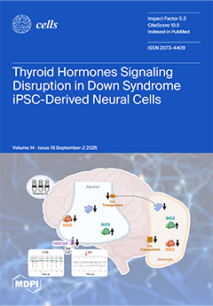
Thyroid hormones (THs) are vital for human brain development, and their dysregulation has been linked to cognitive impairment and neurodevelopmental disorders. Individuals with Down syndrome (DS) frequently exhibit thyroid dysfunction, yet its specific cellular and molecular impacts on neural cells remain underexplored. Here, we used human iPSC-derived NPCs, neurons, and astrocytes to dissect THs signaling across trisomy 21 iPSC-derived neural cells. We identified cell-type-specific alterations in the expression of deiodinases, transporters, receptors, and TH-responsive genes. Functional impairments in glutamatergic signaling were also detected. Our findings highlight THs dysregulation as one of the components of DS neurobiology and suggest novel therapeutic targets for early intervention. View this paper
  • Issues are regarded as officially published after their release is announced to the table of contents alert mailing list.
  • You may sign up for e-mail alerts to receive table of contents of newly released issues.
  • PDF is the official format for papers published in both, html and pdf forms. To view the papers in pdf format, click on the "PDF Full-text" link, and use the free Adobe Reader to open them.
Order results
Result details
Section
Select all
Export citation of selected articles as:
27 pages, 2143 KB  
Review
Targeting the CXCR4/CXCL12 Axis to Overcome Drug Resistance in Triple-Negative Breast Cancer
by Desh Deepak Singh, Dharmendra Kumar Yadav and Dongyun Shin
Cells 2025, 14(18), 1482; https://doi.org/10.3390/cells14181482 - 22 Sep 2025
Viewed by 614
Abstract
Triple-negative breast cancer (TNBC) remains one of the most aggressive and treatment-resistant forms. TNBC is an aggressive and therapeutically resistant subtype of breast cancer, marked by the absence of estrogen, progesterone, and HER2 receptors. The lack of defined molecular targets significantly limits treatment [...] Read more.
Triple-negative breast cancer (TNBC) remains one of the most aggressive and treatment-resistant forms. TNBC is an aggressive and therapeutically resistant subtype of breast cancer, marked by the absence of estrogen, progesterone, and HER2 receptors. The lack of defined molecular targets significantly limits treatment options and contributes to high recurrence rates. Among the key pathways involved in TNBC progression and resistance, the CXCR4/CXCL12 chemokine axis has emerged as a critical player. CXCR4, a G-protein-coupled receptor, binds specifically to its ligand CXCL12, promoting tumour cell proliferation, metastasis, immune evasion, and stromal remodelling. Its overexpression is frequently associated with poor prognosis, disease progression, and resistance to conventional therapies in TNBC. This review explores how the chemokine receptor type 4 (CXCR4/CXCL12) axis facilitates drug resistance through mechanisms such as epithelial–mesenchymal transition (EMT), cancer stemness, and microenvironmental interactions. Notably, CXCR4 antagonists like plerixafor, balixafortide, and POL5551 have shown encouraging preclinical and clinical results, particularly when combined with chemotherapy or immunotherapy. Additionally, innovative strategies, including radiopharmaceuticals, peptide inhibitors, and nanotechnology-based delivery platforms, offer expanded therapeutic avenues. Despite persistent challenges such as tumour heterogeneity and potential toxicity, growing clinical evidence supports the translational relevance of this axis. This manuscript provides an in-depth analysis of CXCR4/CXCL12-mediated drug resistance in TNBC and evaluates current and emerging therapeutic interventions. Full article
(This article belongs to the Special Issue Unlocking the Secrets Behind Drug Resistance at the Cellular Level)
Show Figures

Figure 1

21 pages, 7427 KB  
Article
Effect of Oral Peritumoral Tissue on Infiltration and Differentiation of Tumor-Associated Macrophages in Oral Squamous Cell Carcinoma
by Tianyan Piao, Kiyofumi Takabatake, Takuma Arashima, Yulu Zhao, Hotaka Kawai, Htoo Shwe Eain, Yamin Soe, Zin Zin Min, Keisuke Nakano and Hitoshi Nagatsuka
Cells 2025, 14(18), 1481; https://doi.org/10.3390/cells14181481 - 22 Sep 2025
Viewed by 306
Abstract
The recruitment of tumor-associated macrophages (TAMs) in the tumor microenvironment (TME) of oral squamous carcinoma (OSCC) affects significant cancer invasion; however, in the normal host tissue that is located in the cancer’s surrounding area, this is poorly investigated. In this study, we examined [...] Read more.
The recruitment of tumor-associated macrophages (TAMs) in the tumor microenvironment (TME) of oral squamous carcinoma (OSCC) affects significant cancer invasion; however, in the normal host tissue that is located in the cancer’s surrounding area, this is poorly investigated. In this study, we examined the impact of gingival connective tissue cells (GCTCs) and periodontal ligament cells (PDLCs), which are involved in the invasive pathway of OSCC, on oral cancer invasion via TAMs recruitment. Transwell (migration) assays were used to examine the effects of GCTCs and PDLCs on the migration of macrophages, which indicated that the interaction between GCTCs and HSC-2/HSC-3 (human oral squamous cell carcinoma cell line) promoted the recruitment of macrophages, whereas the interaction between PDLCs was inhibited. An indirect co-culture was then used to examine the effects of GCTCs and PDLCs on the differentiation of macrophages, which indicated that the interaction between GCTCs enhanced their ability to transform into M2-type macrophages. Furthermore, the effects of GCTCs and PDLCs on the recruitment of CD45(+) monocytes, F4/80(+) M0 macrophages, iNOS(+) M1 macrophages, and CD163(+) M2 TAMs were assayed by immunohistochemistry. The results revealed that the interaction between GCTCs and HSC-2/HSC-3 promoted the infiltration of CD45(+) monocytes, F4/80(+) M0 macrophages, and CD163(+) M2 TAMs, whereas the PDLCs inhibited it, while their effect on iNOS(+) M1 macrophages was limited. Collectively, the GCTCs contributed to the infiltration of TAMs into the TME of OSCC cells, whereas the PDLCs exerted an inhibitory effect. These findings suggest a potential regulatory mechanism underlying the progression of OSCC. Full article
Show Figures

Figure 1

48 pages, 7479 KB  
Review
Structural Simplification from Tricyclic to Bicyclic Scaffolds: A Long-Term Investigation in the Field of Adenosine Receptor Antagonists
by Costanza Ceni, Sara Calenda, Giulia Vagnoni, Daniela Catarzi, Flavia Varano and Vittoria Colotta
Cells 2025, 14(18), 1480; https://doi.org/10.3390/cells14181480 - 22 Sep 2025
Viewed by 523
Abstract
Adenosine receptor (AR) antagonists have attracted considerable interest due to their therapeutic potential in a wide range of pathological conditions, including neurological, cardiovascular, and inflammatory disorders. Although a large number of AR antagonists have been developed worldwide, the interest in new derivatives remains [...] Read more.
Adenosine receptor (AR) antagonists have attracted considerable interest due to their therapeutic potential in a wide range of pathological conditions, including neurological, cardiovascular, and inflammatory disorders. Although a large number of AR antagonists have been developed worldwide, the interest in new derivatives remains high, and achieving subtype selectivity continue to be a major challenge. This review summarizes our research on adenosine receptor antagonists, highlighting the discovery of potent and selective compounds for the diverse AR subtypes across various chemical classes. Specifically, the paper focuses on the study of the triazolo[4,3-a]quinoxalin-1-one (TQX) and pyrazolo[3,4-c]quinoline (PQ) series, along with their simplified analogues, which have yielded highly potent and selective AR antagonists. An overview of the structure–activity relationship (SAR) studies and molecular docking investigations is provided, emphasizing the structural requirements for A2A and A3 receptor–ligand interaction. In addition, we present pharmacological studies of selected AR antagonists, in various in vitro and in vivo models of pain, depression, neuroinflammation-related diseases, and cancer. Full article
Show Figures

Graphical abstract

31 pages, 1647 KB  
Review
PCSK9 Regulation of Lipid Metabolism in the Nervous System: Implications for Schwann Cell Function and Peripheral Neuropathy
by Agnieszka Nowacka, Maciej Śniegocki and Ewa A. Ziółkowska
Cells 2025, 14(18), 1479; https://doi.org/10.3390/cells14181479 - 22 Sep 2025
Viewed by 740
Abstract
Neural function relies on tightly regulated lipid metabolism to sustain membrane integrity, synaptic signaling, and energy production. Myelinating glia, particularly Schwann cells, require continuous lipid flux to build and maintain myelin, rendering them vulnerable to imbalances between lipid entry and oxidative capacity. Proprotein [...] Read more.
Neural function relies on tightly regulated lipid metabolism to sustain membrane integrity, synaptic signaling, and energy production. Myelinating glia, particularly Schwann cells, require continuous lipid flux to build and maintain myelin, rendering them vulnerable to imbalances between lipid entry and oxidative capacity. Proprotein convertase subtilisin/kexin type 9 (PCSK9), widely studied in hepatic cholesterol regulation, has emerging roles in the nervous system. In the central nervous system (CNS), local PCSK9 expression influences low-density lipoprotein receptor (LDLR) family abundance, neuronal survival pathways, and neuroinflammatory tone, although circulating PCSK9 has limited parenchymal access due to the blood–brain barrier (BBB). In the peripheral nervous system (PNS), recent evidence highlights a PCSK9–CD36 axis in Schwann cells; genetic Pcsk9 loss elevates CD36, increases fatty-acid influx, promotes lipid droplet expansion and acylcarnitine accumulation, and triggers mitochondrial stress that manifests as hypomyelination, C-fiber pathology, and selective small-fiber neuropathy. These findings suggest that PCSK9 normally restrains CD36-dependent transport to align lipid supply with metabolic demand. Clinically, PCSK9 inhibitors have demonstrated cardiovascular benefit without major neurocognitive signals, yet small-fiber outcomes have not been systematically assessed. This review integrates current evidence on PCSK9 biology across neural compartments, highlights mechanistic links to Schwann cell lipid handling, and outlines research priorities to resolve neural safety and therapeutic potential in lipid-driven neuropathies. Full article
Show Figures

Figure 1

17 pages, 2289 KB  
Article
Ashwagandha Root Extract Mitigates Fibromyalgia-like Symptoms via Neurochemical and Histological Modulation in Mice
by Razan Fawaz Hasanyn, Ashwaq H. Batawi, Mona A. AL-Thepyani, Reham Tash, Asma Almuhammadi, Ashwaq Hassan Alsabban and Badrah S. Alghamdi
Cells 2025, 14(18), 1478; https://doi.org/10.3390/cells14181478 - 22 Sep 2025
Viewed by 630
Abstract
Fibromyalgia syndrome (FMS) is a chronic disorder marked by widespread musculoskeletal pain, fatigue, mood disturbances, and cognitive impairments. Current treatments primarily focus on symptom management. Ashwagandha (Withania somnifera), a traditional Ayurvedic herb, is known for its adaptogenic and neuroprotective properties. This [...] Read more.
Fibromyalgia syndrome (FMS) is a chronic disorder marked by widespread musculoskeletal pain, fatigue, mood disturbances, and cognitive impairments. Current treatments primarily focus on symptom management. Ashwagandha (Withania somnifera), a traditional Ayurvedic herb, is known for its adaptogenic and neuroprotective properties. This study evaluated the protective effects of the methanolic root extract of Ashwagandha (ARE) in a reserpine-induced fibromyalgia model in male Swiss albino mice. Mice received oral ARE (100 mg/kg) for 17 days and reserpine (0.5 mg/kg, subcutaneously) for three consecutive days to induce fibromyalgia-like symptoms. Behavioral assessments included Von Frey, tail suspension, rotarod, and Y-maze tests. Histological analysis was conducted on the hippocampus and thalamus; however, neurochemical analysis focused on markers such as serotonin, norepinephrine, IL-1β, TNFα, MDA, and NO. Results indicated that ARE significantly reduced pain and depressive-like behavior and improved motor function (p < 0.0001); however, no significant changes were observed in open-field locomotion. Histological examination revealed protection of Ashwagandha against neurodegeneration and improved hippocampal integrity, accompanied by increased serotonin and norepinephrine levels and decreased pro-inflammatory cytokines. These findings suggest that Ashwagandha root extract may offer therapeutic benefits for managing fibromyalgia symptoms. Full article
Show Figures

Figure 1

15 pages, 2598 KB  
Article
Use of Amplified Lewy Body Dementia Fibrils and Autoradiography to Characterize Binding of Radioligand Tg-1-90B to Alpha-Synuclein Fibrils in Postmortem Brain Tissue
by Jennifer Y. O’Shea, Dhruva D. Dhavale, Helen Hwang, Zachary Smith, Thomas J. A. Graham, Robert H. Mach and Paul T. Kotzbauer
Cells 2025, 14(18), 1477; https://doi.org/10.3390/cells14181477 - 22 Sep 2025
Viewed by 461
Abstract
Parkinson’s disease (PD) and Lewy Body Dementia (LBD) are defined by accumulation of alpha-synuclein (Asyn) fibrils within Lewy bodies (LBs) and Lewy neurites (LNs). The development of a Positron Emission Tomography (PET) tracer for quantifying Asyn fibrils would improve diagnostic accuracy and provide [...] Read more.
Parkinson’s disease (PD) and Lewy Body Dementia (LBD) are defined by accumulation of alpha-synuclein (Asyn) fibrils within Lewy bodies (LBs) and Lewy neurites (LNs). The development of a Positron Emission Tomography (PET) tracer for quantifying Asyn fibrils would improve diagnostic accuracy and provide a biomarker for disease progression. We previously described radioligand [3H]Tg-1-90B, which binds to in vitro Asyn fibrils (PDB 2N0A) via interactions with residues Y39, S42 and K44. Here, we performed molecular docking studies with Tg1-90B and PD/LBD Asyn fibrils (PDB 8A9L), which predicts interactions with residues Y39 and K43 in a structurally distinct binding site. In radioligand binding assays, Tg-1-90B has moderate to high affinity (Kd 17.5 nM) for amplified LBD fibrils (PDB 8FPT), whose protofilament fold is highly similar to PD/LBD fibrils (PBD 8A9L). Autoradiography confirmed binding of [3H]Tg-1-90B to LBs in PD brain tissue. However, Tg-1-90B also binds to amyloid-beta fibrils in Alzheimer’s disease (AD) tissue, indicating insufficient selectivity for Asyn fibrils. These results indicate that Tg-1-90B binds to Asyn fibrils in PD tissue but needs further structural optimization. Binding assays with amplified LBD fibrils and autoradiography with postmortem PD tissue can guide further development of Asyn fibril PET ligands for PD/LBD. Full article
(This article belongs to the Section Cellular Aging)
Show Figures

Figure 1

17 pages, 1030 KB  
Review
Next-Generation mRNA Vaccines in Melanoma: Advances in Delivery and Combination Strategies
by Stefano Zoroddu and Luigi Bagella
Cells 2025, 14(18), 1476; https://doi.org/10.3390/cells14181476 - 22 Sep 2025
Viewed by 1119
Abstract
Messenger RNA (mRNA) vaccines have redefined cancer immunotherapy, offering unparalleled flexibility to encode tumor-specific antigens and to be adapted to individual mutational landscapes. Melanoma, with its high mutational burden and responsiveness to immune checkpoint blockade, has become the leading model for translating these [...] Read more.
Messenger RNA (mRNA) vaccines have redefined cancer immunotherapy, offering unparalleled flexibility to encode tumor-specific antigens and to be adapted to individual mutational landscapes. Melanoma, with its high mutational burden and responsiveness to immune checkpoint blockade, has become the leading model for translating these advances into clinical benefit. Recent innovations in delivery—ranging from lipid nanoparticles and polymeric carriers to biomimetic hybrids and intratumoral administration—are dismantling long-standing barriers of stability, targeting, and immunogenicity. Clinical milestones, including the randomized phase IIb KEYNOTE-942, show that adding the personalized neoantigen vaccine mRNA-4157 (V940) to pembrolizumab prolonged recurrence-free survival versus pembrolizumab alone (HR 0.561, 95% CI 0.309–1.017; 18-month RFS 79% vs. 62%), with the ASCO 3-year update reporting 2.5-year RFS 74.8% vs. 55.6% and sustained distant metastasis-free survival benefit in resected high-risk melanoma. Parallel preclinical studies highlight the potential of multifunctional platforms co-delivering cytokines or innate agonists to reshape the tumor microenvironment and achieve durable systemic immunity. As artificial intelligence drives epitope selection and modular manufacturing accelerates personalization, mRNA vaccines may have the potential to transition from adjuncts to main therapies in melanoma and beyond. Full article
Show Figures

Figure 1

22 pages, 4275 KB  
Article
Lumpy Skin Disease Virus ORF137 Protein Inhibits Type I Interferon Production by Interacting with and Decreasing the Phosphorylation of IRF3
by Qunhua Ke, Kaishen Yao, Min Qu, Zhengji Liang, Miaomiao Li, Xiangwei Wang, Xiangping Yin and Yuefeng Sun
Cells 2025, 14(18), 1475; https://doi.org/10.3390/cells14181475 - 22 Sep 2025
Viewed by 489
Abstract
Lumpy skin disease (LSD) is an invasive infectious disease caused by the lumpy skin disease virus (LSDV), which is detrimental to the production of cattle. LSDV encodes about 156 proteins, most of whose functions are still unknown. In this study, we found that [...] Read more.
Lumpy skin disease (LSD) is an invasive infectious disease caused by the lumpy skin disease virus (LSDV), which is detrimental to the production of cattle. LSDV encodes about 156 proteins, most of whose functions are still unknown. In this study, we found that the ORF137 protein was identified as one of the strongest inhibitors of IFN-β and ISG expression, determining LSDV ORF137 as a negative regulator of interferon (IFN) β signaling. Further evidence suggests that ORF137 interacts with the signal transduction factor IRF3 and inhibits the activation of IFN-β signaling by reducing Phospho-IRF3 (p-IRF3). Further investigation indicated that overexpression of ORF137 in BMEC could significantly inhibit the transcription of IFN-β and ISGs, thereby promoting the replication of LSDV. More importantly, through homologous recombination, we deleted the ORF137 gene from the LSDV/FJ/CHA/2021 strain and constructed the recombinant strain LSDV-ΔORF137-EGFP. Compared with the parental strain, LSDV-ΔORF137-EGFP showed a weakened effect on inhibiting the transcription of IFN-β and ISGs and a reduced replication level in infected MDBK cells. In summary, ORF137 facilitates LSDV replication by targeting IRF3 to inhibit IFN-β signaling. Our findings reveal a new mechanism by which LSDV suppresses the host antiviral response, which may facilitate the development of attenuated live vaccines for LSDV. Full article
Show Figures

Figure 1

19 pages, 3243 KB  
Article
PF-04691502, a PI3K/mTOR Dual Inhibitor, Ameliorates AD-like Pathology in a Mouse Model of AD
by Marika Lanza, Rossella Basilotta, Antonella Caccamo, Giovanna Casili, Alberto Repici, Salvatore Oddo and Emanuela Esposito
Cells 2025, 14(18), 1474; https://doi.org/10.3390/cells14181474 - 21 Sep 2025
Viewed by 563
Abstract
Alzheimer’s disease (AD) is a neurodegenerative disorder that significantly impacts the lives of patients and their families. The pathological features of AD include the accumulation of amyloid-β (Aβ) and Tau, which disrupt neuronal function and communication, ultimately leading to neuronal loss and brain [...] Read more.
Alzheimer’s disease (AD) is a neurodegenerative disorder that significantly impacts the lives of patients and their families. The pathological features of AD include the accumulation of amyloid-β (Aβ) and Tau, which disrupt neuronal function and communication, ultimately leading to neuronal loss and brain atrophy. Efforts to understand the molecular mechanisms underlying these pathological changes have led to advancements in diagnostic techniques and potential therapeutic interventions. However, the complexity of AD necessitates further research to develop more effective treatments and, ideally, preventive measures. Extensive research suggests that diminishing mTOR signaling increases lifespan and health span across various species. Increased PI3K/mTOR signaling has been linked to the progression of AD pathology, leading to neuronal degeneration and impairments in cognitive function. In this study, we explored the therapeutic potential of PF-04691502, a dual PI3K/mTOR inhibitor, in Alzheimer’s disease (AD)-like pathology using male and female B6.Cg-Tg(APPswe, PSEN1dE9)85Dbo/Mmjax mice (APP/PS1), a well-established transgenic model of AD. Eighteen-month-old APP/PS1 and wild-type mice received oral administration of PF-04691502 at a dose of 1 mg/kg for 12 weeks. Following the treatment period, spatial learning and memory were evaluated using the Morris water maze. Subsequently, the mice brains were collected for neuropathological and biochemical assessments. Our findings showed that PF-04691502 enhanced cognitive performance in APP/PS1 mice and significantly reduced insoluble Aβ accumulation in the brain. Mechanistically, these effects were associated with enhanced autophagy induction. Treatment with PF-04691502 increased the LC3-II/LC3-I ratio, upregulated Beclin-1, and elevated LAMP-2 levels, indicative of stimulated autophagosome formation and lysosomal activity. Overall, these preclinical results suggest that PF-04691502 holds promise as a potential therapeutic agent for AD and other aging-related neurodegenerative diseases involving mTOR pathway dysregulation. Full article
(This article belongs to the Special Issue Ageing and Neurodegenerative Diseases, Second Edition)
Show Figures

Graphical abstract

24 pages, 14557 KB  
Article
Pericyte Expression of VEGF-A Minimally Impacts Ocular Vascular Development and Neovascularization
by Yong-Seok Song, Shoujian Wang, Samay Inampudi, Hope Risa, Christine M. Sorenson and Nader Sheibani
Cells 2025, 14(18), 1473; https://doi.org/10.3390/cells14181473 - 21 Sep 2025
Viewed by 478
Abstract
Pericytes produce vascular endothelial growth factor-A (VEGF-A; hereafter referred to as VEGF). VEGF inhibits pericyte proliferation and migration through enhanced VEGFR2 and PDGFRβ heterodimerization. Heterodimerization of these receptors on perivascular supporting cells, mediated by VEGF in culture, mitigates signaling through these receptors and [...] Read more.
Pericytes produce vascular endothelial growth factor-A (VEGF-A; hereafter referred to as VEGF). VEGF inhibits pericyte proliferation and migration through enhanced VEGFR2 and PDGFRβ heterodimerization. Heterodimerization of these receptors on perivascular supporting cells, mediated by VEGF in culture, mitigates signaling through these receptors and promotes a quiescent phenotype. However, the detailed cellular mechanisms and the significance of these interactions in vivo require further investigation. The cell-autonomous activities of pericyte VEGF expression during vascular development and neovascularization remain unknown. Here we utilized mice conditionally lacking Vegfa in pericytes (VegfaPC) to examine its impact on retinal vascular development and pathological ocular neovascularization. Vascular integrity was also assessed in older mice using fundus imaging and fluorescein angiography. The lack of Vegfa pericyte expression delayed the initial spreading of the superficial layer of the retinal vasculature. Mice lacking Vegfa pericyte expression had similar numbers of retinal endothelial cells and arteries to their wild-type littermates. However, the number of pericytes was significantly reduced in younger VegfaPC mice but increased in more mature mice. In addition, pericyte Vegfa deficiency did not impact responses during oxygen-induced ischemic retinopathy and laser-induced choroidal neovascularization. Thus, pericyte VEGF expression plays a role during early stages of retinal vascular development with limited influence on mature retinal vascularization, its integrity, and neovascularization. Full article
Show Figures

Figure 1

23 pages, 2771 KB  
Article
RNA Sequencing Identified Differentially Expressed Genes in the Mesocorticolimbic and Nigrostriatal Systems of Compulsive METH-Taking Rats
by Nasser Adjei, Bruce Ladenheim, Michael T. McCoy, Vikrant Palande, Jean Lud Cadet and Atul P. Daiwile
Cells 2025, 14(18), 1472; https://doi.org/10.3390/cells14181472 - 20 Sep 2025
Viewed by 426
Abstract
Methamphetamine (METH) is an extremely addictive drug which continues to cause significant harm to individuals and communities. In the present study we trained male rats to self-administer METH for 20 days, followed by 9 days of foot shock exposure. All rats escalated their [...] Read more.
Methamphetamine (METH) is an extremely addictive drug which continues to cause significant harm to individuals and communities. In the present study we trained male rats to self-administer METH for 20 days, followed by 9 days of foot shock exposure. All rats escalated their METH intake during the first 20 days. The rats that continued to self-administer METH in the presence of aversive stimuli were termed shock-resistant (SR), while those that reduced their intake were shock-sensitive (SS). RNA sequencing showed numerous differentially expressed genes (DEGs) in the prefrontal cortex, nucleus accumbens, dorsal striatum, and midbrain. Ingenuity pathway analysis linked DEGs to addiction-related mechanisms. We identified shared genes with similar expression patterns across four brain regions (SR: Fos and Ahsp; SS: Tet1, Cym, and Tmem30c). The identified genes play key roles in addiction-related brain functions, such as neuronal activity, stress response, and epigenetic regulation, and their importance in METH addiction is highlighted. These genes represent promising targets for developing new treatments aimed at reversing neuroadaptations caused by METH use. Full article
Show Figures

Figure 1

26 pages, 1238 KB  
Review
Temporal Dynamics of Extracellular Matrix Remodeling in Anthracycline-Induced Cardiotoxicity
by Fibi Meshrkey, Somaya Y. Ibrahim, Rushita A. Bagchi and William J. Richardson
Cells 2025, 14(18), 1471; https://doi.org/10.3390/cells14181471 - 20 Sep 2025
Viewed by 563
Abstract
Anthracyclines are widely used chemotherapeutic agents with proven efficacy against a broad range of malignancies, but their clinical utility is limited by a well-documented, dose-dependent cardiotoxicity. While this toxicity has traditionally been attributed to direct cardiomyocyte injury, emerging evidence highlights the pivotal role [...] Read more.
Anthracyclines are widely used chemotherapeutic agents with proven efficacy against a broad range of malignancies, but their clinical utility is limited by a well-documented, dose-dependent cardiotoxicity. While this toxicity has traditionally been attributed to direct cardiomyocyte injury, emerging evidence highlights the pivotal role of cardiac fibroblasts (CFs) in the development and progression of anthracycline-induced cardiotoxicity. This review examines the diverse effects of anthracycline focusing on doxorubicin (DOX) and CFs across the temporal phases of cardiac injury. DOX activates fibroblast-driven extracellular matrix remodeling and promotes fibrosis through enhanced collagen production and the induction of cellular senescence, thereby exacerbating early myocardial inflammation and dysfunction. Clinically, anthracycline cardiotoxicity may present as acute (within days), subacute (within weeks), or chronic progressive forms manifesting either early (within one year) or late (up to decades post-treatment). While early manifestations may be reversible with timely detection and management, late-phase cardiotoxicity is often irreversible, characterized by declining left ventricular ejection fraction and heart failure. A deeper understanding of the molecular and cellular contributions of CFs may uncover novel therapeutic targets to prevent or attenuate anthracycline-related cardiac damage. Full article
Show Figures

Figure 1

23 pages, 7000 KB  
Article
Frog Skin Antimicrobial Peptide 3-13 and Its Analogs Alleviate Atherosclerosis Cholesterol Accumulation in Foam Cells via PPARγ Signaling Pathway
by Xue-Feng Yang, Zi-Meng Hao, Xin-Yu Cui, Wan-Qi Liu, Meng-Miao Li and De-Jing Shang
Cells 2025, 14(18), 1470; https://doi.org/10.3390/cells14181470 - 19 Sep 2025
Viewed by 650
Abstract
Atherosclerosis (AS), a major contributor to cardiovascular disease, hypertension, and stroke, is associated with significant morbidity and mortality. Antimicrobial peptides (AMPs) 3-13, W3R6, and chensinin-1b were engineered based on the sequence of chensinin-1, originally isolated from the skin secretion of Rana chensinensis. [...] Read more.
Atherosclerosis (AS), a major contributor to cardiovascular disease, hypertension, and stroke, is associated with significant morbidity and mortality. Antimicrobial peptides (AMPs) 3-13, W3R6, and chensinin-1b were engineered based on the sequence of chensinin-1, originally isolated from the skin secretion of Rana chensinensis. This study investigated their therapeutic potential in ApoE-/- AS mice and THP-1-derived foam cells, focusing on the regulation of cholesterol metabolism. AMP 3-13 markedly reduced body weight gain, aortic root plaque formation, and plasma cholesterol levels in ApoE-/- mice. Transcriptomic analysis revealed that AMP 3-13 significantly altered gene expression related to cholesterol metabolism and the PPAR signaling pathway. Specifically, AMP 3-13 upregulated PPARγ, ABCA1, and ABCG1, while downregulating CD36 in aortic root plaques. In THP-1-derived foam cells, AMP 3-13 and its analogs activated the PPARγ–ABCA1/ABCG1 axis, enhancing cholesterol efflux. Concurrently, they inhibited CD36 expression by competing with PPARγ for promoter binding, thereby reducing ox-LDL uptake. These findings suggested that AMP 3-13 and its analogs represented promising therapeutic agents for AS through their ability to reduce cholesterol accumulation in foam cell. Full article
Show Figures

Figure 1

23 pages, 5765 KB  
Article
Orlistat Confers Neuroprotection in Traumatic Brain Injury by Modulating Microglial Lipid Metabolism
by Chenxuan Yu, Yu Ni, Yuxuan Xiong, Huayu Kang, Zhengqiao Jiang, Yuan Liu, Xincheng Zhang, Yanchao Liu, Kai Zhao, Sheng Wang, Chao Gan and Huaqiu Zhang
Cells 2025, 14(18), 1469; https://doi.org/10.3390/cells14181469 - 19 Sep 2025
Viewed by 435
Abstract
Traumatic brain injury (TBI) represents a major cause of mortality and disability worldwide, particularly affecting young adults and elderly populations. This study investigates the neuroprotective potential of orlistat (ORL), a gastrointestinal lipase inhibitor, in a murine TBI model. Behavioral, histological, and molecular analyses [...] Read more.
Traumatic brain injury (TBI) represents a major cause of mortality and disability worldwide, particularly affecting young adults and elderly populations. This study investigates the neuroprotective potential of orlistat (ORL), a gastrointestinal lipase inhibitor, in a murine TBI model. Behavioral, histological, and molecular analyses demonstrated that ORL significantly attenuated TBI-induced neurological damage. Microglial depletion experiments revealed that ORL’s neuroprotective effects were largely mediated through microglial modulation. In vitro and in vivo studies showed that ORL suppressed microglial activation, phagocytosis, and migration. Single-cell RNA sequencing identified upregulation of lipoprotein lipase (LPL) in a TBI-induced microglial subpopulation. Molecular docking predicted ORL-LPL binding, suggesting direct enzymatic inhibition. Transcriptomic and metabolomic analyses further revealed ORL’s modulation of microglial metabolic pathways and inflammatory responses. Our findings position ORL as a promising repurposed therapeutic for TBI through its novel mechanism of targeting microglial LPL-mediated neuroinflammation. Full article
Show Figures

Figure 1

11 pages, 1321 KB  
Article
Beyond Spherical: Unveiling the Significance of Oval Blastocyst Morphology on Euploidy and Implantation Success
by Jakub Wyroba, Agnieszka Kuczyńska, Klaudia Kasperkowicz, Katarzyna Kostarczyk, Pawel Kordowitzki and Joanna Kochan
Cells 2025, 14(18), 1468; https://doi.org/10.3390/cells14181468 - 19 Sep 2025
Viewed by 373
Abstract
The selection of the most suitable embryo, based on the morphology and shape, for embryo transfer is a critical aspect of the in vitro fertilization (IVF) process, as its precision can significantly enhance the overall effectiveness of IVF and contribute to a healthy [...] Read more.
The selection of the most suitable embryo, based on the morphology and shape, for embryo transfer is a critical aspect of the in vitro fertilization (IVF) process, as its precision can significantly enhance the overall effectiveness of IVF and contribute to a healthy birth. This study aimed to compare the chromosomal status and implantation potential of oval-shaped blastocysts versus normal-shaped blastocysts on day 5 post-ICSI (intracytoplasmic sperm injection). Initially, the frequency of oval blastocysts was assessed by analyzing 1328 embryos from 610 ICSI cycles. Subsequently, 80 patients undergoing ICSI and PGT-A (preimplantation genetic testing for aneuploidy), who had both normal and oval blastocysts in the same cycle, were selected to evaluate the euploid rate relative to blastocyst morphology. Finally, the implantation outcomes of fresh embryo transfers involving oval and normal-shaped blastocysts, neither of which had undergone PGT-A, were analyzed. Half of the blastocysts from each group were transferred after assisted hatching (AH), and the other half were transferred without AH. Blastocyst shape does not appear to correlate with an increased risk of aneuploidy but does influence hatching ability. Following AH, the implantation potential of elongated blastocysts is equivalent to that of normally shaped blastocysts, suggesting AH is beneficial for oval embryos. Consequently, the transfer of oval blastocysts is considered as safe and effective as the transfer of normally shaped embryos. Full article
(This article belongs to the Section Tissues and Organs)
Show Figures

Figure 1

18 pages, 1134 KB  
Review
Role of Nucleotide P2 Receptors in the Immune System: Focus on Effector T Cells
by Romuald Brice Babou Kammoe, Chakib Hamoudi and Fawzi Aoudjit
Cells 2025, 14(18), 1467; https://doi.org/10.3390/cells14181467 - 19 Sep 2025
Viewed by 444
Abstract
The emergence of purinergic signaling has given ATP and other extracellular nucleotides a whole new perspective. This concept refers to the fact that extracellular nucleotides released by most cells act as signaling molecules via specific membrane receptors called purinergic receptors, thus regulating several [...] Read more.
The emergence of purinergic signaling has given ATP and other extracellular nucleotides a whole new perspective. This concept refers to the fact that extracellular nucleotides released by most cells act as signaling molecules via specific membrane receptors called purinergic receptors, thus regulating several cellular functions. Although purinergic signaling has been well studied in several physiological systems, recent work has shown that this signaling pathway is also essential in the immune system. In this review, we will discuss the complex role of purinergic signaling in the regulation of effector T cell functions, including migration, differentiation, and activation. We will also cover the role of P2 receptors in the development of autoimmune diseases. Understanding how P2 receptors regulate effector T cells will likely further our understanding of protective immunity and immune pathogenesis and may lead to new therapeutic approaches and agents in immune diseases Full article
(This article belongs to the Section Cellular Immunology)
Show Figures

Figure 1

28 pages, 1037 KB  
Review
Wings of Discovery: Using Drosophila to Decode Hereditary Spastic Paraplegia and Ataxias
by Rachele Vivarelli, Chiara Vantaggiato, Maria Teresa Bassi, Filippo Maria Santorelli and Maria Marchese
Cells 2025, 14(18), 1466; https://doi.org/10.3390/cells14181466 - 19 Sep 2025
Viewed by 355
Abstract
Hereditary spastic paraplegia (HSP) and hereditary ataxias (HA) are clinically and genetically heterogeneous neurodegenerative disorders that primarily affect motor coordination and neural integrity. Despite distinct pathological features, such as pyramidal tract degeneration in HSP and spinocerebellar pathway involvement in HA, these conditions share [...] Read more.
Hereditary spastic paraplegia (HSP) and hereditary ataxias (HA) are clinically and genetically heterogeneous neurodegenerative disorders that primarily affect motor coordination and neural integrity. Despite distinct pathological features, such as pyramidal tract degeneration in HSP and spinocerebellar pathway involvement in HA, these conditions share overlapping genetic pathways and mechanisms. The fruit fly Drosophila melanogaster has emerged as a powerful model organism for investigating the molecular basis of rare diseases, including HSP and HA. Its genetic tractability, rapid life cycle, and high degree of gene conservation with humans make it a cost-effective and ethically viable platform for disease modelling. In this review, we provide a comprehensive overview of Drosophila-based models for HSP and HA. We highlight the use of advanced genetic tools, including RNA interference, CRISPR/Cas9, and the GAL4/UAS system, as well as behavioral and neuroanatomical assays to model disease features. Furthermore, we discuss the application of genetic “avatars” and high-throughput drug screening platforms to test therapeutic candidates. Collectively, these models have deepened our understanding of the pathophysiology of HSP and HA, offering valuable insights for the development of targeted therapies and approaches to personalized medicine. Full article
Show Figures

Figure 1

22 pages, 6009 KB  
Article
Dicer1 Depletion Leads to DNA Damage Accumulation and Cell Death in a RET/PTC3 Papillary Thyroid Cancer Mouse Model, Thereby Inhibiting Tumor Progression
by Maria Rojo-Pardillo, Alice Augenlicht, Geneviève Dom, Jukka Kero, Bernard Robaye and Carine Maenhaut
Cells 2025, 14(18), 1465; https://doi.org/10.3390/cells14181465 - 19 Sep 2025
Viewed by 504
Abstract
Beyond well-known genetic drivers, microRNA dysregulation has emerged as a key contributor to thyroid tumorigenesis. Central to this process is Dicer1, a ribonuclease essential for microRNA maturation, whose expression is often reduced in papillary thyroid carcinoma (PTC). Evidence from previous studies suggest [...] Read more.
Beyond well-known genetic drivers, microRNA dysregulation has emerged as a key contributor to thyroid tumorigenesis. Central to this process is Dicer1, a ribonuclease essential for microRNA maturation, whose expression is often reduced in papillary thyroid carcinoma (PTC). Evidence from previous studies suggest Dicer1 functions as a context-dependent haplo-insufficient tumor suppressor gene: partial loss may promote tumor development, whereas complete loss may disrupt essential cellular functions, causing cell death and tumor suppression. However, the effects of partial or complete Dicer1 loss in thyroid cancer remain unclear. To explore this, we genetically inactivated one (heterozygous) or both (homozygous) Dicer1 alleles specifically in thyroid follicular cells of a RET/PTC3 transgenic mouse model using an inducible Cre-Lox system. Our findings deepen the current understanding of the RET/PTC3-driven PTC model by revealing an increased number of vimentin-positive cells and disruption in redox homeostasis. Additionally, whereas heterozygous Dicer1 loss did not alter tumor progression in RET/PTC3 mice, total loss reduced tumor growth and led to accumulated DNA damage and cell death. These findings highlight the crucial role of Dicer1 dosage in thyroid cancer progression and underscore its potential as a therapeutic target for aggressive PTC and other malignancies characterized by aberrant Dicer1 expression. Full article
(This article belongs to the Special Issue Molecular Mechanisms of Tumor Pathogenesis)
Show Figures

Figure 1

18 pages, 4346 KB  
Review
Patient-Derived Tumor Organoids to Model Cancer Cell Plasticity and Overcome Therapeutic Resistance
by Roberto Coppo and Masahiro Inoue
Cells 2025, 14(18), 1464; https://doi.org/10.3390/cells14181464 - 18 Sep 2025
Viewed by 556
Abstract
Cancer cell plasticity, defined as the ability of tumor cells to reversibly adopt distinct functional states, plays a central role in tumor heterogeneity, therapy resistance, and disease relapse. This process enables cells to enter stem-like, dormant, or drug-tolerant persister states in response to [...] Read more.
Cancer cell plasticity, defined as the ability of tumor cells to reversibly adopt distinct functional states, plays a central role in tumor heterogeneity, therapy resistance, and disease relapse. This process enables cells to enter stem-like, dormant, or drug-tolerant persister states in response to treatment or environmental stress without undergoing genetic changes. Such reversible transitions complicate and limit current treatments. Conventional cancer models often fail to capture the complexities of these adaptive states. In contrast, patient-derived tumor organoids (PDOs), which retain the cellular diversity and structure of primary tumors, provide a unique system for investigating plasticity. This review describes how PDOs can model cellular plasticity, such as the emergence of drug-tolerant persister cells and the interconversion between cancer stem cell states across multiple tumor types. We particularly focused on colorectal cancer organoids, for which research on the mechanism of plasticity is the most advanced. Combined with single-cell analysis, lineage tracing, and functional assays, PDOs can help identify the molecular pathways that control plasticity. Understanding these mechanisms is important for developing therapies to prevent treatment failure and control disease progression. Full article
Show Figures

Figure 1

12 pages, 1863 KB  
Protocol
Expansion Microscopy of the Enteric Nervous System: A Feasibility Study
by Xin Xu, Wenchuan Zhang and Menachem Hanani
Cells 2025, 14(18), 1463; https://doi.org/10.3390/cells14181463 - 18 Sep 2025
Viewed by 482
Abstract
Expansion microscopy (ExM) enables conventional light microscopes to achieve nanoscale resolution by physically enlarging biological specimens. While ExM has been widely applied in neurobiology, it has not been adapted for the enteric nervous system (ENS). Here, we provide a detailed and reproducible protocol [...] Read more.
Expansion microscopy (ExM) enables conventional light microscopes to achieve nanoscale resolution by physically enlarging biological specimens. While ExM has been widely applied in neurobiology, it has not been adapted for the enteric nervous system (ENS). Here, we provide a detailed and reproducible protocol for applying ExM to mouse colonic ENS tissue. The procedure includes preparation of the external muscle layers with the myenteric plexus, histochemical staining for NADPH-diaphorase, immunostaining for glial fibrillary acidic protein (GFAP), anchoring of biomolecules, gelation, proteinase K digestion, and isotropic expansion in a swellable polymer matrix. Step-by-step instructions, required reagents, and critical parameters are described to ensure robustness and reproducibility. Using this protocol, tissues expand 3–5-fold, allowing neuronal somata, fibers, and glial cell processes to be clearly visualized by standard brightfield or fluorescence microscopy. The tissue architecture is preserved, with distortion in the X–Y plane of about 7%. This protocol provides a reliable framework for high-resolution structural analysis of the ENS and can be readily adapted to other peripheral tissues. Full article
(This article belongs to the Section Cell Methods)
Show Figures

Graphical abstract

14 pages, 2576 KB  
Protocol
A Backwards Approach to GD2 Immunofluorescence in Human Neuroblastoma Tissue Samples: From Staining to Slicing
by Sara Peggion, Clara Volz, Magdalena Trochimiuk, Isabelle Ariane Bley, Júlia Ramos, Konrad Reinshagen and Laia Pagerols Raluy
Cells 2025, 14(18), 1462; https://doi.org/10.3390/cells14181462 - 18 Sep 2025
Viewed by 460
Abstract
Background: The disialoganglioside GD2, located at the plasma membrane, is selectively overexpressed in various solid tumors, where it contributes to tumor growth and the development of an aggressive tumor phenotype. Thus, over the last two decades GD2 has been gaining importance both as [...] Read more.
Background: The disialoganglioside GD2, located at the plasma membrane, is selectively overexpressed in various solid tumors, where it contributes to tumor growth and the development of an aggressive tumor phenotype. Thus, over the last two decades GD2 has been gaining importance both as a tumor marker and a therapy target. In neuroblastoma, anti-GD2 monoclonal antibodies and CAR T-cells have become an integral part of the multimodal treatment for relapsed or refractory high-risk cases, which continue to associate with poor prognosis. GD2 characterization in neuroblastoma is well established for bone marrow aspirates and biopsies, but remains challenging in tumoral tissue samples, mostly due to epitope loss upon fixation. Aims: The aim of our work was to assess a new protocol by staining GD2 in tissue specimens prior to fixation. Methods: Positive controls were tissue specimens from patients with histologically confirmed neuroblastoma and GD2 expression in bone marrow aspirate (n = 5). Nephroblastoma or Hodgkin lymphoma samples were considered as negative controls (n = 5). Tissue staining was performed prior to fixation with either anti-GD2 antibody or isotype control, followed by secondary antibody staining and subsequent paraffinization. To examine GD2 staining before and after paraffinization, fluorescence images were acquired using 3D and 2D immunofluorescence microscopy techniques respectively. Results: GD2 signal was detected in all positive controls, while absent in all negative controls. After fixation, paraffinization and slicing no relevant signal loss was observed. Nevertheless, sufficient staining of 3D specimens required long incubation times, which led to increased cytolysis of the unfixed tissue. Conclusions: We were able to establish and validate a novel protocol to reliably perform immunostaining of the membrane antigen GD2 in unfixed, primary neuroblastoma tissue. Although including few limitations, this staining workflow enables relatively quick assessment of GD2 status and thus, might represent a relevant diagnostic tool within the framework of tumor staging and precision medicine. Full article
(This article belongs to the Special Issue Imaging Methods in Cell Biology)
Show Figures

Graphical abstract

38 pages, 1281 KB  
Review
NUP214 in Acute Myeloid Leukemia
by Øystein Bruserud and Håkon Reikvam
Cells 2025, 14(18), 1461; https://doi.org/10.3390/cells14181461 - 18 Sep 2025
Viewed by 587
Abstract
Nucleoporin 214 (NUP214) is a component of the nucleopore molecular complex, but in addition to this role in nucleocytoplasmic transport it is also involved in the regulation of gene transcription/translation, intracellular signaling, cell cycle progression and programmed cell death. Several uncommon translocations associated [...] Read more.
Nucleoporin 214 (NUP214) is a component of the nucleopore molecular complex, but in addition to this role in nucleocytoplasmic transport it is also involved in the regulation of gene transcription/translation, intracellular signaling, cell cycle progression and programmed cell death. Several uncommon translocations associated with acute myeloid leukemia (AML) involve the NUP214 gene, and the corresponding fusion proteins are involved in leukemic transformation. First, the t(6;9) translocation encodes the DEK-NUP214 fusion protein; this translocation is seen in 1–2% of AML patients and is associated with an adverse prognosis that is improved by allogeneic stem cell transplantation. Second, the SET-NUP214 fusion gene is less common in AML and is formed either by del(9)(q34.11q34.13) or a balanced t(9;9)(q34;q34). This AML variant shows several biological similarities with the DEK-NUP214 variant, but the possible prognostic impact of this fusion protein is not known. Finally, the NUP214-ABL1 and especially the NUP214-SQSTM1 fusions are very uncommon, and only a few case reports have been published. In this article, we review the functions of the genes/proteins formed by these fusion genes, the available studies of molecular mechanisms and biological functions for each fusion protein, the characteristics of the corresponding AML cells, the clinical characteristics of these patients and the possible prognostic impact of the fusion genes/proteins. Full article
(This article belongs to the Special Issue Molecular Mechanisms of Leukemias)
Show Figures

Figure 1

16 pages, 4412 KB  
Article
ODAD4-Related Primary Ciliary Dyskinesia: Report of Five Cases and a Founder Variant in Quebec
by Marie-Hélène Bourassa, Guillaume Sillon, Shuizi Ding, Maurizio Chioccioli, Monkol Lek, Kaiyue Ma, Alejandro Mejia-Garcia, Simon Gravel, Donald C. Vinh, Michael R. Knowles, Margaret W. Leigh, Stephanie D. Davis, Thomas Ferkol, Kenneth N. Olivier, Elizabeth N. Schecterman, Weining Yin, Patrick R. Sears, Martina Gentzsch, Susan E. Boyles, William D. Bennett, Kirby L. Zeman, Lawrence E. Ostrowski, Maimoona A. Zariwala and Adam J. Shapiroadd Show full author list remove Hide full author list
Cells 2025, 14(18), 1460; https://doi.org/10.3390/cells14181460 - 18 Sep 2025
Viewed by 626
Abstract
Pathogenic variants in ODAD4 are an ultra-rare cause of primary ciliary dyskinesia (PCD). Previously reported cases display classic disease phenotypes, including chronic oto-sino-pulmonary disease and development of bronchiectasis by adulthood. We report five individuals with PCD harboring biallelic ODAD4 variants (median age 14, [...] Read more.
Pathogenic variants in ODAD4 are an ultra-rare cause of primary ciliary dyskinesia (PCD). Previously reported cases display classic disease phenotypes, including chronic oto-sino-pulmonary disease and development of bronchiectasis by adulthood. We report five individuals with PCD harboring biallelic ODAD4 variants (median age 14, range 3–41 years). Participants underwent standardized PCD diagnostic evaluations. Three individuals shared the novel homozygous ODAD4 genotype [NM_031421.5: c.245delA, p.(Lys82Argfs*29)], and genealogy analysis highly suggests a founder effect in French-Canadians from two regions of Quebec. All five participants had normal pulmonary function values. Two Quebec participants lacked radiographic pneumonias or bronchiectasis (ages 14 and 38 years) despite life-long suppurative respiratory symptoms, low nasal nitric oxide levels, and outer dynein arm defects on electron microscopy. Reverse transcription polymerase chain reaction of the c.245delA variant showed abnormal splicing with in-frame skipping of exon 2, allowing expression of a mildly shortened mRNA product. However, functional analysis showed overall static cilia, absence of ODAD4 protein on Western blot, and absence of in vivo mucociliary clearance. The reason for a milder pulmonary phenotype with the c.245delA variant in ODAD4 remains unclear, but regional screening for this variant in Quebec may identify more cases and enhance understanding of this mild form of PCD. Full article
(This article belongs to the Special Issue The Role of Cilia in Health and Diseases—2nd Edition)
Show Figures

Figure 1

10 pages, 1444 KB  
Communication
Spike Timing-Dependent Plasticity at Layer 2/3 Horizontal Connections Between Neighboring Columns During Synapse Formation Before the Critical Period in the Developing Barrel Cortex
by Chiaki Itami and Fumitaka Kimura
Cells 2025, 14(18), 1459; https://doi.org/10.3390/cells14181459 - 18 Sep 2025
Viewed by 393
Abstract
The Hebbian type of spike timing-dependent plasticity (STDP) with long-term potentiation and depression (LTP and LTD) plays a crucial role at layer 4 (L4) to L2/3 synapses in deprivation-induced map plasticity. In addition, plasticity at the L2/3 horizontal connection is suggested to play [...] Read more.
The Hebbian type of spike timing-dependent plasticity (STDP) with long-term potentiation and depression (LTP and LTD) plays a crucial role at layer 4 (L4) to L2/3 synapses in deprivation-induced map plasticity. In addition, plasticity at the L2/3 horizontal connection is suggested to play an additional role in map plasticity, especially for “spared whisker response potentiation.” Unimodal STDP with only LTP, or all-LTP STDP drives circuit formation at thalamocortical, as well as L4-L2/3 synapse before the critical period. Here, we first show that the L2/3 horizontal connections exhibit all-LTP STDP when axons are extending during synapse formation before the critical period. LTP-STDP induced by pre-post timing was mediated by NMDA-R because APV blocked the induction. In addition, PKA signaling was involved because PKI 6-22 blocked the induction. However, LTP-STDP induced by post-pre timing was not mediated by NMDA-R, because APV could not block its induction. Nevertheless, PKA signaling was also involved in its induction because PKI 6-22 blocked the induction. Our finding indicates that PKA signaling plays an important role in all-LTP STDP during synaptic formation at the L2/3-L2/3 connection between neighboring columns with a distinct source of Ca2+ influx in the developing mouse barrel cortex. Full article
(This article belongs to the Section Cells of the Nervous System)
Show Figures

Figure 1

19 pages, 1273 KB  
Review
Tumor-Associated Macrophages in Glioblastoma: Mechanisms of Tumor Progression and Therapeutic Strategies
by Jianan Chen, Qiong Wu, Anders E. Berglund, Robert J. Macaulay, James J. Mulé and Arnold B. Etame
Cells 2025, 14(18), 1458; https://doi.org/10.3390/cells14181458 - 18 Sep 2025
Viewed by 946
Abstract
Glioblastoma (GBM) is an aggressive brain tumor with a highly immunosuppressive microenvironment that promotes tumor progression and therapy resistance. Tumor-associated macrophages (TAMs), comprising up to 50% of the tumor mass, are recruited via chemokine axes such as CCL2/CCR2, CX3CL1/CX3CR1, and CXCL12/CXCR4 and adopt [...] Read more.
Glioblastoma (GBM) is an aggressive brain tumor with a highly immunosuppressive microenvironment that promotes tumor progression and therapy resistance. Tumor-associated macrophages (TAMs), comprising up to 50% of the tumor mass, are recruited via chemokine axes such as CCL2/CCR2, CX3CL1/CX3CR1, and CXCL12/CXCR4 and adopt an M2-like immunosuppressive phenotype, facilitating immune escape and angiogenesis. Key signaling pathways, including CSF1R, STAT3, NF-κB, PI3K/Akt, and HIF-1α, regulate TAM function, making them promising therapeutic targets. Strategies such as TAM depletion, reprogramming, and immune checkpoint blockade (PD-1/PD-L1, and CD47-SIRPα) have shown potential in preclinical models. Emerging approaches, including CAR-macrophage (CAR-M) therapy, nanotechnology-based drug delivery, and exosome-mediated modulation, offer new avenues for intervention. However, clinical translation remains challenging due to GBM’s heterogeneity and adaptive resistance mechanisms. Future research should integrate multi-omics profiling and AI-driven drug discovery to refine TAM-targeted therapies and improve patient outcomes. This review provides a comprehensive analysis of TAM-mediated immune regulation in GBM and explores evolving therapeutic strategies aimed at overcoming its treatment barriers. Full article
(This article belongs to the Special Issue Cellular Mechanisms of Anti-Cancer Therapies)
Show Figures

Figure 1

13 pages, 1297 KB  
Article
Dysregulation of SELENOI Is Associated with TDP-43 Neuropathology in Amyotrophic Lateral Sclerosis
by Finula I. Isik, Jasmin Galper, Russell Pickford, Nicolas Dzamko, YuHong Fu and Woojin Scott Kim
Cells 2025, 14(18), 1457; https://doi.org/10.3390/cells14181457 - 17 Sep 2025
Viewed by 453
Abstract
Amyotrophic lateral sclerosis (ALS), also known as motor neuron disease, is characterized by progressive degeneration of motor neurons and accumulation of TAR DNA-binding protein 43 (TDP-43) in the brain. Increasing evidence indicates that aberration in lipid synthesis or regulation underlies neuronal dysfunction and [...] Read more.
Amyotrophic lateral sclerosis (ALS), also known as motor neuron disease, is characterized by progressive degeneration of motor neurons and accumulation of TAR DNA-binding protein 43 (TDP-43) in the brain. Increasing evidence indicates that aberration in lipid synthesis or regulation underlies neuronal dysfunction and degeneration. Phosphatidylethanolmine (PE) is an abundant phospholipid in the brain and is synthesized by the SELENOI gene. SELENOI is important in motor neuron development and function, as demonstrated in hereditary spastic paraplegia, a neurological disorder in which SELENOI is mutated. Despite this, virtually nothing was known about SELENOI in the context of ALS neuropathology. We therefore undertook a comprehensive assessment of PE in ALS brain tissues, using sophisticated liquid chromatography-mass spectrometry, and investigated how SELENOI regulates TDP-43 expression. PE levels were significantly decreased in the disease-affected motor cortex of ALS compared to controls and were inversely associated with disease duration. In contrast, PE levels were unaltered in the disease-unaffected cerebellum. Consistent with this, SELENOI expression was dysregulated only in the motor cortex of ALS. The correlation between SELENOI and TDP-43 was also lost in the motor cortex of ALS. A knockdown of SELENOI expression in neuronal cells caused an upregulation of TDP-43 expression. When put together, these results suggest that SELENOI dysregulation may contribute to TDP-43 pathology in ALS brain. Our study has provided new insights into an unrecognized pathway in ALS brain and revealed new targets for controlling TDP-43 pathology in ALS brain. Full article
(This article belongs to the Section Cells of the Nervous System)
Show Figures

Figure 1

17 pages, 2323 KB  
Article
ATHB1 Interacts with Hormone-Related Gene Regulatory Networks Involved in Biotic and Abiotic Stress Responses in Arabidopsis
by Valentina Forte, Sabrina Lucchetti, Andrea Ciolfi, Barbara Felici, Marco Possenti, Fabio D’Orso, Giorgio Morelli and Simona Baima
Cells 2025, 14(18), 1456; https://doi.org/10.3390/cells14181456 - 17 Sep 2025
Viewed by 464
Abstract
ATHB1, an Arabidopsis thaliana homeodomain-leucine zipper (HD-Zip) transcription factor, is involved in the control of leaf development and hypocotyl elongation under short-day conditions. As growth adaptation to environmental conditions is essential for plant resilience, we investigated the role of ATHB1 in the interaction [...] Read more.
ATHB1, an Arabidopsis thaliana homeodomain-leucine zipper (HD-Zip) transcription factor, is involved in the control of leaf development and hypocotyl elongation under short-day conditions. As growth adaptation to environmental conditions is essential for plant resilience, we investigated the role of ATHB1 in the interaction between transcriptional regulatory networks and hormone signaling pathways. We found that wounding, flooding and ethylene induce ATHB1 expression. In addition, we found that the ethylene signal transduction pathway is also involved in an age-dependent ATHB1 expression increase in leaves. Conversely, methyl jasmonate (MeJA) application decreases the ATHB1 transcript level. By exploiting mutant and over-expressing (OE) lines, we also found that the ATHB1 level influences plant sensitivity to the inhibitory effect of MeJA treatment on growth. To gain deeper insights into the regulatory pathways affected by ATHB1, we performed a microarray analysis comparing the transcriptome of wild-type and athb1 mutant plants following exposure to MeJA. Remarkably, although the response to the MeJA treatment was not impaired in athb1, several genes involved in jasmonate and salycilic acid signaling were already downregulated in athb1 seedlings under normal conditions compared to the wild type. Thus, our study suggests that ATHB1 may integrate different hormone signaling pathways to influence plant growth under various stress conditions. Full article
(This article belongs to the Special Issue Current Advances in Plant Gene Regulatory Networks)
Show Figures

Figure 1

25 pages, 1439 KB  
Review
The Form and Function of Retinal Ganglion Cells in Diabetes
by Alistair J. Barber
Cells 2025, 14(18), 1455; https://doi.org/10.3390/cells14181455 - 17 Sep 2025
Viewed by 667
Abstract
This review examines how diabetes affects the ganglion cells of the retina, including the axons that make up the optic nerve. Links between established changes in the morphology of retinal ganglion cells (RGCs) and vision loss, as well as other functions, such as [...] Read more.
This review examines how diabetes affects the ganglion cells of the retina, including the axons that make up the optic nerve. Links between established changes in the morphology of retinal ganglion cells (RGCs) and vision loss, as well as other functions, such as the pupillary light reflex, are considered. RGC morphology and function are significantly altered in both animal models and humans with diabetes. Diabetes affects all parts of the RGC, including the dendrites, the cell body, the axons making up the nerve fiber layer, and the optic nerve. Subtypes of RGCs appear to be affected differently by diabetes, and the morphology and electrophysiological output are more significantly affected in ON-RGCs than in OFF cells, which may explain part of the mechanism underlying the widely documented diabetes-induced reduction in contrast sensitivity. Furthermore, the morphology of the specialized light-sensitive melanopsin-containing RGCs also appears to be affected by diabetes, which may explain deficits in circadian rhythm and the pupillary light reflex. Potential therapeutic approaches aimed at protecting RGCs in diabetes are also discussed. Overall, strong evidence supports the conclusion that diabetes impacts the form and function of RGCs and their axons within the optic nerve, resulting in deficient regulation of circadian rhythms and the pupillary light reflex, in addition to vision. Full article
(This article belongs to the Special Issue Molecular Mechanisms and Pathophysiology of Diabetic Retinopathy)
Show Figures

Figure 1

30 pages, 724 KB  
Review
Astrocytes and Astrocyte-Derived Extracellular Conduits in Opiate-Mediated Neurological Disorders
by Sudipta Ray, Souvik Datta, Arnab Saha and Susmita Sil
Cells 2025, 14(18), 1454; https://doi.org/10.3390/cells14181454 - 17 Sep 2025
Viewed by 871
Abstract
Opioid-use disorder (OUD) poses a growing global health crisis, with chronic opioid exposure linked not only to addiction but also to enduring neurological impairments. While traditional research has focused primarily on neuronal alterations, emerging evidence underscores the pivotal role of astrocytes, abundant glial [...] Read more.
Opioid-use disorder (OUD) poses a growing global health crisis, with chronic opioid exposure linked not only to addiction but also to enduring neurological impairments. While traditional research has focused primarily on neuronal alterations, emerging evidence underscores the pivotal role of astrocytes, abundant glial cells in the central nervous system, and their secreted extracellular vesicles (EVs) in opioid-mediated neuropathology. This review delineates the mechanistic roles of astrocytes and astrocyte-derived EVs (ADEVs) across a spectrum of opioids, including morphine, heroin, fentanyl, codeine, tramadol, buprenorphine, and methadone. Opioids disrupt astrocytic homeostasis by impairing glutamate regulation, altering the redox balance, and activating pro-inflammatory signaling pathways. In response, astrocytes release EVs enriched with neurotoxic cargo, including amyloidogenic proteins, cytokines, microRNAs, and long non-coding RNAs, that propagate neuroinflammation, compromise blood–brain barrier (BBB) integrity, and exacerbate synaptic dysfunction. Preclinical models and in vitro studies reveal drug-specific astrocytic responses and ADEV profiles, implicating these vesicles in modulating microglial function, neuroimmune signaling, and neuronal viability. Notably, morphine-induced ADEVs promote amyloidosis and inflammatory signaling, while heroin and fentanyl affect glutamatergic and inflammasome pathways. Even opioids used in therapy, such as buprenorphine and methadone, alter astrocyte morphology and EV cargo, particularly during neurodevelopment. Collectively, these findings advance a neuro-glial paradigm for understanding opioid-induced brain injury and highlight ADEVs as both biomarkers and mediators of neuropathology. Targeting astrocyte-EV signaling pathways represents a promising therapeutic avenue to mitigate long-term neurological consequences of opioid exposure and improve outcomes in OUD. Full article
(This article belongs to the Special Issue The Role Glial Cells in Neurodegenerative Disorders)
Show Figures

Figure 1

13 pages, 891 KB  
Review
Advances in Non-Small Cell Lung Cancer Cellular Immunotherapy: A Progress in Dendritic Cell, T-Cell, and NK Cell Vaccines
by Mirza Masroor Ali Beg, Mohammad Aslam, Asma Ayaz, Muhammad Saeed Akhtar and Wajid Zaman
Cells 2025, 14(18), 1453; https://doi.org/10.3390/cells14181453 - 16 Sep 2025
Viewed by 770
Abstract
Over the past decade, cellular immunotherapy has emerged as a transformative strategy for non-small cell lung cancer (NSCLC), with dendritic-cell (DC) vaccines, T-cell vaccines, and natural killer (NK)-cell therapies demonstrating distinct mechanisms and clinical potential. DC vaccines capitalize on antigen presentation to prime [...] Read more.
Over the past decade, cellular immunotherapy has emerged as a transformative strategy for non-small cell lung cancer (NSCLC), with dendritic-cell (DC) vaccines, T-cell vaccines, and natural killer (NK)-cell therapies demonstrating distinct mechanisms and clinical potential. DC vaccines capitalize on antigen presentation to prime tumor-specific T-cell responses, showing excellent safety profiles limited mainly to injection-site reactions and flu-like symptoms. While monotherapy has shown limited efficacy, combinations with checkpoint inhibitors or chemotherapy enhance immune activation and survival outcomes. Recent innovations, including neoantigen-loaded, mRNA-electroporated, and exosome-pulsed DCs, demonstrate improved immunogenicity and personalized approaches. T-cell vaccines, designed to activate cytotoxic CD8+ T-cell responses, have been tested across multiple platforms, including peptide-based (MAGE-A3), viral vector (TG4010/MUC1), and mRNA (CV9201/92) formulations. While the phase III MAGRIT trial presented no disease-free survival (DFS) benefit with adjuvant MAGE-A3 vaccination, the TG4010 vaccine improved progression-free survival (PFS; HR 0.66) and overall survival (OS; HR 0.67) in MUC1-positive NSCLC when combined with chemotherapy. Current strategies focus on personalized neoantigen vaccines and KRAS-targeted approaches (e.g., ELI-002), with ongoing phase III trials evaluating their potential in resectable NSCLC. NK-cell therapies have also shown promise, with early trials establishing the feasibility of autologous and allogeneic infusions, while engineered CAR-NK cells enhance tumor-specific targeting. Combination strategies with checkpoint inhibitors significantly improve response rates and PFS, revealing synergies between innate and adaptive immunity. Recent advances include cytokine-enhanced, memory-like NK cells to overcome immunosuppression and “off-the-shelf” products for broader clinical use. Together, these cellular immunotherapies represent a versatile and evolving frontier in NSCLC treatment, with ongoing research optimizing combinations, delivery platforms, and patient selection to maximize therapeutic benefit. Full article
(This article belongs to the Section Cell Microenvironment)
Show Figures

Figure 1

Previous Issue
Back to TopTop