Next Issue
Volume 16, October
Previous Issue
Volume 16, August


Mar. Drugs, Volume 16, Issue 9 (September 2018) – 53 articles

Cover Story (view full-size image): 
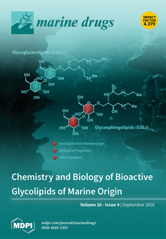
Glycolipids from marine origin represent an outstanding class of natural products with wide structural diversity and a broad range of biological activities. According to the nature of the lipidic fragment, they are classified into three main groups that are glycosphingolipids, glycoglycerolipids, and atypical glycolipids. The extreme difficulty to access these fascinating molecules from natural sources has made their chemical synthesis essential to provide their access for further biological studies and biomedicinal development. The exploration of the chemistry and biology of these fascinating molecules is providing excellent opportunities for the discovery and development of new drugs based on such glycoconjugates. View this paper.
  • Issues are regarded as officially published after their release is announced to the table of contents alert mailing list.
  • You may sign up for e-mail alerts to receive table of contents of newly released issues.
  • PDF is the official format for papers published in both, html and pdf forms. To view the papers in pdf format, click on the "PDF Full-text" link, and use the free Adobe Reader to open them.
Order results
Result details
Section
Select all
Export citation of selected articles as:
12 pages, 1720 KiB  
Article
Cloning of the Bisucaberin B Biosynthetic Gene Cluster from the Marine Bacterium Tenacibaculum mesophilum, and Heterologous Production of Bisucaberin B
by Masaki J. Fujita *, Yusuke Goto and Ryuichi Sakai
Graduate School of Fisheries Sciences, Hokkaido University, Hakodate 041-8611, Japan
Mar. Drugs 2018, 16(9), 342; https://doi.org/10.3390/md16090342 - 19 Sep 2018
Cited by 8 | Viewed by 6078
Abstract
The biosynthetic gene cluster for bisucaberin B (1, bsb gene cluster), an N-hydroxy-N-succinyl diamine (HSD)-based siderophore, was cloned from the marine bacterium Tenacibaculum mesophilum, originated from a marine sponge. The bsb gene cluster consists of six open [...] Read more.
The biosynthetic gene cluster for bisucaberin B (1, bsb gene cluster), an N-hydroxy-N-succinyl diamine (HSD)-based siderophore, was cloned from the marine bacterium Tenacibaculum mesophilum, originated from a marine sponge. The bsb gene cluster consists of six open reading frames (ORFs), in contrast to the four ORFs typically seen in biosynthetic gene clusters of the related molecules. Heterologous expression of the key enzyme, BsbD2, which is responsible for the final biosynthetic step of 1 resulted in production of bisucaberin B (1), but not bisucaberin (2) a macrocyclic counterpart of 1. To date, numbers of related enzymes producing macrocyclic analogues have been reported, but this work represents the first example of the HSD-based siderophore biosynthetic enzyme which exclusively produces a linear molecule rather than macrocyclic counterparts. Full article
Show Figures

Graphical abstract

25 pages, 6117 KiB  
Article
Stress-Induced Mucus Secretion and Its Composition by a Combination of Proteomics and Metabolomics of the Jellyfish Aurelia coerulea
by Wenwen Liu 1,2,†, Fengfeng Mo 3,†, Guixian Jiang 4,†, Hongyu Liang 1,2, Chaoqun Ma 2, Tong Li 5, Lulu Zhang 2, Liyan Xiong 6, Gian Luigi Mariottini 7, Jing Zhang 1,* and Liang Xiao 2,*
1 College of Traditional Chinese Medicine, Jilin Agricultural University, Changchun 130118, China
2 Department of Marine Biotechnology, Faculty of Naval Medicine, Second Military Medical University, Shanghai 200433, China
3 Department of Ship Hygiene, Faculty of Navy Medicine, Second Military Medical University, Shanghai 200433, China
4 Clinical Medicine, Grade 2015, Second Military Medical University, Shanghai 200433, China
5 School of Marine Sciences, Ningbo University, Ningbo 315211, China
6 Department of Traditional Chinese Medicine Identification, School of Pharmacy, Second Military Medical University, Shanghai 200433, China
7 Department of Earth, Environment and Life Sciences (DISTAV), University of Genova, Viale Benedetto XV 5, I-16132 Genova, Italy
These authors contributed equally to this work.
Mar. Drugs 2018, 16(9), 341; https://doi.org/10.3390/md16090341 - 18 Sep 2018
Cited by 31 | Viewed by 7911
Abstract
Background: Jellyfish respond quickly to external stress that stimulates mucus secretion as a defense. Neither the composition of secreted mucus nor the process of secretion are well understood. Methods: Aurelia coerulea jellyfish were stimulated by removing them from environmental seawater. Secreted mucus and [...] Read more.
Background: Jellyfish respond quickly to external stress that stimulates mucus secretion as a defense. Neither the composition of secreted mucus nor the process of secretion are well understood. Methods: Aurelia coerulea jellyfish were stimulated by removing them from environmental seawater. Secreted mucus and tissue samples were then collected within 60 min, and analyzed by a combination of proteomics and metabolomics using liquid chromatography coupled with tandem mass spectrometry (LC-MS/MS) and ultra-performance liquid chromatography/quadrupole time-of-flight mass spectrometry (UPLC-QTOF-MS/MS), respectively. Results: Two phases of sample collection displayed a quick decrease in volume, followed by a gradual increase. A total of 2421 and 1208 proteins were identified in tissue homogenate and secreted mucus, respectively. Gene Ontology (GO) analysis showed that the mucus-enriched proteins are mainly located in extracellular or membrane-associated regions, while the tissue-enriched proteins are distributed throughout intracellular compartments. Tryptamine, among 16 different metabolites, increased with the largest-fold change value of 7.8 in mucus, which is consistent with its involvement in the Kyoto Encyclopedia of Genes and Genomes (KEGG) pathway ‘tryptophan metabolism’. We identified 11 metalloproteinases, four serpins, three superoxide dismutases and three complements, and their presence was speculated to be related to self-protective defense. Conclusions: Our results provide a composition profile of proteins and metabolites in stress-induced mucus and tissue homogenate of A. coerulea. This provides insight for the ongoing endeavors to discover novel bioactive compounds. The large increase of tryptamine in mucus may indicate a strong stress response when jellyfish were taken out of seawater and the active self-protective components such as enzymes, serpins and complements potentially play a key role in innate immunity of jellyfish. Full article
Show Figures

Figure 1

16 pages, 1096 KiB  
Review
Bioactive Compounds from Marine Organisms: Potential for Bone Growth and Healing
by Matthew A. Carson * and Susan A. Clarke
School of Nursing and Midwifery, Queen’s University Belfast, Belfast BT9 7BL, UK
Mar. Drugs 2018, 16(9), 340; https://doi.org/10.3390/md16090340 - 18 Sep 2018
Cited by 72 | Viewed by 13515
Abstract
Marine organisms represent a highly diverse reserve of bioactives which could aid in the treatment of a wide range of diseases, including various musculoskeletal conditions. Osteoporosis in particular would benefit from a novel and effective marine-based treatment, due to its large disease burden [...] Read more.
Marine organisms represent a highly diverse reserve of bioactives which could aid in the treatment of a wide range of diseases, including various musculoskeletal conditions. Osteoporosis in particular would benefit from a novel and effective marine-based treatment, due to its large disease burden and the inefficiencies of current treatment options. Osteogenic bioactives have been isolated from many marine organisms, including nacre powder derived from molluscan shells and fucoidan—the sulphated polysaccharide commonly sourced from brown macroalgae. Such extracts and compounds are known to have a range of osteogenic effects, including stimulation of osteoblast activity and mineralisation, as well as suppression of osteoclast resorption. This review describes currently known soluble osteogenic extracts and compounds from marine invertebrates and algae, and assesses their preclinical potential. Full article
(This article belongs to the Special Issue Marine Organisms for Bone Regeneration)
Show Figures

Figure 1

10 pages, 1952 KiB  
Review
Natural Product Chemistry of Gorgonian Corals of Genus Junceella–Part III
by Hsu-Ming Chung 1,†, Yi-Chen Wang 2,†, Chung-Chih Tseng 3,4, Nan-Fu Chen 5,6, Zhi-Hong Wen 3, Lee-Shing Fang 7,8, Tsong-Long Hwang 9,10,11,12,*, Yang-Chang Wu 13,14,15,16,* and Ping-Jyun Sung 3,13,16,17,18,*
1 Department of Applied Chemistry, National Pingtung University, Pingtung 900, Taiwan
2 Division of Cardiology, Department of Internal Medicine, Kaohsiung Armed Forces General Hospital, Kaohsiung 802, Taiwan
3 Department of Marine Biotechnology and Resources, National Sun Yat-sen University, Kaohsiung 804, Taiwan
4 Division of Dentistry, Zuoying Branch of Kaohsiung Armed Forces General Hospital, Kaohsiung 813, Taiwan
5 Division of Neurosurgery, Department of Surgery, Kaohsiung Armed Forces General Hospital, Kaohsiung 802, Taiwan
6 Department of Neurological Surgery, Tri-Service General Hospital, National Defense Medical Center, Taipei 114, Taiwan
7 Center for Environmental Toxin and Emerging-Contaminant Research, Cheng Shiu University, Kaohsiung 833, Taiwan
8 Super Micro Mass Research and Technology Center, Cheng Shiu University, Kaohsiung 833, Taiwan
9 Graduate Institute of Natural Products, College of Medicine, Chang Gung University, Taoyuan 333, Taiwan
10 Chinese Herbal Medicine Research Team, Healthy Aging Research Center, Chang Gung University, Taoyuan 333, Taiwan
11 Research Center for Chinese Herbal Medicine, Research Center for Food and Cosmetic Safety, Graduate Institute of Healthy Industry Technology, College of Human Ecology, Chang Gung University of Science and Technology, Taoyuan 333, Taiwan
12 Department of Anaesthesiology, Chang Gung Memorial Hospital, Taoyuan 333, Taiwan
13 Graduate Institute of Natural Products, Kaohsiung Medical University, Kaohsiung 807, Taiwan
14 Research Center for Natural Products and Drug Development, Kaohsiung Medical University, Kaohsiung 807, Taiwan
15 Department of Medical Research, Kaohsiung Medical University Hospital, Kaohsiung 807, Taiwan
16 Chinese Medicine Research and Development Center, China Medical University Hospital, Taichung 404, Taiwan
17 National Museum of Marine Biology and Aquarium, Pingtung 944, Taiwan
18 Graduate Institute of Marine Biology, National Dong Hwa University, Pingtung 944, Taiwan
These authors contributed equally to this work.
add Show full affiliation list remove Hide full affiliation list
Mar. Drugs 2018, 16(9), 339; https://doi.org/10.3390/md16090339 - 17 Sep 2018
Cited by 19 | Viewed by 4243
Abstract
The structures, names, bioactivities, and references of 82 natural products, including 48 new metabolites, purified from the gorgonian corals belonging to the genus Junceella are described in this review. All compounds mentioned in this review were obtained from Junceella fragilis, Junceella gemmacea [...] Read more.
The structures, names, bioactivities, and references of 82 natural products, including 48 new metabolites, purified from the gorgonian corals belonging to the genus Junceella are described in this review. All compounds mentioned in this review were obtained from Junceella fragilis, Junceella gemmacea, Junceella juncea, and Junceella sp., collected from tropical Indo-Pacific Ocean. Some of these compounds exhibited potential biomedical activities. Full article
Show Figures

Figure 1

11 pages, 1243 KiB  
Communication
Total Synthesis of the Highly N-Methylated Peptides Carmabin A and Dragomabin
by Baijun Ye 1, Peng Jiang 1, Tingrong Zhang 1, Yuanjun Sun 1, Xin Hao 1, Yingjun Cui 1, Liang Wang 1,* and Yue Chen 1,2,*
1 The State Key Laboratory of Medicinal Chemical Biology, College of Pharmacy and Tianjin Key Laboratory of Molecular Drug Research, Nankai University, Tianjin 300350, China
2 Collaborative Innovation Center of Chemical Science and Engineering, Tianjin 300350, China
Mar. Drugs 2018, 16(9), 338; https://doi.org/10.3390/md16090338 - 17 Sep 2018
Cited by 8 | Viewed by 4185
Abstract
The first total synthesis of carmabin A and dragomabin was achieved at 52.3 mg and 43.8 mg scale, respectively. The synthesis led to determination of the configuration of carmabin A and reassignment of the configuration of dragomabin at the stereogenic centre on the [...] Read more.
The first total synthesis of carmabin A and dragomabin was achieved at 52.3 mg and 43.8 mg scale, respectively. The synthesis led to determination of the configuration of carmabin A and reassignment of the configuration of dragomabin at the stereogenic centre on the alkyne-bearing fragment. Full article
Show Figures

Scheme 1

13 pages, 3621 KiB  
Article
Isolation, Characterization, and Pharmaceutical Applications of an Exopolysaccharide from Aerococcus Uriaeequi
by Chunlei Wang 1, Qiuping Fan 1, Xiaofei Zhang 1, Xiaoping Lu 1, Yanrui Xu 1, Wenxing Zhu 1, Jie Zhang 1, Wen Hao 2,3 and Lujiang Hao 1,*
1 State Key Laboratory of Biobased Material and Green Papermaking, Qilu University of Technology, Shandong Academy of Sciences, Jinan 250353, China
2 Qingdao Municipal Center for Disease Control & Prevention, Qingdao 266033, China
3 Qingdao Institute of Preventive Medicine, Qingdao 266033, China
Mar. Drugs 2018, 16(9), 337; https://doi.org/10.3390/md16090337 - 16 Sep 2018
Cited by 41 | Viewed by 5655
Abstract
Many marine bacteria secrete exopolysaccharides (EPSs), which are made up of a substantial component of the macro-molecules surrounding cells. Recently, the wide demand for EPSs for food, cosmetics, pharmaceutical and other applications has led to great interest in them. In this study, an [...] Read more.
Many marine bacteria secrete exopolysaccharides (EPSs), which are made up of a substantial component of the macro-molecules surrounding cells. Recently, the wide demand for EPSs for food, cosmetics, pharmaceutical and other applications has led to great interest in them. In this study, an EPS produced by marine bacteria Aerococcus uriaeequi HZ strains (EPS-A) was isolated and purified to examine its structure and biological function. The molecular weight of EPS-A analyzed by high-performance liquid gel filtration chromatography (HPGFC) is found to have a number average of 2.22 × 10⁵ and weight average of 2.84 × 10⁵, respectively. High-performance liquid chromatography (HPLC) and Fourier-transform–infrared (FT–IR) analysis indicate that EPS-A was a polysaccharide composed of glucose and a little mannose. In addition, the flocculating rate of sewage of EPS-A was 79.90%. The hygroscopicity studies showed that hygroscopicity of EPS-A was higher than chitosan but lower than that of sodium hyaluronate. The moisture retention of EPS-A showed similar retention activity to both chitosan and sodium hyaluronate. EPS-A also can scavenge free radicals including both OH• free radical and O2 free radical and the activity to O2 free radical is similar to vitamin C. Safety assessment on mice indicated that the EPS-A is safe for external use and oral administration. EPS-A has great potential for applications in medicine due to its characteristics mentioned above. Full article
(This article belongs to the Collection Marine Polysaccharides)
Show Figures

Figure 1

55 pages, 3844 KiB  
Review
UV-Protective Compounds in Marine Organisms from the Southern Ocean
by Laura Núñez-Pons 1, Conxita Avila 2, Giovanna Romano 3, Cinzia Verde 1,4 and Daniela Giordano 1,4,*
1 Department of Biology and Evolution of Marine Organisms (BEOM), Stazione Zoologica Anton Dohrn (SZN), 80121 Villa Comunale, Napoli, Italy
2 Department of Evolutionary Biology, Ecology, and Environmental Sciences, and Biodiversity Research Institute (IrBIO), Faculty of Biology, University of Barcelona, Av. Diagonal 643, 08028 Barcelona, Catalonia, Spain
3 Department of Marine Biotechnology (Biotech), Stazione Zoologica Anton Dohrn (SZN), 80121 Villa Comunale, Napoli, Italy
4 Institute of Biosciences and BioResources (IBBR), CNR, Via Pietro Castellino 111, 80131 Napoli, Italy
Mar. Drugs 2018, 16(9), 336; https://doi.org/10.3390/md16090336 - 14 Sep 2018
Cited by 66 | Viewed by 11992
Abstract
Solar radiation represents a key abiotic factor in the evolution of life in the oceans. In general, marine, biota—particularly in euphotic and dysphotic zones—depends directly or indirectly on light, but ultraviolet radiation (UV-R) can damage vital molecular machineries. UV-R induces the formation of [...] Read more.
Solar radiation represents a key abiotic factor in the evolution of life in the oceans. In general, marine, biota—particularly in euphotic and dysphotic zones—depends directly or indirectly on light, but ultraviolet radiation (UV-R) can damage vital molecular machineries. UV-R induces the formation of reactive oxygen species (ROS) and impairs intracellular structures and enzymatic reactions. It can also affect organismal physiologies and eventually alter trophic chains at the ecosystem level. In Antarctica, physical drivers, such as sunlight, sea-ice, seasonality and low temperature are particularly influencing as compared to other regions. The springtime ozone depletion over the Southern Ocean makes organisms be more vulnerable to UV-R. Nonetheless, Antarctic species seem to possess analogous UV photoprotection and repair mechanisms as those found in organisms from other latitudes. The lack of data on species-specific responses towards increased UV-B still limits the understanding about the ecological impact and the tolerance levels related to ozone depletion in this region. The photobiology of Antarctic biota is largely unknown, in spite of representing a highly promising reservoir in the discovery of novel cosmeceutical products. This review compiles the most relevant information on photoprotection and UV-repair processes described in organisms from the Southern Ocean, in the context of this unique marine polar environment. Full article
Show Figures

Figure 1

8 pages, 1445 KiB  
Article
Cytotoxic Tetrahydroxanthone Dimers from the Mangrove-Associated Fungus Aspergillus versicolor HDN1009
by Guihong Yu 1, Guangwei Wu 1, Zichao Sun 1, Xiaomin Zhang 1, Qian Che 1, Qianqun Gu 1, Tianjiao Zhu 1,2, Dehai Li 1,2,3 and Guojian Zhang 1,2,*
1 Key Laboratory of Marine Drugs, Chinese Ministry of Education, School of Medicine and Pharmacy, Ocean University of China, Qingdao 266003, China
2 Laboratory for Marine Drugs and Bioproducts of Qingdao Pilot National Laboratory for Marine Science and Technology, Qingdao 266237, China
3 Open Studio for Druggability Research of Marine Natural Products, Qingdao Pilot National Laboratory for Marine Science and Technology, Qingdao 266237, China
Mar. Drugs 2018, 16(9), 335; https://doi.org/10.3390/md16090335 - 14 Sep 2018
Cited by 42 | Viewed by 4266
Abstract
Three new tetrahydroxanthone dimers, 5-epi-asperdichrome (1), versixanthones N (2), and O (3), were isolated from the mangrove-derived fungus Aspergillus versicolor HDN1009. Their structures, including the absolute configurations, were elucidated by NMR, HRMS, and circular dichroism [...] Read more.
Three new tetrahydroxanthone dimers, 5-epi-asperdichrome (1), versixanthones N (2), and O (3), were isolated from the mangrove-derived fungus Aspergillus versicolor HDN1009. Their structures, including the absolute configurations, were elucidated by NMR, HRMS, and circular dichroism (CD) experiments. Among them, compound 1 was the second example of tetrahydroxanthone dimers, which dimerized by a rare diaryl ether linkage and showed promising antibacterial activities against Vibrio parahemolyticus, Bacillus subtilis, Mycobacterium phlei, and Pseudomonas aeruginosa, with MIC values ranging from 100 μM to 200 μM; whilst compounds 2 and 3 exhibited extensive cytotoxicities against five cancer cell lines (HL-60, K562, H1975, MGC803, and HO-8910), with IC50 values ranging from 1.7 μM to 16.1 μM. Full article
Show Figures

Graphical abstract

18 pages, 2913 KiB  
Article
Supercritical Carbon Dioxide Extraction of Astaxanthin, Lutein, and Fatty Acids from Haematococcus pluvialis Microalgae
by Giuseppe Di Sanzo 1, Sanjeet Mehariya 2,3, Maria Martino 1, Vincenzo Larocca 1, Patrizia Casella 2, Simeone Chianese 3, Dino Musmarra 3, Roberto Balducchi 1 and Antonio Molino 2,*
1 ENEA, Italian National Agency for New Technologies, Energy and Sustainable Economic Development, Department of Sustainability, CR Trisaia SS Jonica 106, km 419+500, 75026 Rotondella, Italy
2 ENEA, Italian National Agency for New Technologies, Energy and Sustainable Economic Development, Department of Sustainability, CR Portici. P. Enrico Fermi, 1, 80055 Portici, Italy
3 Department of Engineering, Università degli Studi della Campania “L. Vanvitelli”, Real Casa dell’Annunziata, Via Roma 29, 81031 Aversa, Italy
Mar. Drugs 2018, 16(9), 334; https://doi.org/10.3390/md16090334 - 13 Sep 2018
Cited by 132 | Viewed by 9566
Abstract
Haematococcus pluvialis microalgae in the red phase can produce significant amounts of astaxanthin, lutein, and fatty acids (FAs), which are valuable antioxidants in nutraceutics and cosmetics. Extraction of astaxanthin, lutein, and FAs from disrupted biomass of the H. pluvialis red phase using carbon [...] Read more.
Haematococcus pluvialis microalgae in the red phase can produce significant amounts of astaxanthin, lutein, and fatty acids (FAs), which are valuable antioxidants in nutraceutics and cosmetics. Extraction of astaxanthin, lutein, and FAs from disrupted biomass of the H. pluvialis red phase using carbon dioxide (CO2) in supercritical fluid extraction (SFE) conditions was investigated using a bench-scale reactor in a semi-batch configuration. In particular, the effect of extraction time (20, 40, 60, 80, and 120 min), CO2 flow rate (3.62 and 14.48 g/min) temperature (50, 65, and 80 °C), and pressure (100, 400, and 550 bar.) was explored. The results show the maximum recovery of astaxanthin and lutein achieved were 98.6% and 52.3%, respectively, at 50 °C and 550 bars, while the maximum recovery of FAs attained was 93.2% at 65 °C and 550 bars. Full article
Show Figures

Figure 1

10 pages, 1305 KiB  
Article
Influence of Modified Fucoidan and Related Sulfated Oligosaccharides on Hematopoiesis in Cyclophosphamide-Induced Mice
by Natalia Yu. Anisimova 1, Nadezhda E. Ustyuzhanina 2,*, Maria I. Bilan 2, Fedor V. Donenko 1, Natalia A. Ushakova 3, Anatolii I. Usov 2, Mikhail V. Kiselevskiy 1,* and Nikolay E. Nifantiev 2,*
1 N.N. Blokhin National Medical Research Center of Oncology, Kashirskoe shosse, 24, 115478 Moscow, Russia
2 N.D. Zelinsky Institute of Organic Chemistry, Russian Academy of Sciences, Leninsky Prospect 47, 119991 Moscow, Russia
3 V.N. Orekhovich Research Institute of Biomedical Chemistry, Pogodinskaya str. 10, 119121 Moscow, Russia
Mar. Drugs 2018, 16(9), 333; https://doi.org/10.3390/md16090333 - 13 Sep 2018
Cited by 26 | Viewed by 4628
Abstract
Immunosuppression derived after cytostatics application in cancer chemotherapy is considered as an adverse side effect that leads to deterioration of quality of life and risk of infectious diseases. A linear sulfated (1→3)-α-l-fucan M-Fuc prepared by chemical modification of a fucoidan isolated [...] Read more.
Immunosuppression derived after cytostatics application in cancer chemotherapy is considered as an adverse side effect that leads to deterioration of quality of life and risk of infectious diseases. A linear sulfated (1→3)-α-l-fucan M-Fuc prepared by chemical modification of a fucoidan isolated from the brown seaweed Chordaria flagelliformis, along with two structurally related synthetic sulfated oligosaccharides, were studied as stimulators of hematopoiesis on a model of cyclophosphamide immunosuppression in mice. Recombinant granulocyte colony-stimulating factor (r G-CSF), which is currently applied in medicine to treat low blood neutrophils, was used as a reference. Polysaccharide M-Fuc and sulfated difucoside DS did not demonstrate significant effect, while sulfated octasaccharide OS showed higher activity than r G-CSF, causing pronounced neutropoiesis stimulation. In addition, production of erythrocytes and platelets was enhanced after the octasaccharide administration. The assessment of populations of cells in blood and bone marrow of mice revealed the difference in mechanisms of action of OS and r G-CSF. Full article
(This article belongs to the Special Issue Marine Polysaccharides in Pharmaceutical Applications)
Show Figures

Figure 1

17 pages, 1872 KiB  
Article
Characterization of the Microbial Population Inhabiting a Solar Saltern Pond of the Odiel Marshlands (SW Spain)
by Patricia Gómez-Villegas, Javier Vigara and Rosa León *
Laboratory of Biochemistry and Molecular Biology, Faculty of Experimental Sciences, Marine International Campus of Excellence (CEIMAR), University of Huelva, 21071 Huelva, Spain
Mar. Drugs 2018, 16(9), 332; https://doi.org/10.3390/md16090332 - 12 Sep 2018
Cited by 20 | Viewed by 4855
Abstract
The solar salterns located in the Odiel marshlands, in southwest Spain, are an excellent example of a hypersaline environment inhabited by microbial populations specialized in thriving under conditions of high salinity, which remains poorly explored. Traditional culture-dependent taxonomic studies have usually under-estimated the [...] Read more.
The solar salterns located in the Odiel marshlands, in southwest Spain, are an excellent example of a hypersaline environment inhabited by microbial populations specialized in thriving under conditions of high salinity, which remains poorly explored. Traditional culture-dependent taxonomic studies have usually under-estimated the biodiversity in saline environments due to the difficulties that many of these species have to grow at laboratory conditions. Here we compare two molecular methods to profile the microbial population present in the Odiel saltern hypersaline water ponds (33% salinity). On the one hand, the construction and characterization of two clone PCR amplified-16S rRNA libraries, and on the other, a high throughput 16S rRNA sequencing approach based on the Illumina MiSeq platform. The results reveal that both methods are comparable for the estimation of major genera, although massive sequencing provides more information about the less abundant ones. The obtained data indicate that Salinibacter ruber is the most abundant genus, followed by the archaea genera, Halorubrum and Haloquadratum. However, more than 100 additional species can be detected by Next Generation Sequencing (NGS). In addition, a preliminary study to test the biotechnological applications of this microbial population, based on its ability to produce and excrete haloenzymes, is shown. Full article
(This article belongs to the Special Issue Genome Mining and Marine Microbial Natural Products)
Show Figures

Figure 1

16 pages, 1097 KiB  
Article
Stress-Driven Discovery of New Angucycline-Type Antibiotics from a Marine Streptomyces pratensis NA-ZhouS1
by Najeeb Akhter 1, Yaqin Liu 2, Bibi Nazia Auckloo 1, Yutong Shi 1, Kuiwu Wang 3, Juanjuan Chen 4, Xiaodan Wu 5 and Bin Wu 1,*
1 Ocean College, Zhejiang University, Hangzhou 310058, China
2 Department of Chemistry, Zhejiang University, Hangzhou 301000, China
3 Department of Chemistry, Zhejiang Gongshang University, Hangzhou 310012, China
4 Key Laboratory of Applied Marine Biotechnology, Ningbo University, Chinese Ministry of Education, Ningbo 315211, China
5 Centre of Analysis and Measurement, Zhejiang University, Hangzhou 310058, China
Mar. Drugs 2018, 16(9), 331; https://doi.org/10.3390/md16090331 - 12 Sep 2018
Cited by 41 | Viewed by 6429
Abstract
Natural products from marine actinomycetes remain an important resource for drug discovery, many of which are produced by the genus, Streptomyces. However, in standard laboratory conditions, specific gene clusters in microbes have long been considered silent or covert. Thus, various stress techniques activated [...] Read more.
Natural products from marine actinomycetes remain an important resource for drug discovery, many of which are produced by the genus, Streptomyces. However, in standard laboratory conditions, specific gene clusters in microbes have long been considered silent or covert. Thus, various stress techniques activated latent gene clusters leading to isolation of potential metabolites. This study focused on the analysis of two new angucycline antibiotics isolated from the culture filtrate of a marine Streptomyces pratensis strain NA-ZhouS1, named, stremycin A (1) and B (2) which were further determined based on spectroscopic techniques such as high resolution time of flight mass spectrometry (HR-TOF-MS), 1D, and 2D nuclear magnetic resonance (NMR) experiments. In addition, four other known compounds, namely, 2-[2-(3,5-dimethyl-2-oxo-cyclohexyl)-6-oxo-tetrahydro-pyran-4yl]-acetamide (3), cyclo[l-(4-hydroxyprolinyl)-l-leucine] (4), 2-methyl-3H-quinazoline-4-one (5), and menthane derivative, 3-(hydroxymethyl)-6-isopropyl-10,12-dioxatricyclo[7.2.1.0]dodec-4-en-8-one (6) were obtained and elucidated by means of 1D NMR spectrometry. Herein, we describe the “Metal Stress Technique” applied in the discovery of angucyclines, a distinctive class of antibiotics that are commonly encoded in microbiomes but have never been reported in “Metal Stress” based discovery efforts. Novel antibiotics 1 and 2 exhibited antimicrobial activities against Pseudomonas aeruginosa, methicillin resistant Staphylococcus aureus (MRSA), Klebsiella pneumonia, and Escherichia coli with equal minimum inhibitory concentration (MIC) values of 16 µg/mL, while these antibiotics showed inhibition against Bacillus subtilis at MIC value of approximately 8–16 µg/mL, respectively. As a result, the outcome of this investigation revealed that metal stress is an effective technique in unlocking the biosynthetic potential and resulting production of novel antibiotics. Full article
(This article belongs to the Special Issue Genome Mining and Marine Microbial Natural Products)
Show Figures

Graphical abstract

7 pages, 710 KiB  
Communication
New Antibacterial Bagremycins F and G from the Marine-Derived Streptomyces sp. ZZ745
by Di Zhang 1,†, Chenyan Shu 2,†, Xiaoyuan Lian 2,* and Zhizhen Zhang 1,*
1 Ocean College, Zhoushan Campus, Zhejiang University, Zhoushan 316021, China
2 College of Pharmaceutical Sciences, Zhejiang University, Hangzhou 310058, China
These authors contributed equal to this work.
Mar. Drugs 2018, 16(9), 330; https://doi.org/10.3390/md16090330 - 12 Sep 2018
Cited by 26 | Viewed by 4552
Abstract
As part of our research to discover novel bioactive natural products from marine microorganisms, five bagremycin analogues, including the previously unreported bagremycins F (1) and G (2), were isolated from a marine actinomycete Streptomyces sp. ZZ745. The structures of [...] Read more.
As part of our research to discover novel bioactive natural products from marine microorganisms, five bagremycin analogues, including the previously unreported bagremycins F (1) and G (2), were isolated from a marine actinomycete Streptomyces sp. ZZ745. The structures of these compounds were determined by means of NMR spectroscopic analysis, HRESIMS data, and optical rotation. Both bagremycins F (1) and G (2) showed antibacterial activity against Escherichia coli, with MIC values of 41.8 and 61.7 μM, respectively. Full article
(This article belongs to the Special Issue Natural Products from Marine Actinomycetes)
Show Figures

Graphical abstract

12 pages, 1808 KiB  
Article
Highly Substituted Benzophenone Aldehydes and Eremophilane Derivatives from the Deep-Sea Derived Fungus Phomopsis lithocarpus FS508
by Jian-Lin Xu 1,2,†, Hong-Xin Liu 1,3,†, Yu-Chan Chen 1, Hai-Bo Tan 3, Heng Guo 1,2, Li-Qiong Xu 1, Sai-Ni Li 1, Zi-Lei Huang 1, Hao-Hua Li 1, Xiao-Xia Gao 2,* and Wei-Min Zhang 1,*
1 State Key Laboratory of Applied Microbiology Southern China, Guangdong Provincial Key Laboratory of Microbial Culture Collection and Application, Guangdong Open Laboratory of Applied Microbiology, Guangdong Institute of Microbiology, Guangzhou 510070, China
2 College of Pharmacy, Guangdong Pharmaceutical University, Guangzhou 510006, China
3 Program for Natural Products Chemical Biology, Key Laboratory of Plant Resources Conservation and Sustainable Utilization, Guangdong Provincial Key Laboratory of Applied Botany, South China Botanical Garden, Chinese Academy of Sciences, Guangzhou 510650, China
These authors contributed equally to this work.
Mar. Drugs 2018, 16(9), 329; https://doi.org/10.3390/md16090329 - 11 Sep 2018
Cited by 28 | Viewed by 4612
Abstract
Five new benzophenone derivatives named tenellones D–H (15), sharing a rare naturally occurring aldehyde functionality in this family, and a new eremophilane derivative named lithocarin A (7), together with two known compounds (6 and 8), [...] Read more.
Five new benzophenone derivatives named tenellones D–H (15), sharing a rare naturally occurring aldehyde functionality in this family, and a new eremophilane derivative named lithocarin A (7), together with two known compounds (6 and 8), were isolated from the deep marine sediment-derived fungus Phomopsis lithocarpus FS508. All of the structures for these new compounds were fully characterized and established on the basis of extensive spectroscopic interpretation and X-ray crystallographic analysis. Compound 5 exhibited cytotoxic activity against HepG-2 and A549 cell lines with IC50 values of 16.0 and 17.6 μM, respectively. Full article
(This article belongs to the Special Issue Natural Products from Marine Fungi)
Show Figures

Graphical abstract

21 pages, 3901 KiB  
Article
Pyropia yezoensis Protein Supplementation Prevents Dexamethasone-Induced Muscle Atrophy in C57BL/6 Mice
by Min-Kyeong Lee 1, Jeong-Wook Choi 1, Youn Hee Choi 1,2,* and Taek-Jeong Nam 1,3,*
1 Institute of Fisheries Sciences, Pukyong National University, Busan 46041, Korea
2 Department of Marine Bio-Materials & Aquaculture, Pukyong National University, Busan 48513, Korea
3 Department of Food Science and Nutrition, Pukyong National University, Busan 48513, Korea
Mar. Drugs 2018, 16(9), 328; https://doi.org/10.3390/md16090328 - 11 Sep 2018
Cited by 7 | Viewed by 6283
Abstract
We investigated the protective effects of Pyropia yezoensis crude protein (PYCP) against dexamethasone (DEX)-induced myotube atrophy and its underlying mechanisms. DEX (3 mg/kg body weight, intraperitoneal injection) and PYCP (150 and 300 mg/kg body weight, oral) were administrated to mice for 18 days, [...] Read more.
We investigated the protective effects of Pyropia yezoensis crude protein (PYCP) against dexamethasone (DEX)-induced myotube atrophy and its underlying mechanisms. DEX (3 mg/kg body weight, intraperitoneal injection) and PYCP (150 and 300 mg/kg body weight, oral) were administrated to mice for 18 days, and the effects of PYCP on DEX-induced muscle atrophy were evaluated. Body weight, calf thickness, and gastrocnemius and tibialis anterior muscle weight were significantly decreased by DEX administration (p < 0.05), while PYCP supplementation effectively prevented the DEX-induced decrease in body weight, calf thickness, and muscle weight. PYCP supplementation also attenuated the DEX-induced increase in serum glucose, creatine kinase, and lactate dehydrogenase levels. Additionally, PYCP supplementation reversed DEX-induced muscle atrophy via the regulation of the insulin-like growth factor-I/protein kinase B/rapamycin-sensitive mTOR complex I/forkhead box O signaling pathway. The mechanistic investigation revealed that PYCP inhibited the ubiquitin-proteasome and autophagy-lysosome pathways in DEX-administrated C57BL/6 mice. These findings demonstrated that PYCP increased protein synthesis and decreased protein breakdown to prevent muscle atrophy. Therefore, PYCP supplementation appears to be useful for preventing muscle atrophy. Full article
(This article belongs to the Special Issue The Pharmacological Potential of Marine-Derived Peptides and Proteins)
Show Figures

Figure 1

17 pages, 6808 KiB  
Article
Gloeothece sp. as a Nutraceutical Source—An Improved Method of Extraction of Carotenoids and Fatty Acids
by Helena M. Amaro 1,2, A. Catarina Guedes 1,*, Marco A. C. Preto 1, I. Sousa-Pinto 1,3 and F. Xavier Malcata 4,5
1 Interdisciplinary Centre of Marine and Environmental Research (CIIMAR), University of Porto, Terminal de Cruzeiros do Porto de Leixões, Avenida General Norton de Matos, S/N, P-4450-208 Matosinhos, Portugal
2 Institute of Biomedical Sciences Abel Salazar (ICBAS), University of Porto, Rua Jorge Viterbo Ferreira no. 228, P-4050-313 Porto, Portugal
3 FCUP—Faculty of Sciences, University of Porto, Rua do Campo Alegre s/n, 4169-007 Porto, Portugal
4 LEPABE—Laboratory of Process Engineering, Environment, Biotechnology and Energy, Rua Dr. Roberto Frias s/n, P-4200-465 Porto, Portugal
5 Department of Chemical Engineering, University of Porto, Rua Dr. Roberto Frias, s/n 4200-465 Porto, Portugal
Mar. Drugs 2018, 16(9), 327; https://doi.org/10.3390/md16090327 - 11 Sep 2018
Cited by 26 | Viewed by 5085
Abstract
The nutraceutical potential of microalgae boomed with the exploitation of new species and sustainable extraction systems of bioactive compounds. Thus, a laboratory-made continuous pressurized solvent extraction system (CPSE) was built to optimize the extraction of antioxidant compounds, such as carotenoids and PUFA, from [...] Read more.
The nutraceutical potential of microalgae boomed with the exploitation of new species and sustainable extraction systems of bioactive compounds. Thus, a laboratory-made continuous pressurized solvent extraction system (CPSE) was built to optimize the extraction of antioxidant compounds, such as carotenoids and PUFA, from a scarcely studied prokaryotic microalga, Gloeothece sp. Following “green chemical principles” and using a GRAS solvent (ethanol), biomass amount, solvent flow-rate/pressure, temperature and solvent volume—including solvent recirculation—were sequentially optimized, with the carotenoids and PUFA content and antioxidant capacity being the objective functions. Gloeothece sp. bioactive compounds were best extracted at 60 °C and 180 bar. Recirculation of solvent in several cycles (C) led to an 11-fold extraction increase of β-carotene (3C) and 7.4-fold extraction of C18:2 n6 t (5C) when compared to operation in open systems. To fully validate results CPSE, this system was compared to a conventional extraction method, ultrasound assisted extraction (UAE). CPSE proved superior in extraction yield, increasing total carotenoids extraction up 3-fold and total PUFA extraction by ca. 1.5-fold, with particular extraction increase of 18:3 n3 by 9.6-fold. Thus, CPSE proved to be an efficient and greener extraction method to obtain bioactive extract from Gloeothece sp. for nutraceutical purposes—with low levels of resources spent, while lowering costs of production and environmental impacts. Full article
Show Figures

Graphical abstract

11 pages, 3390 KiB  
Article
Bacillamidins A–G from a Marine-Derived Bacillus pumilus
by Si-Yu Zhou 1, Yi-Jie Hu 1, Fan-Cheng Meng 1, Shen-Yue Qu 1, Rui Wang 1, Raymond J. Andersen 2, Zhi-Hua Liao 3 and Min Chen 1,*
1 Key Laboratory of Luminescent and Real-Time Analytical Chemistry (Ministry of Education), College of Pharmaceutical Sciences, Southwest University, Chongqing 400715, China
2 Department of Chemistry, University of British Columbia, Vancouver, BC V6T1Z1, Canada
3 School of Life Sciences, Southwest University, Chongqing 400715, China
Mar. Drugs 2018, 16(9), 326; https://doi.org/10.3390/md16090326 - 11 Sep 2018
Cited by 8 | Viewed by 4369
Abstract
Seven long-chain amides, including five previously undescribed bacillamidins A–E (15) and two previously reported synthetic analogs, bacillamidins F (6) and G (7), were isolated from extracts of the marine-derived Bacillus pumilus strain RJA1515. The structures [...] Read more.
Seven long-chain amides, including five previously undescribed bacillamidins A–E (15) and two previously reported synthetic analogs, bacillamidins F (6) and G (7), were isolated from extracts of the marine-derived Bacillus pumilus strain RJA1515. The structures of the new compounds were established by extensive analysis of 1D and 2D nuclear magnetic resonance (NMR) data as well as high resolution mass spectrometry (HRMS), and the absolute configurations of the stereogenic carbons of 14 were established by comparison of the calculated and the experimental electronic circular dichroism (ECD) spectra. The cytotoxic and antimicrobial activities of 17 were evaluated. Full article
Show Figures

Figure 1

16 pages, 5315 KiB  
Article
Inhibition of Prostate Cancer DU-145 Cells Proliferation by Anthopleura anjunae Oligopeptide (YVPGP) via PI3K/AKT/mTOR Signaling Pathway
by Xiaojuan Li 1, Yunping Tang 1,*, Fangmiao Yu 1, Yu Sun 2, Fangfang Huang 1, Yan Chen 1, Zuisu Yang 1 and Guofang Ding 1,3,*
1 Zhejiang Provincial Engineering Technology Research Center of Marine Biomedical Products, School of Food and Pharmacy, Zhejiang Ocean University, Zhoushan 316022, China
2 Zhejiang Provincial Engineering Technology Research Center of Marine Biomedical Products, Zhejiang Ocean University Donghai Science and Technology College, Zhoushan 316000, China
3 Zhejiang Marine Fisheries Research Institution, Zhoushan 316021, China
Mar. Drugs 2018, 16(9), 325; https://doi.org/10.3390/md16090325 - 11 Sep 2018
Cited by 37 | Viewed by 5841
Abstract
We investigated the antitumor mechanism of Anthopleura anjunae oligopeptide (AAP-H, YVPGP) in prostate cancer DU-145 cells in vitro and in vivo. Results indicated that AAP-H was nontoxic and exhibited antitumor activities. Cell cycle analysis indicated that AAP-H may arrest DU-145 cells in the [...] Read more.
We investigated the antitumor mechanism of Anthopleura anjunae oligopeptide (AAP-H, YVPGP) in prostate cancer DU-145 cells in vitro and in vivo. Results indicated that AAP-H was nontoxic and exhibited antitumor activities. Cell cycle analysis indicated that AAP-H may arrest DU-145 cells in the S phase. The role of the phosphatidylinositol 3-kinase/protein kinase B/mammalian rapamycin target protein (PI3K/AKT/mTOR) signaling pathway in the antitumor mechanism of APP-H was investigated. Results showed that AAP-H treatment led to dose-dependent reduction in the levels of p-AKT (Ser473), p-PI3K (p85), and p-mTOR (Ser2448), whereas t-AKT and t-PI3K levels remained unaltered compared to the untreated DU-145 cells. Inhibition of PI3K/AKT/mTOR signaling pathway in the DU-145 cells by employing inhibitor LY294002 (10 μM) or rapamycin (20 nM) effectively attenuated AAP-H-induced phosphorylation of AKT and mTOR. At the same time, inhibitor addition further elevated AAP-H-induced cleaved-caspase-3 levels. Furthermore, the effect of AAP-H on tumor growth and the role of the PI3K/AKT/mTOR signaling pathway in nude mouse model were also investigated. Immunohistochemical analysis showed that activated AKT, PI3K, and mTOR levels were reduced in DU-145 xenografts. Western blotting showed that AAP-H treatment resulted in dose-dependent reduction in p-AKT (Ser473), p-PI3K (p85), and p-mTOR (Ser2448) levels, whereas t-AKT and t-PI3K levels remained unaltered. Similarly, Bcl-xL levels decreased, whereas that of Bax increased after AAP-H treatment. AAP-H also increased initiator (caspase 8 and 9) and executor caspase (caspase 3 and 7) levels. Therefore, the antitumor mechanism of APP-H on DU-145 cells may involve regulation of the PI3K/AKT/mTOR signaling pathway, which eventually promotes apoptosis via mitochondrial and death receptor pathways. Thus, the hydrophobic oligopeptide (YVPGP) can be developed as an adjuvant for the prevention or treatment of prostate cancer in the future. Full article
Show Figures

Figure 1

13 pages, 3470 KiB  
Article
Liposomal Form of the Echinochrome-Carrageenan Complex
by Irina M. Yermak 1,*, Vladimir I. Gorbach 1, Valery P. Glazunov 1, Anna O. Kravchenko 1, Natalya P. Mishchenko 1, Evgeniya A. Pimenova 2 and Viktoria N. Davydova 1
1 G.B. Elyakov Pacific Institute of Bioorganic Chemistry, Far-Eastern Branch of the Russian Academy of Sciences, 100 Let Vladivostoku Prosp., 159, 690022 Vladivostok, Russia
2 National Scientific Center of Marine Biology, Far-Eastern Branch of the Russian Academy of Sciences, Palchevskogo, 17, 690041 Vladivostok, Russia
Mar. Drugs 2018, 16(9), 324; https://doi.org/10.3390/md16090324 - 10 Sep 2018
Cited by 11 | Viewed by 4555
Abstract
Inclusion of drugs in liposomes offers the potential for localized and sustained delivery to mucosal surfaces. The inclusion of the carrageenan matrix with echinochrome A ((Ech)—the active substance of the drug Histochrome) in liposomes was studied. According to the spectral characteristics, Ech was [...] Read more.
Inclusion of drugs in liposomes offers the potential for localized and sustained delivery to mucosal surfaces. The inclusion of the carrageenan matrix with echinochrome A ((Ech)—the active substance of the drug Histochrome) in liposomes was studied. According to the spectral characteristics, Ech was not oxidized and retained stability after encapsulation in the liposomes and the lyophilization process. Loading the liposomes with negatively charged polysaccharide results in the increase in the zeta potential to more negative values (from −14.6 to −24.4 mV), that together with an increasing in the sizes of liposomes (from 125.6 ± 2.5 nm to 159.3 ± 5.8 nm) propose of the formation of the polymer coating on liposomes. The interactions of liposomes with porcine stomach mucin was determined by the DLS and SEM methods. The changes in the zeta-potential and size of the mucin particles were observed as the result of the interaction of liposomes with mucin. To evaluate the mucoadhesive properties of liposomes and the penetration of Ech in the mucosa, a fresh-frozen inner surface of the small intestine of a pig as a model of mucous tissue was used. Polysaccharide-coated liposomes exhibit very good mucoadhesive properties −50% of Ech remains on the mucosa. Full article
(This article belongs to the Special Issue Marine Drugs and Nanomedicine)
Show Figures

Figure 1

13 pages, 2411 KiB  
Article
Fucoxanthin Inhibits Myofibroblast Differentiation and Extracellular Matrix Production in Nasal Polyp-Derived Fibroblasts via Modulation of Smad-Dependent and Smad-Independent Signaling Pathways
by Hyun Jung 1, Dae-Sung Lee 2, Seong Kook Park 1, Jung Sik Choi 3, Won-Kyo Jung 4, Won Sun Park 5,* and Il-Whan Choi 6,*
1 Department of Otorhinolaryngology-Head & Neck Surgery, Inje University College of Medicine, Busan 47392, Korea
2 Department of Applied Research, National Marine Biodiversity Institute of Korea, Seocheon 33662, Korea
3 Department of Internal Medicine, Busan Paik Hospital, Inje University, Busan 47392, Korea
4 Department of Biomedical Engineering, Center for Marine-Integrated Biomedical Technology (BK21 Plus) Pukyong National University, Busan 48513, Korea
5 Department of Physiology, Kangwon National University School of Medicine, Chuncheon 24341, Korea
6 Department of Microbiology and Immunology, College of Medicine Inje University, Busan 47392, Korea
Mar. Drugs 2018, 16(9), 323; https://doi.org/10.3390/md16090323 - 10 Sep 2018
Cited by 14 | Viewed by 4434
Abstract
Nasal polyps (NPs) are a multifactorial disorder associated with a chronic inflammatory state of the nasal mucosa. Fucoxanthin (Fx) is a characteristic orange carotenoid obtained from brown algae and has diverse immunological properties. The present study investigated whether Fx inhibits fibrosis-related effects in [...] Read more.
Nasal polyps (NPs) are a multifactorial disorder associated with a chronic inflammatory state of the nasal mucosa. Fucoxanthin (Fx) is a characteristic orange carotenoid obtained from brown algae and has diverse immunological properties. The present study investigated whether Fx inhibits fibrosis-related effects in nasal polyp-derived fibroblasts (NPDFs) and elucidated the molecular signaling pathways involved. The production of collagen type I (Col-1) was investigated in NP tissue via immunohistochemistry and western blot analysis. NPDFs were treated with transforming growth factor (TGF)-β1 (1 ng/mL) in the presence or absence of Fx (5–30 µM). The levels of α-smooth muscle actin (α-SMA), Col-1, and phosphorylated (p)-Smad 2/3, signal protein-1 (SP-1), MAPKs (mitogen-activated protein kinases), and Akt were measured by western blot analysis. The expression of Col-1 was detected in NP tissues. TGF-β1 stimulated the production of α-SMA and Col-1, and stimulated the contraction of collagen gel. However, pretreatment with Fx attenuated these effects. Furthermore, these inhibitory effects were mediated through modulation of both Smad 2/3 and Akt/SP-1 signaling pathways in TGF-β1-induced NPDFs. The results from the present study suggest that Fx may be a novel anti-fibrotic agent for the treatment of NP formation. Full article
Show Figures

Figure 1

15 pages, 2767 KiB  
Article
Biogenic Polyphosphate Nanoparticles from a Marine Cyanobacterium Synechococcus sp. PCC 7002: Production, Characterization, and Anti-Inflammatory Properties In Vitro
by Guangxin Feng, Shiyuan Dong, Min Huang, Mingyong Zeng, Zunying Liu, Yuanhui Zhao and Haohao Wu *
1 College of Food Science and Engineering, Ocean University of China, 5 Yushan Road, Qingdao 266003, Shandong Province, China
These authors contributed equally to this work.
Mar. Drugs 2018, 16(9), 322; https://doi.org/10.3390/md16090322 - 10 Sep 2018
Cited by 17 | Viewed by 5767
Abstract
Probiotic-derived polyphosphates have attracted interest as potential therapeutic agents to improve intestinal health. The current study discovered the intracellular accumulation of polyphosphates in a marine cyanobacterium Synechococcus sp. PCC 7002 as nano-sized granules. The maximum accumulation of polyphosphates in Synechococcus sp. PCC 7002 [...] Read more.
Probiotic-derived polyphosphates have attracted interest as potential therapeutic agents to improve intestinal health. The current study discovered the intracellular accumulation of polyphosphates in a marine cyanobacterium Synechococcus sp. PCC 7002 as nano-sized granules. The maximum accumulation of polyphosphates in Synechococcus sp. PCC 7002 was found at the late logarithmic growth phase when the medium contained 0.74 mM of KH2PO4, 11.76 mM of NaNO3, and 30.42 mM of Na2SO4. Biogenic polyphosphate nanoparticles (BPNPs) were obtained intact from the algae cells by hot water extraction, and were purified to remove the organic impurities by Sephadex G-100 gel filtration. By using 100 kDa ultrafiltration, BPNPs were fractionated into the larger and smaller populations with diameters ranging between 30–70 nm and 10–30 nm, respectively. 4′,6-diamidino-2-phenylindole fluorescence and orthophosphate production revealed that a minor portion of BPNPs (about 14–18%) were degraded during simulated gastrointestinal digestion. In vitro studies using lipopolysaccharide-activated RAW264.7 cells showed that BPNPs inhibited cyclooxygenase-2, inducible nitric oxide (NO) synthase expression, and the production of proinflammatory mediators, including NO, tumor necrosis factor-α, interleukin-6, and interleukin-1β through suppressing the Toll-like receptor 4/NF-κB signaling pathway. Overall, there is promise in the use of the marine cyanobacterium Synechococcus sp. PCC 7002 to produce BPNPs, an anti-inflammatory postbiotic. Full article
Show Figures

Graphical abstract

17 pages, 1953 KiB  
Article
Fucoidan Extracted from Undaria pinnatifida: Source for Nutraceuticals/Functional Foods
by Yu Zhao 1,*, Yizhou Zheng 1, Jie Wang 1, Shuyi Ma 1, Yiming Yu 1, William Lindsey White 2, Shiping Yang 1, Fan Yang 1,3 and Jun Lu 1,2,4,5,6,7,*
1 Life and Environment Science College, Shanghai Normal University, 100 Guilin Road, Shanghai 200234, China
2 School of Science, Faculty of Health and Environmental Sciences, Auckland University of Technology, Auckland 1010, New Zealand
3 State Key Laboratory of Bioreactor Engineering, East China University of Science and Technology, 130 Meilong Road, Shanghai 200237, China
4 School of Interprofessional Health Studies, Faculty of Health and Environmental Sciences, Auckland University of Technology, Auckland 1010, New Zealand
5 Institute for Biomedical Technology, Auckland University of Technology, Auckland 1010, New Zealand
6 College of Life and Marine Sciences, Shenzhen University, Shenzhen 518060, China
7 College of Food Engineering and Nutrition Sciences, Shaanxi Normal University, Xi’an 710119, China
Mar. Drugs 2018, 16(9), 321; https://doi.org/10.3390/md16090321 - 9 Sep 2018
Cited by 151 | Viewed by 14597
Abstract
The importance of fucoidan as a functional ingredient in food, health products, and pharmaceutics is well-recognized due to its beneficial biological effects. Fucoidan is usually extracted from brown seaweeds, including Undaria pinnatifida. Fucoidan exhibits beneficial bio-activity and has antioxidant, anticancer, and anticoagulant [...] Read more.
The importance of fucoidan as a functional ingredient in food, health products, and pharmaceutics is well-recognized due to its beneficial biological effects. Fucoidan is usually extracted from brown seaweeds, including Undaria pinnatifida. Fucoidan exhibits beneficial bio-activity and has antioxidant, anticancer, and anticoagulant properties. This review focuses on the biological activity of U. pinnatifida-derived fucoidan and investigates its structure–activity or fraction–activity relationship. It also describes several fucoidan extracts, along with their claimed anticancer effects. It aims to provide information and thoughts for future research such as the development of fucoidan into functional foods or nutraceuticals. Full article
(This article belongs to the Special Issue Pre-Clinical Marine Drug Discovery)
Show Figures

Figure 1

15 pages, 3421 KiB  
Review
Terpenoids from Marine Soft Coral of the Genus Lemnalia: Chemistry and Biological Activities
by Qihao Wu 1,2, Jiadong Sun 3, Jianwei Chen 1, Huawei Zhang 1, Yue-Wei Guo 1,2,* and Hong Wang 1,*
1 College of Pharmaceutical Sciences, Zhejiang University of Technology, Hangzhou 310014, China
2 State Key Laboratory of Drug Research, Shanghai Institute of Materia Medica, Chinese Academy of Sciences, 555 Zu Chong Zhi Road, Zhangjiang Hi-Tech Park, Shanghai 201203, China
3 Laboratory of Bioorganic Chemistry, National Institute of Diabetes and Digestive and Kidney Diseases (NIDDK), National Institutes of Health, Bethesda, MD 20878, USA
Mar. Drugs 2018, 16(9), 320; https://doi.org/10.3390/md16090320 - 9 Sep 2018
Cited by 35 | Viewed by 6701
Abstract
Lemnalia is one of the most widely-distributed marine soft coral in tropical oceans and is known to produce novel terpenoids with a broad spectrum of biological activities. This review provides the first comprehensive overview of terpenoids produced by soft coral Lemnalia since their [...] Read more.
Lemnalia is one of the most widely-distributed marine soft coral in tropical oceans and is known to produce novel terpenoids with a broad spectrum of biological activities. This review provides the first comprehensive overview of terpenoids produced by soft coral Lemnalia since their first discovery in 1974. Full article
(This article belongs to the Special Issue Terpenoids from Marine Organisms)
Show Figures

Figure 1

31 pages, 10466 KiB  
Review
Lead Compounds from Mangrove-Associated Microorganisms
by Elena Ancheeva, Georgios Daletos *,† and Peter Proksch *
1 Institute of Pharmaceutical Biology and Biotechnology, Heinrich-Heine-University, Universitaetsstrasse 1, 40225 Düsseldorf, Germany
These authors contributed equally to this work.
Mar. Drugs 2018, 16(9), 319; https://doi.org/10.3390/md16090319 - 7 Sep 2018
Cited by 76 | Viewed by 9839
Abstract
The mangrove ecosystem is considered as an attractive biodiversity hotspot that is intensively studied in the hope of discovering new useful chemical scaffolds, including those with potential medicinal application. In the past two decades, mangrove-derived microorganisms, along with mangrove plants, proved to be [...] Read more.
The mangrove ecosystem is considered as an attractive biodiversity hotspot that is intensively studied in the hope of discovering new useful chemical scaffolds, including those with potential medicinal application. In the past two decades, mangrove-derived microorganisms, along with mangrove plants, proved to be rich sources of bioactive secondary metabolites as exemplified by the constant rise in the number of publications, which suggests the great potential of this important ecological niche. The present review summarizes selected examples of bioactive compounds either from mangrove endophytes or from soil-derived mangrove fungi and bacteria, covering the literature from 2014 to March 2018. Accordingly, 163 natural products are described in this review, possessing a wide range of potent bioactivities, such as cytotoxic, antibacterial, antifungal, α-glucosidase inhibitory, protein tyrosine phosphatase B inhibitory, and antiviral activities, among others. Full article
(This article belongs to the Special Issue Bioactive Compounds from Mangroves and Their-Associated Microbes)
Show Figures

Graphical abstract

18 pages, 4228 KiB  
Article
The Bisindole Alkaloid Caulerpin, from Seaweeds of the Genus Caulerpa, Attenuated Colon Damage in Murine Colitis Model
by Alessandra M. M. Lucena 1, Cássio R. M. Souza 1, Jéssica T. Jales 1, Paulo M. M. Guedes 1, George E. C. De Miranda 2, Adolpho M. A. De Moura 3, João X. Araújo-Júnior 4, George J. Nascimento 5, Kátia C. Scortecci 6, Barbara V. O. Santos 7 and Janeusa T. Souto 1,*
1 Department of Microbiology and Parasitology, Federal University of Rio Grande do Norte, Avenida Salgado Filho, BR 101, Campus Universitario, Lagoa Nova, Natal, RN 59078-900, Brazil
2 Laboratory of Marine Algae, Department of Systematics and Ecology, Federal University of Paraiba, Joao Pessoa, PB 58051-900, Brazil
3 Laboratory of Animal Experimentation of the Institute of Immunobiological Technology (LAEAN/Bio-Manguinhos), Rio de Janeiro, RJ 21040-900, Brazil
4 School of Nursing and Pharmacy, Federal University of Alagoas, Maceió, AL 57072-900, Brazil
5 Academic Unit of Biological Sciences, Federal University of Campina Grande, Patos, PB 58708-110, Brazil
6 Department of Cell Biology and Genetics, Federal University of Rio Grande do Norte, Avenida Salgado Filho, BR 101, Campus Universitario, Lagoa Nova, Natal, RN 59078-900, Brazil
7 Postgraduate Program in Natural Products and Synthetic Bioactive and Department of Pharmaceutical Sciences, Federal University of Paraíba, João Pessoa, PB 58051-900, Brazil
Mar. Drugs 2018, 16(9), 318; https://doi.org/10.3390/md16090318 - 7 Sep 2018
Cited by 38 | Viewed by 6695
Abstract
Caulerpin (CLP), an alkaloid from algae of the genus Caulerpa, has shown anti-inflammatory activity. Therefore, this study aimed to analyze the effect of CLP in the murine model of peritonitis and ulcerative colitis. Firstly, the mice were submitted to peritonitis to evaluate which [...] Read more.
Caulerpin (CLP), an alkaloid from algae of the genus Caulerpa, has shown anti-inflammatory activity. Therefore, this study aimed to analyze the effect of CLP in the murine model of peritonitis and ulcerative colitis. Firstly, the mice were submitted to peritonitis to evaluate which dose of CLP (40, 4, or 0.4 mg/kg) could decrease the inflammatory infiltration in the peritoneum. The most effective doses were 40 and 4 mg/kg. Then, C57BL/6 mice were submitted to colitis development with 3% dextran sulfate sodium (DSS) and treated with CLP at doses of 40 and 4 mg/kg. The disease development was analyzed through the disease activity index (DAI); furthermore, colonic tissue samples were submitted to histological analysis, NFκB determination, and in vitro culture for cytokines assay. Therefore, CLP at 4 mg/kg presented the best results, triggering improvement of DAI and attenuating the colon shortening and damage. This dose was able to reduce the TNF-α, IFN-γ, IL-6, IL-17, and NFκB p65 levels, and increased the levels of IL-10 in the colon tissue. Thus, CLP mice treatment at a dose of 4 mg/kg showed promising results in ameliorating the damage observed in the ulcerative colitis. Full article
Show Figures

Graphical abstract

12 pages, 5830 KiB  
Article
Marine Microalgae: Promising Source for New Bioactive Compounds
by Caterina R. De Vera 1,2, Guillermo Díaz Crespín 1,2, Antonio Hernández Daranas 1,3, Sofia Montalvão Looga 4,†, Katja-Emilia Lillsunde 4, Päivi Tammela 4, Merja Perälä 5,†, Vesa Hongisto 5,†, Johannes Virtanen 5,†, Heiko Rischer 5, Christian D. Muller 6, Manuel Norte 1,2, José J. Fernández 1,2,* and María L. Souto 1,2,*
1 Instituto Universitario de Bio-Orgánica Antonio González (IUBO AG), Centro de Investigaciones Biomédicas de Canarias (CIBICAN), Universidad de La Laguna (ULL), Avenida Astrofísico Francisco Sánchez 2, 38206 Tenerife, Spain
2 Departamento de Química Orgánica, Universidad de La Laguna (ULL), Avenida Astrofísico Francisco Sánchez 2, 38206 Tenerife, Spain
3 Instituto de Productos Naturales y Agrobiología (IPNA), Consejo Superior de Investigaciones Científicas (CSIC), Avenida Astrofísico Francisco Sánchez 2, 38206 Tenerife, Spain
4 Drug Research Program, Division of Pharmaceutical Biosciences, Faculty of Pharmacy, P.O. Box 56, University of Helsinki, FI-00014 Helsinki, Finland
5 VTT Technical Research Centre of Finland Ltd., P.O. Box 1000, FI-02044 Espoo, Finland
6 Institut Pluridisciplinaire Hubert Curien, UMR 7178 CRNS, Faculté de Pharmacie, Université de Strasbourg, 67401 Illkirch, France
Current Addresses: Borealis Polymers Oy, P.O. Box 330 (Kilpilahti), 06101 Porvoo, Finland (S.M.L.); Auria Biobank, Kiinamyllynkatu 8, P.O. Box 52, FIN-20521 Turku, Finland (M.P.); Misvik Biology, Karjakatu 35 B, 20520 Turku, Finland (V.H.).
Mar. Drugs 2018, 16(9), 317; https://doi.org/10.3390/md16090317 - 6 Sep 2018
Cited by 56 | Viewed by 8639
Abstract
The study of marine natural products for their bioactive potential has gained strength in recent years. Oceans harbor a vast variety of organisms that offer a biological and chemical diversity with metabolic abilities unrivalled in terrestrial systems, which makes them an attractive target [...] Read more.
The study of marine natural products for their bioactive potential has gained strength in recent years. Oceans harbor a vast variety of organisms that offer a biological and chemical diversity with metabolic abilities unrivalled in terrestrial systems, which makes them an attractive target for bioprospecting as an almost untapped resource of biotechnological applications. Among them, there is no doubt that microalgae could become genuine “cell factories” for the biological synthesis of bioactive substances. Thus, in the course of inter-laboratory collaboration sponsored by the European Union (7th FP) into the MAREX Project focused on the discovery of novel bioactive compounds of marine origin for the European industry, a bioprospecting study on 33 microalgae strains was carried out. The strains were cultured at laboratory scale. Two extracts were prepared for each one (biomass and cell free culture medium) and, thus, screened to provide information on the antimicrobial, the anti-proliferative, and the apoptotic potential of the studied extracts. The outcome of this study provides additional scientific data for the selection of Alexandrium tamarensis WE, Gambierdiscus australes, Prorocentrum arenarium, Prorocentrum hoffmannianum, and Prorocentrum reticulatum (Pr-3) for further investigation and offers support for the continued research of new potential drugs for human therapeutics from cultured microalgae. Full article
(This article belongs to the Special Issue Marine Bioactive Natural Product Studies in Europe)
Show Figures

Figure 1

14 pages, 3631 KiB  
Article
(+)-Aeroplysinin-1 Modulates the Redox Balance of Endothelial Cells
by Javier A. García-Vilas 1,†, Beatriz Martínez-Poveda 1, Ana R. Quesada 1,2 and Miguel Ángel Medina 1,2,*
1 Departamento de Biología Molecular y Bioquímica, Facultad de Ciencias, and IBIMA (Biomedical Research Institute of Málaga), Universidad de Málaga, Andalucía Tech, E-29071 Málaga, Spain
2 CIBER de Enfermedades Raras (CIBERER), E-29071 Málaga, Spain
Current address: Department of Pediatrics, University of Alberta, Edmonton, AB T6G 2S2, Canada.
Mar. Drugs 2018, 16(9), 316; https://doi.org/10.3390/md16090316 - 6 Sep 2018
Cited by 8 | Viewed by 3570
Abstract
The bioactive natural compound from marine origin, (+)-aeroplysinin-1, has been shown to exhibit potent anti-inflammatory and anti-angiogenic effects. The aim of the present study was to identify new targets for (+)-aeroplysinin-1 in endothelial cells. The sequential use of 2D-electrophoresis and MALDI-TOF-TOF/MS allowed [...] Read more.
The bioactive natural compound from marine origin, (+)-aeroplysinin-1, has been shown to exhibit potent anti-inflammatory and anti-angiogenic effects. The aim of the present study was to identify new targets for (+)-aeroplysinin-1 in endothelial cells. The sequential use of 2D-electrophoresis and MALDI-TOF-TOF/MS allowed us to identify several differentially expressed proteins. Four of these proteins were involved in redox processes and were validated by Western blot. The effects of (+)-aeroplysinin-1 were further studied by testing the effects of the treatment with this compound on the activity of several anti- and pro-oxidant enzymes, as well as on transcription factors involved in redox homeostasis. Finally, changes in the levels of total reactive oxygen species and mitochondrial membrane potential induced by endothelial cell treatments with (+)-aeroplysinin-1 were also determined. Taken altogether, these findings show that (+)-aeroplysinin-1 has multiple targets involved in endothelial cell redox regulation. Full article
(This article belongs to the Special Issue Halogenated Metabolites)
Show Figures

Figure 1

14 pages, 2982 KiB  
Article
Synthesis, Characterization, and Antifungal Property of Hydroxypropyltrimethyl Ammonium Chitosan Halogenated Acetates
by Yingqi Mi 1,2, Wenqiang Tan 1,2, Jingjing Zhang 1,2, Lijie Wei 1,2, Yuan Chen 1,2, Qing Li 1, Fang Dong 1,* and Zhanyong Guo 1,2,*
1 Key Laboratory of Coastal Biology and Bioresource Utilization, Yantai Institute of Coastal Zone Research, Chinese Academy of Sciences, Yantai 264003, China
2 University of Chinese Academy of Sciences, Beijing 100049, China
Mar. Drugs 2018, 16(9), 315; https://doi.org/10.3390/md16090315 - 5 Sep 2018
Cited by 33 | Viewed by 4382
Abstract
Hydroxypropyltrimethyl ammonium chitosan halogenated acetates were successfully synthesized from six different haloacetic acids and hydroxypropyltrimethyl ammonium chloride chitosan (HACC) with high substitution degree, which are hydroxypropyltrimethyl ammonium chitosan bromacetate (HACBA), hydroxypropyltrimethyl ammonium chitosan chloroacetate (HACCA), hydroxypropyltrimethyl ammonium chitosan dichloroacetate (HACDCA), hydroxypropyltrimethyl ammonium chitosan [...] Read more.
Hydroxypropyltrimethyl ammonium chitosan halogenated acetates were successfully synthesized from six different haloacetic acids and hydroxypropyltrimethyl ammonium chloride chitosan (HACC) with high substitution degree, which are hydroxypropyltrimethyl ammonium chitosan bromacetate (HACBA), hydroxypropyltrimethyl ammonium chitosan chloroacetate (HACCA), hydroxypropyltrimethyl ammonium chitosan dichloroacetate (HACDCA), hydroxypropyltrimethyl ammonium chitosan trichloroacetate (HACTCA), hydroxypropyltrimethyl ammonium chitosan difluoroacetate (HACDFA), and hydroxypropyltrimethyl ammonium chitosan trifluoroacetate (HACTFA). These chitosan derivatives were synthesized by two steps: first, the hydroxypropyltrimethyl ammonium chloride chitosan was synthesized by chitosan and 3-chloro-2-hydroxypropyltrimethyl ammonium chloride. Then, hydroxypropyltrimethyl ammonium chitosan halogenated acetates were synthesized via ion exchange. The structures of chitosan derivatives were characterized by Fourier transform infrared spectroscopy (FTIR), 1H Nuclear magnetic resonance spectrometer (1H NMR), 13C Nuclear magnetic resonance spectrometer (13C NMR), and elemental analysis. Their antifungal activities against Colletotrichum lagenarium, Fusarium graminearum, Botrytis cinerea, and Phomopsis asparagi were investigated by hypha measurement in vitro. The results revealed that hydroxypropyltrimethyl ammonium chitosan halogenated acetates had better antifungal activities than chitosan and HACC. In particular, the inhibitory activity decreased in the order: HACTFA > HACDFA > HACTCA > HACDCA > HACCA > HACBA > HACC > chitosan, which was consistent with the electron-withdrawing property of different halogenated acetates. This experiment provides a potential idea for the preparation of new antifungal drugs by chitosan. Full article
Show Figures

Graphical abstract

16 pages, 4942 KiB  
Review
Metabolic and Biosynthetic Diversity in Marine Myxobacteria
by Katja Gemperlein 1,2, Nestor Zaburannyi 1,2, Ronald Garcia 1,2, James J. La Clair 3,* and Rolf Müller 1,2,4,*
1 Department of Microbial Natural Products (MINS), Helmholtz Institute for Pharmaceutical Research Saarland (HIPS)—Helmholtz Centre for Infection Research (HZI), Campus E8 1, 66123 Saarbrücken, Germany
2 German Center for Infection Research (DZIF), Partner site Hannover-Braunschweig, Inhoffenstr. 7, 38124 Braunschweig, Germany
3 Xenobe Research Institute, P.O. Box 3052, San Diego, CA 92163-1052, USA
4 Department of Pharmacy, Saarland University, Campus E8 1, 66123 Saarbrücken, Germany
Mar. Drugs 2018, 16(9), 314; https://doi.org/10.3390/md16090314 - 5 Sep 2018
Cited by 32 | Viewed by 6225
Abstract
Prior to 2005, the vast majority of characterized myxobacteria were obtained from terrestrial habitats. Since then, several species of halotolerant and even obligate marine myxobacteria have been described. Chemical analyses of extracts from these organisms have confirmed their ability to produce secondary metabolites [...] Read more.
Prior to 2005, the vast majority of characterized myxobacteria were obtained from terrestrial habitats. Since then, several species of halotolerant and even obligate marine myxobacteria have been described. Chemical analyses of extracts from these organisms have confirmed their ability to produce secondary metabolites with unique chemical scaffolds. Indeed, new genera of marine-derived myxobacteria, particularly Enhygromyxa, have been shown to produce novel chemical scaffolds that differ from those observed in soil myxobacteria. Further studies have shown that marine sponges and terrestrial myxobacteria are capable of producing similar or even identical secondary metabolites, suggesting that myxobacterial symbionts may have been the true producers. Recent in silico analysis of the genome sequences available from six marine myxobacteria disclosed a remarkably versatile biosynthetic potential. With access to ever-advancing tools for small molecule and genetic evaluation, these studies suggest a bright future for expeditions into this yet untapped resource for secondary metabolites. Full article
(This article belongs to the Special Issue Marine Myxobacteria: Sea Secrets from the Slime)
Show Figures

Figure 1

20 pages, 1997 KiB  
Review
Cognitive Impairment in Chronic Obstructive Pulmonary Disease (COPD): Possible Utility of Marine Bioactive Compounds
by Giulia Prinzi 1, Alessia Santoro 1, Palma Lamonaca 1, Vittorio Cardaci 2, Massimo Fini 3 and Patrizia Russo 1,*
1 Clinical and Molecular Epidemiology, IRCSS San Raffaele Pisana, Via di Valcannuta 247, I-00166 Rome, Italy
2 Unit of Pulmonary Rehabilitation, IRCCS San Raffaele Pisana, Via della Pisana 235, I-00163 Rome, Italy
3 Scientific Direction, IRCSS San Raffaele Pisana, Via di Valcannuta 247, I-00166 Rome, Italy
Mar. Drugs 2018, 16(9), 313; https://doi.org/10.3390/md16090313 - 4 Sep 2018
Cited by 3 | Viewed by 4935
Abstract
Chronic obstructive pulmonary disease (COPD) is characterized by long-term airflow limitation. Early-onset COPD in non-smoker subjects is ≥60 years and in the elderly is often associated with different comorbidities. Cognitive impairment is one of the most common feature in patients with COPD, and [...] Read more.
Chronic obstructive pulmonary disease (COPD) is characterized by long-term airflow limitation. Early-onset COPD in non-smoker subjects is ≥60 years and in the elderly is often associated with different comorbidities. Cognitive impairment is one of the most common feature in patients with COPD, and is associated with COPD severity and comorbidities. Cognitive impairment in COPD enhances the assistance requirement in different aspects of daily living, treatment adherence, and effectual self-management.This review describes various bioactive compounds of natural marine sources that modulate different targets shared by both COPD and cognitive impairment and hypothesizes a possible link between these two syndromes. Full article
(This article belongs to the Special Issue Marine Compounds in Neurodegenerative Diseases)
Show Figures

Figure 1

Previous Issue
Next Issue
Back to TopTop