Open Access
 - Article

20 Pages
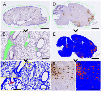
Histopathology and immunohistochemistry (IHC) are central to COVID-19 tissue evaluation. However, conventional manual scoring is limited by its semiquantitative nature and potential observer variability. In this retrospective study of experimentally infected mice, we implemented a semi-automated digital pathology workflow using commercially available software to quantitatively assess SARS-CoV-2 nucleocapsid (N) immunolabeling in lung (n = 135) and brain (n = 67) tissues. The performance of digital quantification was evaluated against conventional manual scoring, and its association with virological and pathological parameters was assessed across different stages of disease progression. Digital IHC quantification demonstrated strong agreement with manual scoring in both lung [R = 0.94, p < 0.0001, concordance correlation coefficient (CCC) = 0.95] and brain [R = 0.98, p < 0.0001; CCC = 0.977], demonstrating concordance between the two approaches. In addition, digital SARS-CoV-2 N immunolabeling quantification showed significant positive correlations with viral RNA levels, infectious viral titers, and histopathological scores, supporting its association with independent measures of SARS-CoV-2 infection and lesions. Although computational image analysis requires additional infrastructure, technical expertise, and increased analysis time, it provides an objective continuous quantitative measure of SARS-CoV-2 N immunolabeling that complements conventional manual assessment and may reduce observer-dependent variability.
Viruses
14 October 2026