


Toxins 2017, 9(10), 308; https://doi.org/10.3390/toxins9100308 - 30 Sep 2017
Cited by 30 | Viewed by 6709
Abstract
►
Show Figures
Extracellular toxins released by marine toxigenic algae into the marine environment have attracted increasing attention in recent years. In this study, profiling, characterization and quantification of extracellular toxin compounds associated with diarrhetic shellfish poison (DSP) in the culture medium of toxin-producing dinoflagellates were
[...] Read more.
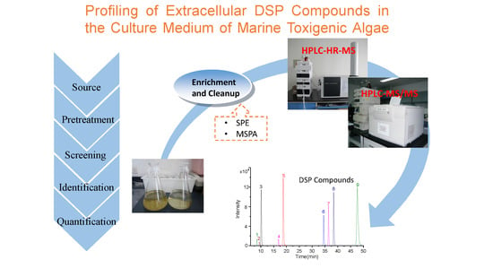
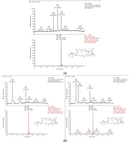
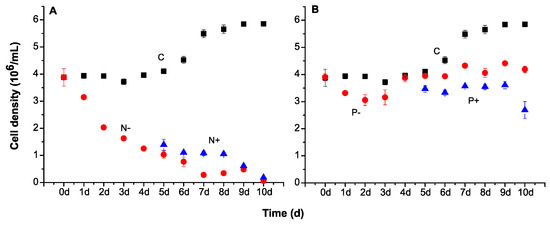
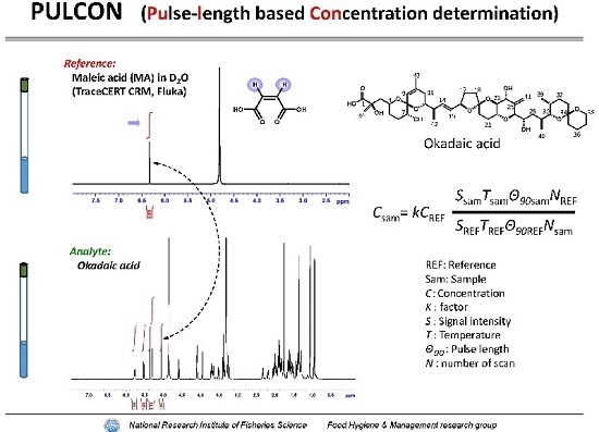
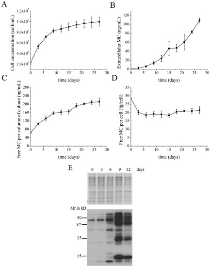
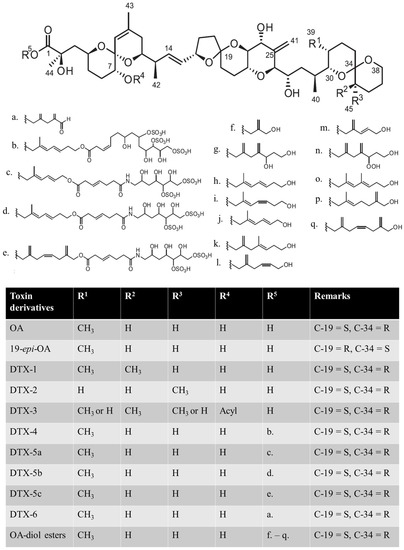
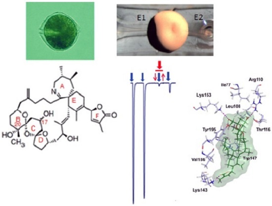
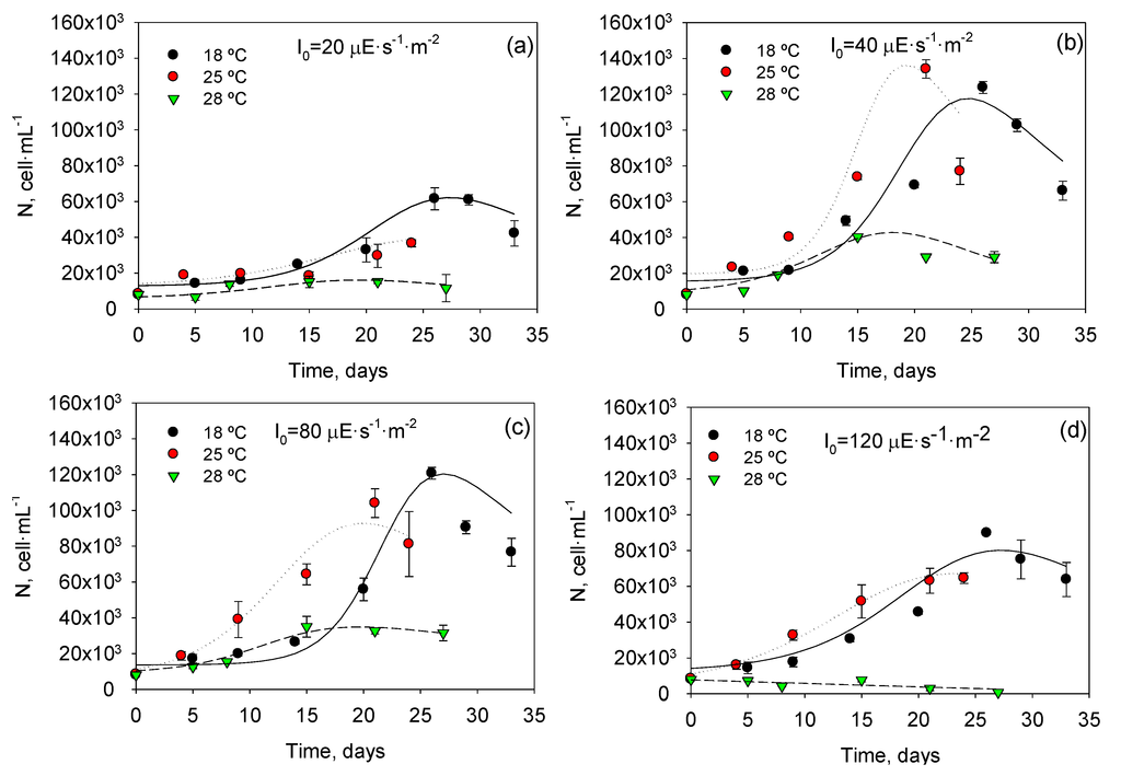
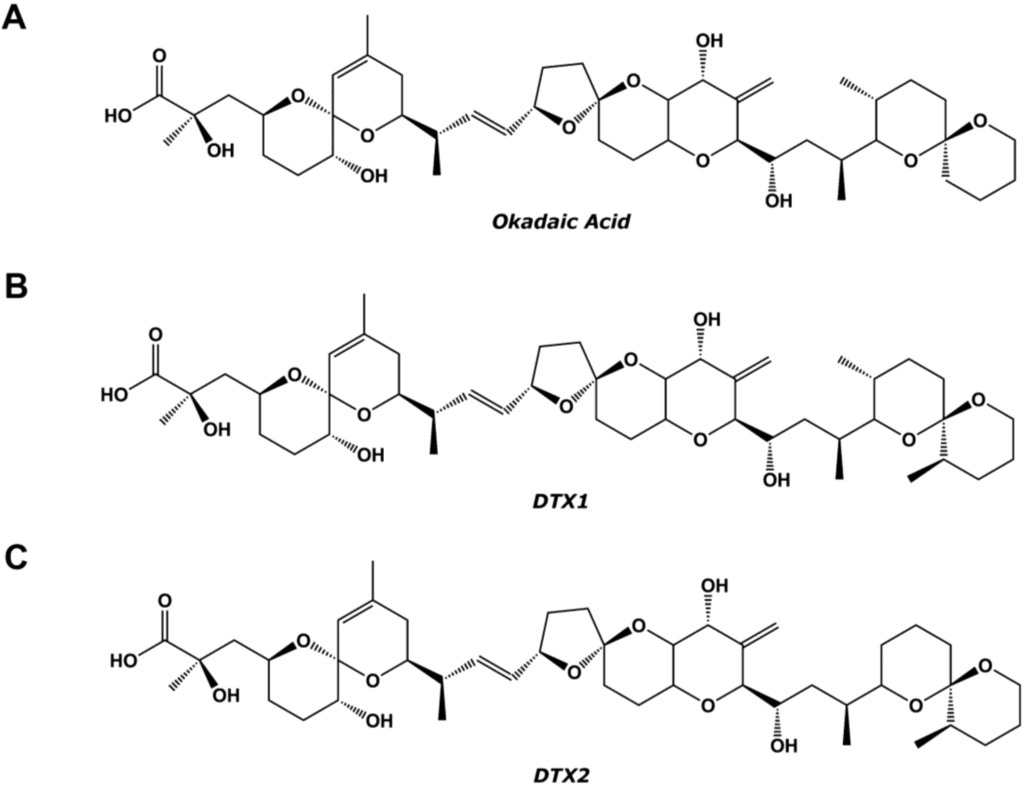
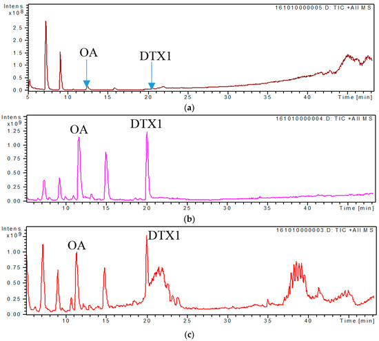
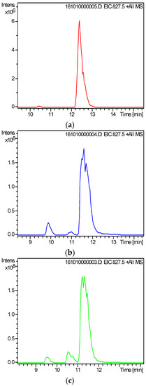
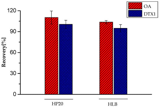
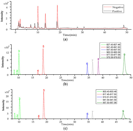
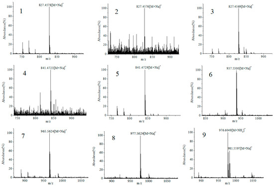
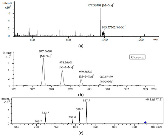
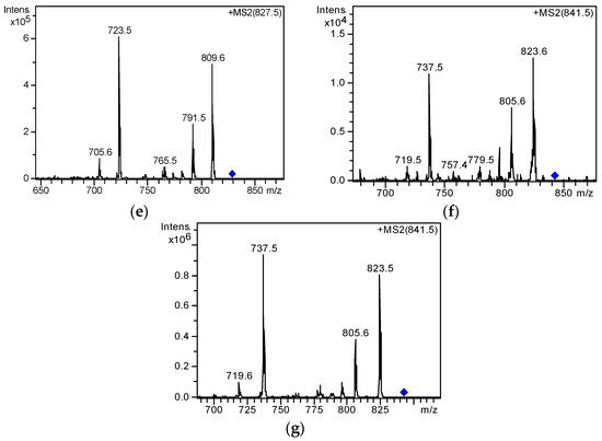
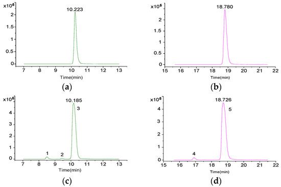
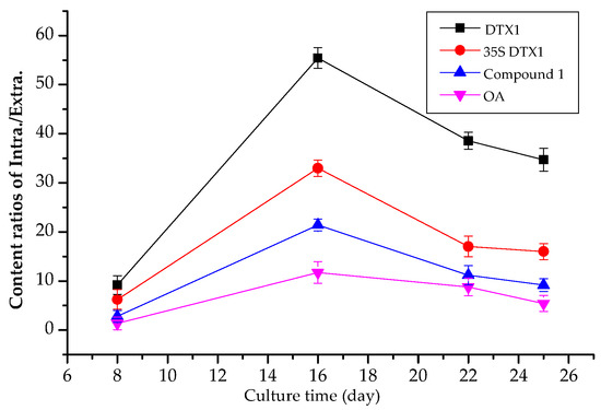
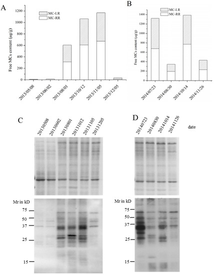
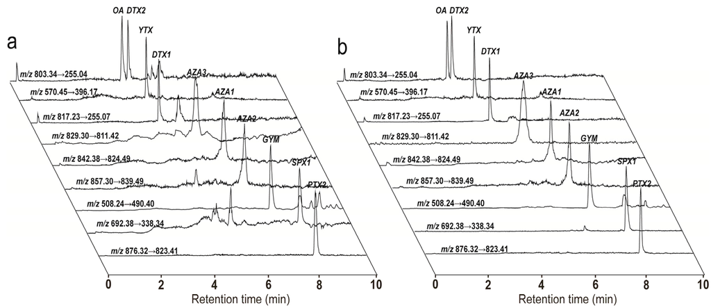
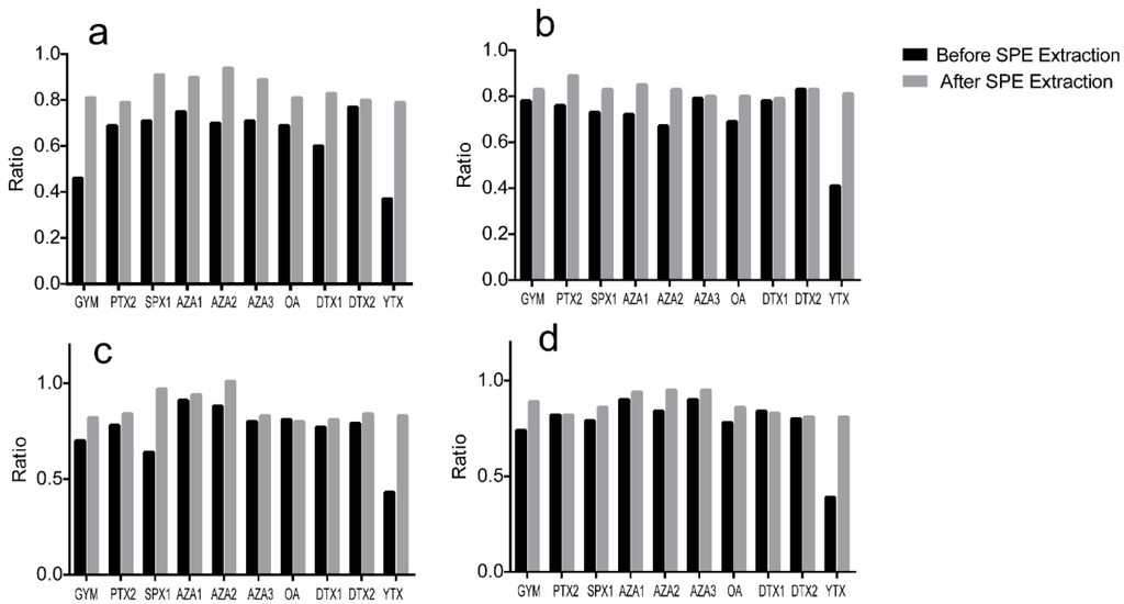
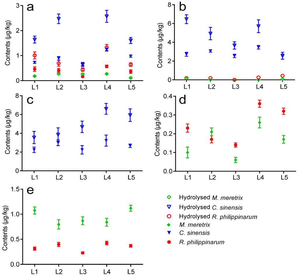
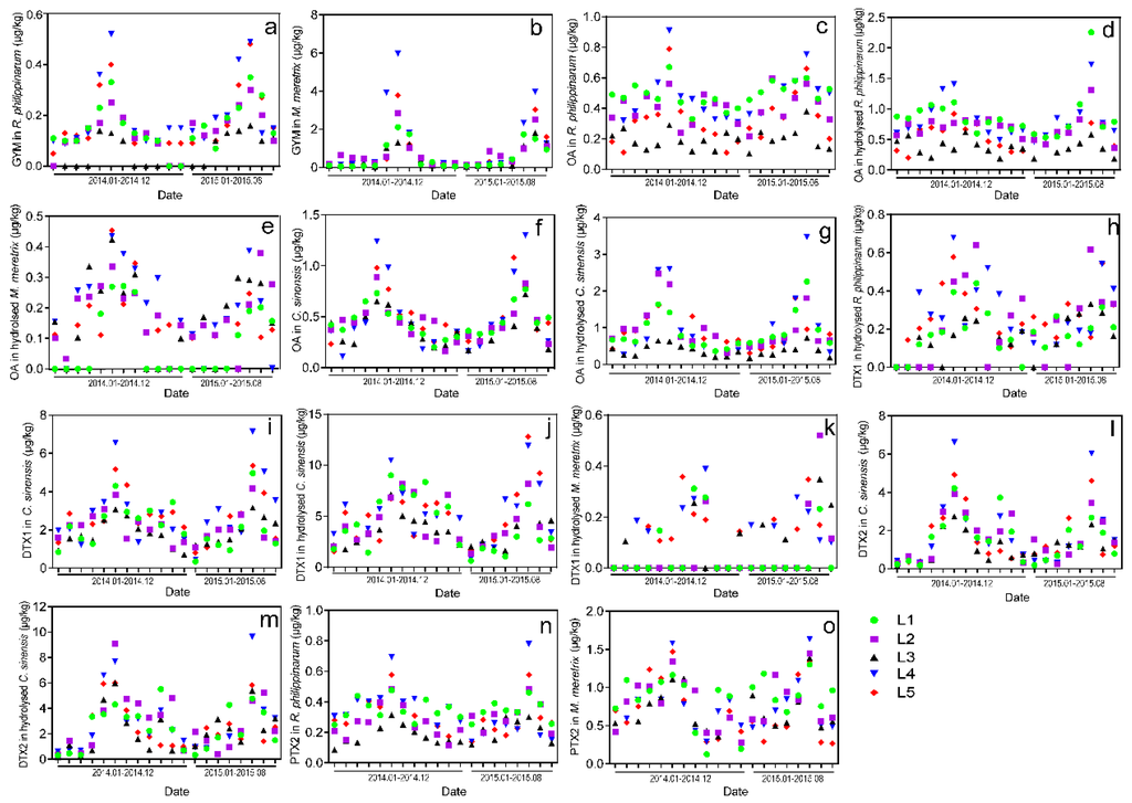
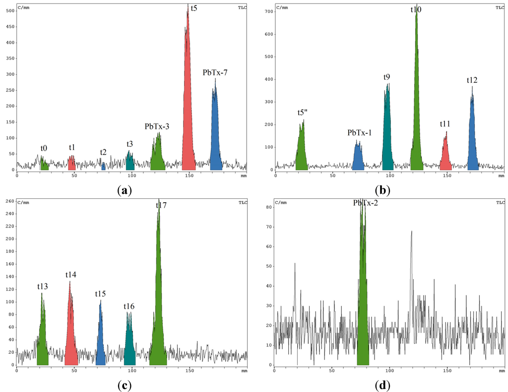
Extracellular toxins released by marine toxigenic algae into the marine environment have attracted increasing attention in recent years. In this study, profiling, characterization and quantification of extracellular toxin compounds associated with diarrhetic shellfish poison (DSP) in the culture medium of toxin-producing dinoflagellates were performed using high-performance liquid chromatography–high-resolution mass spectrometry/tandem mass spectrometry for the first time. Results showed that solid-phase extraction can effectively enrich and clean the DSP compounds in the culture medium of Prorocentrum lima (P. lima), and the proposed method achieved satisfactory recoveries (94.80%–100.58%) and repeatability (relative standard deviation ≤9.27%). Commercial software associated with the accurate mass information of known DSP toxins and their derivatives was used to screen and identify DSP compounds. Nine extracellular DSP compounds were identified, of which seven toxins (including OA-D7b, OA-D9b, OA-D10a/b, and so on) were found in the culture medium of P. lima for the first time. The results of quantitative analysis showed that the contents of extracellular DSP compounds in P. lima culture medium were relatively high, and the types and contents of intracellular and extracellular toxins apparently varied in the different growth stages of P. lima. The concentrations of extracellular okadaic acid and dinophysistoxin-1 were within 19.9–34.0 and 15.2–27.9 μg/L, respectively. The total concentration of the DSP compounds was within the range of 57.70–79.63 μg/L. The results showed that the proposed method is an effective tool for profiling the extracellular DSP compounds in the culture medium of marine toxigenic algae.
Full article
Graphical abstract


