


Biomolecules 2026, 16(5), 723; https://doi.org/10.3390/biom16050723 (registering DOI) - 14 May 2026
Abstract
Background: Hepatocellular carcinoma (HCC) shows a high rate of early recurrence after curative resection, indicating a critical contribution of tumor microenvironment-driven molecular mechanisms. Early recurrence of hepatocellular carcinoma is defined as recurrence within 6 months after curative resection, with a prevalence exceeding 30%.
[...] Read more.
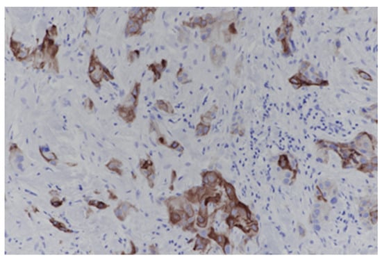
Background: Hepatocellular carcinoma (HCC) shows a high rate of early recurrence after curative resection, indicating a critical contribution of tumor microenvironment-driven molecular mechanisms. Early recurrence of hepatocellular carcinoma is defined as recurrence within 6 months after curative resection, with a prevalence exceeding 30%. Hypoxia signaling and immune dysregulation have been implicated, yet their compartment-specific relevance remains unclear. Methods: This multicenter nested case–control study included 49 HCC patients to evaluate associations between hypoxia-inducible factor-1 alpha (HIF-1α), vascular endothelial growth factor (VEGF), tumor-infiltrating lymphocytes (TILs), CD4+ T cells, CD8+ T cells, regulatory T cells (Tregs), programmed cell death protein 1 (PD-1), and programmed death-ligand 1 (PD-L1) and early recurrence after resection. TIL density was assessed using hematoxylin and eosin staining, while immunohistochemistry was performed to quantify intratumoral and peritumoral expression of the studied markers. Receiver operating characteristic (ROC) curve analysis was used to evaluate the predictive performance. Recurrence-free survival (RFS) was analyzed using the Kaplan–Meier, and independent predictors were identified using multivariate Cox proportional hazards regression. Results: Early recurrence occurred in 11 of 49 patients (22.4%) of Child–Pugh A patients. Recurrent tumors were characterized by elevated VEGF expression despite absent HIF-1α, alongside significant depletion of intratumoral TILs (HR 5.02; 95% CI 1.09–23.26), CD4+ (HR 7.68; 95% CI 1.66–35.60) and CD8+ cells (HR 6.68; 95% CI 1.77–25.23) and reduced peritumoral CD8+ infiltration (HR 4.20; 95% CI 1.11–15.91). Multivariable analysis identified low intratumoral CD4+ (HR 7.98; 95% CI 1.63–39.07) and reduced peritumoral CD8+ expression (HR 4.98; 95% CI 1.14–21.70) as independent predictors, whereas HIF-1α, VEGF, Treg, PD-1, and PD-L1 were not significantly associated. Conclusions: Early HCC recurrence shows HIF-1α-independent angiogenesis alongside spatial immune depletion, supporting integrated immune profiling over single angiogenic markers.
Full article
(This article belongs to the Special Issue Tumor Microenvironment, Immunology and Precision Medicine of Liver Cancer)
►
Show Figures
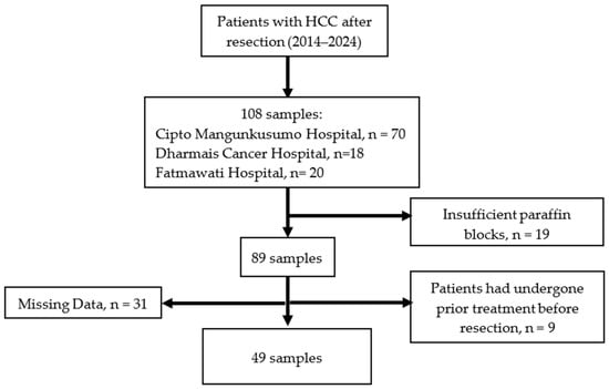
Figure 1


