

Prosthesis, Volume 7, Issue 5 (October 2025) – 28 articles
Cover Story (view full-size image):
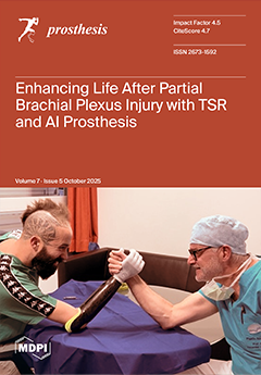
Upper limb amputation imposes major physical and psychological challenges, particularly in young, active individuals. This case describes the rehabilitation of a 33-year-old Italian Paralympic snowboard cross athlete who underwent elective transradial amputation after partial brachial plexus injury, followed by upper-limb-targeted sensory reinnervation (ulTSR) and fitting with the AI-driven Adam’s Hand prosthesis. The integration of ulTSR restored functional sensory feedback and enabled intuitive prosthetic control with minimal effort. Combined surgical and technological neurorehabilitation resulted in regained independence, enhanced dexterity, and markedly improved quality of life. View this paper
- Issues are regarded as officially published after their release is announced to the table of contents alert mailing list.
- You may sign up for e-mail alerts to receive table of contents of newly released issues.
- PDF is the official format for papers published in both, html and pdf forms. To view the papers in pdf format, click on the "PDF Full-text" link, and use the free Adobe Reader to open them.
Previous Issue
Next Issue