Article

Effect of Manual and Electronic Toothbrushes on Color Stability and Contact Profilometry of Different CAD/CAM Ceramic Materials After Immersion in Coffee for Varying Time Intervals

by
Mohammed M. Al Moaleem
1,* and
Manea Musa M. Alahmari
2
1
Department of Prosthetic Dental Science, College of Dentistry, Jazan University, Jazan 45142, Saudi Arabia
2
Department of Periodontics and Community Dental Science, College of Dentistry, King Khalid University, Abha 61421, Saudi Arabia
*
Author to whom correspondence should be addressed.
Prosthesis 2025, 7(5), 110; https://doi.org/10.3390/prosthesis7050110 (registering DOI)
Submission received: 19 July 2025 / Revised: 14 August 2025 / Accepted: 20 August 2025 / Published: 25 August 2025

Abstract

Aim: This study evaluated the effect of manual and electronic toothbrushes on the color stability (∆E*) and surface roughness (Ra) of four CAD/CAM ceramics after their immersion in coffee for 2 and 4 weeks. Methodology: A total of 160 specimens (Vitablocs Mark II, Ceramill Zolid zirconia, Vita Triluxe Forte, and IPS e.max CAD) were divided into four brushing subgroups (manual, sonic, oscillating–rotating, and ionic). The samples underwent daily coffee staining, thermocycling (5–55 °C), and twice-daily brushing. Color parameters (L, a, and b) were assessed and measured utilizing a spectrophotometer (Vita Easyshade) at baseline, 2 weeks, and 4 weeks. ∆E* was calculated using the CIEDE2000 formula, and surface roughness (Ra, µm) was assessed via contact profilometry at the study’s conclusion. Data were analyzed using Kruskal–Wallis and Mann–Whitney tests (α = 0.05). Results: Among the tested samples, IPS e.max ceramic with manual toothbrushing exhibited the highest ΔE* values after 2 and 4 weeks (∆E* = 4.424 and ∆E* = 4.802) of immersion. Moreover, Ceramill Zolid zirconia demonstrated the highest ΔE* values with ionic brushing (∆E* = 4.883 at 2 weeks; ΔE* = 4.760 at 4 weeks). Significant differences were observed among ceramics and cleaning methods, with manual/ionic brushing causing the greatest changes (p < 0.05). IPS e.max had the highest Ra with manual brushing (0.745–0.789 µm), whereas Ceramill Zolid zirconia with ionic brushing showed the highest Ra values among the electric methods (0.745–0.757 µm). Conclusions: Manual brushing induced clinically unacceptable color changes in IPS e.max CAD, whereas ionic brushing adversely affected Ceramill Zolid zirconia. All brushing methods increased surface roughness beyond acceptable limits.

1. Introduction

CAD/CAM dental ceramics are considered fundamental to modern prosthetic and restorative dentistry because of their biocompatibility, esthetic appeal, and durability [1]. Feldspathic porcelain, lithium disilicate glass ceramics, and zirconia-based dental restorations are extensively used in CAD/CAM systems, providing clinicians with reliable solutions for crowns, veneers, and other prostheses. These tooth-colored materials exhibit exceptional optical, esthetic, and mechanical capabilities, allowing them to withstand the challenges of the oral environment [2]. The long-term achievement of these restorations depends not only on their initial properties but also on their resistance to environmental challenges, including dietary stains and oral hygiene practices [3,4,5].
Mechanical toothbrushing is essential for oral health and stain removal, but it may affect ceramic surfaces [6]. Research has shown that manual and electronic toothbrushing can alter the surface characteristics and color stability of dental ceramics, but the results differ. Al Ahmari et al. found significant color variation in Vita Triluxe, minimal changes in IPS e.max CAD, and increased roughness in Vita Enamic after regular brushing [4]. Kaynak Öztürk et al. observed that coffee immersion causes significant color changes, but subsequent brushing reduces color parameters such as ΔE*, L, a, and b [7]. Al Anazi reported that coffee immersion followed by brushing decreases ceramic crown staining to an acceptable level (ΔE = 2.7) [8].
Extrinsically stained feldspathic porcelain shows significant roughness and color changes after simulated brushing equivalent to 8.5 years of use [9,10]. Pouranfar et al. suggested that brushing removes extrinsic stains from pressable ceramics over a simulated duration of 10–12 years, unless protected by glaze [11]. Other studies found no significant effect of simulated brushing (equivalent to 5 years) on the roughness, microhardness, or optical stability of lithium disilicate prostheses [12,13].
The emergence of electronic toothbrushes, featuring technologies such as oscillating–rotating, sonic, or ionic action, has introduced an additional variable [14]. Studies comparing electronic toothbrushes with manual toothbrushes often report the superior efficacy of modern electronic toothbrushes, particularly oscillating–rotating models, in reducing plaque and gingivitis [4,15]. A recent systematic review highlighted the significant beneficial effect of oscillating–rotating electronic toothbrushes on plaque removal compared with high-frequency sonic electronic toothbrushes [16]. Singh et al. observed no significant differences in plaque reduction, gingivitis, or sulcular bleeding between sonic and ionic electronic toothbrushes [17]. However, reports comparing different types of oral hygiene tools, which affect the mechanical and color stability of CAD/CAM ceramic materials, are lacking; thus, data on the effects of different toothbrushing devices and current developments in CAD/CAM materials are limited, even though patients frequently change between manual, sonic, oscillating–rotating, and ionic brushes without being aware of possible long-term effects on ceramic restorations [18,19].
The stability of the optical parameters and properties of dental ceramics, which is crucial for esthetic longevity and clinical success, is significantly influenced by surface characteristics such as roughness and free energy [20]. Accurate color assessment relies on modern instruments such as colorimeters and spectrophotometers, utilizing the standardized CIE L*a*b* system, where L* denotes lightness, and a* and b* represent chromaticity coordinates [21]. However, understanding isolated changes in L, a, or b can be challenging, leading to the widespread use of the mean color difference (ΔE), a single metric derived from these coordinates, to measure observable variations [22,23]. Although the CIELAB (ΔE*ab) and CIEDE2000 (ΔE00) formulas are frequently employed in the field, the latter is more accurate and clinically relevant, incorporating perceptual weighting functions. A ΔE00 below 1.72 is generally imperceptible, while values above 4.08 are considered clinically unacceptable [24,25].
Among the most common dietary challenges to color stability is coffee consumption, a globally prevalent habit [26]. Numerous studies have confirmed that coffee significantly affects color stability in different ceramic types, regardless of whether they are glazed or polished [27,28]. Furthermore, multiple investigations have reported that coffee immersion significantly alters the surface properties of zirconia-based ceramics [29,30]. Coffee contains potent chromogens and tannins that readily adhere to dental surfaces, leading to progressive extrinsic discoloration [31].
This study aimed to investigate and compare the influence of simulated brushing with manual and electronic toothbrushes on the color stability (ΔE*) and contact profilometry (Ra) of four types of CAD/CAM ceramic materials (Mark II, Ceramill Zolid zirconia, Triluxe, and IPS e.max) at different time intervals. The null hypothesis is the absence of significant differences in ΔE* between manual and electronic toothbrushes at 2 and 4 weeks.

2. Materials and Methods

2.1. Study Design and Sample Size Calculation

This study was carried out to investigate and compare the influence of simulated brushing with manual and electronic toothbrushes on the color stability (ΔE*) and contact profilometry (Ra) of four types of CAD/CAM ceramic materials at 2 and 4 weeks. The specimen size and number were calculated based on the estimated effect size between groups, according to the literature for color stability (ΔE*) [3,4]. Contact profilometry (Ra) [32] for each group (n = 40) was estimated using G*Power software (version 3.1; University of Dusseldorf), with an effect size (d) of 0.5, α of 0.05, and 1-β (power) of 0.85.

2.2. Sample Selection Rationale

The four CAD/CAM materials, Vitablocs Mark II, Ceramill Zolid zirconia, Vita Triluxe Forte, and IPS e.max CAD, were selected for their broad clinical use in anterior and posterior prostheses [1,2] and distinct microstructures (Table 1). Mark II (feldspathic glass) and IPS e.max (lithium disilicate) represent highly esthetic options with varying stain susceptibility [3,5], while Ceramill Zolid (zirconia) and Triluxe Forte (feldspathic hybrid) offer high strength but differ in abrasion resistance [9]. This selection enables direct comparison of how material composition influences color stability and surface roughness under mechanical and chemical challenges [3,5,9].

2.3. Sample Preparations

A total of 160 specimens were prepared from four different CAD/CAM prosthetic materials (Vitablocs Mark II, Ceramill Zolid multilayer PS, Vita Triluxe Forte, and IPS e.max CAD). The dimensions of round specimens were standardized to a uniform size of 10 ± 0.25 mm in diameter and a thickness of 2.0 ± 0.25 mm. The samples were finished and polished using 300–800-grit silicon carbide paper (Dentsply Prosthetics Brasseler, Savannah, GA, USA). After cleaning with distilled water in an ultrasonic machine, the circular specimens were washed with isopropanol alcohol for 5 min to remove any grease residue and then dried with compressed air.
The specimens were manufactured from CAD/CAM blocks and processed according to the respective manufacturers’ instructions. VITABLOCS Mark II and VITA Triluxe Forte specimens were glazed as recommended by the manufacturer, while VITA Suprinity and the lithium disilicate glass ceramic blocks were crystallized using the manufacturer-specified firing programs in a Programat P310 dental furnace (Ivoclar Vivadent AG, Schaan, Liechtenstein). All specimens were milled using a CAD/CAM system (Amann Girrbach GmbH, Durrenweg 40, 75177 Pforzheim, Germany). Prior to staining and brushing, specimens were ultrasonically cleaned in sterile water for 10 min and dried with compressed air for 20 s to remove residual debris.

2.4. Sample Grouping

On the basis of the type of oral hygiene aids (manual and regular toothbrushes, sonic toothbrushes, oscillating toothbrushes, and ionic electric toothbrushes), circular specimens for each material group were further divided into four equal subgroups of 10 specimens each.

2.5. Mean Color Evaluations

M. A. A measured the color parameters under uniform measurement settings with a spectrophotometer (Vita Easyshade Compact version V; Vita Zahnfabrik H. Rauter GmbH & Co. KG, Bad Säckingen, Germany) with a 4 mm diameter tip.
Color change (ΔE*) was calculated utilizing the CIEDE2000 color evaluation system at each evaluation time point. ΔE* between the time points was calculated and expressed as the change between baseline and 2 weeks (ΔE* for 2 weeks) and between baseline and 4 weeks (ΔE* for 4 weeks). The following equation was used for the calculation, where ΔL’, ΔC’, and ΔH’ represent luminosity, chroma, and hue, respectively; RT refers to the interaction between chroma and hue in the blue spectrum; SL, SC, and SH are weights utilized to adjust the overall color difference in L*, a*, and b* coordinates; and KL, KC, and KH are correction terms for investigational settings [32]. In this laboratory study, the parametric factors of the ΔE* difference formula were set to 1. Each color parameter was measured 3 times for each sample at a consistent daily time prior to simulating brushing with different oral hygiene aids, and the average was recorded to characterize the different color parameters [3,4]. The ΔE* color difference was calculated by means of the following equation [32]:
C I E D E   2000 = Δ L K L S L 2 + Δ C K C S C 2 + Δ H K H S H 2 + R T Δ C K C S C + Δ H K H S H
The tip of the handpiece was placed at 90° relative to the stained and brushed surface of each circular CAD/CAM disk, and all measurements were conducted under similar lighting settings. The device’s probe tip was inserted into its calibration orifice after each measurement to calibrate the device according to the manufacturer’s recommendations [3,4,32].

2.6. Coffee Staining

Throughout the 2- and 4-week brushing periods, all samples were immersed in a coffee solution, which was changed every 12 h, coinciding with the brushing time [5]. Moreover, the samples underwent thermocycling with 100 cycles per day. After undergoing brushing simulation, the circular samples in each ceramic group were removed from the coffee containers and subjected to thermocycling, alternating between 5 °C cold water and 55 °C hot water for staining durations of 2 and 4 weeks.

2.7. Brushing Protocol for Specimens

Four oral hygiene tools (one manual and three electronic) were applied according to the manufacturers’ specifications. The manual toothbrush utilized was the Oral-B Soft Manual Toothbrush, Charcoal (Procter & Gamble Co., Cincinnati, OH, USA), functioning via manual circular motion for 20 s [3,4]. The following electronic devices were used: (1) Sonicare® Technology (Philips Oral Healthcare, Snoqualmie, WA, USA) with side-to-side oscillation; (2) Oral-B Smart Series 5000 iO (Procter & Gamble Co., Cincinnati, OH, USA), employing oscillating–rotating pulsation; and (3) HyG ionic (Hukuba Dental Corporation, Nagareyama, Chiba, Japan), functioning via ionic technology. All devices were utilized twice every 24 h for 60 s with dentifrice (Signal® toothpaste) with a Relative Dentin Abrasivity (RDA) value of 70. Each 30 s brushing segment utilized was performed using a toothpaste slurry prepared by mixing 0.25 g of toothpaste with deionized water at a 1:3 ratio for 20 s [3,4].
Brushing was performed using a V8 brushing simulator (JWE GmbH, Eschborn, Germany). The simulator arm was fitted with an inline load cell and force was recorded continuously; the load cell was calibrated with standard masses prior to each session and the brushing force was set to 2.0 ± 0.2 kg in unidirectional circular motions, performed every 12 h (simulating morning/evening intervals) [3,4]. Following brushing, specimens were rinsed with running water, immersed and stained in coffee solution (which was changed every 12 h in accordance with the brushing time), and stored in dark glass containers at room temperature.
All immersed samples from the different simulated brushing groups were extracted from their respective staining media. The circular CAD/CAM disks were immersed in distilled water 10 times, wiped with tissue paper, and left to dry before contact profilometry assessment.

2.8. Contact Profilometry Assessment

Surface profilometry analyses of the CAD/CAM specimens were conducted using a profilometer (Contour GT-K1, Bruker Nano GmbH, Berlin, Germany). Three samples from each ceramic group and brushing simulation type were selected randomly for Ra assessment at the end of the study. The assessments were performed at a speed of 0.25 mm/sn and with a cutoff of 0.80 mm. Three readings were obtained at the center of each circular sample, and the average of these values was recorded as Ra in micrometers [32]. The details of the materials and devices used in this laboratory study are presented in Table 1.

2.9. Statistical Analysis

The mean ΔL, Δa, Δb, and ΔE* values after 2 and 4 weeks were calculated. Statistical Package for Social Science (SPSS) version 26.0 (SPSS Inc., Chicago, IL, USA) was used to input and analyze the data. The Shapiro–Wilk test indicated that the majority of the data were not normally distributed (p < 0.05). Therefore, non-parametric tests were used. The Kruskal–Wallis test was used to compare the ΔE* values between ceramic and brushing systems. The Mann–Whitney post hoc test was used for pairwise comparisons between every ceramic–brushing combination. p < 0.05 was considered a cutoff point for statistical significance.

3. Results

3.1. Mean Color Change

The IPS e.max ceramic with the manual toothbrush and the Ceramill Zolid zirconia ceramic with the ionic electric toothbrush exceeded the upper limit of the clinically acceptable ΔE* value (4.2) after 2 and 4 weeks. The Mark II ceramic with the sonic electric toothbrush and the IPS e.max ceramic with the oscillating electric and ionic electric toothbrushes had ΔE* values that fell into a clinically acceptable range (2.8 and 4.2) after 4 weeks. However, for the other ceramic and cleaning type combinations, ΔE* fell below the lower limit (2.5) of acceptable color change, indicating relatively minor color changes (Figure 1).
After 2 weeks, the Kruskal–Wallis test indicated statistically significant differences in the mean color change among ceramic types when cleaned with the manual toothbrush (p = 0.014), sonic electric toothbrush (p = 0.041), oscillating electric toothbrush (p = 0.001), and ionic electric toothbrush (p < 0.001). The same test also indicated statistically significant differences in ΔE* values among the ceramic types when cleaned with the manual toothbrush (p = 0.040) and sonic electric toothbrush (p = 0.026), as shown in Table 2.
After both 2 and 4 weeks of cleaning with various toothbrush types, significant differences in color changes (ΔE*) were observed among the different ceramic materials as shown in Table 3. With the manual toothbrush, Mark II showed significant differences in ΔE* compared to Ceramill Zolid zirconia, IPS e.max, and Triluxe at both time points. The sonic electric toothbrush revealed significant differences between Mark II and Triluxe, as well as between Ceramill Zolid zirconia and both Triluxe and IPS e.max. The oscillating electric toothbrush resulted in several highly significant differences, notably between Mark II and Ceramill Zolid zirconia, Mark II and Triluxe, and between Triluxe and IPS e.max. The ionic electric toothbrush also showed significant differences between Mark II and Ceramill Zolid zirconia, Mark II and Triluxe, and between Triluxe and IPS e.max.
After 2 weeks, the Kruskal–Wallis test indicated statistically significant differences in ΔE* of the Mark II (p = 0.036), Ceramill Zolid zirconia (p = 0.042), and Triluxe ceramics (p = 0.004) among the cleaning types. After 4 weeks, the Mark II and IPS e.max ceramic continued to show statistically significant differences in ΔE* between the cleaning types (p = 0.002 and 0.026, respectively), as shown in Table 4.
After 4 weeks, pairwise comparisons using the Mann–Whitney U test revealed statistically significant differences in ΔE* of the Mark II ceramic between the manual and sonic electric toothbrushes (p = 0.027), between the sonic electric and oscillating electric toothbrushes (p = 0.012), and between the sonic electric and ionic electric toothbrushes (p = 0.027). The ionic electric toothbrush exhibited significant differences in ΔE* of Ceramill Zolid zirconia when compared with the manual (p = 0.048) and sonic electric toothbrushes (p = 0.040).
Moreover, the manual toothbrush exhibited significant differences in ΔE* of Triluxe when compared with the sonic electric (p = 0.008) and oscillating electric toothbrushes (p = 0.002). The oscillating electric toothbrush exhibited a significant difference in ΔE* of Triluxe when compared with the ionic electric toothbrush (p = 0.048). The manual toothbrush exhibited significant differences in ΔE* of IPS e.max when compared with the sonic electric, oscillating electric, and ionic electric toothbrushes (all p = 0.048; Table 5).
A comparison of ΔE* after 2 and 4 weeks of immersion in coffee revealed significant differences in the Mark II ceramic when cleaned with the manual (p < 0.001), oscillating electric (p < 0.001), and ionic electric toothbrushes (p = 0.001). In the zircon ceramic, significant differences in ΔE* were found between 2 and 4 weeks when subjected to cleaning with the manual (p = 0.048), sonic electric (p = 0.010), and oscillating electric toothbrushes (p = 0.010). Similarly, the Triluxe ceramic exhibited significant differences in ΔE* between 2 and 4 weeks when cleaned with the sonic electric (p = 0.010), oscillating electric (p = 0.027), and ionic electric toothbrushes (p = 0.010). Finally, the IPS e.max ceramic showed significant differences in ΔE* between 2 and 4 weeks when cleaned with the oscillating electric (p = 0.006) and ionic electric toothbrushes (p = 0.008), as shown in Table 6.

3.2. Contact Profilometry

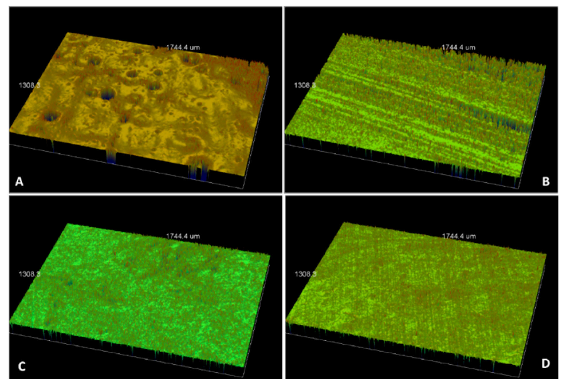
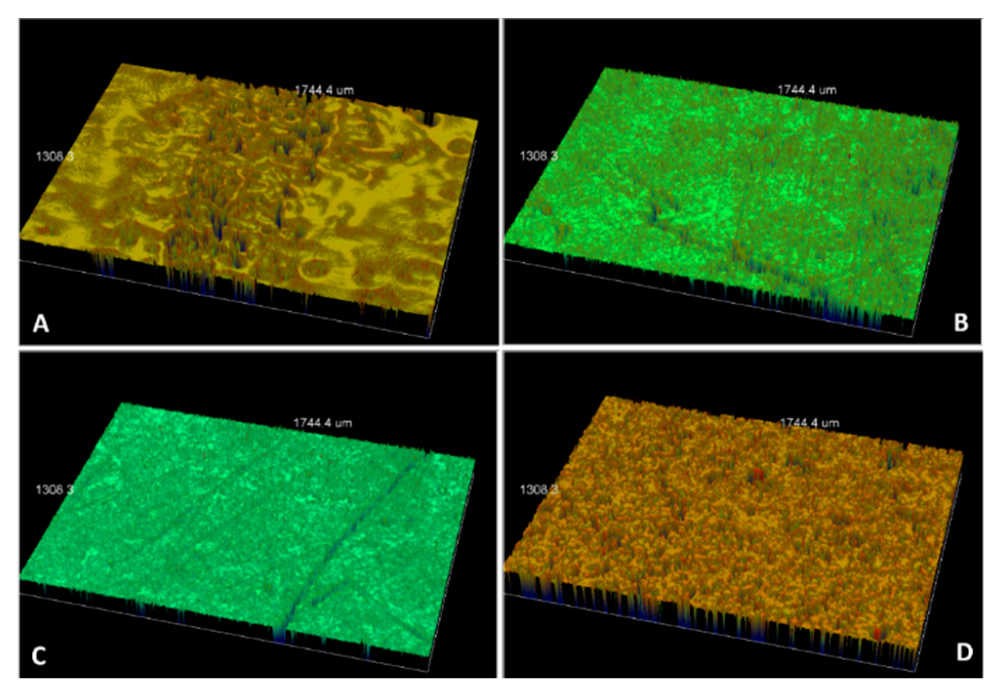
For contact profilometry (Ra) in micrometers, the manual toothbrush exhibited the highest Ra (0.745 and 0.789) for IPS e.max. Conversely, the HyG ionic electric toothbrush for Ceramill Zolid zirconia documented the highest Ra values (0.745 and 0.757) at 2 and 4 weeks, respectively. For the sonic and oscillating electric toothbrushes, the Vita Triluxe samples demonstrated the highest Ra values of 0.483 and 0.682 at 2 weeks, respectively, and 0.438 and 0.453 at 4 weeks, respectively, as shown in Table 7 Overall, all the recorded values exceeded the acceptable Ra value of 20 µm (Table 7). Figure 2 and Figure 3 present Ra images after 2 and 4 weeks of manual toothbrushing.

4. Discussion

This study was conducted to investigate and compare the effects of simulated brushing with manual and electronic toothbrushes on the color stability (ΔE*) and contact profilometry (Ra) of a wide range of commonly used CAD/CAM ceramic materials available in the local market.
Different toothbrushing systems are included in several laboratory aging protocols designed to replicate clinical conditions that may affect the esthetics and durability of ceramic restorations [33]. Evaluating material performance after simulated toothbrushing can provide valuable insights into their clinical behavior [34]. Although this method often evaluates material wear, its influence on surface roughness requires further attention [9,10]. Dental restoration surfaces, exposed to the oral environment, are susceptible to changes induced by prolonged brushing [9,10,33]. Nonetheless, there are limited data evaluating the effects of different brushing devices on the color stability and surface profilometry of diverse CAD/CAM ceramic materials across various time intervals.
Color changes were assessed using a spectrophotometer to minimize human error and ensure objective measurements, with clinically acceptable ΔE* thresholds ranging from 1.7 units to 3.3 units, whereas values exceeding 3.3 were deemed unacceptable. Conversely, other studies indicated that the perceptible and clinically acceptable ΔE00 ranges from 2.8 units to 4.2 units [5,26,27]. In this investigation, for most of the ceramic–cleaning combinations, the materials exhibited ΔE00 values within the acceptable range, except for IPS e.max CAD and Ceramill Zolid zirconia when subjected to manual and ionic brushing, respectively. These groups demonstrated ΔE* values exceeding the acceptable limit (ΔE* ≥ 4) (Figure 1).
Contrary to the null hypothesis, which posited no significant differences in ΔE* between manual and electronic toothbrushes at 2 and 4 weeks, the current results demonstrated statistically significant differences (p < 0.05) for specific combinations of ceramic–brushing systems. These differences may be linked to the optical properties of the ceramics, which are affected by factors such as processing methods, microstructure, chemical composition, and physical characteristics—including crystal size, homogeneity, refractive index, and surface microporosity [35]. Given these outcomes, the null hypothesis was rejected, highlighting that the toothbrush type significantly influences the color stability of CAD/CAM ceramics.
This result was consistent with previous studies indicating that ΔE* values fluctuate across various types of ceramics after toothbrushing simulation [3,4,9,10,12,13]. Anazi et al. noted that coffee immersion followed by brushing reduces the staining of ceramic crowns to an acceptable ΔE* threshold (2.7), with a statistically significant increase in luminosity post-brushing (p < 0.05) [8]. Conversely, Al-Ahmari reported that IPS e.max CAD displays enhanced color stability when brushed with different Miswak-derived oral hygiene products [3]. Such discrepancies may arise from differences in experimental conditions, material composition, or brushing protocols.
Toothbrushing is the predominant oral hygiene practice, but its effectiveness in plaque removal varies depending on factors such as the brush design, technique, duration, and frequency. Since their introduction in the 1960s, power toothbrushes have evolved from simple mechanical movements to advanced sonic and oscillating–rotating technologies [15,16]. Although studies suggest that manual and electric toothbrushes are similarly effective in plaque removal [36,37], the findings of this study on cleaning methods revealed significant differences in their effects on dental ceramics, as seen in Table 5. Ignoring these material-specific vulnerabilities risks the durability and esthetics of restorations, necessitating immediate attention in clinical practice [12,13]. Other studies have found no significant differences in color stability (ΔE*) among various CAD/CAM ceramic materials after simulated toothbrushing, with values remaining within clinically acceptable limits [4,6,9]. These discrepancies likely stem from methodological variation—staining solution composition and pH, dentifrice abrasivity (RDA), brushing force, bristle type, test duration, and surface condition (glazed versus polished or unglazed) Notably, Mahrous et al. observed significant ΔE* changes when non-glazed surfaces were tested, which supports the influence of surface finish on stain uptake and wear and is consistent with our observations [18].
A microscopically smooth surface is essential for achieving color stability in dental materials, as it may cause enhanced stain accumulation [5]. Restorations with increased roughness enhance plaque accumulation, and the increased adhesion of oral microorganisms, which in turn contribute to extrinsic staining and increase the risk of localized gingival inflammation or soft-tissue recession over time [17,36]. The generally accepted threshold for surface roughness (Ra) is 0.2 µm [5,6]. In the present study, most of the tested ceramics resulted in Ra values exceeding this limit across various toothbrushing methods, with Vitablocs Mark II demonstrating the lowest Ra value (≥0.257 µm). These results support the conclusion that toothbrushing adversely impacts glazed ceramics, consistent with the findings of Al Ahmari et al., who observed significant Ra variations in Vita Enamic when different oral hygiene tools were used [4].
The outcome of the current study differs from that of Kaynak Ozturk et al., who examined how surface finishing, coffee immersion, and toothbrushing affect the surface roughness, gloss, and color stability of resin matrix ceramics. They found that toothbrushing produces smooth surfaces and increased gloss, as shown by AFM images [7]. Similarly, Mahrous et al. observed reduced surface roughness for Emax CAD, Emax Press, and LiSi Press specimens after electric toothbrushing, although no significant differences were noted among groups after mechanical brushing (p = 0.842) [18]. These results were consistent with previous studies indicating minimal changes in the roughness of stained lithium disilicate ceramics after brushing [38,39]. The differences observed in this study may arise from variations in experimental conditions, such as high brushing forces or the use of stiff nylon toothbrushes.
The European Federation of Periodontology (EFP) emphasizes that mechanical plaque control is fundamental to the prevention and management of gingival and periodontal diseases, and recommends twice-daily mechanical toothbrushing (manual or powered) as part of routine oral hygiene [40]. Clinicians should balance the EFP-recommended goals of effective plaque removal with the preservation of restorative surfaces by advising a gentle brushing technique, selecting brush heads and modes that minimize abrasive contact with ceramic restorations, reinforcing interdental cleaning, and monitoring restorations for early surface degradation. Incorporating these recommendations into patient education can help optimize periodontal outcomes especially near restoration margins while mitigating adverse effects on ceramic restorations [40,41].
A certain limitation of the present study was its in vitro design. The laboratory environment cannot fully replicate complex oral conditions, such as saliva, biofilm dynamics, and variable masticatory forces. The use of flat, polished ceramic disks might be a disadvantage, as they lack the geometric complexity and occlusal adjustments of clinical restorations. Staining relied solely on immersion in Nescafé 3-in-1 coffee, which cannot reflect the diverse dietary chromogens or intermittent exposure patterns encountered clinically. We used a 4-week evaluation period, which was not sufficient to predict long-term clinical outcomes. The findings are also specific to the four tested CAD/CAM ceramics and the particular manual/electric toothbrush models used, limiting broad applicability. Consequently, extrapolating these results to clinical performance requires caution. Further research should incorporate natural oral environments, diverse restoration geometries, complex staining protocols, long observation periods, and additional ceramic/toothbrush combinations to validate these findings. Further research should also should assess microbial adhesion alongside color change and surface roughness, to better understand the interplay between mechanical cleaning, ceramic degradation, and periodontal health.

5. Conclusions

Considering the limitations of this in-vitro study, our findings show material- and toothbrush-dependent differences in color change and surface alteration after simulated staining and brushing; specifically, IPS e.max CAD and Ceramill Zolid zirconia exhibited greater, perceptible discoloration and surface alteration with certain brush types. Clinicians should therefore consider these differential susceptibilities when selecting restorative materials for patients with high exposure to staining beverages and recognize toothbrush choice and brushing technique as modifiable factors that may influence long-term esthetic outcomes.

Author Contributions

Conceptualization, M.M.A.M. and M.M.M.A.; methodology, M.M.A.M.; software, M.M.M.A.; validation, M.M.A.M. and M.M.M.A.; formal analysis, M.M.A.M.; investigation, M.M.M.A.; resources, M.M.M.A.; data curation, M.M.A.M.; writing—original draft preparation, M.M.A.M.; writing—review and editing, M.M.A.M.; visualization, M.M.M.A.; project administration, M.M.M.A.; funding acquisition, M.M.M.A. All authors have read and agreed to the published version of the manuscript.

Funding

No fund was received for this research.

Institutional Review Board Statement

Not applicable.

Data Availability Statement

All data will be made available upon request.

Conflicts of Interest

The authors declare no conflicts of interest.

Abbreviations

The following abbreviations are used in this manuscript:
ΔE*Mean color change
RaSurface roughness

References

  1. Al-Haj Husain, N.; Dürr, T.; Özcan, M.; Brägger, U.; Joda, T. Mechanical stability of dental CAD-CAM restoration materials made of monolithic zirconia, lithium disilicate, and lithium disilicate-strengthened aluminosilicate glass-ceramic with and without fatigue conditions. J. Prosthet. Dent. 2022, 128, 73–78. [Google Scholar] [CrossRef]
  2. Tanthanuch, S.; Kukiattrakoon, B.; Thongsroi, T.; Saesaw, P.; Pongpaiboon, N.; Saewong, S. In vitro surface and color changes of tooth-colored restorative materials after sport and energy drink cyclic immersions. BMC Oral Health 2022, 22, 578. [Google Scholar] [CrossRef]
  3. Al-Ahmari, M.M.; Alzahrani, A.H.; Al-Qatarneh, F.A.; Al Moaleem, M.M.; Shariff, M.; Alqahtani, S.M.; Porwal, A.; Al-Sanabani, F.A.; AlDhelai, T.A. Effect of Miswak Derivatives on Color Changes and Mechanical Properties of Polymer-Based Computer-Aided Design and Computer-Aided Manufactured (CAD/CAM) Dental Ceramic Materials. Med. Sci. Monit. 2022, 28, e936892. [Google Scholar] [CrossRef]
  4. Al Ahmari, N.M.; Alahmari, M.A.; Al Moaleem, M.M.; Alshahrani, R.S.A.; Alqahtani, F.F.; Mohammed, W.S.; Al-Makramani, B.M.A.; Mehta, V.; Meto, A.; Meto, A. Physical, Optical, and Mechanical Properties of Ceramic Materials after Coffee Immersion and Evaluation of Cleaning Impact with Different Oral Hygiene Tools. Int. J. Environ. Res. Public Health 2022, 19, 15047. [Google Scholar] [CrossRef] [PubMed]
  5. Aldosari, L.I.; Alshadidi, A.A.; Porwal, A.; Ahmari, N.M.; Al Moaleem, M.M.; Suhluli, A.M.; Shariff, M.; Shami, A.O. Surface roughness and color measurements of glazed or polished hybrid, feldspathic, and zirconia CAD/ CAM restorative materials after hot and cold coffee immersion. BMC Oral Health 2021, 21, 422. [Google Scholar] [CrossRef]
  6. Garza, L.A.; Thompson, G.; Cho, S.H.; Berzins, D.W. Effect of toothbrushing on shade and surface roughness of extrinsically stained pressable ceramics. J. Prosthet. Dent. 2016, 115, 489–494. [Google Scholar] [CrossRef]
  7. Kaynak Öztürk, E.; Binici Aygün, E.; Çiçek, E.S.; Sağlam, G.; Turhan Bal, B.; Karakoca Nemli, S.; Bankoğlu Güngör, M. Effects of surface finishing procedures, coffee immersion, and simulated tooth-brushing on the surface roughness, surface gloss, and color stability of a resin matrix ceramic. Coatings 2025, 15, 627. [Google Scholar] [CrossRef]
  8. Anazi, A.A.; Sultan, S. The Effect of Brushing on Coffee Stainability of Ceramic Crowns Constructed from Repeatedly Processed Lithium Disilicate Ceramic Ingots: An In Vitro Study. Appl. Sci. 2023, 13, 7398. [Google Scholar] [CrossRef]
  9. Yuan, J.C.; Barão, V.A.R.; Wee, A.G.; Alfaro, M.F.; Afshari, F.S.; Sukotjo, C. Effect of brushing and thermocycling on the shade and surface roughness of CAD-CAM ceramic restorations. J. Prosthet. Dent. 2018, 119, 1000–1006. [Google Scholar] [CrossRef]
  10. Rodrigues, C.R.T.; Turssi, C.P.; Amaral, F.L.B.; Basting, R.T.; França, F.M.G. Changes to Glazed Dental Ceramic Shade, Roughness, and Microhardness after Bleaching and Simulated Brushing. J. Prosthodont. 2019, 28, e59–e67. [Google Scholar] [CrossRef]
  11. Pouranfar, F.L.; Sheridan, R.; Salmon, C.; Vandewalle, K.S. Effect of Toothbrushing on Surface Color of Ceramic-polymer Materials: An In Vitro Study. J. Contemp. Dent. Pract. 2020, 21, 1054–1058. [Google Scholar] [PubMed]
  12. Lee, W.F.; Iwasaki, N.; Peng, P.W.; Takahashi, H. Effect of toothbrushing on the optical properties and surface roughness of extrinsically stained high-translucency zirconia. Clin. Oral Investig. 2022, 26, 3041–3048. [Google Scholar] [CrossRef]
  13. Alencar-Silva, F.J.; Barreto, J.O.; Negreiros, W.A.; Silva, P.G.B.; Pinto-Fiamengui, L.M.S.; Regis, R.R. Effect of beverage solutions and toothbrushing on the surface roughness, microhardness, and color stainability of a vitreous CAD-CAM lithium disilicate ceramic. J. Prosthet. Dent. 2019, 121, e1–e711. [Google Scholar] [CrossRef]
  14. Di Fiore, A.; Stellini, E.; Basilicata, M.; Bollero, P.; Monaco, C. Effect of Toothpaste on the Surface Roughness of the Resin-Contained CAD/CAM Dental Materials: A Systematic Review. J. Clin. Med. 2022, 11, 767. [Google Scholar] [CrossRef]
  15. Elkerbout, T.A.; Slot, D.E.; Rosema, N.A.M.; Van der Weijden, G.A. How effective is a powered toothbrush as compared to a manual toothbrush? A systematic review and meta-analysis of single brushing exercises. Int. J. Dent. Hyg. 2020, 18, 17–26. [Google Scholar] [CrossRef] [PubMed]
  16. van der Sluijs, E.; Slot, D.E.; Hennequin-Hoenderdos, N.L.; Valkenburg, C.; van der Weijden, F. Dental plaque score reduction with an oscillating-rotating power toothbrush and a high-frequency sonic power toothbrush: A systematic review and meta-analysis of single-brushing exercises. Int. J. Dent. Hyg. 2021, 19, 78–92. [Google Scholar] [CrossRef] [PubMed]
  17. Singh, G.; Mehta, D.S.; Chopra, S.; Khatri, M. Comparison of sonic and ionic toothbrush in reduction in plaque and gingivitis. J. Indian Soc. Periodontol. 2011, 15, 210–214. [Google Scholar] [CrossRef]
  18. Mahrous, A.A.; Alhammad, A.; Alqahtani, F.; Aljar, Y.; Alkadi, A.; Taymour, N.; Alotaibi, A.; Akhtar, S.; Gad, M.M. The Toothbrushing Effects on Surface Properties and Color Stability of CAD/CAM and Pressable Ceramic Fixed Restorations-An In Vitro Study. Materials 2023, 16, 2950. [Google Scholar] [CrossRef] [PubMed]
  19. Tribst, J.P.M.; Maria de Oliveira Dal Piva, A.; Werner, A.; Sampaio Silva, L.T.; Anami, L.C.; Bottino, M.A.; Kleverlaan, C.J. Effect of surface treatment and glaze application on shade characterized resin-modified ceramic after toothbrushing. J. Prosthet. Dent. 2021, 125, 691.e1–691.e7. [Google Scholar] [CrossRef]
  20. Rashid, H. The effect of surface roughness on ceramics used in dentistry: A review of literature. Eur. J. Dent. 2014, 8, 571–579. [Google Scholar] [CrossRef]
  21. CIE. CIE 015:2018 Colorimetry, 4th ed.; International Commission on Illumination (CIE): Vienna, Austria, 2018; ISBN 978-3-902842-13-8. [Google Scholar] [CrossRef]
  22. Alghazali, N.; Burnside, G.; Moallem, M.; Smith, P.; Preston, A.; Jarad, F.D. Assessment of perceptibility and acceptability of color difference of denture teeth. J. Dent. 2012, 40 (Suppl. S1), e10–e17. [Google Scholar] [CrossRef] [PubMed]
  23. Mirjalili, F.; Luo, M.R.; Cui, G.; Morovic, J. Color-difference formula for evaluating color pairs with no separation: ΔENS. J. Opt. Soc. Am. A Opt. Image Sci. Vis. 2019, 36, 789–799. [Google Scholar] [CrossRef] [PubMed]
  24. Tejada-Casado, M.; Herrera, L.J.; Carrillo-Perez, F.; Ruiz-López, J.; Ghinea, R.I.; Pérez, M.M. Exploring the CIEDE2000 thresholds for lightness, chroma, and hue differences in dentistry. J. Dent. 2024, 150, 105327. [Google Scholar] [CrossRef]
  25. Pérez, M.M.; Carrillo-Perez, F.; Tejada-Casado, M.; Ruiz-López, J.; Benavides-Reyes, C.; Herrera, L.J. CIEDE2000 lightness, chroma and hue human gingiva thresholds. J. Dent. 2022, 124, 104213. [Google Scholar] [CrossRef] [PubMed]
  26. Alzahrani, A.H.; Aldosari, L.I.N.; Alshadidi, A.A.F.; Al Moaleem, M.M.; Dhamiri, R.A.A.; Aldossary, M.B.F.; Hazazi, Y.Y.; Awaji, F.A.M.; Ageeli, A.M. Influence of Surface Type with Coffee Immersion on Surface Topography and Optical and Mechanical Properties of Selected Ceramic Materials. Med. Sci. Monit. 2022, 28, e938354. [Google Scholar] [CrossRef]
  27. Alghazali, N.; Hakami, A.A.; AlAjlan, G.A.; Alotaibi, R.M.; Alabdulwahab, F.N.; AlQuraishi, L.A.; Abdalkadeer, H.; Al Moaleem, M.M. Influence of the Arabic-coffee on the overall color of glazed or polished porcelain veneers-study. Open Dent. J. 2019, 13, 364–370. [Google Scholar] [CrossRef]
  28. Azmy, E.; Al-Kholy, M.R.Z.; Gad, M.M.; Al-Thobity, A.M.; Emam, A.M.; Helal, M.A. Influence of Different Beverages on the Color Stability of Nanocomposite Denture Base Materials. Int. J. Dent. 2021, 2021, 5861848. [Google Scholar] [CrossRef] [PubMed]
  29. Alp, G.; Subasi, M.G.; Johnston, W.M.; Yilmaz, B. Effect of surface treatments and coffee thermocycling on the color and translucency of CAD-CAM monolithic glass-ceramic. J. Prosthet. Dent. 2018, 120, 263–268. [Google Scholar] [CrossRef]
  30. Haralur, S.B.; Raqe, S.; Alqahtani, N.; Alhassan Mujayri, F. Effect of Hydrothermal Aging and Beverages on Color Stability of Lithium Disilicate and Zirconia Based Ceramics. Medicina 2019, 55, 749. [Google Scholar] [CrossRef]
  31. Subaşı, M.G.; Alp, G.; Johnston, W.M.; Yilmaz, B. Effects of fabrication and shading technique on the color and translucency of new-generation translucent zirconia after coffee thermocycling. J. Prosthet. Dent. 2018, 120, 603–608. [Google Scholar] [CrossRef]
  32. Meniawi, M.; Şirinsükan, N.; Can, E. Color stability, surface roughness, and surface morphology of universal composites. Odontology 2025, 113, 1–9. [Google Scholar] [CrossRef]
  33. Sulaiman, T.A.; Camino, R.N.; Cook, R.; Delgado, A.J.; Roulet, J.F.; Clark, W.A. Time-lasting ceramic stains and glaze: A toothbrush simulation study. J. Esthet. Restor. Dent. 2020, 32, 581–585. [Google Scholar] [CrossRef]
  34. Bataweel, O.O.; Roulet, J.F.; Rocha, M.G.; Zoidis, P.; Pereira, P.; Delgado, A.J. Effect of Simulated Tooth Brushing on Surface Roughness, Gloss, and Color Stability of Milled and Printed Permanent Restorative Materials. J. Esthet. Restor. Dent. 2025, 37, 1773–1783. [Google Scholar] [CrossRef]
  35. Karpukhina, N.; Hill, R.G.; Law, R.V. Crystallisation in oxide glasses–a tutorial review. Chem. Soc. Rev. 2014, 43, 2174–2186. [Google Scholar] [CrossRef]
  36. Vibhute, A.; Vandana, K.L. The effectiveness of manual versus powered toothbrushes for plaque removal and gingival health: A meta-analysis. J. Indian Soc. Periodontol. 2012, 16, 156–160. [Google Scholar] [CrossRef] [PubMed]
  37. Jain, Y. A comparison of the efficacy of powered and manual toothbrushes in controlling plaque and gingivitis: A clinical study. Clin. Cosmet. Investig. Dent. 2013, 5, 3–9. [Google Scholar] [CrossRef]
  38. Floriani, F.; Jabr, B.; Rojas-Rueda, S.; Garcia-Contreras, R.; Jurado, C.A.; Alshabib, A. Surface Analysis of Lithium Disilicate Ceramics After Use of Charcoal-Containing Toothpastes. J. Funct. Biomater. 2025, 16, 183. [Google Scholar] [CrossRef] [PubMed]
  39. Dal Piva, A.M.d.O.; Bottino, M.A.; Anami, L.C.; Werner, A.; Kleverlaan, C.J.; Lo Giudice, R.; Famà, F.; Silva-Concilio, L.R.d.; Tribst, J.P.M. Toothbrushing wear resistance of stained CAD/CAM ceramics. Coatings 2021, 11, 224. [Google Scholar] [CrossRef]
  40. Van der Weijden, G.A.F.; van Loveren, C. Mechanical plaque removal in step-1 of care. Periodontol. 2000 2023, 95, 1–8. [Google Scholar] [CrossRef]
  41. Needleman, I.; Nibali, L.; Di Iorio, A. Professional mechanical plaque removal for prevention of periodontal diseases in adults--systematic review update. J. Clin. Periodontol. 2015, 42 (Suppl. 16), S12–S35. [Google Scholar] [CrossRef]
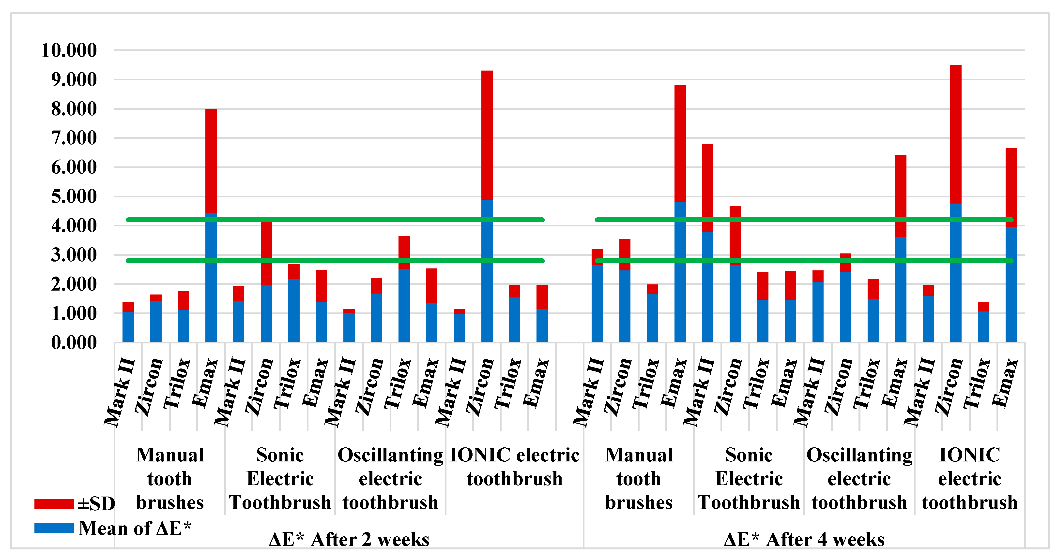
Figure 1. Mean ± SD of color change for different ceramic types after 2 and 4 weeks of immersion in coffee according to the cleaning system.
Prosthesis 07 00110 g001
Figure 2. Ra images after manual toothbrushing for 2 weeks for Vitablocs Mark II (A), Ceramill Zolid zirconia (B), Vita Triluxe (C), and IPS e.max (D).
Prosthesis 07 00110 g002
Figure 3. Ra images after manual toothbrushing for 4 weeks for Vitablocs Mark II (A), Ceramill Zolid zirconia (B), Vita Triluxe (C), and IPS e.max (D).
Prosthesis 07 00110 g003
Table 1. Details of the materials and devices used in this laboratory study.
Material/Device TypeBrand NameManufacturerComposition/Description
Feldspathic porcelain CAD/CAM blocksVitablocs Mark IIVITA Zahnfabrik, Bad Säckingen, GermanyFine-particle feldspar glass ceramic, low-to-moderate < 50% % leucite-containing
Zircon CAD/CAMCeramill Zolid multilayer PSAmann Girrbach, Pforzheim, GermanyZrO2 + HfO2 + Y2O3: ≥99.0; Y2O3: 8.5–9.5; HfO2: ≤5; Al2O3: ≤0.5; other oxides: ≤1
Feldspathic ceramicVita Triluxe ForteVITA Zahnfabrik H. Rauter GmbH & Co. KG, Bad Sackingen, GermanySiO2: 56–64; Al2O3: 20–23; Na2O: 6–9; K2O: 6–8; CaO: 0.3–0.6; TiO2: ≤0.1; other oxides: ≤11.7
Lithium disilicate glass ceramicIPS E.max CAD,Ivoclar
Vivadent, Schaan,
Liechtenstein.
SiO2, Li2O, K2O, P2O5, ZrO2, ZnO, Al2O3, MgO
Oral-B Manual ToothbrushOral-B Charcoal/00000Procter & Gamble Co., Cincinnati, OH, USA
Sonic Electric Toothbrush TechnologySonicare®/Side-to-sidePhilips Oral Healthcare, Snoqualmie, WA, USA
Oral-B Smart Series 5000 Power ToothbrushOral-B iO/Oscillating-rotating pulsatingProcter & Gamble Co., Cincinnati, OH, USA
HyG Ionic Electric ToothbrushIONICKISS/IonicHukuba Dental Corporation, 914-1 Nazukari, Nagareyama, Chiba, Japan.
NESCAFENESCAFE, 3 in 1 (STRONG)Nescafe, Riyadh, Saudi ArabiaSugar, glucose syrup, instant coffee (11%), palm kernel oil, soluble fiber, skimmed MILK powder (0.7%), MILK protein, salt, stabilizers, lactose (MILK), acidity regulator, emulsifiers, natural flavorings, MILK fat
SpectrophotometerVITA Easyshade® Compact version VVITA Zahnfabrik H. Rauter GmbH & Co. KG, Bad Sackingen, GermanyDevice used to measure wavelength transmitted from one object at a time, without being affected by subjective interferences of color
Surface roughness and topography testerWhite Light Interferometry MicroscopeContour GT-K1, Bruker Nano GmbH, Berlin, Germany3D printer of brushed surface characteristics
Table 2. Comparison of the mean color change (ΔE*) values between ceramic types for different cleaning types after immersion in coffee.
Cleaning TypeCeramic TypeAfter 2 WeeksAfter 4 Weeks
Mean RankKruskal–Wallis Hp Value aMean RankKruskal–Wallis Hp Value a
Manual toothbrushMark II14.8010.580.01424.508.290.040
Ceramill Zolid zirconia22.4020.90
Triluxe15.3011.70
IPS e.max29.5024.90
Sonic electric toothbrushMark II18.808.230.04127.309.220.026
Ceramill Zolid zirconia16.7024.70
Triluxe29.5514.35
IPS e.max16.9515.65
Oscillating electric toothbrush Mark II13.6016.940.00121.006.980.072
Ceramill Zolid zirconia24.3025.80
Triluxe31.1012.60
IPS e.max13.0022.60
IONIC electric toothbrush Mark II13.0017.99<0.00121.506.500.089
Ceramill Zolid zirconia31.5019.90
Triluxe24.3013.70
IPS e.max13.2026.90
a: Kruskal-Wallis test to compare between groups.
Table 3. Pairwise comparisons between different ceramic types for varying cleaning types after immersion in coffee.
Cleaning TypeCeramic TypeAfter 2 WeeksAfter 4 Weeks
Mann–Whitney Up Value aMann–Whitney Up Value a
Manual toothbrushMark II vs. Ceramill Zolid zirconia21.000.02750.001.000
Mark II vs. Triluxe48.000.8790.00<0.001
Mark II vs. IPS e.max20.000.02240.000.446
Ceramill Zolid zirconia vs. Triluxe30.000.12830.000.128
Ceramill Zolid zirconia vs. IPS e.max20.000.02234.000.223
Triluxe vs. IPS e.max20.000.02232.000.170
Sonic electric toothbrush Mark II vs. Ceramill Zolid zirconia41.000.49330.000.128
Mark II vs. Triluxe15.000.00822.000.033
Mark II vs. IPS e.max41.000.49330.000.128
Ceramill Zolid zirconia vs. Triluxe20.000.02222.000.033
Ceramill Zolid zirconia vs. IPS e.max49.000.93916.000.010
Triluxe vs. IPS e.max24.500.05044.500.672
Oscillating electric toothbrush Mark II vs. Ceramill Zolid zirconia10.000.00235.000.253
Mark II vs. Triluxe6.000.00126.000.068
Mark II vs. IPS e.max35.000.25346.000.761
Ceramill Zolid zirconia vs. Triluxe24.000.04814.000.006
Ceramill Zolid zirconia vs. IPS e.max26.000.06848.000.879
Triluxe vs. IPS e.max14.000.00631.000.148
IONIC electric toothbrush Mark II vs. Ceramill Zolid zirconia0.00<0.00150.001.000
Mark II vs. Triluxe16.000.01020.000.022
Mark II vs. IPS e.max41.000.49330.000.128
Ceramill Zolid zirconia vs. Triluxe30.000.12850.001.000
Ceramill Zolid zirconia vs. IPS e.max10.000.00244.000.648
Triluxe vs. IPS e.max26.000.06812.000.004
a: Mann–Whitney U test for pairwise comparison.
Table 4. Comparison of the mean color change (ΔE*) values between different cleaning types for varying ceramic types after immersion in coffee.
Ceramic TypeCleaning TypeAfter 2 WeeksAfter 4 Weeks
Mean RankKruskal–Wallis Hp Value aMean RankKruskal–Wallis Hp Value a
Mark IIManual toothbrush17.508.560.03627.8014.520.002
Sonic electric toothbrush29.6025.80
Oscillating electric toothbrush 19.2018.50
Ionic electric toothbrush 15.709.90
Ceramill Zolid zirconiaManual toothbrush17.608.220.04221.500.340.951
Sonic electric toothbrush13.6018.90
Oscillating electric toothbrush 23.4021.50
Ionic electric toothbrush 27.4020.10
TriluxeManual toothbrush10.9013.200.00425.804.250.235
Sonic electric toothbrush24.8018.10
Oscillating electric toothbrush 28.4022.20
IONIC electric toothbrush 17.9015.90
IPS e.maxManual toothbrush28.306.390.09425.109.240.026
Sonic electric toothbrush16.9010.90
Oscillating electric toothbrush 19.9022.90
Ionic electric toothbrush 16.9023.10
a: Kruskal-Wallis test to compare between groups
Table 5. Pairwise comparisons between different cleaning types for various ceramic types after immersion in coffee.
Ceramic TypeCleaning TypeAfter 2 WeeksAfter 4 Weeks
Mann–Whitney Up Value aMann–Whitney Up Value a
Mark IIManual vs. sonic electric toothbrush21.000.02748.000.879
Manual vs. oscillating electric toothbrush40.000.44621.000.027
Manual vs. ionic electric toothbrush41.000.4934.00< 0.001
Sonic electric vs. oscillating electric toothbrush17.000.01229.000.110
Sonic electric vs. ionic electric toothbrush21.000.02720.000.022
Oscillating electric vs. ionic electric toothbrush40.000.44620.000.022
Ceramill Zolid zirconiaManual vs. sonic electric toothbrush32.000.17044.000.648
Manual vs. oscillating electric toothbrush29.000.11046.000.761
Manual vs. ionic electric toothbrush24.000.04850.001.000
Sonic electric vs. oscillating electric toothbrush26.000.06836.000.286
Sonic electric vs. ionic electric toothbrush23.000.04046.000.761
Oscillating electric vs. ionic electric toothbrush34.000.22350.001.000
TriluxeManual vs. sonic electric toothbrush15.000.00832.000.170
Manual vs. oscillating electric toothbrush9.000.00249.000.939
Manual vs. ionic electric toothbrush30.000.12816.000.010
Sonic electric vs. oscillating electric toothbrush38.000.36144.000.648
Sonic electric vs. ionic electric toothbrush30.000.12850.001.000
Oscillating electric vs. ionic electric toothbrush24.000.04838.000.361
IPS e.maxManual vs. sonic electric toothbrush24.000.04818.000.015
Manual vs. oscillating electric toothbrush24.000.04843.000.594
Manual vs. ionic electric toothbrush24.000.04843.000.594
Sonic electric vs. oscillating electric toothbrush40.000.44620.000.022
Sonic electric vs. ionic electric toothbrush50.001.00016.000.010
Oscillating electric vs. ionic electric toothbrush40.000.44649.000.939
a: Mann–Whitney U test for pairwise comparison.
Table 6. Comparison of the mean color change (ΔE*) values between times after immersion in coffee for different ceramic and cleaning types.
Ceramic TypeCleaning TypeManual ToothbrushSonic Electric ToothbrushOscillating Electric ToothbrushIonic Electric Toothbrush
Mann–Whitney Up Value aMann–Whitney Up Value aMann–Whitney Up Value aMann–Whitney Up Value a
Mark IIAfter 2 weeks0.00<0.00129.000.1100.00<0.0016.000.001
After 4 weeks
Ceramill Zolid zirconiaAfter 2 weeks24.000.04816.000.01016.000.01035.000.253
After 4 weeks
TriluxeAfter 2 weeks30.000.12816.000.01021.000.02716.000.010
After 4 weeks
IPS e.maxAfter 2 weeks41.000.49340.000.44614.000.00615.000.008
After 4 weeks
a: Kruskal-Wallis test to compare between groups
Table 7. Mean contact profilometry (µm) values after staining periods for different cleaning and ceramic systems.
TimeAfter 2 WeeksAfter 4 Weeks
Brushing/Ceramic TypesVitablocs Mark IICeramill Zolid zirconiaVita TriluxeIPS e.maxVitablocs Mark IICeramill Zolid zirconiaVita TriluxeIPS e.max
Manual Toothbrush0.2980.3610.4210.7450.3080.3980.4640.789
Sonic Electric Toothbrush 0.3020.4350.4830.3840.3280.4730.6820.420
Oscillating Electric Toothbrush 0.2880.4250.4380.3510.2970.43804.530.372
HyG Ionic Electric Toothbrush0.2570.7450.3830.3560.2850.7570.4010.489
Disclaimer/Publisher’s Note: The statements, opinions and data contained in all publications are solely those of the individual author(s) and contributor(s) and not of MDPI and/or the editor(s). MDPI and/or the editor(s) disclaim responsibility for any injury to people or property resulting from any ideas, methods, instructions or products referred to in the content.