Case Report

Discovery of a Three-Piece Inflatable Penile Prosthesis Implant During Donor Dissection: Anatomical Case Study

1
Oakland University William Beaumont School of Medicine, Rochester, MI 48309, USA
2
Department of Foundational Medical Studies, Oakland University William Beaumont School of Medicine, Rochester, MI 48309, USA
*
Author to whom correspondence should be addressed.
Prosthesis 2025, 7(5), 127; https://doi.org/10.3390/prosthesis7050127 (registering DOI)
Submission received: 31 July 2025 / Revised: 8 September 2025 / Accepted: 30 September 2025 / Published: 13 October 2025

Abstract

Penile prosthetic implants (PPIs) provide a definitive surgical solution for individuals requiring restoration of erectile function, most commonly due to medication-refractory erectile dysfunction (ED) or as part of gender-affirming surgical care. During the Anatomical Foundations of Clinical Practice (AFCP) course at Oakland University William Beaumont (OUWB) School of Medicine, a complete three-piece inflatable PPI was identified in a 66-year-old male donor with a medical history of congestive heart failure, hypertension, and diabetes mellitus type 2. The prosthesis included a fluid reservoir positioned in the lower abdominal cavity in the retropubic space, a scrotal pump with a release valve, and paired inflatable cylinders embedded within the penile shaft. This uncommon finding provided first-year medical students with a hands-on opportunity to examine the structure, placement, and function of a modern PPI. In addition to reinforcing foundational knowledge of ED treatment, the case highlighted the expanding clinical indications for penile implants, including their potential role in gender-affirming procedures. Integrating such real-world surgical findings into anatomical education enriches the learning experience of students and highlights the evolving scope of prosthetic urology across diverse patient populations.

1. Introduction

Erectile function depends on the coordinated interaction of vascular, neural, hormonal, and psychological systems. Adequate arterial inflow and nitric oxide-mediated cyclic guanosine monophosphate (cGMP) signaling are essential for penile rigidity, while psychogenic, reflex, and nocturnal pathways regulate initiation. Testosterone supports libido and nitric oxide synthase expression. Disruption of any of these systems—such as from vascular disease, neuropathy, spinal injury, or psychological factors—can impair erectile function and compromise treatment outcomes [1].
Erectile dysfunction (ED) is a prevalent condition characterized by a persistent inability to achieve or maintain an erection sufficient for sexual activity. Its incidence increases with age and is strongly associated with comorbidities such as cardiovascular disease, diabetes, hypertension, and metabolic syndrome. According to the Massachusetts Male Aging Study, approximately 52% of men aged 40–70 experience some degree of ED [2]. Beyond its physical impact, ED significantly affects quality of life and intimate relationships. The condition is strongly linked to psychological consequences, including decreased self-esteem, anxiety, depression, and interpersonal strain, which may further exacerbate sexual dysfunction [3]. In addition, ED is increasingly recognized as a sentinel marker of systemic vascular disease, with several studies demonstrating its role as an early indicator of subclinical atherosclerosis and future cardiovascular events. In many cases, ED precedes the onset of overt coronary artery disease by several years, underscoring its importance as both a quality-of-life condition and a clinical marker for cardiovascular risk stratification [4].
Several treatment options have been developed to address ED, with varying efficacy and patient satisfaction. First-line management typically involves lifestyle modifications and oral phosphodiesterase type 5 (PDE5) inhibitors such as sildenafil, tadalafil, vardenafil, and avanafil. These medications enhance penile blood flow in response to sexual stimulation by inhibiting PDE5-mediated breakdown of cGMP. For patients unresponsive to oral therapy, alternatives include vacuum erection devices, intracavernosal injections, or intraurethral suppositories. Emerging approaches such as penile shockwave therapy and stem cell–based regenerative techniques are under investigation but remain experimental [5].
For patients who fail conservative management, penile prosthetic implants (PPIs) provide a durable and highly effective solution, with satisfaction rates exceeding those of other therapies [5,6,7]. The history of these devices reflects decades of development within the field of urologic surgery. Early acrylic and polyurethane rod implants provided rudimentary rigidity sufficient for sexual intercourse, however, were limited by poor concealability. Advances in materials science, including the advent of silicone, paved the way for the first inflatable penile prosthesis, developed by F. Brantley Scott and colleagues in 1973 [8]. Contemporary three-piece inflatable PPIs—comprising paired intracavernosal cylinders, a fluid reservoir in the lower abdomen, and a scrotal pump—remain the most widely implanted devices and have been further optimized with infection-resistant coatings, kink-resistant tubing, and improved pump durability [9].
Despite being chosen by fewer than 5% of eligible candidates, over 25,000 prostheses are implanted annually in North America, and utilization is expected to rise with continued technological advancements [5,9]. On a global scale, the estimated number of annual penile prosthesis procedures exceeds 60,000, with roughly half performed in the United States and the remainder distributed across Europe, Asia, and other regions [10]. Historical data illustrate this disparity: between 2005 and 2012, Baas et al. reported 63,013 implants worldwide, with nearly 86% taking place in the United States, underscoring the heavy concentration of this procedure in North American practice [11]. Taken together, these findings demonstrate that while penile prosthesis surgery is relatively common in high-income countries, its global reach remains constrained by access to surgical expertise, device cost, and sociocultural stigma surrounding ED and prosthetic intervention. As awareness and availability expand, utilization may increase internationally, particularly as indications broaden to include gender-affirming phalloplasty.
Multiple surgical approaches are available for inflatable penile prosthesis placement, each with distinct advantages and limitations. The penoscrotal approach provides excellent exposure of the corpora cavernosa and straightforward pump placement, while minimizing risk to dorsal sensory nerves; however, reservoir placement is typically blind through the inguinal canal, raising the potential for vascular injury [12,13]. The infrapubic approach offers faster reservoir placement under direct visualization, reducing iliac vessel risk, but carries a higher likelihood of dorsal nerve injury and can make pump positioning more difficult [14,15]. More recently, the subcoronal approach has been described, which uses a single distal penile incision that facilitates simultaneous correction of penile deformity, provides improved visualization of corporal structures, and allows for procedures under local anesthesia; nonetheless, this technique requires longer operative times and may increase infection risk [16,17]. The choice of approach is therefore individualized, balancing technical considerations with patient comorbidities and surgical goals.

2. Detailed Case Description

2.1. Donor Selection and Dissection Protocol

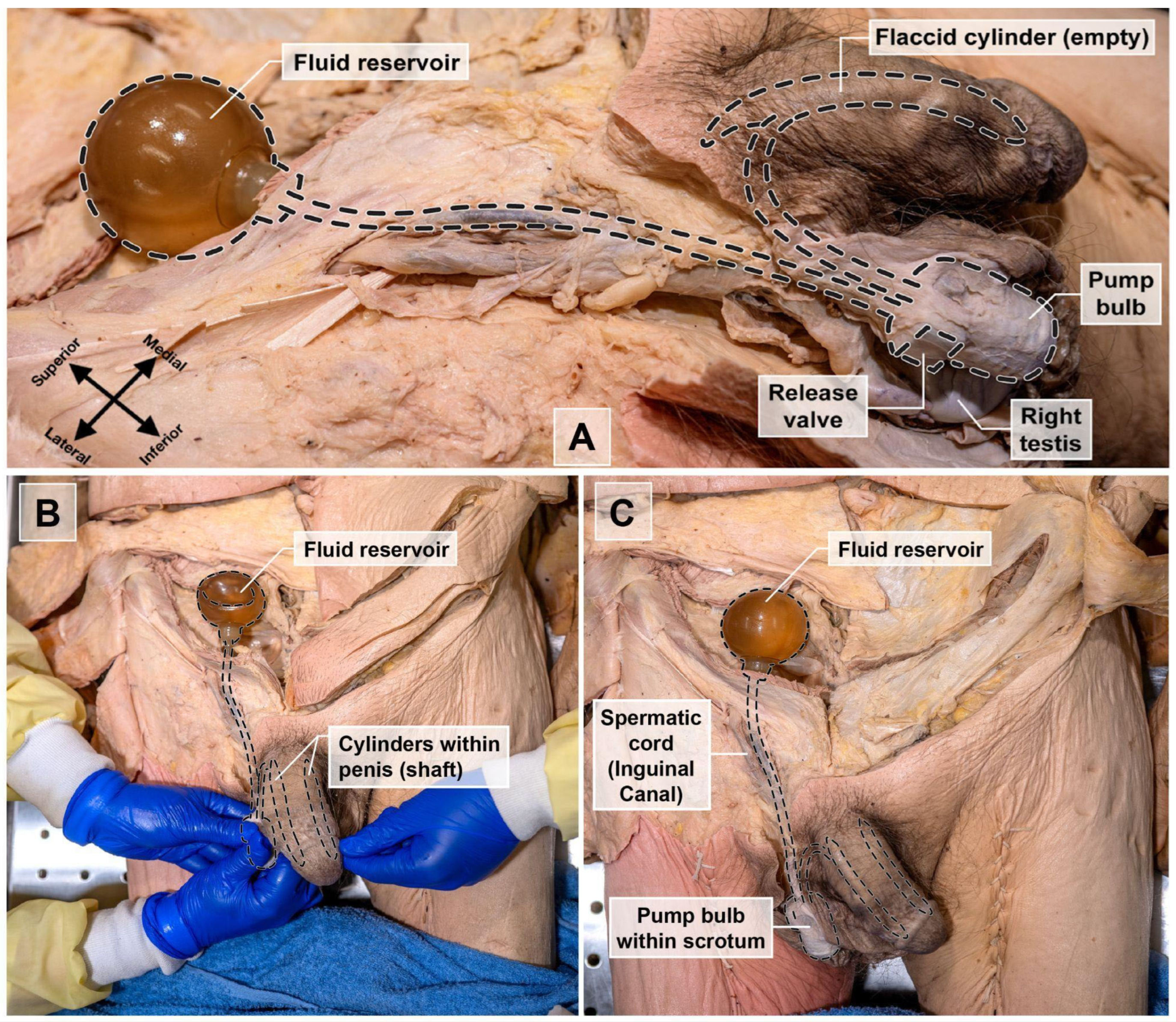
As part of the Anatomical Foundations of Clinical Practice (AFCP) course at Oakland University William Beaumont School of Medicine (OUWB) School of Medicine, first-year medical students perform dissection on 21 donors (10 male, 11 female). Each group of 5–6 students performed full-body dissections using in-house dissection manuals as their guide. During the dissection of the abdominal region, a PPI was discovered in a 66-year-old male donor with a history of congestive heart failure, hypertension, and diabetes mellitus type 2. The device was identified as a three-piece inflatable prosthesis consisting of a fluid reservoir located in the retropubic space (Space of Retzius), a pump bulb with release valve housed within the subdartos pouch of the scrotum, and bilateral inflatable cylinders embedded within the corpora cavernosa of the penis (Figure 1 and Figure 2).

2.2. Discovery of a Penile Prosthetic Implant During Donor Dissection

The three-piece penile prosthesis was discovered during the abdominal and pelvic dissection portion of the AFCP course. The main dissection techniques used to find and isolate the penile implant include layer-by-layer dissection, blunt dissection, using anatomical landmarks, tissue reflection and retraction, and tracing the tubing of the implant. Dissection began with reflection of the anterior abdominal wall musculature to expose intra-abdominal structures. During exploration of the pelvic cavity, a cylindrical structure was found within the retropubic space. The unusual texture and uniform diameter of the components provided the first cue that the finding was prosthetic rather than organic. Careful blunt and sharp dissection was used to isolate this structure, revealing it to be a fluid reservoir with attached tubing. Further investigation through the scrotal region, following standard dissection through the skin, Dartos fascia, and underlying tissue layers, led to the identification of a pump bulb and anteriorly oriented release valve housed within the subdartos pouch at the midline of the scrotum. Structures were carefully dissected and preserved to maintain the integrity of the prosthetic components and associated anatomical relationships. Subsequently, dissection of the penile shaft was performed to preserve erectile tissues. As the cylinders came into view, students realized the importance of handling the surrounding tissue delicately to avoid puncturing the implant. Using meticulous longitudinal incisions, bilateral inflatable cylinders were visualized within the corpora cavernosa. Close observation revealed subtle differences between the tactile feel of the cylinders and the natural corpora cavernosa. The distal tips of each cylinder were positioned near the glans penis. The tubing connecting the pump, reservoir, and cylinders was traced and preserved. When the tubing connections were fully mapped, the group recognized the complete system and confirmed its identity as a penile prosthesis. The PPI was found to be intact and fully functional, achieving erection when the pump bulb was compressed during dissection by an inquisitive student. This prompted the search for a release valve by the group, which was found to be a button on the superior aspect of the pump bulb (Figure 2B). This discovery initiated engagement within the dissection group, extended to the entire class, and prompted discussion with the faculty.

2.3. Anatomical Identification and Functional Analysis

When the fluid reservoir was found in the retropubic region, its purpose was initially unclear, prompting discussion among the students and faculty. The retropubic space is bordered anteriorly by the pubic symphysis, posteriorly by the urinary bladder, laterally by the tendinous arch of the pelvic fascia, superiorly by the transversalis fascia, and inferiorly by the bladder neck, anterior urethra, and pubocervical fascia [18]. The purpose of the reservoir is to store normal saline solution before and after an erection.
The second component, consisting of the pump bulb and release valve, was identified during scrotal dissection. The pump was situated within the subdartos pouch—a surgically created space within the Dartos fascia at the midline of the scrotum. The release valve (deflation button), oriented anteriorly, was positioned just superior to the pump bulb [19]. The purpose of the pump bulb and release valve is for direct alteration between flaccid and erect positions of the penis.
The final component identified during penile dissection was the pair of inflatable cylinders. Each cylinder was embedded within a corpus cavernosum [20], with the distal tips positioned just beneath the glans penis [21]. In cases of erectile dysfunction, the corpus cavernosum fails to fill with blood; thus, the cylinders functionally replace this process to produce an erection. Normal saline is transferred between the reservoir and the cylinders via connecting tubes, enabling inflation and deflation. Upon identification of all three components, the device was tested and found to be intact, fully functional, without signs of degradation.
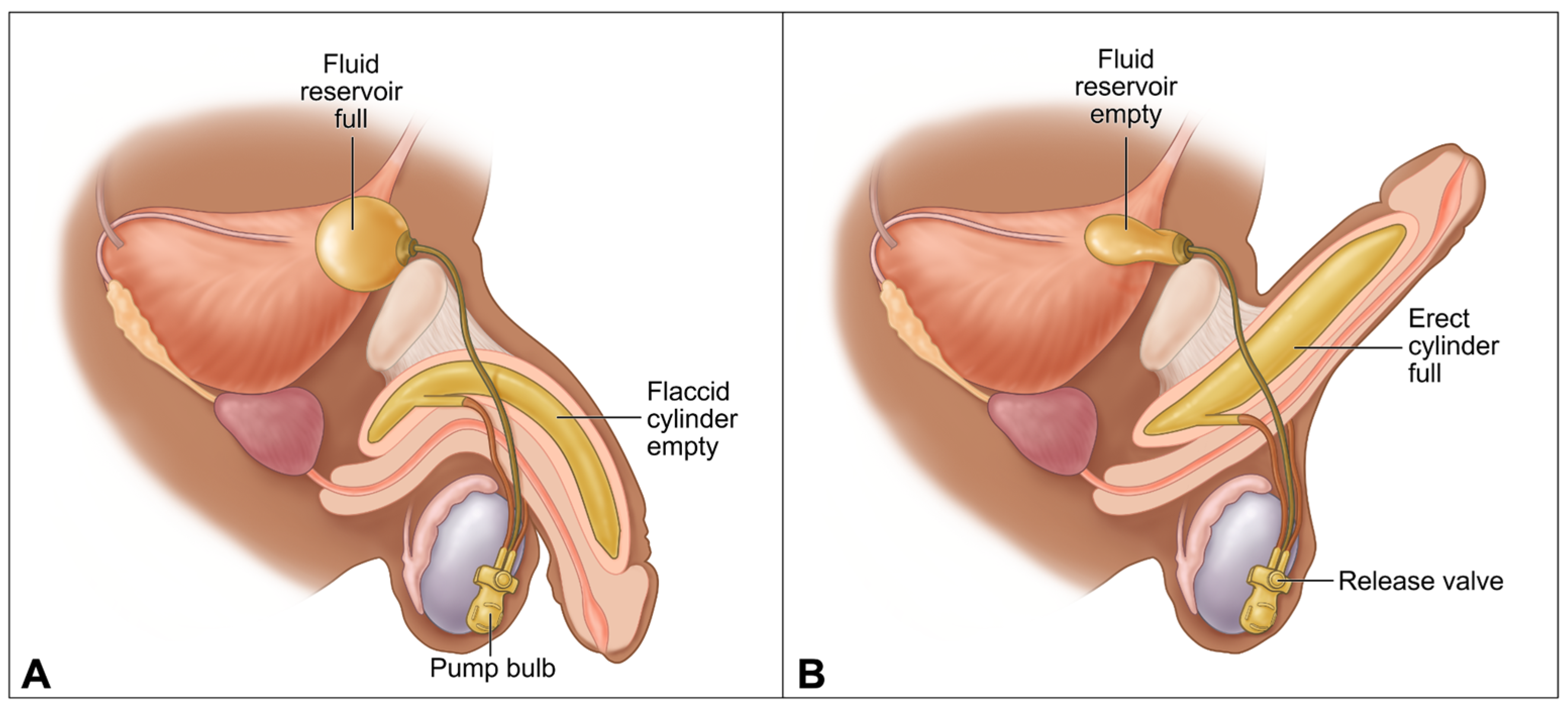
When the pump bulb and release valve were compressed, the fluid reservoir and cylinders changed shape, resulting in the change between flaccid and erect positions of the penis of the donor body. In the flaccid state of the penis, the fluid reservoir was completely filled with normal saline solution, while the cylinders and connecting tubes contained little to no solution. The cylinders were completely deflated and the corpus cavernosum was constricted (Figure 3A). At this stage, the penile shaft maintained a soft, pliable consistency, reflecting the lack of fluid tension within the corporal bodies. The surrounding tissue offered minimal resistance to manual manipulation, highlighting the inactive state of the implant system.
To change the penis from the flaccid position to the erect position, the pump bulb was manually compressed. This resulted in positive pressure in the fluid reservoir that forced the saline solution into the connecting tubes and cylinders. The transfer of solution was bidirectional, guided by pressure differences within the pump mechanism. As fluid entered the cylinders, they gradually elongated and expanded, mimicking the hemodynamic changes in natural penile erection.
In the erect state of the penis, the cylinders were completely filled with saline solution, the reservoir contained little solution, and the connecting tubes contained a moderate amount of solution. The cylinders became completely inflated, which resulted in dilation of the corpus cavernosum (Figure 3B). This dilation produced sufficient rigidity for penetration and functional sexual activity. Additionally, the firmness achieved was dependent on the complete transfer of solution and the integrity of the cylinder walls.
To change the penis from the erect position to the flaccid position, the release valve was firmly pressed. This resulted in negative pressure in the fluid reservoir that drew the saline solution from the cylinders and connecting tubes back into the reservoir. As the fluid was withdrawn, the cylinders collapsed, and the penile shaft returned to a flexible, resting state. This transition occurred smoothly within seconds, allowing controlled cycling between flaccidity and erection. The system design ensured reversibility and repeatability, essential for practical use.

3. Discussion

The discovery of a three-piece PPI during first-year donor dissection at OUWB School of Medicine proved to be a highly valuable and memorable educational experience. The initial finding of the spherical structure in the abdominal cavity sparked genuine curiosity among students and prompted collaborative exploration with faculty. As the dissection progressed and the components of the prosthesis were revealed, culminating in the identification of a fully intact and functional device, the learning experience deepened beyond standard anatomical instruction. This encounter allowed students to connect theoretical knowledge of pelvic and perineal anatomy to its real-world surgical application in a way that is rarely possible in the preclinical setting.
This case reinforced the clinical relevance of spatial relationships in pelvic anatomy, particularly as they pertain to the placement of complex surgical implants. It also emphasized the importance of recognizing that anatomical donors are former patients who may have undergone significant medical or surgical interventions. The identification of the penile prosthesis prompted widespread interest among the first-year class, with nearly all students taking the opportunity to visit the donor and observe the device in context. This heightened engagement situated the experience of learning pelvic and genital anatomy within the clinical realities of disease, surgical care, and patient experience, rather than isolated memorization.
Furthermore, the educational value of this case can be meaningfully framed through the lens of experiential learning theory. Kolb’s Experiential Learning Cycle (1984) describes four stages: concrete experience, reflective observation, abstract conceptualization, and active experimentation [22]. In the first-year AFCP anatomy lab at OUWB, the pelvic dissection and discovery of a three-piece inflatable PPI provided the concrete experience. Reflective observation followed as students engaged in discussion with faculty regarding the anatomical placement and functional features of the device. Guided dialog then prompted students to situate the prosthesis within the broader clinical context of erectile dysfunction treatment, stimulating integration of prior knowledge in anatomy, physiology, and therapeutics—constituting the stage of abstract conceptualization. Finally, testing the device by compressing the pump bulb represented active experimentation, completing the cycle and reinforcing learning through direct interaction with the prosthesis.
Importantly, this finding also fostered early professional identity formation. Engaging with an advanced medical device in situ helped students appreciate the broader context of surgical innovation, the interdisciplinary nature of patient care, and the real-world implications of anatomy in medical decision-making. Additionally, it prompted thoughtful consideration of quality-of-life issues for patients living with erectile dysfunction, as well as the psychological and relational aspects of penile prosthesis implantation.
While our case featured a penile prosthesis in a cisgender male, it also created space for discussions about the expanding role of inflatable penile implants in gender-affirming surgery. Three-piece PPIs are commonly utilized following phalloplasty in transgender men to provide rigidity for penetrative intercourse [23]. Introducing this application early in medical education helps normalize transgender health care and reduce the stigma often associated with discussing sexual function and genital surgery. Preclinical exposure to these topics can help future physicians navigate sensitive conversations with confidence, empathy, and professionalism.
Comparable cadaveric reports of penile prosthesis identification remain absent in the literature to our knowledge, underscoring the rarity and significance of the present case. A literature search on this topic primarily identifies workshop-based training using donors to enhance surgical confidence with penile prosthesis implantation, as well as ex vivo implantation and testing procedures [24,25]. In contrast, our report documents the incidental discovery of a fully intact three-piece PPI during routine donor dissection and emphasizes its pedagogical value by linking anatomical education to surgical practice and patient-centered discussions. This positions our study as a unique contribution to the limited cadaveric literature, reinforcing the importance of integrating such real-world findings into medical education.
In summary, this case highlights the pedagogical power of encountering post-surgical anatomy during donor dissection. Real-world findings like this one elevate the learning experience, bridging the gap between preclinical coursework and clinical realities. They provide opportunities for students to develop not only anatomical knowledge but also curiosity, critical thinking, cultural humility, and respect for the lived experiences represented in the donor body. As penile prosthesis implantation continues to evolve in both technique and indication, integrating such discoveries into medical education supports the development of well-rounded, patient-centered physicians.

4. Conclusions

This discovery marks the first instance at OUWB School of Medicine in which students encountered and studied a three-piece penile prosthesis during donor dissection. While clinically rare, the case served as a powerful educational tool, prompting in-depth review and discussion of male pelvic anatomy, vascular and neural structures, and the surgical techniques involved in prosthetic implantation.
This case exemplifies the pedagogical value of integrating real-world clinical findings into preclinical education. By linking anatomical dissection with actual therapeutic interventions, students are better equipped to contextualize their learning, ask clinically relevant questions, and appreciate the functional implications of anatomical structures. Experiences like this enrich the preclinical curriculum by fostering early clinical reasoning, encouraging interdisciplinary dialog, and deepening students’ understanding of how anatomy informs surgical care. Most importantly, it promotes a patient-centered approach to learning, highlighting how anatomy education can lay the foundation for thoughtful, evidence-based medical practice.
Positioning this case within the framework of experiential learning theory underscores its impact: the dissection provided students with a concrete experience that was followed by reflection, conceptual integration with clinical knowledge, and active experimentation through device testing. In this way, the incidental discovery not only enriched anatomical understanding but also modeled the iterative cycle of learning that underpins effective medical education. Furthermore, by documenting a rare incidental finding scarcely reported in cadaveric literature, this case adds a unique contribution to anatomical and surgical education, reinforcing the unique value of such discoveries in bridging anatomy education with clinical practice.

Author Contributions

Conceptualization, methodology, B.H., A.P., J.T., S.T., M.B. and J.G.; investigation, B.H., A.P., J.T., S.T., M.B. and J.G.; writing—original draft preparation, B.H., A.P. and J.T.; writing—review and editing, B.H., A.P., J.T., S.T., M.B. and J.G.; resources, supervision, M.B. and J.G.; funding acquisition, J.G., project administration, B.H.; references and citation management, S.T. All authors have read and agreed to the published version of the manuscript.

Funding

This research received no external funding.

Institutional Review Board Statement

This study involved the use of human cadavers that were voluntarily donated for educational and research purposes. As the study did not include living human subjects, it was exempt from institutional review board (IRB) approval in accordance with Oakland University policies. No identifiable personal or health information was collected.

Informed Consent Statement

The donor body used in this case report was obtained through a willed body donation program in accordance with institutional and legal ethical guidelines. All donors provided informed consent for the use of their bodies in medical education and research prior to death. No identifiable personal information is disclosed in this report. This study was conducted with respect and gratitude for the donor’s contribution to medical education.

Data Availability Statement

The data supporting the findings of this study were obtained from donor records and contain confidential personal information. Due to privacy and ethical considerations, these data are not publicly available.

Acknowledgments

The authors express their sincere gratitude to the individuals who generously donated their bodies to science, making this anatomical study possible. Their selfless contributions continue to advance medical education and research, ultimately enhancing patient care. The authors also extend their appreciation to the anatomy faculty and lab manager, Dan Schlegel, for their invaluable support and guidance in facilitating the success of this project and to Kellyn Sanders, OUWB Medical Illustrator, for creating the illustrations featured in this article.

Conflicts of Interest

The authors declare no conflicts of interest.

Abbreviations

The following abbreviations are used in this manuscript:
PPIPenile Prosthetic Implant
EDErectile Dysfunction
AFCPAnatomic Foundations of Clinical Practice
OUWBOakland University William Beaumont

References

  1. Wang, C.-M.; Wu, B.-R.; Xiang, P.; Xiao, J.; Hu, X.-C. Management of male erectile dysfunction: From the past to the future. Front. Endocrinol. 2023, 14, 1148834. [Google Scholar] [CrossRef]
  2. Feldman, H.A.; Goldstein, I.; Hatzichristou, D.G.; Krane, R.J.; McKinlay, J.B. Impotence and its medical and psychosocial correlates: Results of the Massachusetts Male Aging Study. J. Urol. 1994, 151, 54–61. [Google Scholar] [CrossRef]
  3. Melnik, T.; Soares, B.G.O.; Nasello, A.G. The effectiveness of psychological interventions for the treatment of erectile dysfunction: Systematic review and meta-analysis, including comparisons to sildenafil treatment, intracavernosal injection, and vacuum devices. J. Sex. Med. 2009, 5, 2562–2574. [Google Scholar] [CrossRef]
  4. Montorsi, P.; Montorsi, F.; Schulman, C.C. Is erectile dysfunction the "tip of the iceberg" of a systemic vascular disorder? Eur. Urol. 2003, 44, 352–354. [Google Scholar] [CrossRef]
  5. Krzastek, S.C.; Bopp, J.; Smith, R.P.; Kovac, J.R. Recent advances in the understanding and management of erectile dysfunction. F1000Research 2019, 8, 102. [Google Scholar] [CrossRef]
  6. Cakir, O.O.; Lee, W.G.; Ralph, P.; Megson, M.; Ralph, D.J. Advances in penile prosthesis surgery: A clinical update. Türk Urol. Derg./Turk. J. Urol. 2022, 48, 393–397. [Google Scholar] [CrossRef] [PubMed]
  7. Chung, E.; Bettocchi, C.; Egydio, P.; Love, C.; Osmonov, D.; Park, S.; Ralph, D.; Xin, Z.C.; Brock, G. The International Penile Prosthesis Implant Consensus Forum: Clinical Recommendations and surgical principles on the inflatable 3-piece penile prosthesis implant. Nat. Rev. Urol. 2022, 19, 534–546. [Google Scholar] [CrossRef] [PubMed]
  8. Le, B.; Burnett, A.L. Evolution of penile prosthetic devices. Korean J. Urol. 2015, 56, 179–186. [Google Scholar] [CrossRef]
  9. Cavayero, C.T.; Leslie, S.W.; McIntosh, G.V. Penile Prosthesis Implantation; StatPearls Publishing: St. Petersburg, FL, USA, 2024. Available online: https://www.ncbi.nlm.nih.gov/books/NBK558980/ (accessed on 7 September 2025).
  10. Rodriguez, K.M.; Pastuszak, A.W. A history of penile implants. Transl. Androl. Urol. 2017, 6 (Suppl. S5), S851–S857. [Google Scholar] [CrossRef]
  11. Baas, W.; O’Connor, B.; Welliver, C.; Stahl, P.J.; Stember, D.S.; Wilson, S.K.; Köhler, T.S. Worldwide trends in penile implantation surgery: Data from over 63,000 implants. Transl. Androl. Urol. 2020, 9, 31–37. [Google Scholar] [CrossRef] [PubMed]
  12. Montague, D.K.; Angermeier, K.W. Surgical approaches for penile prosthesis implantation: Penoscrotal vs infrapubic. penile prosthesis implantation: Penoscrotal vs infrapubic. Int. J. Impot. Res. 2003, 15, S134–S136. [Google Scholar] [CrossRef] [PubMed]
  13. Kwan, L.; Zhao, L.C. Penoscrotal approach for inflatable penile prosthesis implantation. Asian J. Androl. 2011, 13, 711–715. [Google Scholar] [CrossRef]
  14. Palmisano, F.; Boeri, L.; Cristini, C.; Antonini, G.; Spinelli, M.G.; Franco, G.; Longo, F.; Gadda, F.; Colombo, F.; Montanari, E. Comparison of infrapubic vs penoscrotal approach for inflatable penile prosthesis placement: A systematic review and meta-analysis. J. Sex. Med. 2018, 15, 1712–1719. [Google Scholar] [CrossRef]
  15. Bertero, E.B. Infrapubic approach to penile prosthesis placement: Indications and outcomes. Urology 2015, 86, 553–557. [Google Scholar] [CrossRef]
  16. Park, S.H. Subcoronal inflatable penile prosthesis under local anesthesia. J. Vis. Surg. 2019, 5, 65. [Google Scholar] [CrossRef]
  17. Feng, C.L.; Langbo, W.A.; Anderson, L.K.; Cao, D.; Bajic, P.; Amarasekera, C.; Wang, V.; Levine, L.A. Subcoronal inflatable penile prosthesis implantation: Indications and outcomes. J. Sex. Med. 2023, 20, 888–892. [Google Scholar] [CrossRef]
  18. Osmonov, D.; Chomicz, A.; Tropmann-Frick, M.; Arndt, K.M.; Jünemann, K.P. High-submuscular vs. space of Retzius reservoir placement during implantation of inflatable penile implants. Int. J. Impot. Res. 2020, 32, 18–23. [Google Scholar] [CrossRef]
  19. Al-Enezi, A.; Al-Khadhari, S.; Al-Shaiji, T.F. Three-piece inflatable penile prosthesis: Surgical techniques and pitfalls. J. Surg. Tech. Case Rep. 2011, 3, 76–83. [Google Scholar] [CrossRef]
  20. Pacheco Usmayo, A.; Torregrosa Andrés, A.; Flores Méndez, J.; Luján Marco, S. Inflatable penile prosthesis (IPP): Diagnosis of complications and component imaging. Radiol./Diagn. Imaging 2021, 63, 504–510. [Google Scholar] [CrossRef]
  21. Otero, J.R.; Manfredi, C.; Wilson, S.K. The good, the bad, and the ugly about surgical approaches for inflatable penile prosthesis implantation. Int. J. Impot. Res. 2020, 34, 128–137. [Google Scholar] [CrossRef] [PubMed]
  22. Kolb, D.A. Experiential Learning: Experience as the Source of Learning and Development; Prentice Hall: Englewood Cliffs, NJ, USA, 1984. [Google Scholar]
  23. Dy, G.W.; Dubin, J.M.; Morey, A.F. Penile prosthesis implantation after phalloplasty: A review of techniques and outcomes. Transl. Androl. Urol. 2019, 8, 208–215. [Google Scholar] [CrossRef]
  24. Krishnappa, P.; Srini, V.S.; Shah, R.; Lentz, A.C.; Garaffa, G.; Martinez-Salamanca, J.I.; Moncada, I. Cadaveric penile prosthesis workshop training improves surgical confidence levels of urologists: South Asian Society for Sexual Medicine course survey. Int. J. Urol. 2020, 27, 1032–1037. [Google Scholar] [CrossRef] [PubMed]
  25. Khorshidi, M.A. Ex-vivo IPP implantation in cadaveric human penis with paired computational modeling. Eur. Urol. Open Sci. 2024, 67, S3. [Google Scholar] [CrossRef]
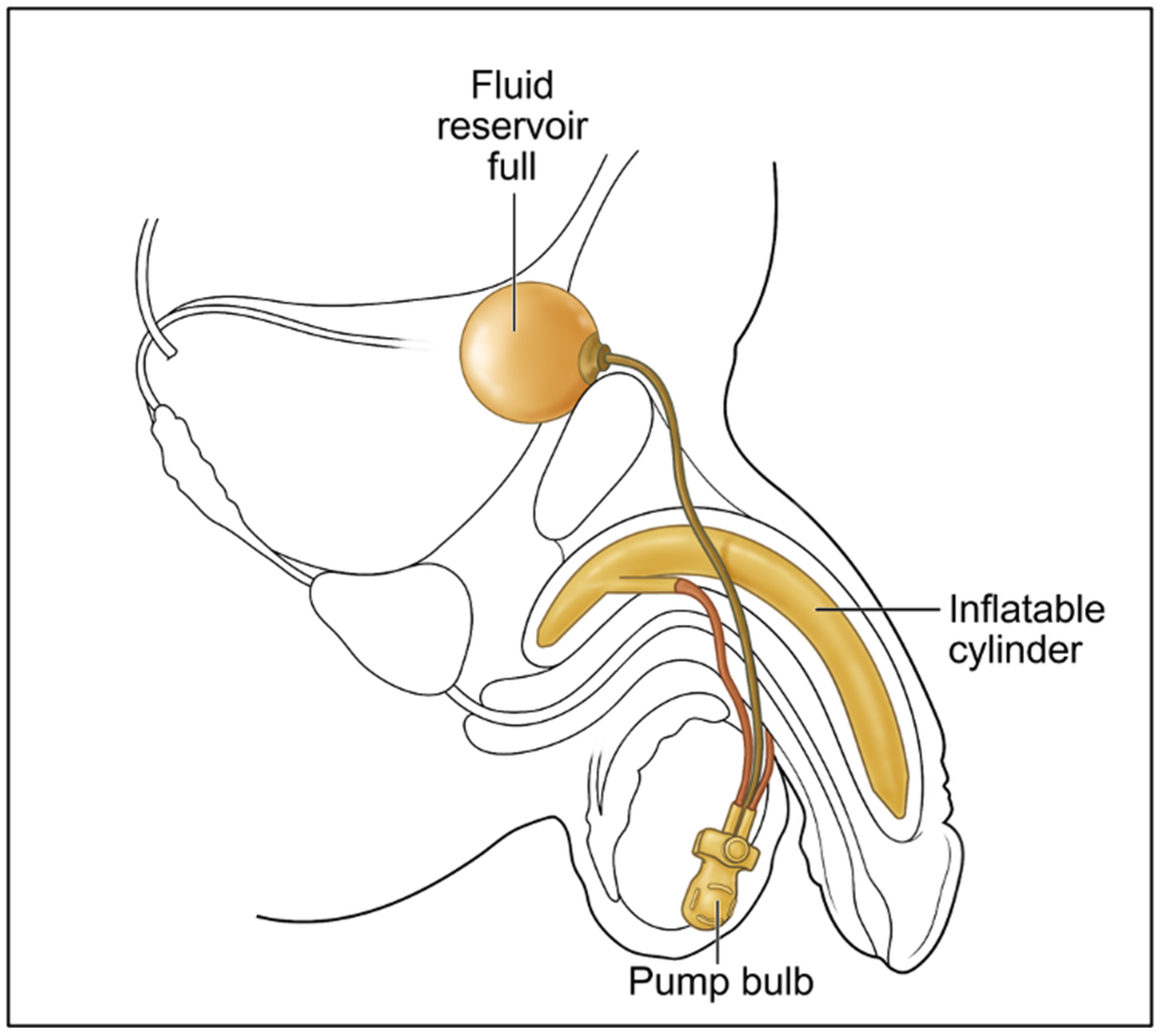
Figure 1. Illustration of a three-piece penile prosthetic implant (PPI), showing two inflatable cylinders placed within the corpora cavernosa of the penis, a fluid reservoir positioned in the retropubic space of the lower abdominal cavity, and a pump bulb located within the scrotum.
Prosthesis 07 00127 g001
Figure 2. Donor images with a three-piece penile prosthetic implant (PPI) seen from the lateral view (A), frontal view (B,C). In the flaccid state of the penis, the fluid reservoir is filled with normal saline solution and tucked within the retropubic space; the pump bulb and release valve were positioned between the two testicles; the cylinders were deflated and situated within each corpus cavernosum; the connecting tubes convey saline solution between the reservoir and cylinders. (A) Lateral view demonstrating the fluid reservoir in situ. (B) Frontal view (intact scrotum) showing the fluid reservoir in relation to the surrounding structure and demonstrating student exploration of functionality. (C) Frontal view (dissected scrotum) exposing the pump bulb with a release valve.
Prosthesis 07 00127 g002
Figure 3. Illustration showing the appearance of the fluid reservoir and cylinder, in the flaccid state (A), and in the erect state (B) of the penis.
Prosthesis 07 00127 g003
Disclaimer/Publisher’s Note: The statements, opinions and data contained in all publications are solely those of the individual author(s) and contributor(s) and not of MDPI and/or the editor(s). MDPI and/or the editor(s) disclaim responsibility for any injury to people or property resulting from any ideas, methods, instructions or products referred to in the content.